American Journal of Otolaryngology and Head and Neck Surgery
Long-term Hearing Preservation with Slim Perimodiolar CI532 ® Cochlear Implant Array
OPEN ACCESS
*Correspondence:
Roland Nagy, Department of Otorhinolaryngology, Head and Neck Surgery, University of Szeged, H-6725 Szeged, Tisza Lajos krt. 111, Hungary, Tel: +3662 545310; +3630 4089088;
Fax: +3662 545848;
E-mail: nagy.roland@med.u-szeged.hu Received Date: 14 Aug 2018 Accepted Date: 18 Sep 2018 Published Date: 21 Sep 2018
Citation:
Nagy R, Jarabin JA, Perényi Á, Dimák B, Tóth F, Jóri J, et al. Long- term Hearing Preservation with Slim Perimodiolar CI532® Cochlear Implant Array. Am J Otolaryngol Head Neck Surg. 2018; 1(4): 1019.
Copyright © 2018 Roland Nagy. This is an open access article distributed under the Creative Commons Attribution License, which permits unrestricted use, distribution, and reproduction in any medium, provided the original work is properly cited.
Published: 21 Sep, 2018
Abstract
By using sophisticated surgical techniques in combination with the Slim Perimodiolar cochlear implant electrode array a hitherto unattained high rate of residual hearing preservation in cochlear implantation has been observed that makes potential for electric acoustic stimulation. One of the primary aims of cochlear implant system engineering is to promote atraumatic electrode insertion to maintain optimal postoperative hearing sensitivity by protecting and preserving the delicate inner ear structures.
The study aimed to collect pre-, and postoperative audiological and surgical results from the experience gained from the applied cochlear implant configuration.
About 30 patients (aged 43.32 ± 24 years) with partial hearing loss were supplied with this atraumatic perimodiolar thin electrode which was designed to preserve residual hearing despite intracochlear insertion of an electrode array. All patients were implanted with consentaneous CI system and surgery technique.
The use of new electrode array profiles in cochlear implantation plays a fundamental role in minimally invasive soft surgery, taking into individual needs, and providing long-term acoustic hearing preservation. Hearing preservation was achieved in most cases (partial residual hearing preservation) after a long-term follow-up period (preoperation, at least one year).
Keywords: Cochlear implantation; Hearing preservation; Soft surgery; Perimodiolar electrode profile
Introduction
Competing companies (Advanced Bionics, Cochlear, Med-El and Oticon, etc.) provide different types of receiver-stimulators, implant electrodes and speech processors. There are several pros and cons when opting for an electrode profile (straight or perimodiolar), cochlear coverage (total or partial), receiver-stimulator (physical attributes) and speech processor (electric or electroacoustic stimulation), that meet the individual needs. One of the primary aims of cochlear implant system engineering is to promote atraumatic electrode insertion to maintain optimal postoperative hearing sensitivity by protecting and preserving the delicate inner ear structures.
Residual hearing sensitivity may deteriorate due to perioperative traumas or complications with delayed onset. The applied surgical approach (Round Window (RW), Extended Round Window (ERW), Cochleostomy (CS)) and the implanted electrode profile mainly lead to immediate or short- term damage, while delayed alteration in cochlear function usually derives from the fibrous or bony remodelling of the endocochlear compartments.
Surgically important properties are the physical attributes of the electrode configuration (perimodiolar vs. straight; rounded vs. smoothened tip; short vs. regular; with or without stylet, etc.), the type of cochlear fenestration (RW, ERW, CS), the method of electrode insertion (standard vs. soft surgery with advance-off-stylet), the use of lubricants or drugs in the cochlea (e.g. intrascalar corticosteroids) and the intrascalar position of the electrode array (perimodiolar, mid-scala, lateral- wall) [1-3].
However, the possible disproportion between the physical dimensions of the electrode profile and the endocochlear compartments (diameter, shape, length of scala tympani) play a significant role in preserving inner ear structures and functions, too.
Roland Nagy*, János András Jarabin, Ádám Perényi, Balázs Dimák, Ferenc Tóth, József Jóri, József Géza Kiss and László Rovó
Department of Otorhinolaryngology, Head and Neck Surgery, University of Szeged, Hungary
Minimizing the damage in the inner ear enhances the possibility for hearing preservation, thus leading to better hearing performance.
Systemic and/or intratympanic administration of steroids may contribute to hearing preservation. The beneficial effects of glucocorticoids are thought to be mediated through several different pathways: the anti-inflammatory effects; the down-regulation of production of inducible nitric-oxide synthase; and direct inhibition of the MAP/JNK cell death signal cascade [2-5].
We aimed to study long-term hearing preservation in a non- randomized, prospective clinical cohort with cochlear implant systems, limited to ones produced by Australian and Austrian leader companies, provided and fully financed by the Hungarian National Health Insurance.
Materials and Methods
Study cohort
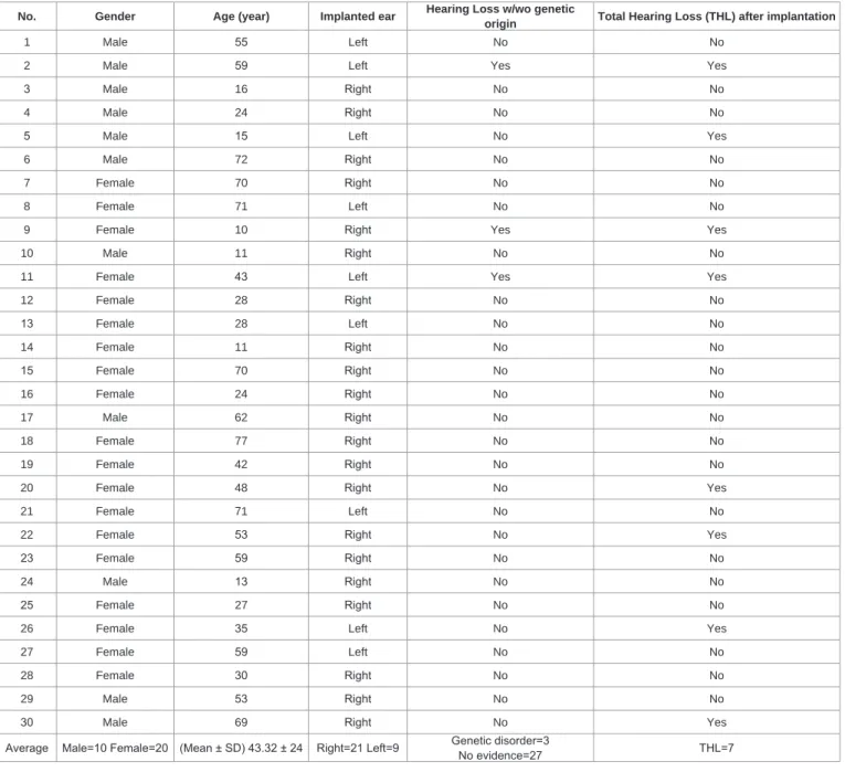
Out of the total number of cochlear implantees with slim perimodiolar implant system (n=94) our study population was recruited on the basis of the following criteria: (1) patient with good compliance; (2) measureable preoperative hearing threshold; (3) slim perimodiolar electrode array implant system; (4) minimum one-year follow-up period. Thirty consecutive subjects were enrolled into this prospective, non-randomized clinical study. Twenty females and ten males with mean age at implantation of 43.32 years, ranged between 10 years to 77 years. All subjects were implanted at the University of Szeged from 2015 until 2017. The postoperative follow-up duration lasted 1.72 years at average (ranged between 1.1 and 2.55 years). All
subjects met the official indication criteria of Cochlear Implantation (CI). Anatomical / structural malformation was not revealed by the preoperative radiological examinations. For detailed patient data, please see Table 1.
Implant configuration
The studied cochlear implant system has a slim, full-length perimodiolar electrode (Figure 1). The thin implant body has no pedestal and it is designed to minimize bone excavation and skin protrusion. At the implant coil the implant measures 3.7 mm and the implant main body measures 3.9 mm in thickness. The side-by-side symmetrical shape makes the implantation easier for the surgeon.
The titanium casing has been used for high impact resistance, and the smooth external geometry to minimize biofilm formation, that reduces the risk of infection. The 98 mm total length of electrode array helps to insert it in a better position, but the main handle assist tool is the reloadable sheath for the smooth electrode insertion. The thin electrode array allows unobstructed access to the scala tympani that has a tip diameter of 0.35 × 0.4 mm and 0.45 × 0.5 mm at the base. At the last edge of the electrode array there are three white marker rings for controlling the insertion depth that are followed by 22 half banded platinum electrode contacts. These properties make this implant configuration easier to use with shorter incision and surgery time. The insertion assistant sheath platform and the physical attributes of the electrode array facilitate to proximate the modiolus and thus prevent the electrode from dislocation into the scalae media or vestibuli [6-8].
Surgical technique
Preserving the residual hearing requires minimally invasive techniques of (1) cochlear fenestration, (2) management of endocochlear fluid compartments and (3) atraumatic electrode insertion, known as soft surgery. Thinner and atraumatic electrode arrays are also designed to accomplish these aims, as postoperative hearing performance can be maximized by minimizing the insertion trauma [3,8-11].
Several important factors contribute to intracochlear damage during implantation: (1) direct physical trauma, (2) pressure wave propagation in the perilymphatic fluid, (3) vibration and/
or heat trauma from drilling, (4) loss of perilymph, (5) changes
Figure 1: (A) A traumatic electrode insertion in optimal position with the reloadable sheath. (B) Slim, perimodiolar electrode configuration with total cochlear coverage.
Figure 2: Skull AP axial on the first postoperative day that confirms the proper in situ electrode position. The depicted subject (not included into the present study due to completely missing preoperative hearing) was chosen to interpret the differences between sequentially implanted systems (A:
right ear: CI512 Contour Advanced; B: left ear: CI532 Slim Perimodiolar).
A decreased electrode array curvature is seen with the slim perimodiolar system (B).
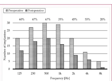
Figure 3: Number of implantees with measurable hearing threshold at different frequencies. Preoperative (striped pattern columns); postoperative (checked pattern columns). On the top horizontal axis, the frequency-specific success rate of hearing preservation is showed in percentages.
in homeosthasis/hydrodynamics of the endocochlear fluid compartments, (6) delayed fibrotic alteration and new bone formation within the cochlear lumen [3,12-16].
The physical attributes (length and diameter) of the electrode array may each limit the postoperatively achieved residual hearing [17].
Comprehensive analysis of imaging diagnostics of the middle and inner ear provide indispensable information for planning the proper surgical access route and electrode [17,18].
Soft surgery
The term soft surgery was introduced by Lehnhardt in 1993 and it provided basis for numerous publications [9,19].
Our routinely applied minimally invasive surgical technique involved electrode insertion via the Round Window (RW). In order to reduce bleeding and to prevent blood from accessing the cochlea, we filled the tympanic cavity with adrenaline solution after having the posterior tympanotomy been completed. To prevent bone fragments entering the cochlea, the tympanic and mastoid cavity were flushed with abundant amount of saline. To remove the bony overhang of the round window, we used a 1 mm diamond burr at low speed (max.
350 rpm) in order to avoid noise and heat injury. We opened the RW membrane with a microscopic needle or hook. After opening the inner ear, suction was applied with care in order to avoid reducing the amount of perilymph. Furthermore, the scala tympani was left
open for the shortest possible period, to prevent bone fragments, blood or other substances entering the inner ear, which might have been sources of primary and/or secondary injuries that finally would lead to loss of residual hearing. As a sort of prevention, after having opened the RW, we placed a piece of gel-foam soaked in corticosteroid solution into the RW niche.
The slim modiolar electrode of the CI532 implant was soaked into methylprednisolone solution (40 mg powder dissolved in 10 ml saline) and it was retracted into the insertion sheath. The insertion sheath together with the electrode array was inserted into the scala tympani with the lowest possible force. Any minute resistance felt by the surgeon would have indicated physical contact of the electrode array to the basilar membrane or the lateral wall of the scala tympani or stria vascularis and possible injury of these structures. After the electrode had been inserted in full length, indicated by the 1st marker ring, the RW was immediately sealed with an autologous tissue (e.g.
fascia or muscle) in order to prevent loss of perilymph [9].
Radiological validation
Radiography (skull AP axial/Towne view) was performed on the first postoperative day to confirm the proper intracochlear electrode position (Figure 2).
Pure-tone audiometry
Pure-tone air-conduction thresholds were used to register residual hearing with the ascending method, with 5 dBHL intensity steps. The audiometer (GSI 61 Clinical Audiometer; Grason- Stadler, MN USA) was calibrated according to the standards of the International Organization for Standardization (ISO 389-1:2017).
THD-50P (Telephonics Corporation/Griffon Company, NY USA) headphone was used for air conduction hearing measurements.
Results
Pre- and postoperative pure tone hearing threshold measurements were completed for all the 30 recruited subjects. Figure 3 frequency-dependently illustrates the number of patients pre- and postoperatively, where hearing sensitivity was measurable. It is well demonstrated that hearing is the most stable within the 250 to 1000 Hz range, and the least is beyond 4 kHz. This statement is true either pre-, or postoperatively.
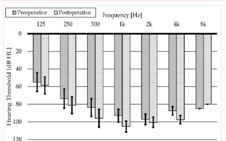
Figure 4: Preoperative (striped pattern columns) and postoperative (dotted pattern columns) hearing thresholds in dBHL at the measured frequencies (*p<0.05).
Figure 5: Loss of acoustic sensitivity interpreted in dBHL ranges, while exhibiting the number of implantees frequency-specifically (with different patterns of columns).
Figure 6: Preoperative threshold of HP (striped pattern columns, n=number of patients) and THL (squared pattern columns, m=number of patients) patients (Continuous line measureable threshold level).
The average preoperative thresholds of the hearing within the lower frequency range were 61.75 dBHL at 125 Hz (no response from 10 patients); 78.52 dBHL at 250 Hz (no response from 3 patients). At the middle frequency range, mean values were 88.67 dBHL at 500 Hz (response from all patients); 97.07 dBHL at 1 kHz (no response from 1 patient) and 100.50 dBHL at 2 kHz (no response from 10 patients).
At the higher frequencies, the average values were 91.36 dBHL at 4 kHz (no response from 19 patients) and 84.00 dBHL at 8 kHz (no response from 25 patients).
The difference in height between the striped and checked pattern columns represents the percentage of successful hearing preservation at specific frequencies.
One year postoperatively the average values of the hearing thresholds at the lower frequency range were: 93.89 dBHL at 125 Hz (no response from 17 patients); 87.86 dBHL at 250 Hz (no response from 10 patients). At the middle frequencies mean values were 102.86 dBHL at 500 Hz (no response from 10 patients); 111.61 dBHL at 1
kHz (no response from 14 patients) and 113.75 dBHL at 2 kHz (no response from 21 patients). At the higher frequencies, average values were 115.18 dBHL at 4 kHz (no response from 24 patients) and 99.29 dBHL at 8 kHz (no response from 29 patients).
Figure 4 illustrates the preoperative (striped pattern columns) and the postoperative (dotted pattern columns) hearing thresholds in dBHL at the measured frequencies. Decrease was detected at each examined frequencies but the grade of it varied. The highest decrease was measured at 500 Hz with an average decrease of 14.19 dBHL and at 1000 Hz with an average decrease of 13.77 dBHL. At the lower frequency range, hearing remained substantially stable. At 125 Hz only 3.06 dBHL, while at 250 Hz only 7.19 dBHL loss was detected. At the high frequencies, from 2 to 8 kHz preoperative hearing sensitivity had been already proved to be rather poor, thus further loss had just little consequences.
Figure 5 frequency-specifically demonstrates the degree of loss of acoustic sensitivity grouped into dBHL ranges, while exhibiting the
No. Gender Age (year) Implanted ear Hearing Loss w/wo genetic
origin Total Hearing Loss (THL) after implantation
1 Male 55 Left No No
2 Male 59 Left Yes Yes
3 Male 16 Right No No
4 Male 24 Right No No
5 Male 15 Left No Yes
6 Male 72 Right No No
7 Female 70 Right No No
8 Female 71 Left No No
9 Female 10 Right Yes Yes
10 Male 11 Right No No
11 Female 43 Left Yes Yes
12 Female 28 Right No No
13 Female 28 Left No No
14 Female 11 Right No No
15 Female 70 Right No No
16 Female 24 Right No No
17 Male 62 Right No No
18 Female 77 Right No No
19 Female 42 Right No No
20 Female 48 Right No Yes
21 Female 71 Left No No
22 Female 53 Right No Yes
23 Female 59 Right No No
24 Male 13 Right No No
25 Female 27 Right No No
26 Female 35 Left No Yes
27 Female 59 Left No No
28 Female 30 Right No No
29 Male 53 Right No No
30 Male 69 Right No Yes
Average Male=10 Female=20 (Mean ± SD) 43.32 ± 24 Right=21 Left=9 Genetic disorder=3
No evidence=27 THL=7
Table 1: Population of study patients.
number of implantees. It is clearly shown that only minute threshold decay with less than 5 dBHL loss is the most frequently found one, while prominent postoperative loss of hearing appears less often.
Subjects with complete loss of hearing following surgery Nine implantees (9/30=30%) showed up with total loss of residual hearing at every measured frequency following surgery.
Their preoperative hearing sensitivity is presented in Figure 6. It is clearly seen that within this subgroup of this cohort the measured average hearing threshold have been already poorer prior to surgery compared to those with preserved hearing. Genetic screening of the 30 recruited subjects revealed mutations in three cases in the background of hearing loss. All of these subjects suffered complete hearing loss postoperatively (3/3=100%), that genetic alteration may serve as a predictor when opting for an electro-acoustic/hybrid device, should be taken into consideration when indicating these systems [20].
Discussion
Preservation of acoustic hearing associated with cochlear implantation improves the postoperatively achievable periodicity and spectral resolution, which improves the patient's speech comprehension and the localization of the tone in particularly difficult conditions [21-26].
The effects of cochlear implantation on residual hearing have been discussed in several studies in which a number of surgical and technical factors have been identified [27]. There are some surgical techniques of approaching the scala tympani (i.e., RW, ERW, CS) with varying risks of harming the fine structures of the cochlea with prompt or delayed onset [13]. Such late complications, like the appearance of endocochlear connective tissue or new bone formation, may lead to a gradual partial or complete loss of residual acoustic hearing [28]. This is most likely to be seen when the round window is extendedly exposed, where endothelial lesions trigger new tissue proliferation. The slightest is the tendency to harm the endocochlear structures when minimally invasive, soft surgery is applied [13].
Physical attributes of the electrode profile may also interfere with postoperative cochlear function. Theoretically, the endocochlear hydrodynamics may also be altered, as the vibration of the basilar membrane is restricted due to the presence of an electrode array. At this point, as the travelling waves to the apical region are modified, the basilar membrane would react to sounds differently, leading to an endocochlear “conductive” hearing loss [29, 30].
The new type of thin-diameter electrode arrays close to the modiolus are expected to have a lower hydrodynamic load, since the bony spiral lamina is attached from below, thus the basilar membrane vibrations remain unrestricted. However, the perimodiolar position of the electrode array allows the adjacent nerve elements of the spiral ganglion to be stimulated with a lower electrical intensity and through a smaller surface.
Cadaver experiments demonstrated that a force, applied to the basilar membrane with an average of 88 mN (42 mN to 122 mN) would be sufficient to accomplish the interscalar dislocation of the electrode, of which manual perceptibility is questionable [31]. Studies with large case numbers (n=100) have shown that the probability of the electrode line being located in the scala vestibuli significantly increased during CS, which also manifested itself in the absence of improvement in speech comprehension [32].
In a number of studies, intraoperatively performed electro- cochleography is used to track the electrode insertional trauma, furthermore to postoperative residual hearing follow-ups [33-35].
For the implementation of Electro-Acoustic (EAS) or hybrid speech processors the long-term preservation of residual acoustic hearing is inherently inevitable, thus application of atraumatic surgical techniques and electrode arrays is essential.
Our study cohort obviously demonstrates that by the application of appropriate soft surgery techniques and atraumatic electrodes are able to retain residual hearing on a long run. The positive experience gained with the new type of CI532 Slim Modiolar electrode predicts the possibility for the preservation of structural and functional integrity of all cochlear regions. Furthermore, a prompt, definitive solution could be provided for a possible late hearing loss progression, where only a psychophysical reprogramming of the implant would be enough.
On the basis of our results, if the acoustic hearing loss can be preserved with the assurance and efficacy of the initial experience, we will be able to provide sustained prominent hearing rehabilitation even in the indication of EAS that results in significant improvement in the life quality of many implantees.
In addition, long-term residual hearing loss may be of crucial importance in the subsequent feasibility of regenerative procedures and medical treatments [3,7,36,37].
Conclusion
In cochlear implantation, the use of new electrode array profiles plays a fundamental role in minimally invasive soft surgery, taking into individual needs, and providing long-term acoustic hearing preservation. Our study demonstrates the efficacy of the Nucleus CI532 Slim Modiolar electrode profile and it has the potential for granting residual hearing, which predicts the possible use of this configuration as part of EAS systems and makes it available for future treatments, i.e., the regeneration-based new therapeutic approaches of intracochlear hair-cells.
References
1. Mistrík P, Jolly C. Optimal electrode length to match patient specific cochlear anatomy. Eur Ann Otorhinolaryngol Head Neck Dis.
2016;133:S68-71.
2. Jolly C, Garnham C, Mirzadeh H, Truy E, Martini A, Kiefer J, et al.
Electrode features for hearing preservation and drug delivery strategies.
Cochlear Implants Hearing Preservation. 2010;67:28-42.
3. Nagy R, Jarabin JA, Dimák B, Perényi A, Tóth F, Szuts V, et al. Possibilities for residual hearing preservation with Nucleus CI532 Slim Modiolar electrode array – case report. Orvosi Hetilap.
4. Lyu AR, Kim DH, Lee SH, Shin DS, Shin SA, Park YH. Effects of dexamethasone on intracochlear inflammation and residual hearing after cochleostomy: A comparison of administration routes. PLoS One.
2018;13(3):e0195230.
5. Zine A, van de Water TR. The MAPK/JNK signalling pathway offers potential therapeutic targets for the prevention of acquired deafness. Curr Drug Targets CNS Neurol Disord. 2004;3(4):325-32.
6. McJunkin JL, Durakovic N, Herzog J, Buchman CA. Early Outcomes With a Slim, Modiolar Cochlear Implant Electrode Array. Otol Neurotol.
2018;39(1):e28-e33.
7. Cuda D, Murri A. Cochlear implantation with the nucleus slim
modiolar electrode (CI532): a preliminary experience. Eur Arch Otorhinolaryngology. 2017;274(12):4141-8.
8. Ramos-Macías A, Borkoski-Barreiro SA, Falcón-González JC, Ramos-de Miguel A. Hearing Preservation with the Slim Modiolar Electrode Nucleus CI532® Cochlear Implant: A Preliminary Experience. Audiol Neurootol.
2017;22(6):317-25.
9. Friedland DR, Runge-Samuelson C. Soft Cochlear Implantation: Rationale for the Surgical Approach. Trends Amplif. 2009;13(2):124-38.
10. Cohen NL. Cochlear implant soft surgery: fact or fantasy? Otolaryngol Head Neck Surg. 1997;117(3):214-6.
11. Carlson ML, Driscoll CL, Gifford RH, Service GJ, Tombers NM, Hughes- Borst BJ, et al. Implications of minimizing trauma during conventional cochlear implantation. Otol Neurotol. 2011;32(6):962-8.
12. Adunka O, Unkelbach MH, Mack M, Hambek M, Gstoettner W, Kiefer J. Cochlear implantation via the round window membrane minimizes trauma to cochlear structures: A histologically controlled insertion study.
Acta Otolaryngol. 2004;124(7):807-12.
13. Richard C, Fayad JN, Doherty J, Linthicum FH. Round window versus cochleostomy technique in cochlear implantation: Histologic findings.
Otol Neurotol. 2012;33(7):1181-7.
14. Jiam NT, Limb CJ. The impact of round window vs cochleostomy surgical approaches on interscalar excursions in the cochlea: Preliminary results from a flat-panel computed tomography study. World J Otorhinolaryngol Head Neck Surg. 2016;2(3):142-7.
15. Khater A, El-Anwar MW. Methods of Hearing Preservation during Cochlear Implantation. Int Arch Otorhinolaryngol. 2017;21(3):297-301.
16. Koch RW, Ladak HM, Elfarnawany M, et al. Measuring Cochlear Duct Length - a historical analysis of methods and results. J Otolaryngol Head Neck Surg. 2017;46(1):19.
17. Karkas A, Champfleur NM, Uziel A, Mondain M, et al. Benefit of Preoperative Temporal Bone CT for Atraumatic Cochlear Implantation.
Otol Neurotol. 2018;39(3):e186-e194.
18. Perényi Á, Bella Zs, Baráth Z, Magyar P, Nagy K, Rovó L. [Role of cone- beam computed tomography in diagnostic otorhinolaryngological imaging]. Orv Hetil. 2016;157(2):52-8.
19. Lehnhardt E. [Intracochlear placement of cochlear implant electrodes in soft surgery technique]. HNO. 1993;41(7):356-9.
20. Szüts V, Jarabin JA, Nagy N, Ötvös F, Nagy R, Nagy A, et al. Altered potassium ion homeostasis in hearing loss. In: Shad Kaneez F, editor. Ion Channel. London: IntechOpen.
21. Dunn CC, Perreau A, Gantz B, Tyler RS. Benefits of Localization and Speech Perception with Multiple Noise Sources in Listeners with a Short- Electrode Cochlear Implant. J Am Acad Audiol. 2010;21(1):44-51.
22. Gifford RH, Dorman MF, Skarzynski H, Lorens A, Polak M, Driscoll CL, et al. Cochlear implantation with hearing preservation yields significant benefit for speech recognition in complex listening environments. Ear Hear. 2013;34(4):413-25.
23. Gifford RH, Driscoll CLW, Davis TJ, Fiebig P, Micco A, Dorman MF.
A Within-Subject Comparison of Bimodal Hearing, Bilateral Cochlear Implantation, and Bilateral Cochlear Implantation with Bilateral Hearing Preservation: High-Performing Patients. Otol Neurotol. 2015;36(8):1331- 7.
24. Gifford RH, Grantham DW, Sheffield SW, Davis TJ, Dwyer R, Dorman MF.
Localization and interaural time difference (ITD) thresholds for cochlear implant recipients with preserved acoustic hearing in the implanted ear.
Hear Res. 2014;312:28-37.
25. Loiselle LH, Dorman MF, Yost WA, Cook SJ, Gifford RH. Using ILD or ITD Cues for Sound Source Localization and Speech Understanding in a Complex Listening Environment by Listeners With Bilateral and With Hearing-Preservation Cochlear Implants. J Speech Lang Hear Res.
2016;59(4):810-8.
26. Loiselle LH, Dorman MF, Yost WA, Gifford RH. Sound source localization by hearing preservation patients with and without symmetric, low- frequency acoustic hearing. Audiol Neurootol. 2015;20(3):167-71.
27. Sweeney AD, Hunter JB, Carlson ML, Rivas A, Bennett ML, Gifford RH, et al. Durability of Hearing Preservation after Cochlear Implantation with Conventional-Length Electrodes and Scala Tympani Insertion.
Otolaryngol Head Neck Surg. 2016;154(5):907-13.
28. Fayad JN, Makarem AO, Linthicum FH. Histopathologic assessment of fibrosis and new bone formation in implanted human temporal bones using 3D reconstruction. Otolaryngol Head Neck Surg. 2009;141(2):247- 52.
29. Banakis Hartl RM, Mattingly JK, Greene NT, Jenkins HA, Cass SP, Tollin DJ. A preliminary investigation of the air-bone gap: Changes in intracochlear sound pressure with air- and bone-conducted stimuli after cochlear implantation. Otol Neurotol. 2016;37(9):1291-9.
30. Chole RA, Hullar TE, Potts LG. Conductive Component After Cochlear Implantation in Patients With Residual Hearing Conservation. Am J Audiol. 2014;23(4):359-64.
31. Schuster D, Kratchman LB, Labadie RF. Characterization of intracochlear rupture forces in fresh human cadaveric cochleae. Otol Neurotol.
2015;36(4):657-61.
32. Wanna GB, Noble JH, Carlson ML, Gifford RH, Dietrich MS, Haynes DS, et al. Impact of electrode design and surgical approach on scalar location and cochlear implant outcomes. Laryngoscope. 2014;124:S1-7.
33. O’Connell BP, Hunter JB, Wanna GB. The importance of electrode location in cochlear implantation. Laryngoscope Investig Otolaryngol.
2016;1(6):169-74.
34. Koka K, Litvak LM. Feasibility of using electrocochleography for objective estimation of electro-acoustic interactions in cochlear implant recipients with residual hearing. Front Neurosci. 2017;11:337.
35. Dalbert A, Pfiffner F, Hoesli M, Koka K, Veraguth D, Roosli C, et al.
Assessment of cochlear function during cochlear implantation by extra- and intracochlear electrocochleography. Front Neurosci. 2018;12:18.
36. Plontke SK, Götze G, Rahne T, Liebau A. Intracochlear drug delivery in combination with cochlear implants: Current aspects. HNO. 2017;65:19- 28.
37. Mittal R, Nguyen D, Patel AP, Debs LH, Mittal J, Yan D, et al. Recent Advancements in the Regeneration of Auditory Hair Cells and Hearing Restoration. Front Mol Neurosci. 2017;10:236.