Association between the C(-1019)G functional polymorphism of the HTR1A gene and impulsivity and subthreshold
depression
Doctoral dissertation
ANITA BENKŐ
Semmelweis University, Ph.D. School of Mental Health Sciences
Supervisor: György Bagdy, Ph.D., D.sc.
László Tóthfalusi, Ph.D.
Director of Comprehensive exam: Mária Kopp, Ph.D., D.sc.
Memberes of Comprehensive exam: Zsuzsanna Mirnics, Ph.D.
György Purebl, Ph.D.
Official reviewers: Róbert Bódizs, Ph.D.
Viktor Vörös, Ph.D.
Budapest
2011
2
TABLE OF CONTENT
1. ABBREVIATIONS 5
2. INTRODUCTION 8
2.1 Neurobiology of impulsivity 10
2.1.1 Neuroanatomy of impulsivity 10
2.1.2 Neurochemical background of impulsivity 12
2.1.2.1 5-HT1A receptor 12
2.1.2.2 Serotonergic system 14
2.1.2.3 Dopaminergic system 16
2.1.2.4 Noradrenergic system 17
2.1.2.5 Other neurotransmitter systems in the regulation of impulsivity 17
2.1.3 Genetic background of impulsivity 18
2.2 Impulsivity in psychiatry 19
2.3 Neurobiology of depression 25
2.3.1 Neuroanatomy of depression 25
2.3.2 Neurochemistry of depression 27
2.3.3 Neurotrophic factors associated with depression 29
2.3.4 Neuroendocrine system 29
2.3.5 Genetics of depression 30
2.4 Clinical and subclinical manifestations of depression 32
2.4.1 Major depression 32
2.4.2 Subthreshold depression 35
3. OBJECTIVES 38
4. MATERIALS AND METHODS 39
4.1 Subjects 39
4.2 Procedures and Measures 39
4.2.1 Background information 39
4.2.2 IVE-I and BIS-11 40
4.2.3 Zung Self-Rating Depression Scale 40
4.2 Genotyping 41
4.3 Statistical analysis 41
4.3.1. Genetic association analysis 41
3
4.3.2. Structural Equation Modeling 42
5. RESULTS 47
5.1 The association of IVE-I and BIS-11 Scales with genotypes 48 5.2 The association of the Zung Self-rating Depression Scale scores
with genotype 50
5.3 The association between impulsivity and subthreshold depression and the effect of genotype on the relationship between impulsivity and
subthreshold depression 51
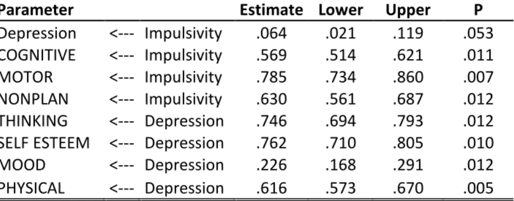
5.3.1 Model for the whole study cohort: association between the BIS-11
and the ZSDS 53
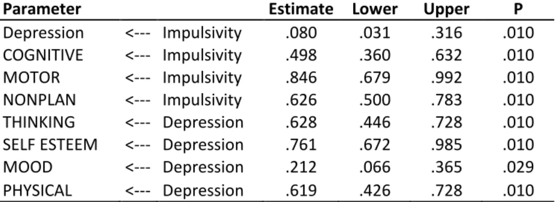
5.3.2 SEM statistics for the CC group 56
5.3.3 SEM statistics for the GG group 59
5.3.4 SEM statistics for the GC group 61
6. DISCUSSION 63
6.1 The C(-1019)G functional polymorphism of the HTR1A gene and
impulsivity 63
6.2 The C(-1019)G functional polymorphism of the HTR1A gene and
subthreshold depression 66
6.3 The latent relationship between impulsivity and subthreshold
depression, and the effect of genotype on this relationship 68
7. CONCLUSIONS 71
8. SUMMARY 73
8.1 Summary 73
8.2 Összefoglalás 74
9. REFERENCES 76
10. PUBLICATIONS 108
10.1 Publications relevant to the dissertation 108
10.2 Other publications 108
10.3 Book chapters 109
11. ACKNOWLEDGEMENT 111
12. APPENDIX 112
12.1 Background information 113
12.2 IVE-I Scale 116
4
12.3 BIS-11 Scale 117
12.3 Zung Self-rating Depression Scale 118
5
1. ABBREVIATIONS
5CSRTT = 5-choice serial reaction time task 5-HT = serotonin
5HTTLPR = serotonin transporter length polymorphic region AcbC = nucleus accumbens core
ACC = anterior cingulate cortex ACh = acetylcholine
ACTH = adrenocorticotropic hormone
ADHD = attention deficit hyperactivity disorder ANOVA = analysis of variance
APA = American Psychiatric Association BDNF = brain-derived neurotrophic factor BIS-11 = Barratt Impulsiveness Scale BLA = basolateral amygdala
CB1 = cannabinoid receptor 1 CI = confidence interval C–I = compulsive–impulsive CNS = central nervous system
COMT = catechol-O-methyl transferase
CPP = 3-(2-Carboxypiperazin-4-yl)propyl-1-phosphonic acid DAT = dopamine transporter
DEAF-1 = deformed epidermal autoregulatory factor-1 DF = degree of freedom
DNA = deoxyribonucleic acid
DSM-IV = Diagnostic and Statistical Manual of Mental Disorders version IV DLPFC = dorsolateral prefrontal cortex
DRD4 = dopamine receptor 4 GLS = generalized least squares
6 HAB = habenula
Hes5 = hairy and enhancer of split 5 HPC = hippocampus
ICDs = impulse control disorders
ICD-NOS = impulse control disorders not otherwise specified IL = infralimbic cortex
IRS = Impulsivity Rating Scale
IVE-I = Impulsiveness subscale of the Eysenck Impulsiveness, Venturesomeness, and Empathy scale
KO = knock out
LOPFC = lateral orbital prefrontal cortex MAO = monoamine oxidase
MAOA = monoamine oxidase A
MAOIs = monoamine oxidase inhibitors MD = major depression
MDMA = 3,4-methylenedioxy-methamphetamine MinD = minor depression
ML = maximum likelihood MS = medial striatum
NAC = nucleus accumbens core region;
NAS = nucleus accumbens shell region
NEO-PI-R = Neuroticism-Extroversion-Openness Personality Inventory - revised NMDA = N-Methyl-D-aspartate
NS = Novelty Seeking
NUDR = nuclear deformed epidermal autoregulatory factor OCD = obsessive–compulsive disorder
OCSD = obsessive–compulsive spectrum disorders OFC = orbitofrontal cortex
PET = positron emission tomography
7 PFC = prefrontal cortex
PL = prelimbic cortex SE = standard error
SEM = structural equation modelling SERT = serotonin transporter
SD = standard deviation
SNP = single nucleotid polymorphism
SSD = subsyndromal symptomatic depression sgACC = subgenual anterior cingulate cortex SSRIs = selective serotonin reuptake inhibitors STAXI = State Trait Anger Expression Inventory STN = subthalamic nucleus
TCI = Temperament and Character Inventory TCAs = tricyclic antidepressants
TPH = tryptophan hydroxylase TPH1 = tryptophan hydroxylase TPH2 = tryptophan hydroxylase
TPQ = Tridimentional Personality Questionnaire VNTR = variable number tandem repeats
WIN552122 = [(R)-(+)-[2,3-dihydro-5-methyl-3[(4-morpholinyl)methyl]pyrrolo[1,2,3- de]-1,4 benzoxazinyl]-(1-naphthalenyl)methanone mesylate salt]
ZSDS = Zung Self-Rating Depression Scale
8
2. INTRODUCTION
Serotonin-1A (5-HT1A) receptors are known to play a role in impulsivity-related behaviour. The C(-1019)G functional polymorphism (rs6295) has been suggested to regulate the 5-HT1A receptor gene (HTR1A) expression in presynaptic raphe neurons, namely, increased receptor concentration and reduced neuronal firing could be associated with the G allele. Previous studies indicate that this polymorphism is associated with aggression, suicide, and several psychiatric disorders, yet its association with impulsivity has rarely been investigated. Furthermore, studies reported that dysfunction of the serotonin 1A receptor may play a role in the background of depression. Some authors suggested a direct relationship between depression and impulsivity. It was noted that there are phenotypic associations occurring in childhood and adolescence between constructs relating to impulsivity and depression and impulsivity shares similar biological background, both are have been shown to be related to decreased serotonin levels. Despite these associations, it remains unclear whether impulsivity is a predictor of the onset of depression in adulthood. Prior research on this issue has typically focused on the relationship between impulsivity and suicide in clinical populations or in children or adolescents with attention deficit hyperactivity disorder (ADHD) or conduct disorders. There have been only a few studies addressing the question of associations between impulsivity and depressive symptoms in subclinical depressive or non-clinical populations.
Our study is part of a 5-year research project called NewMood, which is short for New Molecules in Mood Disorders. It is a Europe-wide research study, a collaboration between 13 research groups in 10 EU countries. Research groups aimed to investigate the possible genetic and phenotypic background of mood disorders and also, to discover new mechanisms of effective drug treatment leading to the development of novel antidepressant therapies in animal models and human studies. Our human study group consisting of pharmacists, psychologists, physicians and biologists, collected data using paper questionnaires in collaboration with the group of Manchester, developing a questionnaire booklet together.
9
In the present study, as part of the NewMood research, we investigated the relationship between impulsivity and depression and the C(-1019)G polymorphism of the HTR1A in a non-clinical population sample of 725 volunteers using the Impulsiveness subscale (IVE-I) of the Eysenck Impulsiveness, Venturesomeness, and Empathy scale, the Barratt Impulsiveness Scale (BIS-11) and the Zung Self-Rating Depression Scale (ZSDS). We studied the association between impulsivity and ZSDS scores below the level indicating major depression and the affect of the C(-1019)G polymorphism on this relationship using the structural equation model.
The biological and psychiatric background of impulsivity and depression is discussed in the following chapters.
10 2.1 Neurobiology of impulsivity
2.1.1 Neuroanatomy of impulsivity
Impulsivity can be characterized as a personality trait, and it is an immediate response to thoughts or deeds without any consideration of the appropriateness or consequences. A growing body of data has indicated that impulsivity is heterogeneous:
it consists of several distinct behavioural phenomena that are dissociable at the neuroanatomical as well as neuropharmacological levels (1).
Consistent with human imaging and brain damage data (2), prefrontal cortical, striatal and limbic brain regions have been found to play an important role in impulsivity in rodents.
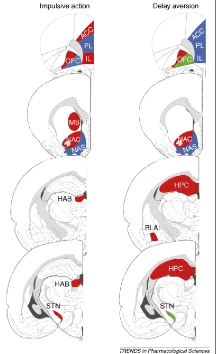
Functional differences have been reported between subregions of the nucleus accumbens in modulating impulsivity, suggesting the involvement of the core and not shell portion of the nucleus accumbens in impulsive action (3). It is innervated by dopamine neurons that respond to errors in reward prediction in a manner appropriate for a teaching signal (4). Causal experimental studies have shown that lesions of the nucleus accumbens core (AcbC) produce impulsive choice, reducing rats‘ preference for large/delayed rewards, compared to small/immediate rewards (5), and AcbC lesions have also been found to impair performance on a task requiring rats to choose between an uncertain immediate reward and a certain delayed reward (3). Figure 1. shows the neuroanatomical regions in the brain involved in impulsive action, in inhibitory control processes and delay aversion.
11
Figure 1. The neuroanatomical regions in the brain involved in impulsive action, in inhibitory control processes and delay aversion. Lesion studies demonstrated that there is considerable overlap in brain areas, including cortical and limbic regions, that modulate impulsive action and delay aversion in rodents. Red indicates that lesions of these regions increase impulsive action or delay aversion. Green indicates beneficial effects of lesions on impulsivity and in particular more self- controlled choice. Blue indicates that lesions of these brain regions did not affect impulsive action nor delay aversion; dark grey/black areas indicate ventricles in the brain, and light grey areas indicate fibre tracts in the brain. ACC, anterior cingulate cortex; BLA, basolateral amygdala; HAB, habenula; HPC, hippocampus; IL, infralimbic cortex; MS, medial striatum; NAC, nucleus accumbens core region; NAS, nucleus accumbens shell region; OFC, orbitofrontal cortex; PL, prelimbic cortex; STN, subthalamic nucleus. (6)
12
The orbitofrontal cortex (OFC) is a region of the prefrontal cortex (PFC) that projects to the AcbC and is strongly implicated in the assessment of reward value.
Choice between small, likely rewards and large, unlikely rewards increases cerebral blood flow in orbital and inferior PFC (7), with lesioned subjects deciding slowly and failing to choose the optimal, most likely outcome (8).
A role for limbic regions such as the habenula and hippocampus in impulsive action (9) has been more firmly established. The precise mechanisms by which these limbic structures affect impulsivity are not completely understood, although both regions project to striatal brain areas including the nucleus accumbens. The subthalamic nucleus (STN) is a component of the basal ganglia that receives projections both from the globus pallidus (pallidum) and the cerebral cortex (10). Lesions of the STN decreased impulsive choice in a task (11), a task in which OFC lesions had the same effect (12). There is good evidence that the hippocampus contributes to the representation of context and contextual conditioning is important in learning with delays. Lesions of the hippocampal formation have been shown to impair Pavlovian conditioning to a contextual conditioned stimulus in rats (13).
2.1.2 Neurochemical background of impulsivity 2.1.2.1 5-HT1A receptor
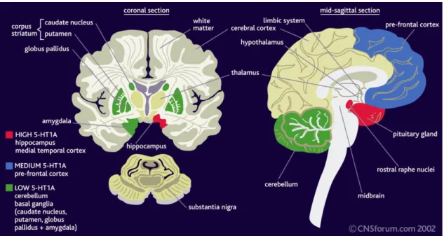
The 5-HT1A receptor consists of a protein of 422 amino acids and it contains seven putative transmembrane domains and belongs to the G-protein-coupled receptor (GPCR) family (14). The intronless gene encoding the 5-HT1A receptor is located on the human chromosome 5 at the locus 5q11.2-q13 (15). Studies show that the 5-HT1A
binding sites are especially abundant in the hippocampus, the lateral septum, the frontal and entorhinal cortex and the anterial raphe nuclei. Besides, the amygdala, the dorsal horn of the spinal cord, some thalamic and hypothalamic nuclei and the lateral part of the caude-putamen also express 5-HT1A receptors in a lower level (Figure 2.).
13
However, 5-HT1A receptors hardly or not at all can be found in the cerebellum, the substantia nigra and in the globus pallidus (16-18).
Figure 2. Distribution of 5-HT1A receptors.
5-HT1A receptors are localized both on serotonergic neurons where they act as somato-dendritic autoreceptors and on the targets of serotonergic projections where they correspond to postsynaptic receptors (17), hence, different effects can be expected from their stimulation in the raphe versus other regions. Indeed, by acting at somato-dendritic autoreceptors, 5-HT agonists inhibit the electrical activity of serotonergic neurons reducing serotonergic neurotransmission. In contrast, at postsynaptic targets, 5-HT1A agonists reproduce the effect of 5-HT which is released from serotonergic terminals therefore enhance 5-HT1A receptor-dependent serotonergic neurotransmission (19).
The HTR1A gene is located on the long arm of chromosome 5 (5q11.2-13) (15).
The functional C(-1019)G polymorphism (also labeled as rs6295) is a common single nucleotid polymorphism (SNP) in the promoter region of the gene (20). The polymorphism is located within a 26 bp palindromic region, which binds the nuclear DEAF-1-related (NUDR) protein and Hes5; the G allele abolishes repression by NUDR, resulting in higher expression of HTR1A enhancing the negative feedback inhibition of serotonergic raphe neurons exerted by HTR1A autoreceptors and leading to lower serotonergic neurotransmission (21).
14
Previous studies indicate that the C(-1019)G polymorphism of the HTR1A gene is associated with several psychiatric disorders including major depression (21) and anxiety disorders such as panic disorder with agoraphobia (22). It has also been found to be associated with suicide: Lemonde et al. (21) found an association between the G allele and completed suicide in an isolated population of French-Canadian origin and results from Sawiniec et al. (23) also indicated a significant role of the C(-1019)G polymorphism in the risk of suicide attempt. However, in the study of Serretti et al.
(24), haplotype analysis in relation to suicidal behaviour did not reveal any significant association, although suicidal attempter females homozygous for the G allele scored significantly higher on the STAXI state anger scale. Studies investigating the possible association with suicide focused on both attempted and completed suicide, however suicidal behaviour is a more complex phenomenon. Besides its association with the serotonergic system (25, 26), suicide has also been linked to aggression and impulsivity (27); attempted suicides are related to impulsivity (28), while completed suicides to aggressiveness (29). Previously, both impulsivity and aggressiveness have also been associated with the serotonergic system (for a review, see Lee and Coccaro, 2001(30)).
Several studies have investigated a potential association between the C(-1019)G polymorphism and various personality traits; using the revised five-factor Personality Inventory (NEO-PI-R) and the Tridimensional Personality Questionnaire (TPQ), higher scores for Neuroticism and Harm Avoidance were found in carriers of the G allele compared with C allele carriers (31), although other studies did not find any significant association between neuroticism and this particular SNP (32, 33). Serretti et al. (34) failed to find any association between three SNPs in the HTR1A gene or six SNPs in the HTR2C gene and personality dimensions, measured by Cloninger's Temperament and Character Inventory (TCI).
2.1.2.2 Serotonergic system
The serotonergic system plays an important role in various physiological functions (35, 36), psychiatric disorders (e.g., anxiety disorders, depression, and
15
schizophrenia) (37, 38), and regulates complex functions related to cognition and emotions (39). At present, there are 14 different known serotonin receptors divided into 7 classes (36).
Serotonin (5-hydroxytryptamine, 5-HT) is among the oldest known biogenic amines (40-43). Serotonin is synthesised from the essential amino acid tryptophan, which is hydroxylated to 5-hydroxytryptophan (5-HTP) by tryptophan hydroxylase, and in a second step 5-HTP is decarboxylated to form 5-HT (44). The negative feedback created by stimulation of the 5-HT autoreceptor decreases further release of serotonin, while the serotonin transporter (SERT) removes serotonin from the synaptic cleft.
SERT has been identified in the central nervous system (CNS), gastrointestinal tract, pulmonary and peripheral vasculature, and platelets (45).
Within the CNS, serotonin is synthesized and stored in presynaptic neurons.
Serotonin is located in groups of cell bodies isolated to the pons and midbrain (46). The raphe nuclei represent the major nuclei with both ascending serotonergic fibers projecting to the forebrain and descending fibers that extend to the medulla and spinal cord. Furthermore, from the median raphe nuclei axons project to the limbic system, the hippocampus and the septum, and axons from the dorsal raphe nuclei provide innervation of the striatum and the thalamus (46). Serotonin synthesis outside the CNS is limited to enterochromaffin cells and to a lesser extent platelets, however, platelets are a major storage site for serotonin outside the CNS. About 90–95% of the body‘s serotonin is located in the periphery, mostly stored in platelets and enterochromaffin cells (47).
The serotonergic system plays an important role in various functions such as appetite (48), thermoregulation (49), sexual function (50), neuroendocrine regulation (51), motor activity (52), pain (53), memory and learning (54), sleep-wake cycle (55), aggression (56), mood (57), impulsivity (58), anxiety (59) and regulates complex functions related to cognition and emotions (39).
Previous animal and human research showed that serotonin is implicated in impulsivity (60, 61). Its interaction with other neurotransmitters, including dopamine, noradrenaline and glutamate, has been investigated as well.
Low brain serotonin level has been associated with increased impulsive choice in animals (62) and in humans (63), but contradictory findings have also been described
16
(64, 65). The role of 5-HT receptors, mainly 5-HT1 and 5-HT2 receptors, have been well studied in the regulation of impulsivity. These receptors can be located presynaptically on 5-HT neurons, where their activation inhibits the release of 5-HT. Thus, treatment with 5-HT1A agonists would result in decreased 5-HT efflux (66). Selective 5-HT1B
receptor agonists and antagonists have been ineffective in altering impulsivity (67) while antagonists of the 5-HT2A receptor have been shown to reduce impulsivity (58, 68).
In brain areas where postsynaptic 5-HT1A receptors are located, such as the amygdala and frontal cortex, the density of 5-HT1A receptors was found to be decreased in aggressive rats (69).
2.1.2.3 Dopaminergic system
The role of dopamine neurotransmission in impulsivity is quite well established.
Psychostimulant drugs, such as amphetamine and methylphenidate has been successful in the treatment of attention deficit hyperactivity disorder (ADHD) (70), however psychostimulant drugs do not reduce all forms of impulsivity giving a showing its heterogeneous nature. Amphetamine has been found to enhance impulsive action in the 5-choice serial reaction time task (5CSRTT, more about the task see Robbins 2002 (71)), via the nucleus accumbens, because 6-hydroxydopamine lesions of the nucleus accumbens prevent the effect of amphetamine (72). In most studies, treatment with amphetamine has been found to reduce delay aversion (more about the task see Cardinal 2006 (73) in humans and rodents (74, 75), in contrast to the effects of amphetamine on impulsive action in the 5CSRTT, it is the orbitofrontal cortex that might play an important modulatory role in delay aversion (76, 77).
17 2.1.2.4 Noradrenergic system
Noradrenaline neurotransmission also plays a role in impulsive action.
Enhancing noradrenaline signaling decreases impulsive action, in the 5CSRTT and in the stop signal tasks, as well as in delay aversion via different mechanisms. In the 5CSRTT, this effect might be mediated through α1 or α2 adrenoceptors, which have been shown to be involved in impulsive action in this test (78). The α2 adrenoceptor agonist clonidine, which decreases noradrenaline release by stimulating presynaptic α2 autoreceptors, increased delay aversion (75). Furthermore, the effects of amphetamine in the stop signal task are mediated by increased noradrenaline neurotransmission (79, 80).
2.1.2.5 Other neurotransmitter systems in the regulation of impulsivity
The nonselective N-Methyl-D-aspartate (NMDA) receptor antagonists 3-(2- Carboxypiperazin-4-yl)propyl-1-phosphonic acid (CPP) have been reported to increase impulsive action in the 5CSRTT (81) as well as delay aversion (82) suggesting the role of the glutamatergic system in impulsivity. Altered glutamate transmission in the medial prefrontal cortex has been associated with impulsive action as demonstrated by CPP infusions into these regions (83).
The cannabinoid system and particularly cannabinoid receptor 1 (CB1) receptors have been implicated in impulsivity. In a preclinical study the CB1 agonist WIN552122 [(R)-(+)-[2,3-dihydro-5-methyl-3[(4-morpholinyl)methyl]pyrrolo[1,2,3-de]-1,4
benzoxazinyl]-(1-naphthalenyl)methanone mesylate salt] was found to impair response inhibition in the stop signal task, whereas the CB1 antagonist rimonabant reduced impulsive action in the 5CSRTT but did not change delay aversion (84).
18 2.1.3 Genetic background of impulsivity
The genetic background of impulsivity has long been investigated. Paaver et al.
(85) reported that subjects with low platelet monoamine oxidase (MAO) activity carrying the s allele of the serotonin transporter length polymorphic region (5HTTLPR) showed a higher mean score of self-reported impulsivity as measured by BIS-11.
Knockout mice lacking monoamine oxidase A (MAOA) differed from wild types by increased aggression (69). Of the 14 different known subtypes of serotonin receptors, the 5-HT1A and 5-HT1B receptor genes have been investigated more thoroughly.
Enhanced aggression was revealed in 5-HT1B receptor knockout mice (86). At the same time 5-HT1A receptor knockout mice showed anxiety-related behaviour (87) rather than enhanced aggression (88). In contrast, low aggression and high social anxiety parallel with rapid desensitization of 5-HT1A receptors during chronic selective serotonin reuptake inhibitor (SSRI) treatment were found in Fawn- Hooded rats compared to other rat strains (89, 90).
Nomura et al. (91) investigated the relationship between a 5-HT2A receptor gene polymorphism and impulsive behaviour in healthy subjects by using a behavioural task (go/no-go task) and found that the A-1438A polymorphism of this gene is possibly involved in impulsive behaviour.
There are certain gene polymorphisms within the dopaminergic system that may influence impulsivity including polymorphisms of the genes coding for the D4 dopamine receptor (DRD4), the dopamine transporter (DAT), and the catechol-o- methyltransferase enzyme (COMT).
The D4 receptor is expressed in the cerebral cortex, amygdala, hypothalamus, hippocampus, pituitary, and basal ganglia (92). The D4 receptor gene expression in the human brain is most abundant in the prefrontal cortex and low in the striatum (93, 94).
The gene coding the D4 receptor contains a variable number tandem repeats (VNTR) polymorphism that comes in a number of variants, ranging from two to ten repeats, (92).
19
Positive association has been reported between novelty seeking and the presence of the 7-repeat allele (95, 96), however contradictory results can be found too due to methodological differences, small samples and selection of trait questionnaires (97).
The DRD4 polymorphism is also associated with ADHD which is characterized by high impulsivity; Faraone et al. found a small but significant association between the 7- repeat allele of the DRD4 and ADHD (98).
The DAT is a protein that plays a role in dopaminergic neurotransmission and it is responsible for removing dopamine from the extracellular space (99). The DAT contains a VNTR polymorphism, resulting in variants that range from 3- to 13-repeats (99). The striatum plays a critical role in impulsivity, such as behavioural inhibition (100, 101). Excessive amounts of the DAT could lead to an overly efficient reuptake of dopamine, reducing extracellular dopamine level. Therefore, studies suggest an increase in expression of the DAT in individuals with the 10-repeat variant, and that the 10- repeat variant can be associated with impaired inhibitory control (102).
The role of COMT in regulating dopamine in the frontal cortex is important, because the frontal cortex lacks the DAT (103), leaving dopaminergic disposal to COMT. The COMT gene contains a functional SNP, which results in the substitution of the amino acid methionine (met) for valine (val) of the val enzyme (104). The met variant (associated with low enzymatic activity) results in high levels of extrasynaptic dopamine, whereas the val variant (associated with high enzymatic activity) results in low levels of extrasynaptic dopamine (103-105). The COMT polymorphism has been associated with with novelty seeking (106, 107), with aggressive behaviour (108-111), and with ADHD (112, 113).
2.2 Impulsivity in psychiatry
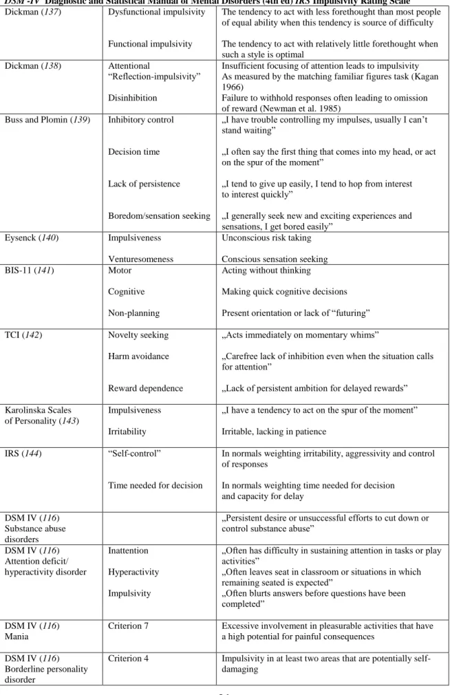
Impulsivity is a heterogeneous behavioural phenomenon that has various definitions (Table 1), the main element being that impulsivity is a human behaviour without adequate thought; the tendency to act without taking into consideration the
20
consequences of action, or a general predisposition toward rapid, unplanned reactions to internal or external stimuli without regard to the possible negative consequences of these reactions (1).
In everyday life we all engage from time to time in impulsive acts, such as saying out critical comments without thinking, or buying expensive items on the spur of the moment. It plays a role in normal behaviour, as well as, in a pathological form of behaviour. Although impulsivity can be present in any individual with or without a DSM-IV axis I or II diagnosis, it is more likely to be present in individuals with certain psychiatric disorders. The association between these disorders and impulsivity is at least partly due to the manner in which these disorders have been conceptualized, with a lack of behavioural inhibition being an element of all of these disorders. Impulsivity may be related to an underlying mechanism of behavioural inhibition (114).
In the Diagnostic and Statistical Manual of Mental Disorders version IV (DSM- IV) several neuropsychiatric disorders are either classified as impulse control conditions or encompass impulsive symptoms in the diagnostic criteria. These extreme pathological manifestations of impulsivity impair quality of life and everyday functioning, and as such represent important targets for treatment intervention (115).
Impulse control disorders, characterized by an impaired ability to resist impulses to engage in self-destructive behaviour, have been categorized in DSM-IV. These disorders include pathological gambling (persistent and maladaptive pattern of gambling), kleptomania (involves the failure to resist impulses to steal things), intermitted explosive disorder (frequent and often unpredictable episodes of extreme anger or physical outbursts), trichotillomania (recurrent pulling out of one‘s own hair which results in significant hair loss), pyromania (involves deliberate and purposeful fire setting on at least two occasions) and impulse control disorder not otherwise specified (116). Researchers have suggested that the impulse control disorders (ICDs) might be a part of an obsessive–compulsive spectrum based on their clinical characteristics, familial transmission, and response to pharmacological and psychosocial treatment (117, 118). Two important changes are considered in DSM-V: separating obsessive–compulsive disorder (OCD) from the anxiety disorders and placing it in an autonomous category—the obsessive–compulsive spectrum disorders (OCSD); and
21
creating several new autonomous disorders from those currently belong to ICD‘s not otherwise specified (ICD-NOS) (116) including four new impulsive disorders, namely compulsive–impulsive (C–I) internet usage disorder, C–I sexual behaviours, C–I skin picking and C–I shopping. They are called compulsive–impulsive disorders due to their impulsive features and the compulsive drive that causes the behaviours to persist over time.
Impulse control disorders are treated with medication, psychotherapy, behaviour modification, anger and stress management. If these disorders are occurring in conjunction with another condition, such as ADHD, medication and therapy for that condition often helps alleviate the impulse control disorder. Depression and anxiety are often underlying factor in some impulse control disorders, therefore, treatment with antidepressants and anxiolytics may be helpful. Long-term counseling, support groups is usually necessary as well (119).
Impulsivity is one of the DSM-IV diagnostic criteria for borderline personality disorder as are affective instability and identity disturbance (120). The study of Dougherty et al. (121) supported a higher level of impulsivity in patients with borderline personality disorder using questionnaire and laboratory measures. Several studies have found a relationship between suicidality and impulsivity in patients with borderline personality disorder. In the study of Soloff et al. (122), patients with borderline personality disorder were compared to patients with major depression alone on measures of depressed mood, hopelessness, impulsive aggression, and suicidal behaviour. A higher level of impulsive aggression or hopelessness or a diagnosis of borderline personality disorder predicted a greater number of suicide attempts.
Similarly, in a previous study by Soloff and colleagues (123), borderline personality disorder patients with a history of suicide attempts had more impulsive actions, antisocial personality disorder comorbidity, and depression than those without a history of suicide attempts.
Psychotherapy is necessary for individuals with borderline personality disorder.
The treatment framework includes discussion and clarification of the goals of treatment and the expected roles of the patient and therapist in achieving these goals.
Treatment goals such as symptom reduction, improved relationships, and ability to
22
maintain constancy at work should be made explicit. Psychodynamic therapy, dialectical behaviour therapy, and psychoeducational approaches are all helpful in working with patients with borderline personality disorder. Common to all successful therapies is the need for a strong therapeutic alliance (124).
Studies measuring impulsivity in substance-dependent individuals have also supported a link between impulsivity and substance abuse. Most studies that use questionnaire measures of impulsivity find higher levels of impulsivity in substance- dependent individuals than in healthy comparison subjects (125, 126). Medication behavioural therapy and support groups especially when combined, are important elements of an overall therapeutic process that often begins with detoxification, followed by treatment and relapse prevention (127).
Studies that have used human behavioural laboratory tasks to measure impulsivity have found high levels of impulsivity in ADHD and conduct disorder.
Treatments include medication, various types of psychotherapy, education or training, or a combination of treatments. Evidences suggest that this increase in impulsivity may be related to dopamine function, which has an impact on treatment of these disorders.
These evidences in humans, supporting a role for dopamine has come from treatment studies in which psychostimulants are used to treat ADHD and conduct disorder.
Psychostimulants are dopamine-releasing agents, although they also lead to increases in the levels of other neurotransmitters, including serotonin and noradrenalin (128).
Moreover, genetic studies found a relationship between dopamine transporter and D4 receptor alleles and ADHD (129, 130) and findings indicate increased activity of dopamine-synthesizing enzymes in brains of children with ADHD (131).
Antisocial personality disorder is defined in DSM-IV as ―a consistent pattern of disregard for and violation of the rights of other occurring since age 15‖; one of the possible criteria for the disorder is ―impulsivity or failure to plan ahead‖. It is likely that individuals with antisocial personality disorder, as categorized by DSM-IV, vary in impulsivity. Barratt et al. (132) studied aggression among subjects who met DSM-IV criteria for antisocial personality disorder. Responses to a structured interview were used to classify subjects into two groups on the basis of whether they committed impulsive aggressive acts or premeditated aggressive acts, the remainder had a mixture
23
of impulsive and premeditated aggressive acts. People with antisocial personality disorder often lack the motivation to improve and are notoriously poor self-observers.
Antisocials who seek care do so for problems such as marital discord, alcohol or drug abuse or suicidal thoughts. Psychotherapy for people with antisocial personality disorder should focus on helping the individual understand the nature and consequences of his disorder so he can be helped to control his behaviour. The treatment and management of the symptoms and behaviours associated with antisocial personality disorder, such as impulsivity and aggression is also needed (133).
Personality traits associated with impulsivity normally decrease during emerging and young adulthood. For example, a study has found that problem alcohol use in a 18- to-25-years-of-age group exhibited the largest declines in impulsivity as well as the sharpest decreases in alcohol consumption suggesting that impulsivity can be viewed as a dynamic construct and developmental covariate of alcohol involvement (134). Risk- taking behaviour (n=177; ages 17–73) as one component of the general concept of impulsivity decreased with age, demonstrated by performance on a computer based gambling task (135). Furthermore in children with ADHD, from seven to ten years of age, attention increased and a decrease in impulsivity, response time, and its variability were revealed (136).
24
Table 1. Definitions of impulsivity. APA American Psychiatric Association, BIS Barratt Impulsiveness Scale, DSM -IV Diagnostic and Statistical Manual of Mental Disorders (4th ed) IRS Impulsivity Rating Scale Dickman (137) Dysfunctional impulsivity
Functional impulsivity
The tendency to act with less forethought than most people of equal ability when this tendency is source of difficulty The tendency to act with relatively little forethought when such a style is optimal
Dickman (138) Attentional
―Reflection-impulsivity‖
Disinhibition
Insufficient focusing of attention leads to impulsivity As measured by the matching familiar figures task (Kagan 1966)
Failure to withhold responses often leading to omission of reward (Newman et al. 1985)
Buss and Plomin (139) Inhibitory control
Decision time
Lack of persistence
Boredom/sensation seeking
„I have trouble controlling my impulses, usually I can‘t stand waiting‖
„I often say the first thing that comes into my head, or act on the spur of the moment‖
„I tend to give up easily, I tend to hop from interest to interest quickly‖
„I generally seek new and exciting experiences and sensations, I get bored easily‖
Eysenck (140) Impulsiveness Venturesomeness
Unconscious risk taking Conscious sensation seeking
BIS-11 (141) Motor
Cognitive Non-planning
Acting without thinking
Making quick cognitive decisions Present orientation or lack of ―futuring‖
TCI (142) Novelty seeking
Harm avoidance
Reward dependence
„Acts immediately on momentary whims‖
„Carefree lack of inhibition even when the situation calls for attention‖
„Lack of persistent ambition for delayed rewards‖
Karolinska Scales of Personality (143)
Impulsiveness Irritability
„I have a tendency to act on the spur of the moment‖
Irritable, lacking in patience
IRS (144) ―Self-control‖
Time needed for decision
In normals weighting irritability, aggressivity and control of responses
In normals weighting time needed for decision and capacity for delay
DSM IV (116) Substance abuse disorders
„Persistent desire or unsuccessful efforts to cut down or control substance abuse‖
DSM IV (116) Attention deficit/
hyperactivity disorder
Inattention Hyperactivity Impulsivity
„Often has difficulty in sustaining attention in tasks or play activities‖
„Often leaves seat in classroom or situations in which remaining seated is expected‖
„Often blurts answers before questions have been completed‖
DSM IV (116) Mania
Criterion 7 Excessive involvement in pleasurable activities that have a high potential for painful consequences
DSM IV (116) Borderline personality disorder
Criterion 4 Impulsivity in at least two areas that are potentially self- damaging
25 2.3 Neurobiology of depression
2.3.1 Neuroanatomy of depression
Mood disorders such as major depression and bipolar disorders are the most common psychiatric disorders in modern society. About 16% of the population is estimated to be affected by major depression one or more times during their life time, respectively (145). Most of the major symptoms of depression observed today were recognized in ancient times. The term melancholia (which means black bile in Greek) was first used by Hippocrates around 400 B.C. (146). Major depression is a diagnostic category within the mood disorders, which also include dysthymia, cyclothymia, minor depression and bipolar disorder (DSM-IV).
Neuroimaging studies have demonstrated the role of several brain areas in mediating the symptoms of depression, including the prefrontal cortex, anterior cingulate cortex, hippocampus, amygdala, insular cortex, striatum and thalamus.
Subregions of the prefrontal cortex most often implicated in depression are the ventromedial prefrontal cortex (VMPFC), the lateral orbital prefrontal cortex (LOPFC) and the dorsolateral prefrontal cortex (DLPFC). VMPFC has rich reciprocal connections with limbic formations and the hypothalamus (147) and also modulates amygdala and hippocampal activity through complex feedback mechanisms (148). Increased VMPFC activity in major depression (MD) has been associated with ruminations and intensity of negative affect (149). The LOPFC have a major role in the regulation of emotion and cognitive reappraisal (150) but it is also involved in involved suppressing maladaptive and perseverative emotional responses (151). Significant reduction has been found in LOPFC gray matter volume of MD patients compared with healthy subjects (152).
Decreased activity in DLPFC contributing to the compromised working memory, impaired sustained attention and executive dysfunction has been seen in the disorder (153).
The subgenual anterior cingulate cortex (sgACC) has a role in assessing the salience of emotional and motivational information and making necessary adjustments
26
in behaviour. It is also involved in modulation of sympathetic and neuroendocrine responses. Functional imaging studies suggest increased metabolism in this area in depressed patients (154). Studies have noted significantly decreased volume of sgACC in MD subjects that can explain the disturbances of motivation, limbic regulation, and neuroendocrine function, commonly seen in patients with MD (155).
The hippocampus plays an important role in mood modulation and memory formation (156). Frodl et al. found a significant decrease over a three-year period in hippocampal gray matter volume in MD patients compared to healthy controls.
Successful treatment had a protective effect, given that remitted patients had a significantly greater hippocampal density than non-remitted ones (157).
The amygdala plays a role in conditioned fear (158) and emotional regulation and it was noted that patients with MD respond to angry and fearful faces with increased amygdala activity (159). Neuroimaging studies suggest that functional abnormalities of the amygdala may contribute to depressive symptom development and that pathophysiological processes inherent to depression may damage this brain structure. Functional neuroimaging studies have found increased activity in the amygdala of depressed patients (160).
The insula plays an essential role in sensory-affective integration that creates a bodily sense of self and it is also involved in modulating the influence of sensory and emotional distractors (161). MD patients appear to have decreased activity of insula, which can be improved with antidepressant treatment (162).
The hypothalamus is known to mediate several neuroendocrine and neurovegetative functions. Studies suggest that dysfunction of orexinergic neurons may be involved in the pathology of depression. Orexin-producing neurons are specifically localized in the lateral hypothalamic area and in the posterior hypothalamus (163, 164).
Decrease in orexin-A levels has been reported to be associated with depression (165, 166).
27 2.3.2 Neurochemistry of depression
The monoamine hypothesis of depression, that depression is caused by decreased monoamine function in the brain, originated from early clinical researches (167, 168). Two compounds, namely iproniazide and imipramine, had potent antidepressant effects in humans by enhancing serotonin and noradrenalin transmission.
This view was supported by the pharmacological action of both tricyclic antidepressants (TCAs) and monoamine oxidase inhibitors (MAOIs), able to acutely increase synaptic levels of monoamines, and by drugs, such as reserpine, to induce depression (169).
Rapid dietary depletion of the precursor of serotonin synthesis, tryptophan, caused a transient return of depression in 67% of patients who have had a therapeutic antidepressant response, underlying that serotonin has a role in the background of depression (170). Although the monoaminergic hypothesis does not provide a complete explanation for the pathophysiology of depression, investigating the role of serotonin and noradrenalin in the treatment of depression is still in the centre of attention.
Antidepressant drugs modulate monoamine neurotransmission through the inhibition of the serotonin transporter, thus increasing synaptic levels of serotonin (SSRIs), although their therapeutic effects need as long as 6 to 8 weeks to develop, and each drug is efficacious in only 60–70% of patients (171). Inhibitors of both serotonin and noradrenaline (SNRI) are also in use (172). Both SSRIs and SNRIs produce side effects due to an increase in noradrenalin and serotonin turnover which effects multiple noradrenalin and serotonin receptors including 5-HT2 and 5-HT3 receptors. Thus inhibitors of the serotonin reuptake that block 5HT2 receptors (SARI) at the same time can produce less side effects (173). Other drugs such as mirtazapine (NaSSA) that act by increasing both serotonergic and noradrenergic neurotransmission by blocking central α2 auto- and heteroreceptors as well as 5HT2 and 5HT3 receptors are also in use (174) while bupropion acts through the inhibition of dopamine and noradrenaline reuptake (173).
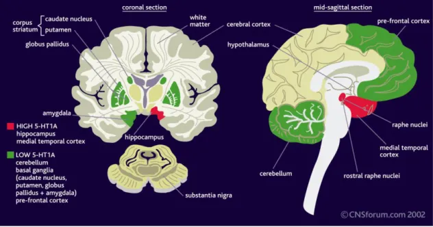
In major depression the density of 5-HT1A receptors is altered compared with the normal brain (Figure 3.). The 5-HT1A receptor density is increased in the hippocampus
28
and medial temporal cortex, while the density of these receptors is reduced in the cerebellum, basal ganglia and prefrontal cortex (175).
Figure 3. The density of 5-HT1A receptors in depression.
Other neurotransmitters also play a role in depression. Hamner et al. (176) found that dopaminergic dysregulation in depression is responsible for anhedonia, loss of motivation, and psychomotor slowdown.
Cholinergic systems appear to be associated with depression as well.
Acetylcholine (ACh) plays a significant role in mediating neuroendocrine, emotional, and physiological responses to stress (177). Central ACh turnover is increased following stress (178) and ACh facilitates the release of several stress-sensitive neurohormones and peptides including corticosterone, adrenocorticotropin (ACTH), and corticotropin-releasing factor (CRF) (179) suggesting an interaction between cholinergic and monoaminergic systems in the regulation of mood.
Glutamate is the major mediator of excitatory synaptic transmission in the brain (180). Glutamatergic abnormalities have been reported in plasma (181), serum (182) cerebrospinal fluid (183), and brain tissue (184) of subjects affected by mood disorders.
29
2.3.3. Neurotrophic factors associated with depression
Brain-derived neurotrophic factor (BDNF) was found to play a major role not only in the birth, survival and maturation of brain cells during development but also in cell growth and in allowing changes in the synapses between neurons (synaptic plasticity) throughout life. BDNF activates DNA-binding factors that stimulate gene transcription. In the raphe nuclei located in the brain stem, BDNF stimulates transcription of genes involved in serotonin function, such as tryptophan hydroxylase and the serotonin transporter (185). A polymorphism in the gene that codes for BDNF produces alleles called ―Val‖ and ―Met.‖ Studies suggest that having the Met allele, having the short allele of the serotonin transporter and psychosocial stress, increases vulnerability to depression more than having the short allele of the serotonin transporter and psychosocial stress alone (186).
Further evidences supporting the role of BDNF in the pathophysiology of depression come from postmortem studies, which have found low levels of BDNF in the hippocampus and prefrontal cortex of depressed patients (187, 188).
There is also evidence that antidepressants increase hippocampal BDNF levels in humans (189) and this antidepressant-induced upregulation of BDNF could help repair some stress-induced damage to hippocampal neurons and protect vulnerable neurons from further damage (190).
2.3.4 Neuroendocrine system
Psychosocial stress can influence multiple neurobiological systems relevant to major depressive disorder. Stress may activate can activate the hypothalamic–pituitary–
adrenal axis by stimulating local synthesis and release of corticotropinreleasing factor
30
(191) in addition it also activate this axis by releasing corticotropin-releasing factor from neurons in other regions of the brain, including the amygdala (192). These neurons may also contribute to activation of the serotonergic and noradrenergic systems (193, 194).
Dysregulation of the hypothalamic and extrahypothalamic corticotropin- releasing factor systems may gives an explanation why patients with major depressive disorder often have high levels of corticotropin-releasing factor and elevated levels of noradrenaline in their blood plasma and cerebrospinal fluid, and why they exaggerated stress reactions. Bradley et al (195) suggests that the impact of childhood abuse on a person‘s vulnerability for depression may be moderated by polymorphisms in the corticotropin-releasing factor type 1 receptor gene.
2.3.5 Genetics of depression
Depression is a complex phenomenon with many genes possibly involved.
Researchers have not identified a single gene or a series of genes that cause depression, rather, certain variations in genes, called polymorphisms, may increase risk for depression. Genes can predispose individuals to major depressive disorder in many ways (196). Among the many genes that have been identified to play a role in the background of the disorder, studies have most consistently supported a role for polymorphisms in genes that regulate the serotonin transporter promoter locus (5HTTPR), catechol-o-methyl transferase (COMT), monoamine oxidase (MAO), brain- derived neurotrophic factor (BDNF) and glutamate receptors.
The serotonin transporter gene is one of the most studied genetic polymorphisms in major depressive disorder. This gene contains a polymorphism (5-HTTLPR) that gives rise to 2 different alleles, the long and short one. People usually have 2 copies of each gene in their deoxyribonucleic acid (DNA); therefore, a person can be homozygous for the long allele, homozygous for the short allele or heterozygous. The short allele slows down the synthesis of the serotonin transporter. This is thought to
31
reduce the speed with which serotonin neurons can adapt to changes in their stimulation (197). Several studies found association between the 5HTTLPR and major depression mainly in interaction with stressful life events (198-200).
Studies reported an association between the Val/Val genotype of the COMT enzyme and early onset major depressive disorder (104, 201) suggesting the involvement of this genotype in the background of depression.
Tryptophan hydroxylase (TPH) is the enzyme involved in the biosynthesis of serotonin (202). Two genes have been discovered which encode for the TPH isoforms (TPH1 and TPH2). Zill et al (203) reported that MD was associated with 10 SNP haplotypes in TPH2 in 300 MD patients and 265 control subjects.
Brain-derived neurotrophic factor (BDNF) is a neuroprotective protein, and reduced serum BDNF was reported in MD (204). The activity of genes known to modulate neurotrophic factors, neuroplasticity and neurogenesis may be compromised in MD patients (205). The val66met allele confers reduced BDNF functioning and has been associated with structural brain changes common in MD (206).
Depression aggregates in families. Family studies observed an increase in the risk of developing major depressive disorder in the relatives of individuals with major depressive disorder (207-210). Twin studies consistently support genetic effects in the development of depression as well (211, 212). Expression of major depression‘s heritability is also influenced by the family environment and ‖parental coldness‖ was found to be associated with a 38% increased risk for developing this disease (213).
The lifetime prevalence of major depression is at least 10%, with the risk in women twice that in men (214, 215). Epidemiologic studies show that approximately 40%–50% of the risk for depression is genetic (216, 217) so vulnerability to depression is only partly genetic, with nongenetic factors also being important such as stress and emotional trauma or viral (e.g., Borna virus) infections (146, 216).
32
2.4 Clinical and subclinical manifestations of depression
2.4.1 Major depression
Major depression, a common (218, 219) and recurrent disorder (220), is associated with considerable morbidity (221) and excess mortality (222). Major depression has been projected to become the second leading cause of disability worldwide by 2020 (223). Moreover, major depression is of increasing importance in clinical psychiatry (224).
Studies investigated that depressive symptoms are common in the Hungarian population as well. In a study Szádóczky et al. reported the lifetime rate for major depression in the Hungarian population was 15.1%, which was similar to the data from the Western countries (225). According to earlier studies, 24%, 31% and 27.3% of the Hungarian population complained of mild depressive symptoms in 1988, 1995 and 2002, respectively (226).
Kopp et al. reported that 30.6% of the Hungarian adult population had complained of depressive symptoms; and the rate of severe depression was 7% (227).
Even though our knowledge about the basis and treatment of depression increased, similar results were established in 2002, 27.3% of the Hungarian population suffered from depressive symptom; in which 7.3% reported severe depressive symptoms (228).
According to the DSM-IV (116), a person who suffers from major depressive disorder must either have a depressed mood or a loss of interest or pleasure in daily activities consistently for at least a two week period. This mood must represent a change from the person's normal mood; social, occupational, educational or other important functioning must also be negatively impaired by the change in mood. A depressed mood caused by substances (such as drugs, alcohol, medications) or which is part of a general medical condition is not considered to be major depressive disorder. Major depressive disorder cannot be diagnosed if a person has a history of manic, hypomanic, or mixed episodes or if the depressed mood is better accounted for by schizoaffective disorder and is not superimposed on schizophrenia, schizophreniform disorder, delusional disorder or psychotic disorder. Further, the symptoms are not better accounted for by
33
bereavement (i.e., after the loss of a loved one) and the symptoms persist for longer than two months or are characterized by marked functional impairment, morbid preoccupation with worthlessness, suicidal ideation, psychotic symptoms, or psychomotor retardation.
Five (or more) of the following symptoms have been present during the same 2- week period and represent a change from previous functioning; at least one of the symptoms is either depressed mood or loss of interest or pleasure:
(1) depressed mood most of the day, nearly every day, as indicated by either subjective report (e.g., feels sad or empty) or observation made by others (e.g., appears tearful). Note: In children and adolescents, can be irritable mood.
(2) markedly diminished interest or pleasure in all, or almost all, activities most of the day, nearly every day (as indicated by either subjective account or observation made by others)
(3) significant weight loss when not dieting or weight gain (e.g., a change of more than 5% of body weight in a month), or decrease or increase in appetite nearly every day. Note: In children, consider failure to make expected weight gains.
(4) insomnia or hypersomnia nearly every day
(5) psychomotor agitation or retardation nearly every day (observable by others, not merely subjective feelings of restlessness or being slowed down)
(6) fatigue or loss of energy nearly every day
(7) feelings of worthlessness or excessive or inappropriate guilt (which may be delusional) nearly every day (not merely self-reproach or guilt about being sick)
(8) diminished ability to think or concentrate, or indecisiveness, nearly every day (either by subjective account or as observed by others)
34
(9) recurrent thoughts of death (not just fear of dying), recurrent suicidal ideation without a specific plan, or a suicide attempt or a specific plan for committing suicide
Major depression is a multifactorial disorder that is influenced by several risk factors. Epidemiologic data indicate that gender and age are two independent risk factors for the development of MD. Lifetime episodes of MD have high heritability, and MD is 1.5 to 3 times more common among first-degree biological relatives of people with this disorder than in the general population (229). Socioeconomic status (e.g., income and education), and marital status, ethnicity, urbanicity, and geographic region also affects mental health (219). Kessler et al. (230) noted that adult gender-role stresses contribute to the greater risk of adult-onset depression in women.
Childhood sexual abuse is an important early stressor that can predispose individuals to adult-onset depression, just like other types of childhood trauma, such as parental loss, poor parenting, parental drinking, mental illness, and family violence (231).
Kendler et al. (232) found that recent stressful events can be the single most powerful risk factor for 1-year prevalence of MD, followed by genetic factors, previous history of MD, and temperament. Furthermore, the subjective interpretation of a situation depends on the early environmental influences on development, both in respect to the development of the brain structures and psychological coping abilities. The
―social stress model‖ of depression can be characterised with an early life chronic stress situation which is the result of the disruption of mother–infant or peer bonding, which seems to resemble human depression or vulnerability to depression (233). Learned helplessness is a chronic stress situation, when feeling of total lack of control makes the avoidance of an emotionally negative situation impossible. In such a state, the hippocampus is affected by the long-lasting elevations of circulating corticosteroids resulting from uncontrollable stress. Severe stress for a long period causes damage in hippocampal pyramidal neurons, mainly in the CA3 and CA4 region and reductions in the length and arborization of their dendrites. In connection with the physiological consequences of chronic stress the feeling of lack of control has central importance (234).
35
Psychotherapy is necessary for individuals suffering from depression.
Interpersonal therapy is one of the most promising types of psychotherapies. It is a short-term psychotherapy, normally consisting of 12 to 16 weekly sessions. It was developed specifically for the treatment of major depression, and focuses on correcting current social dysfunction (235).
Cognitive behavioural therapy is also widely used in the treatment of depression.
The cognitive behavioural theory of depression states that the patient's self-rejection and self-criticism causes major depression. This therapy seeks to correct these negative thoughts or dysfunctional attitudes in order to overcome the patient's pessimism and hopelessness and tries to break the depressed patient's vicious cycle of increased negative thinking leading to increased social isolation which further increases the negative thinking (236).
Psychoanalytic psychotherapy for major depression usually continues with one or more weekly visits for several years. The psychoanalytic approach to treating major depression focuses on hypothesized unconscious phenomena, such as defense mechanisms or internal conflicts and it focuses on the patient's past analyzing the historical reasons why the patient has "turned anger inwards against the self" in becoming depressed. A modified form of this technique is the short-term psychodynamic psychotherapy, that was scientifically proven effective (237).
Family therapy can be a crucial and effective modality in the treatment of mood disorders in cases when the depression appears to be seriously jeopardizing the patient's marriage and family functioning. Family therapy examines the role of the depressed member in the psychological well-being of the family and it also examines the role of the entire family in the maintenance of the depression (238).
2.4.2 Subthreshold depression
Depression can best be conceptualized within a continuum from normality to full blown depression (239, 240). According to a 2009 research (241), subthreshold
36
depression is associated with functional impairment. This research showed that functional impairment becomes apparent early on the continuum.
Subthreshold depression is generally defined to represent patients who suffer from depressive symptoms, in which the number, duration, or quality of symptoms do not meet the DSM criteria necessary for a diagnosis of major depression (242). This category includes patients who complain of depressive symptoms, but do not complain of depressed mood or anhedonia. Therefore, they lack a necessary criterion for all DSM-IV depressive categories. Judd et al. (243) labeled the cluster as subsyndromal symptomatic depression (SSD). Depressive symptoms in SSD can either be operationalized as scoring above a cut-off score of a self-rating depression scale or meeting the criteria of minor depression (244).
It has been known since Kraepelin, that fluctuating affective manifestations occur prior and after major affective episodes. Paskind noted relatively minor affective symptoms which recurred on a cyclical basis, as well as neurovegetative symptoms such as anergia, headaches, insomnia, sexual disturbances, gastro-intestinal disturbances, palpitations, and anxiety symptoms (245).
SSD shares four out of the five most common symptoms of MD and minor depression (MinD): insomnia, tiredness, frequent thoughts of death, and decrease in concentration (243). The risk of suicide is significantly higher in patients with SSD as compared to normal subjects (246), but lower than in MD and MinD. SSD has greater prevalence than MinD of significant weight gain, slowed thinking, and hypersomnia, however, decreased prevalence of poor appetite compared with MinD. After clinical researchers started to study and identify the features of subthreshold depressive states, three additional categories were added to the DSM as subthreshold forms of major depression: dysthymia, minor depression and recurrent brief depression. Dysthymic disorder is characterised by a reduced number of symptoms showing a minimal duration of 2 years. Minor depression is diagnosed in patients who have more than two, but less than five, symptoms of depression for at least 2 weeks duration. Recurrent brief depression is diagnosed in patients who suffer from recurrent episodes of depressive symptoms that are identical to major depressive episodes in number and quality of symptoms but do not meet the 2-week duration requirement. The episodes recur at least once per month for a period of one year.