Chemical Injuries
A. W. Α. BROWN
Department of Zoology, University of Western Ontario, London, Canada
I. Nerve Poisons 67 A. Narcotics 67 B. Axonic Poisons 72 C. Ganglionic Poisons 86 II. Tissue Poisons 102
A. Necrotic Poisons 102 B. Oxidative-Enzyme Inhibitors 107
C. Muscle Poisons I l l D. Neuromuscular-Junction Poisons 114
References 116
This chapter deals with the pathological changes induced in insects by chemicals employed as insecticides. Insect toxicology is becoming ever more relevant to insect pathology, with the characterization of tox
ins produced by entomogenous bacteria such as Bacillus thuringiensis Berliner, and the discovery that the toxin of the diphtheria bacillus is an inhibitor of cytochrome oxidase in insects (Pappenheimer and Wil
liams, 1952).
T h e nature of the poisoning process and the cause of death becomes intelligible to the insect pathologist when visible changes develop in cells and tissues. T h e insect toxicologist, however, attains understand
ing of the mode of action of a chemical with discovery of inhibition of particular enzymes or of characteristic changes in the electrophysiology of insect nerve. But since these changes are reversible, many workers consider that the biochemical or electrophysiological lesion, although the effective cause of ins^cticidal action, is not the final cause of death.
For insects, which constitutionally can take "an unconscionable time
65
dying," the final stages of morbidity involve an array of pathological conditions—disbalance of neuroactive chemicals in the hemolymph, crises in secretory glands, depletion of cytoplasmic reserves, drastic loss of water—which it is more convenient to regard as secondary. Never
theless these changes are relevant to the irreversibility that means death, and so also are visible histological changes to the extent that they are consistent and peculiar to the particular insecticide.
This chapter will omit a number of considerations pertinent in insect toxicology, such as the penetration of the insecticide and its metabolic degradation, considerations so important in the relative re
sistance of species and intraspecific strains, and in discovering synergists or more effective analogs. Chemicals which are of interest only in insect pharmacology, for their action on insect nerve, heart or muscle, will also be omitted.
T h e pathological effects of the many diverse chemicals which have been used as insecticides will be discussed in each case usually in the following order: external symptoms and signs, effect on respiration and heart, changes in nerve electrophysiology, inhibition of enzymes, and visible histological changes. T h e chemicals are grouped and arranged for convenience according to the pattern of effects in the accompanying tabulation. T h e urethanes, inhibitors of dehydrogenases in grasshopper embryos (Bodine and Fitzgerald, 1949), and the haloacetates, inhib
itors of triosephosphate dehydrogenase in house flies (Bettini and Boc- cacci, 1955) are omitted because they have not been employed as insecticides.
I. Nerve Poisons A. Narcotics
1. Hydrocarbon Oils 2. Narcotic Gases B. Axonic Derivatives
1. Benzene Derivatives 2. DDT and Relatives 3. Pyrethroids
a. Pyrethrin synergists 4. Veratrine Alkaloids C. Ganglionic Poisons
1. Non-anticholinesterases a. v-Benzene hexachloride b. Cyclodiene derivatives c. Nicotine alkaloids d. Organic thiocyanates 2. Anticholinesterases
a. Organophosphorus insecticides b. Carbamate insecticides
II. Tissue Poisons A. Necrotic Poisons
1. Arsenicals
2. Inorganic Fluorine Compounds 3. Heavy-Metal Ions
4. Fatty Acids
B. Oxidative-Enzyme Inhibitors 1. Sulfur and Sulfides 2. Hydrogen Cyanide 3. Rotenone
4. Thiourea and Phenylthiourea C. Muscle Poisons
1. Non-narcotic Gases 2. Dinitro Compounds 3. Ryanodine
D. Neuromuscular-Junction Poisons 1. Phenothiazine
2. Wasp Venoms
I . N E R V E POISONS
A. Narcotics
1. Hydrocarbon Oils
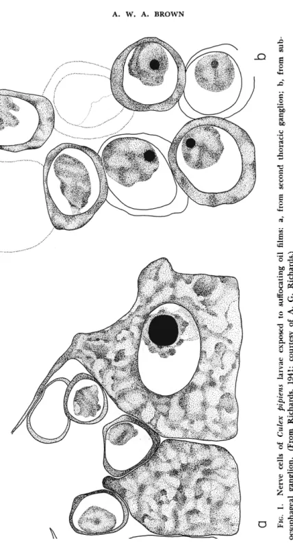
Nontoxic oils are able to narcotize insects into immobility and cause reversible histological changes in the nerve cells by denying them oxygen.
Larvae of the mosquito Culex pipiens pipiens Linnaeus respond to a film of refined kerosene on the water surface by becoming lethargic and sinking to the bottom in 10 to 20 minutes (Richards, 1941). I f the narcotized larvae are examined histologically, it is found that their nerve cells show nuclear pycnosis and reticulation of the Nissl granules (Fig.
1 ) . This condition has been exactly reproduced in larvae of several dipterous species by denying them oxygen (Buck and Boche, 1938).
Thus it is concluded that narcosis involves anoxia of the nerve cells, with consequent nerve-cell changes which are normally reversible and not a cause of death (Richards, 1941). However, anoxia long main
tained may eventually kill; the scale insect Phenacoccus colemani (Ehr
horn) is killed by a highly refined heavy oil in 5 to 15 days, the period also necessary for death from asphyxiation by anoxia (deOng et al, 1927).
Whereas a refined oil such as medicinal paraffin does not even affect the ultrastructure of axon sheaths (Richards and Cutkomp, 1945b), un
refined oils contain unsaturated toxicants. These toxic oils are often volatile and usually kill by entry as a vapor into the tracheae, not only in house flies (Moore and Graham, 1918) but also in mosquito larvae (Freeborn and Atsatt, 1918). T h e olefins in unrefined kerosene kill the scale P. colemani three times as fast as kerosene does (deOng et al., 1927).
Larvae of C. pipiens are killed by a film of unrefined kerosene or fuel oil even if the water underneath is constantly oxygenated; their death is preceded by convulsions and twitches, and premortem examination of the nerves reveals a degeneration of the fibers and their separation from the cellular layer (Richards, 1941). T h e fat body does not change until after death, when it begins to disintegrate (Richards, 1941) ; eventual dissolution of the fat body has been observed in the beetle Passalus cor- nutus Fabricius fumigated with gasoline vapor (Shafer, 1911).
2. Narcotic Gases
Vapors of diethyl ether or chloroform quickly anesthetize the house fly Musca domestica Linnaeus, the paralysis extending from the legs to the wings and then to the proboscis and antennae; recovery occurs first in the head appendages, then the legs and wings, full muscle tonus being somewhat delayed (Hiestand, 1932). Vinegar flies, such as Drosophila
FIG. 1. Nerve cells of Culex pipiens larvae exposed to suffocating oil films: a, from second thoracic ganglion; b, from sub- oesophageal ganglion. (From Richards, 1941; courtesy of A. G. Richards.)
virilis Sturtevant, may withstand several successive periods of anesthesia with diethyl ether, but if the period is so long as to prohibit recovery the legs and wings abandon their normal position of rest and come to be held stiffly away from the body (Bodenstein, 1946). Whereas diethyl ether causes a flaccid paralysis in the silkworm, Bombyx mori (Linnaeus), chloroform causes a reversible paralysis characterized by muscular con
tracture (Grandori and Reali, 1950). Thus among the narcotic gases, as in the hydrocarbon oils, there are those which have additional toxic effects. Some narcotic fumigants are indeed neurotoxic; for example, trichloro-i-butyl ketone (Chloretone) interposes ataxia and spasms be
tween hyperexcitation and paralysis in the house fly, and if recovery occurs it is ataxic (Hiestand, 1932).
T h e aphid Macrosiphum tulipae Boyer, fumigated with high con
centrations of carbon tetrachloride or carbon disulfide, is paralyzed within 1 minute, but the heart continues to beat for about 10 minutes
(Kirschner, 1932). Introduction of xylene into the siphon of C. pipiens paralyzes the larva in 1 minute, and the heart stops in 15 minutes
(Richards, 1943). Most fumigant gases when employed at the median lethal dose paralyze insects such as the beetle Tribolium confusum duVal within 1 hour (Sun, 1947), and in the survivors the narcosis is reversi
ble. During the initial stages of narcosis, diethyl ether and other fumi
gants cause a transient increase in C 02 output of beetles and grasshop
pers (Shafer, 1911; Bodine, 1923), and a fleeting acceleration of the heartbeat at first (Kirschner, 1932).
Narcotics such as chloroform, diethyl ether, carbon tetrachloride, carbon disulfide, and ethylene dichloride accumulate in the nervous system, as shown by experimentally injecting them into the respiratory siphon of the mosquitoes Aedes aegypti (Linnaeus) and C. pipiens (Rich
ard and Weygandt, 1945). When injected as liquids into the spiracles of the American cockroach, Periplaneta americana (Linnaeus), these narcotics destroyed the normal birefringent state of the lipid nerve sheaths; however, no change in optical properties of the nerve sheaths ensued when the narcotics were applied as vapors, even when in lethal concentrations (Richards and Cutkomp, 1945b).
Chloroform, carbon tetrachloride, and carbon disulfide were reported by Shafer (1915) to have an inhibitory effect on the dehydrogenases of Passalus cornutus both in vivo and in vitro. T h e gross symptoms of poi
soning by chloroform, diethyl ether, and other narcotics, which in these beetles were successively excitation, ataxia, and paralysis, were identical with those obtained in oxygen-free nitrogen. It was therefore suggested that narcosis of these insects involved tissue anoxia, especially in the nerves.
On the other hand, Hurst (1945) reported that larvae of the yellow mealworm Tenebrio molitor Linnaeus and of Musca domestica, im
mersed in chloroform or cyclohexane, showed an increase in dehydro
genase and phenoloxidase activity during the narcotic period before death, and suggested that these solvent chemicals had removed the pro
tective lipoid from these enzymes. Indeed the vapors of chloroform, ethylene dichloride, or diethyl ether, in artificially breaking the prepupal diapause in the beet webworm, Loxostege sticticalis (Linnaeus), may in
duce the liquefaction of the fat body which normally terminates diapause (Pepper, 1937). Insects in advanced stages of poisoning by carbon di
sulfide suffer a dissolution of their body fat (Peters, 1936). This nar
cotic fumigant also has been reported to react with protein, particularly in ganglionic cells (Shafer, 1915; Peters, 1936; Trappmann, 1938).
Recently assays have been made of the activity of cellular-oxidation enzymes in the thoracic muscles of house flies previously exposed to these narcotics. Ethylene dichloride resulted in stimulation of succinic dehy
drogenase, cytochrome oxidase, and overall glycolytic activity. On the other hand, carbon disulfide, though stimulating succinic dehydrogenase, inhibited cytochrome ovidase and overall glycolysis (Pant and Dahm, 1957).
When insects such as house flies are under anesthesia from cyclopro
pane (Winteringham et al., 1955), there is no effect on the level of adenosine triphosphate (ATP) or of arginine phosphate (Arg-P) in the thoracic muscle, but there is a gradual depletion of phosphoglyeerie acid ( P G A ) . Under prostration from ethylene dibromide, A T P and Arg-P are unaffected, but PGA is greatly depleted (Winteringham and Hellyer, 1954). Ethylene dichloride does not affect the PGA level; the reduction of A T P and Arg-P is only slight with narcotic doses (Winteringham et al., 1958) but considerable with lethal doses of ethylene dichloride (Winter
ingham and Hellyer, 1954).
Carbon disulfide vapor causes the American cockroach to lose very much of its blood volume (Shull et al., 1932). However, C S2 did not reduce the hemolymph cell count of the oriental cockroach Blatta orien-
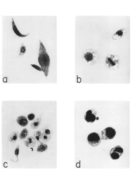
talis Linnaeus (Fisher, 1936), but diethyl ether slightly inhibited the coagulability of the hemolymph of this species without diminishing the cell count. Dichlorodiethyl ether and carbon tetrachloride, however, caused a reduction in the cell count of larvae of the Mediterranean flour moth, Anagasta kühniella (Zeller), which was most striking in the plasmatocytes and least in the spheroidocytes (Arnold, 1952). T h e plasmatocytes lost their fusiform shape and the swollen cells often ag
glomerated; they developed vacuoles and their bounding membrane eventually disintegrated. With sublethal doses the recovery period was
FIG. 2. Hemolymph cells of Anagasta kiihniella: a, normal plasmatocytes;
b, normal spheroidocytes; c, hemocytes from larva exposed to methyl bromide;
d, spheroidocytes from larva recovering from carbon tetrachloride poisoning. (From Arnold, 1952; courtesy of J . W . Arnold.)
marked by an increase in cell number and by the spheroidocytes becom
ing filled with neutral fat (Fig. 2 ) . Carbon tetrachloride or ethylene trichloride applied to the body louse, Pediculus humanus humanus Lin
naeus, caused nuclear enlargement and chromatin swelling especially in the hemocytes, but also in cells of the fat body, Malpighian tubes, and ganglia; in advanced stages of poisoning a general lysis of nuclei and cytoplasm ensued (Hopp, 1953).
B. Axonic Poisons
1. Benzene Derivatives
A useful insecticide in this group is ^-dichlorobenzene ( P D B ) ; when injected into Periplaneta americana (Munson and Yeager, 1945a) or con
tact-applied to Pediculus humanus (Hopp, 1953), P D B causes hyper
activity, tremors, and then paralysis. Insects fumigated with P D B show a normal respiratory rate at first, but it subsequently increases in the excitation phase and decreases at paralysis, as shown by determinations of C 02 production (Punt, 1950) and 02 consumption (Harvey and Brown, 1951). Applied to a crayfish nerve preparation, P D B causes re
petitive discharge of action potentials (Welsh and Gordon, 1947).
These symptoms resemble those of D D T poisoning, and are induced in Periplaneta americana by injection of yet other derivatives of ben
zene, such as phenol, aniline, hydroquinone and its tetrachloro deriva
tive (Munson and Yeager, 1945a). T h e injection of o-dichlorobenzene (ODB) induces tremors in many insects (Läuger et al., 1946). Larvae of A. kiihniella exposed to O D B vapor enter a rigor, with head held high, accompanied by increased 02 consumption and not fully relaxed for 6 hours (Payne, 1937). Chlorobenzene vapor causes silkworms to show spastic tetanic contractions and paralyzes them in a contorted posture (Grandori and Reali, 1950). It usually induces tremors (Läuger et al., 1946), but in body lice the activity simply falls to paralysis (Hopp, 1953). Benzene itself when injected causes tremors in insects (Läuger et al., 1946).
Naphthalene vapor leads directly to paralysis whereas injected naph
thalene causes dissolution of the fat body (Pyenson and MacLeod, 1936).
Applied to crayfish nerve, naphthalene evokes repetitive discharge of action potentials (Welsh and Gordon, 1947).
Direct application of chlorobenzene or P D B to Pediculus humanus induces cellular changes in epidermis, hemocytes and other tissue; either there is a burst of mitosis or there is nuclear enlargement and hyper- chromatosis, leading eventually to lysis of cells and nuclei (Hopp, 1953).
In advanced stages of poisoning, P D B causes dissolution of the body lipids (Trappmann, 1938). Larvae of T. molitor injected or fumigated
with naphthalene show darkening of the hemolymph and a progressive disintegration of the fat body and eventually the muscles (Pyenson and MacLeod, 1936).
2. DDT and Relatives
T h e action of this residual-contact insecticide is characterized by the
" D D T jitters," a continuing tremulousness of the entire body and ap
pendages of the poisoned insect. DDT-poisoned cockroaches (P. amer
icana) become unusually active and irritable, their gait becomes unco
ordinated, and they fall on their back time and again until they can no longer right themselves. T h e legs continue to show high-frequency twitches superimposed on slow spasmodic movements, the result of the two kinds (slow and fast) of muscle innervation. T h e twitches cease first, the slow movements then vanish, later the heart stops, and finally the insect gives no response to galvanic stimulation (Tobias and Koll- ros, 1946). T h e cockroach dies in a desiccated condition, with the body muscles contracted (Heslop and Ray, 1959).
Adults of the honey bee (Apis mellifera Linnaeus) contact-poisoned with D D T develop an ascending paralysis, the insects rolling and falling until death (Filmer and Smith, 1944). Stomach poisoning is slower, gradually resulting in excitation, then ataxia, then a succession of fall
ing and rising; finally the bee lies paralyzed on its back, only the legs quivering (Hoist, 1944). T h e black blow fly, Phormia regina (Meigen), is prostrated within 15 to 30 minutes of contact with D D T , first having shown hyperactivity and ataxia; high-frequency tremors and violent
spasms precede its death (Buck and Keister, 1949). Adult moths and mosquitoes show such tremors in the legs that they fall off, and the autotomized limbs continue to show the tremors (Wiesmann and Fen- jues, 1944). T h e boll worm, Heliothis zea (Boddie) ( = H. armigera Hüb
ner) , ceases feeding 2 hours after contact poisoning, and develops trem
ors and convulsions 2 hours later. In the prostrate condition 8 hours later the mouthparts are in constant motion and the alimentary canal frequently convulsed; the larva shrivels up before death (Chadbourne and Rainwater, 1953). Larvae of the greater wax moth, Galleria mel- lonella (Linnaeus), develop convulsive snakelike flexing movements, while regurgitating copious viscous liquid due to spasms of the foregut (Beard, 1958). Silkworm larvae in convulsions from injected D D T expel large quantities of fluid from the mouth, and the water loss results in a progressive shortening and mummification (Reali, 1951). Many spe
cies of insects exposed to residual deposits of D F D T (Gix) expelled a proteinaceous liquid from their tarsi and intersegmental membranes
(Bott, 1948).
Injection of D D T into larvae of the vinegar fly D. virilis induces within 20 seconds successive waves of contraction passing along the longitudinal muscles; the rate of heartbeat is also accelerated (Boden- stein, 1946). However, the rate was increased only slightly by injected D D T in Periplaneta americana (Orser and Brown, 1951) and by D D T in the aqueous environment of larvae of the mosquito Anopheles quad- rimaculatus Say (Jones, 1957b). T h e isolated heart of P. americana is unsettled and retarded by D D T , an effect which is not antagonized by atropine (Naidu, 1955).
As the insect enters the condition of tremors, its respiratory consump
tion of oxygen increases to some five times the normal rate, as shown by tests on the Japanese beetle Popillia japonica Newman (Ludwig,
1946, the saw-toothed grain beetle Oryzaephilus surinamensis (Lin
naeus) (Lord, 1949), the red flour bettle Tribolium castaneum (Herbst) (Lord, 1950), the house fly and the black blow fly (Buck and Keister, 1949), and the German cockroach Blattella germanica (Linnaeus)
(Harvey and Brown, 1951). T h a t this increase is due to the neurotoxic effect of D D T in increasing muscular activity is proved by the fact that larvae of Phormia regina in which the brain has been protected by liga
tion do not show the respiratory increase that D D T evokes from non- ligated larvae (Buck et al, 1952).
Poisoned insects rapidly lose weight because of tissue oxidation and water loss. DDT-poisoned house flies lost more weight and retained less glucose than starved flies (Dahm and Kearns, 1951). Poisoned larvae of Popillia japonica may consume all their glycogen and draw on theii fat (Ludwig, 1946). German cockroaches in the 3rd day of poisoning have consumed 70 percent of their carbohydrates and 15 percent of their fat (Clark and Butz, 1961). American cockroaches before death lose nearly all their carbohydrate and much of their fat, their blood showing ketone bodies (Merrill et ah, 1946). Adult Phormia regina during con
vulsions show the high respiratory quotient characteristic of carbohy
drate utilization, but death occurs before the caloric reserves decrease down to starvation level, and acidosis does not develop in the hemo
lymph. Larvae of Popillia japonica also died before the starvation level of fat was reached (Ludwig and Bartolotta, 1953).
T h e neuromuscular symptoms of D D T poisoning have been pre
vented in Periplaneta americana and M. domestica by anesthesia with cyclopropane (Merrill et ah, 1946; Winteringham, 1956), and in G.
mellonella by paralysis with Habrobracon venom (Beard, 1958) ; how
ever they resume as soon as the paralyzant wears off. Ether anesthesia has an absolute protective effect for larvae of Anopheles quadrimaculatus exposed to D D T (Jones, 1958). Phenobarbital protected larvae of Dro-
sophila virilis from D D T symptoms, even when injected after the con
vulsions have already commenced (Bodenstein, 1946). T h e muscles of prostrated house flies and wax-moth larvae show a decrease in A T P con
tent (Winteringham, 1956; Beard, 1958).
Qualitative changes have been detected in the lipids of house flies exposed to deposits of D D T . T h e lipoprotein in the tarsi becomes dis
sociated, to produce free lipid (Reiff, 1955). T h e free lipid content of the body as a whole increases, although it decreases in the thoracic ganglia and hemolymph (Reiff and Beye, 1960). T h e tarsal lipid de
creases in its solvent power for D D T in normal flies, but increases in resistant flies (Wiesmann and Reiff, 1956).
DDT-poisoned larvae of Popillia japonica showed a notable increase in the hemolymph content of reducing substances, urea, and amino acids (Ludwig and Bartolotta, 1953). On the other hand, Reiff (1956) had found a decrease in amino acid level of the hemolymph of adult house flies. Corrigan and Kearns (1958) reported that in DDT-poisoned Peri
planeta americana the major amino acids did not decrease, but that proline progressively decreased as prostration proceeded. In larvae of T. molitor about to die from residual D D T poisoning, norleucine and taurine were the only amino acids that decreased in the hemolymph, the others increasing in concentration as in starvation (Joseph, 1958).
In adult house flies topically treated there was an increase in free glu- tamine, possibly as a result of the increased respiration (Winteringham, 1959).
It has been found that D D T inhibits cytochrome oxidase when in
cubated with homogenates of M. domestica (Anderson et ah, 1954), T . molitor (Ludwig et al., 1955) or P. americana (Morrison and Brown, 1954) and with isolated leg muscles of Periplaneta (Ludwig et al., 1955).
However, noninsecticidal analogs of D D T also were inhibitory in vitro.
Moreover, there was no reduction in cytochrome oxidase activity in DDT-poisoned P. americana even by the time of prostration (Brown and Brown, 1956). Succinic dehydrogenase was the only one of the nine dehydrogenases tested to be inhibited by D D T in homogenates of M.
domestica or T. molitor (Barsa and Ludwig, 1959). Moreover, there was reduction in succinic dehydrogenase activity in thoracic muscle of D D T - poisoned house flies, accompanied by increased overall glycolytic activity
(Pant and Dahm, 1957). But the respiratory enzyme activity in nerve and muscle of P. americana and Locusta migratoria (Linnaeus) was not inhibited by D D T in vivo (Fukami, 1956). D D T has also been reported to inhibit, in homogenates of house flies, the oxidation of Krebs-cycle compounds and the process of oxidative phosphorylation (Sacklin et al.,
1955). It also uncoupled phosphorylation in suspensions of mitochon-
dria of Aedes aegypti by inhibiting phosphate uptake more than oxygen consumption (Gonda et al., 1957), but this effect was also obtained with the noninsecticidal tetrachloroethane analog of D D T (Gonda et al., 1959). D D T , and also endrin and toxaphene, inhibited the transami
nase conversion of α-ketoglutarate to glutamate in homogenates of P.
americana, but not in vivo (McAllan and Brown, 1960).
D D T is well known for its affinity for chitin and its ability to pene
trate the epicuticle and cuticle. After topical application, D D T be
comes abundant in the hemolymph (Lindquist et al., 1951) and appears in all tissues of the house fly (Tahori and Hoskins, 1953). Restriction of hemolymph flow to the brain reduces by nine-tenths the amount of D D T reaching that site from elsewhere (Morrison and LeRoux, 1954).
Nevertheless, D D T is more effective when applied to the cuticular sur
face than when injected into internal tissues (Fisher, 1952). This in
secticide may be detoxified in the bodies of resistant species or strains of insects by the enzyme DDT-dehydrochlorinase (Sternburg et al., 1954), and the process may be inhibited by synergists such as piperonyl- cyclonene (Perry and Hoskins, 1950).
D D T normally contact-poisons insects by affecting the sensory neu
rons, the cell bodies of which are located in the integument (Roeder and Weiant, 1946) ; in P. americana the campaniform sensilla at the base of the legs are especially vulnerable sites (Roeder and Weiant, 1948). At higher concentrations D D T may act on the motor nerves (Yeager and Munson, 1945), and has even caused repeated contractions in the iso
lated musculature of Drosophila virilis (Bodenstein, 1946) and Dytiscus marginalis Linnaeus (Fritsch, 1952).
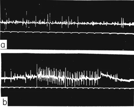
T h e effect of D D T on an arthropod nerve is to cause repetitive dis
charge (Fig. 3 ) , the normal single spikes being replaced by a train of multiple spikes, at a frequency of from 250 to 500 per second and a duration of from 0.1 to 1.0 seconds (Welsh and Gordon, 1947; Roeder and Weiant, 1946, 1948). Similar effects are produced by methoxy chlor, D D T , and D F D T (Welsh and Gordon, 1947). Repetitive discharge is a characteristic of nerve when there is a deficiency of calcium at the axon surface. DDT-poisoned insect axons do not repolarize immediately, but show a prolonged negative afterpotential, and in this condition of instability they are liable to show repetitive discharge (Yamasaki and Narahashi, 1960a) ; it is thus considered that the effect of D D T at the axon surface is to inhibit the extra-rapid permeability of potassium necessary for immediate repolarization.
Typically, D D T acts through a reflex arc (Yeager and Munson, 1945), and blocking the ganglion inhibits the effect of low doses (Tobias and Kollros, 1946). It was therefore considered by Dresden (1948) that
the characteristic effect of D D T was ganglionic, and that it was at the synapse that the change to repetitive discharge occurred. Such a change in trans-synaptic conduction has been demonstrated in P. americana by Smyth (1960), but only if the ganglion has been partially desheathed.
T h e axonic effects of D D T in multiplying discharges of sensory neurons are the most important, though evidently it is possible to have such multiplication at the ganglion. Poisoning of motor nerves by applica
tion of D D T to the last abdominal ganglion of Dytiscus marginalis has
FIG. 3. Action potentials in metathoracic crural nerve of Periplaneta americana: a, normal cockroach; b, cockroach injected with DDT. (Courtesy of K. D. Roeder.)
been obtained by Fritsch (1952), who considers that normally the D D T symptoms may have a ganglionic as well as a peripheral origin.
D D T is not an inhibitor in vitro for Cholinesterase from Apis melli- fera or P. americana (Richards and Cutkomp, 1945a; Hartley and Brown, 1955). However, increase of free acetylcholine (ACh) in the nerve of poisoned P. americana in the first stages of prostration has been observed by Tobias et al. (1946). T h i s is considered to be an artifact by Lewis (1953), who found that extracts of DDT-poisoned Calliphora erythrocephala (Meigen) and Lucilla (Phaenicia) sericata (Meigen) taken at this stage of poisoning rapidly synthesized ACh. Colhoun
(1959a) has demonstrated that American cockroaches showing the first symptoms of poisoning also show a transient increase in free ACh which returns to the normal level; it is only subsequent to prostration that the really substantial increase in free ACh occurs. T h e nerve cords of cockroaches poisoned by D D T or T E P P , or deranged by electric shock, release a toxin into the hemolymph (Sternburg et al., 1959). T h i s ma
terial, which is probably a primary aromatic amine, induces continuous increased activity in healthy nerve cords (Sternburg, 1960). Adult male P. americana tied down on their backs for days frequently exhibited the initial rise in 02 consumption, the appearance of repetitive discharge in the nerve cord, and final paralysis, that is characteristic of D D T poisoning (Heslop and Ray, 1959), and appeared to liberate a paralyz
ing neurotoxin as a result of this stress (Beament, 1958). Other neuro- active substances, including catecholamine from the corpus cardiacum, are also released into the blood of P. americana poisoned by D D T (Col- houn, 1960). ACh itself, a highly polar compound, is inactive when applied to nerve by injection into the hemolymph (Hopf, 1952).
D D T has no detectable effect on the ultrastructure of insect neurons (Richards and Cutkomp, 1945b). It does not alter in any way the growth of imaginal discs in fly larvae (Bodenstein, 1946) or cells in tissue culture (Lewis and Richards, 1945). No histopathological changes were observed in DDT-poisoned C. erythrocephala (Witt, 1947), P.
americana ^Richards and Cutkomp, 1945b), and Anopheles quadrimacu- latus (Jones, 1953). With DDT-poisoned larvae of H. armigera the only detectable effect was a slight nuclear pycnosis in fat-body cells (Chad- bourne and Rainwater, 1953), which could have been due to secondary anoxia (Richards and Cutkomp, 1945b).
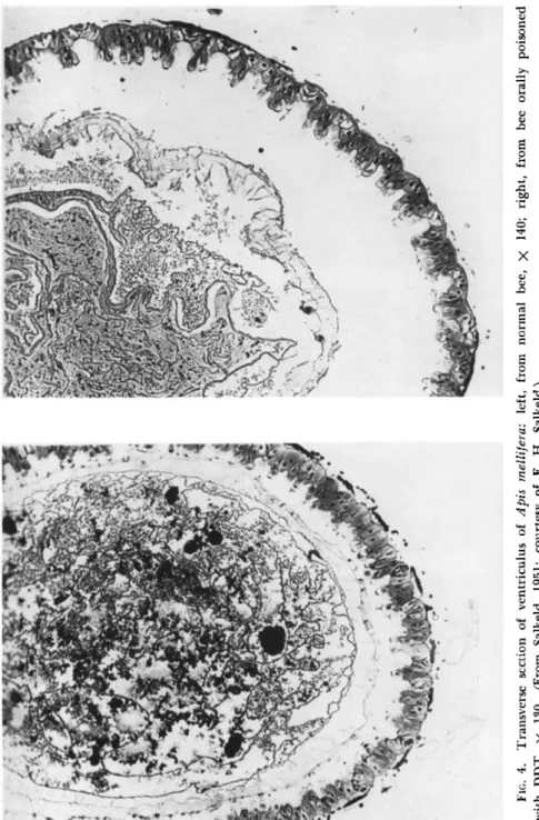
However, definite histological change was noted in the midgut epithe
lium of honey bees orally poisoned with D D T (Salkeld, 1950, 1951).
T h e cells showed severe vacuolization, and proliferation at the tips (Fig.
4 ) , and this glandular hyperactivity was considered due to the stimula
tion by D D T of the stomatogastric nerve branches. A large gas bubble was usually present in the lumen of the gut, possibly derived from air swallowed by the poisoned bee. In silkworms contact-treated with D D T , the marked muscular contracture, caused either directly or through the nerve, resulted in the separation of the intestinal epithelium from its greatly contracted muscular coat (Grandori and Reali, 1950). Effects on the midgut epithelium also were noted in the body louse contact- treated with D D T and taken as paralysis commenced. T h e cells became vacuolized, the nuclei swollen, and the walls indistinct; these effects were even more pronounced in the labial glands. Similar cell changes were observed in the hemocytes, epidermis, and Malpighian tubes, while
FIG. 4. Transverse section of ventriculus of Apis mellifera: left, from normal bee, χ 140; right, from bee orally poisoned with DDT, χ 130. (From Salkeld, 1951; courtesy of Ε. H. Salkeld.)
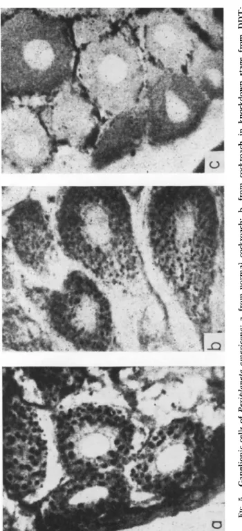
FIG. 5. Ganglionic cells of Periplaneta americana: a, from normal cockroach; b, from cockroach in knockdown stage from DDT; c, from cockroach at time of death. (From Chang, 1951; courtesy of J. P. Chang.)
in the ganglia the neuron cell-bodies shrank and lost their Nissl gran
ules (Hopp, 1953).
House flies taken 10 minutes after being sprayed, and while still capable of movement, were found by Hartzell (1945) to show slight pycnosis of muscle nuclei, and partial cytolysis and nuclear degeneration in the brain tissue; these may be interpreted as secondary effects of pros
tration. However, by means of silver-nitrate impregnation of ganglionic nerve cells in P. americana and Apis mellifera in the knockdown stage, Chang (1951) was able to detect the dissolution of Golgi bodies (Fig. 5 ) , and their almost complete disappearance at the time of death.
Toumanoff and Lapied (1950) had detected an increase in plasma- tocyte count in DDT-poisoned larvae of Galleria mellonella, and inter
preted it as a defense mechanism. However, Jones (1957a) found that, when cell counts were made on material not fixed by heat, DDT-poisoned larvae of T. molitor showed a decrease in plasmatocytes. Arvy et al.
(1950) orally poisoned with D D T larvae and adults of the potato beetle Leptinotarsa decemlineata (Say) and subsequently observed nuclear changes and cytolysis in the hemocytes, and the presence of their debris in the hemolymph; but the effects were not as great with D D T and with B H C or lead arsenate.
3. Pyrethroids
Contact or oral administration of Pyrethrins to the honey bee results in: (i) hyperactivity, restless running and frantic flying; (ii) ataxia, with zigzag flight and locomotion; (iii) paralysis ascending from legs to still the buzzing wings; and (iv) complete paralysis with opisthotonus of abdomen (Böttcher, 1939). Caterpillars of many phytophagous species contact-poisoned with Pyrethrins at the L D5 0 develop within 1 to 5 minutes the following symptoms: (i) rapid restless locomotion, prolegs strongly prehensile, head swaying, mandibles snapping and mouth regurgitating;
(ii) body writhing and often rolling over, curling ventrally and snapping straight dorsally, occasional prolapse of rectum; (iii) twitches of trunk and appendages become weaker and less frequent, reactions to stimuli cease and a flaccid paralysis ensues in which the heart beats weakly for several days (Klinger, 1936). Larvae of G. mellonella injected with Pyrethrins show similar symptoms, and dosage levels can be found which induce recovery from the excitation phase, from the ataxic phase, and even from paralysis (Belleuvre, 1938). House flies that had been com
pletely paralyzed by pyrethrum spray could recover in 5 to 15 hours and lay the normal amount of fertile eggs (Richardson, 1931). However, leaf-feeding caterpillars may survive pyrethrin poisoning only to fail to pupate or emerge (Klinger, 1936). T h e initial excitation in contact-
poisoned Β. germanica is due to stimulation of sensory endings, since it is not inhibited by transverse section of the nerve cord (Hutzel, 1942b).
T h e heart of insects fatally poisoned by Pyrethrins may continue to beat weakly for days with caterpillars (Klinger, 1936) or even weeks in Cimex lectularius Linnaeus and Rhodnius prolixus (Stäl) (Wigglesworth, 1941). In P. americana, sublethal doses stimulate; lethal doses steadily depress the heartbeat (Coon, 1944). Retardation was also observed in pyrethrinized Corethra plumicornis (Fabricius) (Krüger, 1931) and
Chaoborus astictopus Dyar & Shannon (Deonier and Lindquist, 1942).
T h e isolated heart of P. americana is also accelerated by low, and de
pressed by higher, concentrations, the effect being antagonized by atro
pine (Naidu, 1955). Pyrethrins stop the isolated heart of G. mellonella in diastole (Belleuvre, 1938), and of B. germanica in systole (Yeager et al,
1935). During the initial excitatory phase there is a transient rise of oxygen consumption in Oryzaephilus surinamensis (Lord, 1949) and B.
germanica (Harvey and Brown, 1951). There is no abnormal weight loss in poisoned R. prolixus, and the spiracles of pyrethrinized Cimex lectularius remain closed (Wigglesworth, 1941). However, abnormal weight losses have been reported for pyrethrin-treated Popillia japonica (Ludwig, 1946), Phormia regina larvae (Buck et al., 1952) and Tribo- lium castaneum (Hewlett and Gostick, 1955).
T h e conductivity of the ventral nerve cord of poisoned Porthetria dispar (Linnaeus) is greatly reduced before paralysis supervenes (Klin
ger, 1936). When the nerve cord of the German cockroach was perfused with Pyrethrins, it showed massive discharge, then trains of impulses, and finally block even to external stimulation; the same picture was shown by the nerve cord of pyrethrin-poisoned cockroaches (Lowenstein,
1942). Application of Pyrethrins to nerves of crayfish and of Periplaneta americana evoke trains of repetitive discharge (Ellis et al., 1942; Welsh and Gordon, 1947; Lalonde and Brown, 1954). Pyrethrins have an un- stabilizing effect on nerve axons similar to that of D D T (Roeder, 1953).
Like D D T , the Pyrethrins are not inhibitors of insect Cholinesterase (Hartley and Brown, 1955) ; moreover, allethrin induces a negative after- potential in the poisoned axons of P. americana that is even more pro
nounced than with D D T (Narahashi, 1961). In high concentrations, Pyrethrins have blocked conduction in the giant axons of P. americana
(Narahashi, 1961) and peripheral nerve of the crayfish (Welsh and Gordon, 1947). Like D D T , Pyrethrins caused the release of a neuroac- tive toxin into the hemolymph of P. americana at low temperatures but not at high (Blum and Kearns, 1956).
Unlike D D T , which had no effect, Pyrethrins injected into the tra
cheae of P. americana destroyed the birefringence (oriented anisotropy)
of the protein in the axis cylinders, and subsequently the birefringence of the lipid sheaths; these changes coincided with loss of irritability of the nerve cord, long before all general symptoms ceased (Richards and Cutkomp, 1945b).
T h e Pyrethrins also have a direct effect on muscle, since in poi
soned Acheta domesticns (Linnaeus) the Chronaxie of muscle but not of nerve is changed, and perfused Pyrethrins can paralyze stom
ach muscle isolated from nerve (Rigal and Gautrelet, 1932). It is perhaps of interest that Pyrethrins and allethrin inhibited cytochrome oxidase in homogenates of P. americana (Morrison and Brown, 1954).
But the oxygen consumption of P. americana muscle and the stainability of its nerve with T T C was not reduced by Pyrethrins in vivo or in vitro
(Fukami, 1956).
Although there is evidence that Pyrethrins can act on sensory nerve endings after penetrating only the cuticle (Page et ah, 1949), and that further penetration is only along the nerves (Hutzel, 1942b), yet there is good evidence that it is also transported in the hemolymph (Roy et al., 1943). Pyrethrins and their metabolites after injection into P. ameri
cana accumulate in the foregut (Zeid et al., 1953); in some species such as the cabbageworm Pier is rapae (Linnaeus) (Swingle, 1934) and to a less extent Prodenia eridania (Woke, 1939) orally administered Pyre
thrins are detoxified in the alimentary canal. T h e detoxification of Py
rethrins by Cochylis caterpillars is accelerated by raising the temperature to 37°C, and they thus survive moderate doses that would normally have been lethal; thus the poikilothermy of insects is a handicap in
creasing their susceptibility to Pyrethrins (Chevalier, 1930).
Many workers have observed histopathological changes in insects poisoned by Pyrethrins. T h e transparent larvae of the aquatic midge Corethra plumicornis could be seen in vivo to develop vacuoles in the nerve connectives and ganglia during the convulsive stage, even when immobilized by ether; doses of Pyrethrins that proved sublethal did not induce vacuolization (Krüger, 1931). Pyrethrins injected into the tra
cheae of Periplaneta americana induced nuclear pycnosis in the nerve cord quite rapidly, but vacuolization and chromatolysis did not develop until paralysis was complete and irreversible (Richards and Cutkomp, 1945b). Larvae of Tenebrio molitor (Wilcoxon and Hartzell, 1933) and adults of Melanoplus femnr-rubrum (De Geer) (Hartzell, 1934) contact- poisoned with Pyrethrins showed histological changes in preparations fixed 16 hours later; the brain, ganglia and to some extent the connec
tives showed vacuolization, tigrolysis of the Nissl granules, and tissue disintegration. T h e thoracic and fused abdominal ganglia of Cimex lec- tularius and R. prolixus, which had been paralyzed by Pyrethrins for 10
days but still showed heartbeat and leg movements, were found to be shrunken and vacuolated, and their cells degenerated (Wigglesworth, 1941). Larvae of the gypsy moth Porthetria dispar, prostrated by high doses of pyrethrum but still living 24 hours later (Klinger, 1936), showed extensive destruction of nerve cell bodies in the peripheral layer of the abdominal ganglia (Fig. 6 ) . House flies taken 4 hours after knockdown by Pyrethrins showed pycnosis in the nervous system, an annular vacuole surrounding the condensed nucleus (Hartzell and Scud- der, 1942); the brains of house flies taken before paralysis was complete showed nuclear pycnosis, tigrolysis, and fenestration and lysis of nerve fibers (Hartzell, 1945). These lesions, caused by Pyrethrins but not by
FIG. 6. Transverse section of abdominal ganglion of Porthetria dispar: left, normal larva; right, larva pyrethrum-poisoned for 24 hours. (From Klinger, 1936; courtesy of John Wiley & Sons Inc., New York.)
rotenone, are attributed by Richards and Cutkomp (1945b) to anoxia and autolysis and are considered to be the effect rather than the cause of the lethal paralysis.
Muscle tissue of larvae of Corethra plumicornis showed vacuoles within 15 minutes of exposure to Pyrethrins, and within 1 day the hypodernal cells began to degenerate, vacuolize, and separate from the cuticle (Krüger, 1931). Muscles of poisoned house flies showed clumping of the nuclei into dense rods, loss of striation, and fenestration between the fibers (Hartzell, 1945); cells of the fat body also showed pycnosis and separation (Hartzell and Scudder, 1942).
a. Pyrethrin synergists. Nontoxic concentrations of these compounds when added to nontoxic concentrations of Pyrethrins make a highly in- secticidal mixture, mainly through inhibiting recovery from knockdown.
Examples are: (i) methylenedioxyphenyl compounds such as sesamin,
piperine, piperonylbutoxide and piperonylcyclonene, (ii) 2V-substituted amides such as IN 930 (N-diisobutylundecylenamide), and (iii) terpenes such as DHS activator and Thanite. Piperonyl compounds have also shown some synergism with rotenone, ryanodine, gamma-BHC, and or- ganophosphorus compounds, and with D D T only against DDT-resistant flies. Synergism is shown in house flies if the synergist is applied before the Pyrethrins, but not after (Lindquist et al., 1947; Yates and Lindquist, 1950), even if the applications are on separate parts of the body (Wil
son, 1949). Piperonyl butoxide was found partially to inhibit the lipase activity of homogenates of P. americana and M. domestica (Chamberlain, 1950). Enzymatic detoxication of pyrethrins by tissues of house flies was inhibited by synergists, but not by related nonsynergists (Matsubara, 1955). However, lipase preparations from house-fly abdomens were relatively inactive in detoxifying allethrin and were unaffected by piper
onylcyclonene (Bridges, 1957). But in P. americana the detoxication of pyrethrins is indeed mainly hydrolytic rather than oxidative (Zeid et al, 1953).
Pyrethrin synergists in lethal concentrations were found to result in histological changes in moribund flies. iV-Isobutylundecylenamide caused vacuolation and chromatolysis of nuclei in fat cells and muscles
(Hartzell and Scudder, 1942). Piperine induced vacuolation and de
struction of the fiber tracts in the brain, and enlargement of Krause's membrane in the head muscles (Hartzell and Strong, 1944). Sesamin like sesame oil resulted in vacuolation of brain tissue around the larger nerve cells, and destruction of the fiber tracts; in muscles, the nodes and Krause's membrane became accentuated (Hartzell and Wexler, 1946).
DHS activator caused lysis of the nonfibrous cellular components of the brain, and accentuation of muscle nodes and Krause's membrane (Hart
zell, 1945). When flies were killed by mixtures of these synergists with py
rethrins, the characteristic pyrethrin lesion of pycnosis (nuclear chro
matin clumping) was combined with the above-described synergist lesions (Hartzell and Scudder, 1942; Hartzell, 1945). Many toxicologists however (Richards, 1943; Roeder, 1953; Metcalf, 1955) consider these histological changes to be of secondary importance.
4. Veratrine Alkaloids
T h e action of pyrethrins and of the veratrine alkaloids on mamma
lian nerve are very similar, and the insecticidal symptoms are similar.
T h e sabadilla alkaloids (cevadine and veratridine) induce repetitive discharge in the leg nerve of the spider crab, their negative afterpotential being prolonged as in D D T poisoning (Shanes, 1952). Sabadilla ex
tract injected into B. germanica (Harvey and Brown, 1951) or contact-
applied to Oncopeltus fasciatus (Dallas) (Collias et al., 1952) increased the oxygen consumption at 23°C slightly. Adult O. fasciatus treated with sabadilla showed a much greater cytochrome oxidase activity than nor
mal in their bodies (Collias et al, 1952).
C. Ganglionic Poisons
1. Non-anticholinesterases
a. y-Benzene hexachloride. T h i s potent insecticide, possessing both residual-contact and vapor toxicity, is so rapid in action that in many species the symptomatic stages are telescoped (Velbinger, 1949). In P.
americana the symptoms of poisoning are tremors, ataxia, convulsions, falling, and prostration (Savit et al., 1946). House flies knocked down by gamma-BHC show characteristic symptoms: the tarsi and legs are strongly flexed inward, the wings are spread wide and bent ventrally, the ovipositor is much protruded, and the abdomen shows strong con
vulsions (Wiesmann, 1951). In the desert locust Schistocerca gregaria (Forskäl) the first symptoms are telescopic movements of the abdomen and hyperexcitability; after ataxic dancing of legs and wings, the locust is prostrated, tremors develop in legs and mouthparts, and the abdomen may be distended to the point of rupture (Pasquier, 1946). In the southern armyworm, Prodenia eridania (Cramer), the BHC-treated cat
erpillar assumes the shape of a dumbbell due to contraction of the middle and swelling of either end of the body, while a green fluid is expressed from mouth and anus (Sherman, 1948). In the final paralysis, as observed in Pediculus humanus, B H C poisoning leaves the muscles decidedly relaxed, in contrast to D D T which leaves them strongly con
tracted (Hopp, 1953). Gravid female salt-marsh mosquitoes, Aedes sol- licitans (Walker), poisoned by B H C oviposited freely under death stress;
the same effect was produced by D D T and certain other insecticides, and by destruction of the head (DeCoursey and Webster, 1952).
During the initial hyperactive and convulsive periods in B. germanica (Harvey and Brown, 1951) and Oryzaephilus surinamensis (Lord, 1949), the respiratory rate as measured by oxygen consumption increases by at least five times. T h e heartbeat in BHC-treated American cockroaches becomes slightly irregular (Orser and Brown, 1951).
Gamma-BHC has no direct effect on motor nerves, but an intact reflex arc is required for development of the characteristic leg tremors of Periplaneta americana and Calliphora erythrocephala (Bot, 1952).
Sensory fibers afferent from the chordotonal organs in the leg of P. amer
icana, which showed repetitive discharge in DDT-poisoned individuals, showed no change in action potentials in cockroaches poisoned with
gamma-BHC (Becht, 1958). From experiments on Pyrrhocoris apterus (Linnaeus), Acheta domesticus, and Dytiscus marginalis, Fritsch (1952) concluded that gamma-BHC resembled parathion in having a central effect on the ganglia. It was discovered by Yamasaki and Narahashi
(1958) in BHC-poisoned Periplaneta americana that the normal single presynaptic discharges in the cereal nerve give rise to prolonged post
synaptic discharges in the giant fibers of the nerve cord. Although a ganglionic poison, gamma-BHC has no effect on insect Cholinesterase (Hartley and Brown, 1955); however, poisoned cockroaches show an increase of free acetylcholine in their nervous system (Tobias et al., 1946).
T h e stimulant effect of the gamma isomer is antagonized to some extent by the other isomers of B H C , in P. americana as in mammals (van Asperen, 1954), although comparative tolerance levels indicate joint action also (van Asperen, 1955). M y o i n o s i t o l , a dietary factor of similar molecular configuration, exerted a protectant effect against B H C poisoning in B. germanica (Srivastava, 1952), but not in P. americana
(Dresden and Krijgsman, 1948), Heliothrips haemorrhoidalis (Bouche) (Metcalf, 1947), Culex pipiens quinquefasciatus Say, and Cimex lectula- rius (Thorp and de Meillon, 1947). Gamma-BHC may be quite rapidly detoxified by house flies, the catabolites including pentachlorocyclohex- ene (Sternburg and Kearns, 1956) and dichlorothiophenols (Bradbury and Standen, 1959).
BHC-poisoned larvae and adults of Leptinotarsa decemlineata have shown changes in the hemolymph; the hemocytes have suffered nuclear changes, vacuolization, and cytolysis, the debris passing into the plasma (Arvy et. al., 1950). In the body louse the cells of the midgut swell, become vacuolated, secrete simultaneously, and disintegrate; similar changes occur to a less extent in the labial glands (Hopp, 1953). BHC- poisoned B. germanica show an aggregation and accumulation of free fat droplets not only in the fat-body cells but also in the neuropile of the mesothoracic ganglion (Srivastava, 1951). In Pediculus humanus the ganglionic cell-bodies show shrinkage, hyperchromatosis, and loss of Nissl granules (Hopp, 1953). Evidently gamma-BHC acts by deranging the ganglionic cells, by an unknown mechanism unconnected with Cholinesterase.
b. Cyclodiene derivatives. T h e action of these residual-contact in
secticides is characteristically slow. American cockroaches poisoned by dieldrin develop in succession the stages of ataxia, convulsions, and a flaccid paralysis (Yamasaki and Narahashi, 1958). House flies poisoned with dieldrin show characteristic "fanning" movements of the wings, observed also with gamma-BHC but not with D D T (Busvine, 1954).
Larvae of Heliothis armigera contact-poisoned with dieldrin cease feed
ing after 2 hours, develop tremors and convulsions after 4 hours which may cause prolapse of the hindgut through the anus, accompanied by expulsion of liquid through mouth and anus and even through the body wall; from 8 to 16 hours the larvae are moribund, shriveling up, suffering peristaltic movements and constantly moving the mouthparts;
death is evident at 24 hours (Chadbourne and Rainwater, 1953). In some insects the mortality may be delayed; anophelines treated with dieldrin in the larval stage frequently fail to emerge from the pupa
(Rehm et al, 1958).
T h e effect of chlordane on the American cockroach is to decrease its muscle tonus so that movements first become weak though still co
ordinated (Brown, 1951); in this passive condition the roach shows exaggerated responses to stimuli, due probably to the presence of the hexachloro analog in the technical grade of chlordane. By contrast the honey bee becomes highly agitated on contact with chlordane dust, its movements becoming uncoordinated in 4 hours, and death supervening at 8 hours (Eckert, 1948). Adults of the pomace fly, Drosophila mela
nogaster Meigen, pass through alternating periods of frenzy and normal
ity, just as they do when poisoned with gamma-BHC, and do not show a prolonged period of prostration before death (Eichler, 1953).
Poisoning by chlordane and toxaphene increases the respiratory rate of Tribolium castaneum (Lord, 1950). This increase in B. germanica was about four times the normal rate with pure α-chlordane and ß-chlor- dane, and with heptachlor, aldrin, dieldrin, and toxaphene (Harvey and Brown, 1951); the increase coincided with the onset of convulsions, following a latent period of 2 to 8 hours' normal respiration. A small dose of dieldrin, sufficient to kill B. germanica in about a week, did not induce abnormal consumption of carbohydrate or fat reserves, while a similar dose of Strobane greatly increased it; both insecticides caused a great loss of water content (Clark and Butz, 1961). T h e heart rate is only slightly increased with chlordane or toxaphene in P. americana (Orser and Brown, 1951) and with dieldrin or toxaphene in larvae of Anopheles quadrimaculatus (Jones, 1957b). However the activity of the Malpighian tubes of P. americana in excreting indigo carmine is sig
nificantly inhibited by heptachlor, dieldrin, or endrin (Patton et al, 1959).
On entry into the insect, aldrin is oxidized to dieldrin, and hepta
chlor to heptachlor epoxide. T h e toxicants are distributed in the hemo
lymph rather than along the nerves (Bot, 1952). Dieldrin accumulates in the fat body of P. americana (Gianotti et al., 1956), while fat-body
expenditure increased the toxicity of heptachlor epoxide in M. domes
tica (Perry et. ah, 1958).
Chlordane applied to the legs of P. americana or Calliphora erythro- cephala has no effect on the motor nerves, but acts through the reflex arc (Bot, 1952). Dieldrin, although not an inhibitor of Cholinesterase, is a ganglionic poison; for it causes prolonged post-synaptic discharges to appear in the nerve cord of P. americana whenever normal impulses pass along the cereal nerve into the last abdominal ganglion (Yamasaki and Narahashi, 1958). A comparatively long latent period elapses between the first application of the various cyclodiene insecticides to the nerve of P. americana and the final development of multiple discharge (La- londe and Brown, 1954).
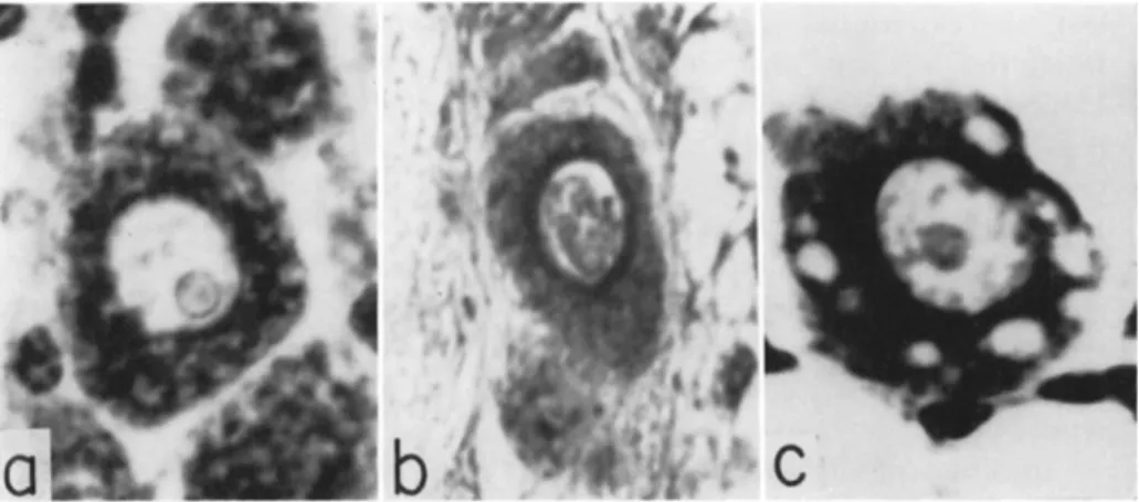
FIG. 7. Motor neuron cell-bodies in thoracic ganglia of Blattella germanica: a, normal cockroach; b, aldrin-poisoned cockroach; c, parathion-poisoned cockroach.
(From Roche and Lhoste, 1958; courtesy of J . Lhoste.)
Dieldrin-poisoned larvae of H. armigera taken in the moribund con
dition show no histopathology of the nervous system; but the walls of the midgut have become indistinct and their nuclear chromatin has compacted. Nuclear pyenosis is also evident in fat-body cells and in the muscles of the hindgut, which latter show vacuolization (Chadbourne and Rainwater, 1953). Adults of D. melanogaster and B. germanica contact-poisoned with aldrin, endrin, heptachlor, or chlordane show changes in the cell bodies of the motor neurons in the thoracic ganglia
(Fig. 7 ) ; the Nissl granules disappear from the cell periphery, while the perinuclear zone liquifies and is replaced by material chromophilic to methylene blue (Roche and Lhoste, 1958). However, these changes develop in B. germanica only when intoxication is prolonged for more than 1 day. T h e cockroach Blabera fusca Brunner, contact-poisoned with
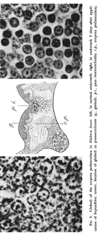
heptachlor so that it takes about a week to die, develops cytolysis in many organs during the late prostrate stage. But a symptom quite typical and independent of general degenerative change is to be found in the corpora pedunculata of the forebrain (Fig. 8 ) ; their numerous nuclei (globuli) have become pycnotic by the 3rd day, and this change in the nervous controlling center could be responsible for the ataxia caused by heptachlor (Lhoste and Roche, 1956). It would appear that the cyclodiene derivatives, which fall in the same resistance group as gamma-BHC (Busvine, 1954), act upon the ganglionic cells in some way unconnected with Cholinesterase.
c. Nicotine alkaloids. Leaf-feeding aphids contact-poisoned with nicotine quickly withdraw their proboscis, develop ataxia and a paraly
sis progressing from the hind legs to the antennae, and fall from the leaf; the extremities continue twitching until death 30 minutes later, when the legs curl up, and the cuticle has become completely dry.
Honey bees orally poisoned with nicotine show a similar ascending paralysis, the mouthparts and antennae not succumbing until after prostration, and occasional twitches of extremities preceding death (Mc- Indoo, 1916). Cockroaches (Blattella germanica, Blatta orientalis and P. americana) contact-poisoned with nicotine in the dorsal cervical re
gion develop convulsions in 2, 3, and 6 minutes, respectively, about ten times as fast as with pyrethrum; during the convulsive period before paralysis, the American cockroach swallows air and becomes greatly dis
tended (O'Kane et al., 1933).
Injection of nicotine into the blow fly Phormia regina caused an almost immediate quivering of body and appendages; the proboscis was retracted after preliminary extension, the wings were bent toward the body and the legs were usually folded together. Nicotine injected into silkworms caused expulsion of liquid from mouth and anus; convulsive movements were local or lacking in the four kinds of caterpillars tested (Mclndoo, 1937). In P. regina the injection was the more effective the closer it was to the ventral ganglion; in the larva of the white-lined sphinx Celerio lineata (Fabricius), the paralysis was faster the closer the injection was to the brain (Hockenyos and Lilly, 1932). Nicotine injected into B. germanica increased the oxygen consumption 2 to 3 times during the convulsive period before paralysis (Harvey and Brown, 1951).
T h e heart of contact-poisoned P. americana is initially accelerated but then its pulsations decline steadily, occasionally showing temporary arrests and reversals of the segmental sequence (Coon, 1944; O'Kane et.
al., 1933). With heart preparations isolated from segmental nerves, low doses of nicotine were stimulatory in B. germanica (Yeager and
FIG. 8. Globuli of the corpora pedunculata in Blabera fusca: left, in normal cockroach; right, in cockroach 3 days after appli cation of heptachlor; center, location of globuli in protocerebrum (g., globuli; p.i., pars intercerebralis; c.p., corpora pedunculata). (From Lhoste and Roche, 1956; courtesy of J. Lhoste.)
Gahan, 1937) and Melanoplus differentialis (Thomas) (Hamilton, 1939), and in P. americana the acceleration was antagonized by atropine
(Naidu, 1955). High concentrations depressed the frequency of beat and eventually arrested the heart in systole; in Stenopelmatus longispina Brunner the systolic arrest occurred during a period of acceleration and was tetanic (Davenport, 1949). T h e arrest in B. germanica and M.
differentialis could be terminated by washing the nicotine away (Yeager, 1938; Hamilton, 1939).
Nicotine characteristically acts on ganglia. Application to a thoracic ganglion of P. americana induces tremors in the leg, which disappear on severing the base of the nerve; application to the brain induces tremors throughout the body, which cease on decapitation (Yeager and Munson, 1945). Perfusion of the nerve cord itself with nicotine did result in bursts of activity (Roeder and Roeder, 1939), while treatment of the chela "slow" nerve of the crayfish resulted in multiplication of spikes into trains (Welsh and Gordon, 1947). These axonic effects are nonspecific for nicotine, seldom lead to block, and are readily reversible.
T h e main effect of nicotine in P. americana is on trans-synaptic conduc
tion, which it initially facilitates and eventually blocks (Roeder, 1953).
Nicotine is a ganglionic poison without any effects on Cholinesterase;
it did not inhibit this enzyme in homogenates from Apis mellifera (Richards and Cutkomp, 1945a) or P. americana (Hartley and Brown, 1955). It failed to inhibit dehydrogenase activity in homogenates of Passahis cornutus (Shafer, 1915) or cytochrome oxidase taken from P.
americana (Morrison and Brown, 1954). No histological lesions have been found, and nicotine-poisoned Prodenia eridania did not even show changes in the hemocytes (Yeager and Munson, 1942). However, pro
longed exposure to nicotine vapor has induced vacuolization in these hemocytes; and direct addition of nicotine to suspended fat cells and oenocytes of A. mellifera has caused degeneration of cytoplasm and cell walls (Melndoo, 1916).
Anabasine is a closely related alkaloid which facilitates and blocks synaptic transmission; experiments on crayfish have shown that it is less active than nicotine and may protect the ganglion against nicotine sub
sequently applied (Wiersma and Schallek, 1947). Contact application of anabasine to Scirtothrips citri (Moulton) results in ataxia and con
vulsions, followed in 10 minutes by a prolonged paralysis with slight movement of the extremities (McGregor, 1944). Larvae of the sawfly Nematus ribesii (Scopoli) dipped in anabasine showed marked kinesis, emitted fluid from mouth and anus, and became paralyzed in about 10 minutes; the heartbeat was very greatly increased (Tarasova, 1936) and so was the respiratory rate (Rotman, 1936).
d. Organic thiocyanates. These compounds cause rapid knockdown of insects; however, unlike pyrethrins, they do not induce a preceding hyperactive stage, as exemplified by the action of Lethane 384 (butoxy- thiocyanodiethyl ether) on Blatta orientalis (Hutzel, 1942a). Contact- applied to P. americana, thiocyanopropylphenyl ether caused longitu
dinal convulsions of the body and progressive paralysis of the legs (Hartzell and Wilcoxon, 1934). Injected into Blattella germanica, Le
thane 60 (thiocyanoethyl laurate) caused a brief period of excitement, then extension of the legs and twitches of the extremities before paraly
sis became complete (Brown, 1951).
T h e thiocyanates have a depressing action on the heart of P. ameri
cana, as found after contact application of Lethane 384 (Coon, 1944) and injection of high doses of Lethane 60 (Orser and Brown, 1951).
This depression was produced by 10 different aliphatic thiocyanates applied to the heart of B. orientalis isolated from its segmental nerves, and stoppage occurred in diastole with the alary muscles contracted (Yeager et al., 1935). Thiocyanate poisoning also depresses the respir
atory rate, as found with injection of Lethane 60 into B. germanica (Harvey and Brown, 1951) and contact application of Lethane B71 (dithiocyanodiethyl ether) to Oryzaephilus surinamensis (Lord, 1949).
T h e gross symptoms suggest a paralytic poison of the central nervous system and particularly the ganglia, as found by Taubmann (1930) for propyl thiocyanate on both warm-blooded and cold-blooded animals.
However, the thiocyanates do not inhibit insect cholinesterases (Rich
ards and Cutkomp, 1945a; Hartley and Brown, 1955). Larvae of Culex pipiens poisoned by Thanite (mainly isobornyl thiocyanoacetate) showed vacuolization of the nerve cord while still capable of move
ment (Richards and Cutkomp, 1945b). Larvae of Tenebrio molitor contact-poisoned with thiocyanopropyl phenyl ether, taken at death 16 hours later, showed histopathological changes in the ventral nerve gan
glia; the cells were degenerated so that they were stained with toluidine blue, while the tigroid of the Nissl granules was disrupted, and vacuo
lization was extensive (Hartzell and Wilcoxon, 1934). House flies sprayed with thiocyanates and fixed 10 minutes later showed histopatho
logical changes in the brain. With Lethane 384 the cells were destroyed, their nuclei became more chromophilic, and the fibers became more prominent; with Thanite at high concentrations the larger cells became vacuolated. A muscular change was detected with Lethane 384, the muscles at the back of the head showing destruction of the nuclear membrane ( Hartzell, 1945). T h e evidence does suggest that the thio
cyanates act as ganglionic poisons by disrupting the nerve cells.
2. Anticholinesterases
a. Organophosphorus insecticides. Injection of simpler organic phosphates such as D F P or T E P P ( H E T P ) into P. americana produces the successive symptoms of hyperactivity and hyperexcitability, exagger
ated tonus, ataxia, clonic and tonic convulsions, paralysis and death (Chadwick and Hill, 1947). T h e symptoms with T E P P commence in 10 to 30 minutes, and are particularly characterized by violent tremors of the body and appendages; on the other hand with parathion they do not commence for 2 to 7 hours after injection; in both cases death is delayed for 24 to 36 hours (Chamberlain and Hoskins, 1951). With malathion, death is very slow, being delayed for about 5 days (O'Brien, 1956). In house flies contact-treated with parathion, hyperactivity de
velops in 30 minutes, paralysis in 3 hours, and death in 24 hours (Men- gle and Casida, 1958); during the convulsions the ovipositor is strongly extruded and the legs and tarsi are extended, finally to become flexed (Wiesmann, 1951). In silkworms topically treated with parathion, a swelling develops at the point of application and passes backward, while the larvae make snakelike winding movements, expressing much fluid from mouth and anus; the movements become stiffer, finally to relax and cease in 12 hours (Jochum, 1956).
During the hyperactive period before paralysis, German cockroaches injected with parathion or T E P P show a threefold increase in oxygen consumption (Harvey and Brown, 1951). Malathion, however, injected into P. americana did not increase respiratory intake (O'Brien, 1956).
Injected parathion immediately increased the rate of heartbeat in P.
americana (Orser and Brown, 1951) and Anopheles quadrimaculatus (Jones, 1957b); the isolated heart of the former species was stimulated by paraoxon but not by parathion (Naidu, 1955). Parathion and re
lated organophosphorus compounds had no effect on the heartbeat of adult Oncopeltus fasciatus and larvae of Galleria mellonella (Beard, 1953).
In parathion-poisoned silkworms and larvae of Dendrolimus pini (Linnaeus), there is such an excessive secretion of fluid into the gut and thence voided from mouth and anus in the first 2 hours that the hemolymph loses one-third of its water content and the body one- quarter of its weight (Jochum, 1956). Similar loss of free body fluids was observed in P. americana prostrated by T E P P (Roan et al., 1950).
In B. germanica residually poisoned by methylparathion there is little loss of water, less than with D D T ; however, there is considerable expen
diture of carbohydrate, but not of fat (Clark and Butz, 1961).
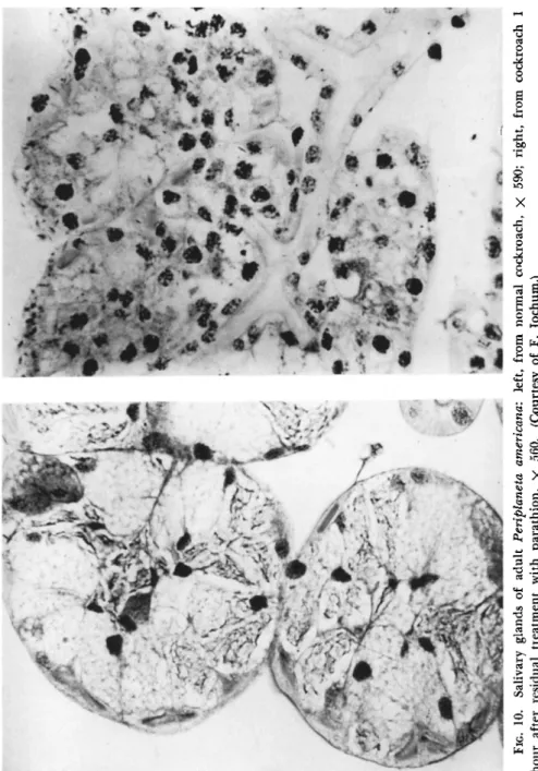
When radioactive T E P P or parathion is topically applied to P. amer-