RESTORATION OF PIGMENT SYNTHESIS IN MUTANT MELANOCYTES
AFTER FUSION WITH CHICK EMBRYO ERYTHROCYTES
D. G. SCHALL J. A. BRUMBAUGH
School of Life Sciences University of Nebraska
Lincoln, Nebraska
I. INTRODUCTION
When chick erythrocyte nuclei are fused with various estab- lished mammalian cell lines, they often resume DNA and RNA syn- thesis (Harris, 1967; Johnson and Harris, 1969). When a reacti- vating erythrocyte nucleus develops a nucleolus and synthesizes RNA (Sidebottom and Harris, 1969), it begins the synthesis of chick specific proteins (Harris et al., 1966; Cook, 1970). When the host nucleus of a heterokaryon enters the cell cycle, the chick nucleus may also begin DNA synthesis, but out of phase with the host nucleus. This asynchrony of DNA synthesis causes "pre- mature chromosome condensation" or "chromosome pulverization" in the chick nucleus (Schwartz et al., 1971; Johnson et al., 1970;
Kato and Sandberg, 1968). It is possible for these chromatin
491
fragments to become incorporated into the nucleus of the host cell (Schwartz et al., 1971) but remain karyotypically undetectable (Boyd and Harris, 1973). Cook (1970), Schwartz et al. (1971) and Klinger and Shin (1974) showed that mouse A9 cells defective for inosinic acid pyrophosphorylase (IMP: pyrophosphate phosphori- bosyl transferase, E. C. 2.4.2.8), can produce clones of IMP- producing cells after fusion with chick embryo erythrocyte nuclei.
Chick specific IMP was produced by these stable, reproducing, postfusion cells.
In this study chick embryo neural crest melanocytes, defective in pigment production, were fused with inactive erythrocyte nuclei from normally pigmented embryos. Colonies of pigment producing cells were recovered indicating restoration of normal pigment synthesis.
II. MATERIALS AND METHODS
A. Melanocyte Cultures from Neural Crest
The posterior two-thirds of the somites of 72 hour chick em- bryos were dissected, trypsinized and plated out in 60 mm culture dishes at 2.0-3.0 x 10^ cells per dish. They were grown in medi- um F-12 (GIBCO) with 1% bovine serum albumin, 2% fetal calf serum, and antibiotics at 38.5°C in a 5% C 02 incubator. On the third and fourth culture days, small colonies of melanocytes became evident which proliferated so that on days 5 and 6 melanocytes accounted for approximately 70-90% of the cell population. In this study wild type (normal; +P k/ +P k) melanocytes produced dark pigment granules as early as days 4 and 5 while pinkeye (pk/pk) melanocytes never produced overt pigment. Pinkeye melanocytes both in vivo and in vitro fail to deposit melanin upon the forming pigment granule (prernelanosome) matrices (Brumbaugh, 1968; Brum- baugh et al., 1973; Brumbaugh and Lee, 1975). Some cultures were labeled with 3H-thymidine (1.0 μ Ci/ml) during the 24 hours prior
PIGMENT SYNTHESIS IN MELANOCYTES 493
to harvesting for fusion. Fertile eggs for the cultures were from genetic stocks maintained at the School of Life Sciences, Univer- sity of Nebraska-Lincoln.
B. Erythrocytes
Erythrocytes were collected from 9-10 day chick embryos of the wild type (pigmented; +P k/+Pk) genotype by cutting the allan- toic blood vessels and subsequently collecting the allantoic fluid
(Bolund et al., 1969). The cells were centrifuged and washed twice in Hank's solution without glucose and counted. This prep- aration contains essentially only erythrocytes since granulocytes do not normally enter the circulation until the 14th day of incu- bation. Thrombocytes are rarely encountered (Lucas and Jamroz, 1961).
C. Cell Fusion
Five or 6-day melanocyte cultures were trypsinized, counted, and pelleted into a centrifuge tube. They were then mixed thor- oughly with 0.5 ml of the erythrocyte preparation so that the ratio of erythrocytes to melanocytes was 10:1. U. V. inactivated Sendai virus preparation (0.5 ml; Connaught Laboratories, Wil- lowdale, Ontario) was then added so that a final concentration of 12-16,000 HAU1 s per ml was achieved. A typical experiment in- volved 1.5-2.0 x 1 06 melanocytes and 1.5-2.0 x 1 07 erythrocytes.
The remainder of the fusion procedure was a modification of Harris et al. (1966). After fusion the cells were replated in standard medium on the basis of 1.0 x 10^ melanocytes per dish.
D. Microscopy
At 24 and 48 hours post-fusion sample dishes were fixed and stained with Giemsa for light microscopic determination of fusion rate and plating efficiency.
Living, post-fusion cultures were periodically screened for heterokaryons and/or pigment production with the light microscope.
When desired cells or colonies were located they were fixed in situ for electron microscopy and embedded. Ultrathin sections made parallel to the plane of each culture dish were stained with uranyl acetate and lead citrate, then viewed and photographed using a Philips 201C electron microscope.
Electron microscopic autoradiography was performed according to the method of Brumbaugh and Lee (1975) using Ilford L-4 emul- sion and an exposure time of 12 days.
III. RESULTS
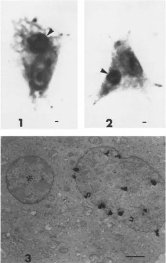
Twenty-four hours after fusion, Giemsa stained cultures showed that approximately 13.0% of the melanocytes contained one or more erythrocyte nuclei. Erythrocyte nuclei were readily dis- cerned because of their small size and condensed chromatin. Fig- ure 1 shows a heterokaryon between a wild type pigmented melano- cyte containing a few pigment granules and an erythrocyte nucleus
(Fig. 1, arrow). Figure 2 shows a similar heterokaryon but the melanocyte is of the amelanotic pinkeye genotype, hence no pig- ment granules. Further verification of erythrocyte fusion is
shown in the high resolution autoradiograph (Fig. 3) prepared 24 hours after fusion. The larger, less condensed, labeled melano- cyte nucleus is clearly distinguished from the smaller, condensed, unlabeled erythrocyte nucleus (*, Fig. 3). The erythrocyte nu- cleus is completely surrounded by melanocyte cytoplasm and is clearly an integral part of the cell.
Melanocyte homokaryons were also classified in the Giemsa stained preparation at a frequency of 32.5%. Not all of these are true homokaryons since suspected multinucleated cells fre- quently contained separating cell membranes when observed with the electron microscope.
PIGMENT SYNTHESIS IN MELANOCYTES 495
FIGURE 1 Heterokaryon of a wild type (+Pk/+Pk) chick melano- cyte and a chick embryo erythrocyte nucleus (arrow) 24 hr post fusion. ¢ few pigment granules are present in the cytoplasm.
2225X. Scale equals 1 micron.
FIGURE 2 Heterokaryon of a pinkeye (pk/pk) chick melanocyte and a chick embryo erythrocyte nucleus (arrow) 48 hr post fusion.
No pigment granules are present as is characteristic of the pink- eye genotype. 2500X. Scale equals 1 micron.
FIGURE 3 High resolution autoradiograph of a 3H-thymidine labeled melanocyte and a chick embryo erythrocyte nucleus (*) 24 hr post fusion. Silver grains are evident over the melanocyte nucleus but not the erythrocyte nucleus. 14,000X. Scale equals 1 micron.
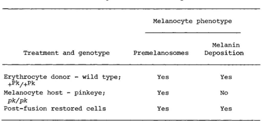
TABLE I Summary of Restoration Experiments
Melanocyte phenotype
Melanin Treatment and genotype Pr erne lano some s Deposition
Erythrocyte donor - wild type? Yes Yes
+P k/ +P k
Melanocyte host - pinkeye; Yes No
pk/pk
Post-fusion restored cells Yes Yes
Two fusion experiments using a total of 3 . 4 x 1 0 ^ melanocytes of the amelanotic pinkeye (pk/pk) genotype and 3 . 4 x 1 07 erythro- cytes from the pigmented, wild type (+P k/+Pk) genotype were con- ducted. Three pigment producing colonies, one in the first exper- iment and two in the second, were detected after visual screening.
Each colony occurred in a separate dish. One colony was detected on the fourth post-fusion day and was followed for 32 additional days during which time it divided several times. All of the daughter cells appeared to be pigment producers. The second and third colonies were detected on days 10 and 24 after fusion and were subsequently fixed for electron microscopy. Table I sum- marizes the genetic design of the experiments and the results.
Figure 4 is a portion of the cytoplasm from a typical, syn- thetically active, pinkeye melanocyte. Even though the cell has been subjected to the cytochemical test for dopa oxidase (notice the darkened Golgi system), the forming pigment granules (pre- melanosomes) show no sign of melanin deposition (see arrow. Fig.
4 ). This cell is to be contrasted with the 24 day, post-fusion cell in Fig. 5 , which contains several frank pigment granules in various stages of melanin deposition.
PIGMENT SYNTHESIS IN MELANOCYTES 497
FIGURE 4 Cytoplasm of a pinkeye (pk/pk) melanocyte in cul- ture. The Golgi system has been cytochemically stained. The ar- row locates a solitary unpigmented prernelanosome. 21,000X.
Scale equals 1 micron.
FIGURE 5 ¢ melanogenically active cell from a colony of such cells present in a pinkeye (pk/pk) culture 24 days after fusion with wild type erythrocyte nuclei. Note the several electron dense pigment granules. 22,750X. Scale equals 1 micron.
The 24 hour post-fusion plating efficiency, as determined by counting attached cells in Giemsa stained preparations, was ap- proximately 20%. This means that only 20% of the 3.4 x 1 06
melanocytes fused (6.8 χ 105) were recovered. Thus approximately one pigment colony was produced per 2.3 x 10^ recovered melano- cytes.
IV. DISCUSSION
There are several ways of explaining the restoration of pig- ment synthesis in the pinkeye melanocytes. Since only mononu- cleated dividing pigment cells were recovered, it is not possible to suggest that a reactivated erythrocyte nucleus in a persisting heterokaryon produced the factor necessary for pigment synthesis.
The rate of restoration (1 in 2.3 x 10$) seems too high to be due to back mutation. This is corroborated by the fact that pinkeye adult feathers do not show wild type flecks or "ticks". Integra- tion of the erythrocyte genome and the melanocyte genome to pro- duce a tetraploid cell is a plausible explanation since karyo- types of the pigmented clones were not determined. Such integra- tions have not been reported in chick erythrocyte-mammalian cell fusions, however (Boyd and Harris, 1973; Schwartz, et al., 1971;
Klinger and Shin, 1974.
The differentiating melanocytes used in these experiments were mitotically very active, dividing both before and after fusion.
This lends credence to the hypothesis that "premature chromosome condensation" or "chromosome pulverization" (Johnson, et al., 1970) occurs in the erythrocyte nucleus of the heterokaryons as they attempt to divide in synchrony with the melanocyte nucleus.
An erythrocyte-derived chromosome fragment, bearing the wild type allele of the pinkeye lesion could explain the restoration of pigment production if it became incorporated into the melanocyte nucleus. A similar hypothesis is used to explain the production of chick IMP in IMP deficient mouse A9 cells after fusion with chick erythrocytes (Schwartz, et al., 1971; Boyd and Harris, 1973). Like Boyd and Harris (1973) and Schwartz (1971) we also
P I G M E N T S Y N T H E S I S I N M E L A N O C Y T E S 499
obtained a phenotypically stable, dividing population of restored cells.
Several investigators (Cook, 1970; Schwartz, et al., 1971;
Boyd and Harris, 1973; Klinger and Shin, 1974) have shown that it is possible to correct genetically defective mammalian cells by fusion with chick erythrocytes. The correcting molecules were usually chick specific. All of these systems depended on some type of cell selection and usually involved ubiquitous enzymes necessary for cell survival and/or growth. In this study restora- tion required the synthesis of a specific product of differentia- tion-melanin. Thus a pigment gene normally inactive in the erythrocyte is apparently activated after exposure to the nuclear or cytoplasmic environment of the melanocyte.
V. SUMMARY
Five to 6-day cultures of chick embryo melanocytes of the amelanotic pinkeye genotype were fused with 9-10 day chick embryo erythrocytes from pigment-producing, wild type embryos. Three colonies of pigment producing cells were produced in two different experiments at a rate of one per 2.3 x 1 05 melanocytes recovered after fusion. These results strongly suggest that the wild type pigment gene from the erythrocyte nucleus is activated after ex- posure to the nuclear and/or cytoplasmic environment of the melanocyte.
REFERENCES
Bolund, L., Ringertz, N. R., Harris, Ç., (1969), J. Cell Sei. 4, 71-87.
Boyd, Y. L., Harris, Ç., (1973), J. Cell Sei. 13, 841-861.
Brumbaugh, J., (1968), Devep. Biol. 18, 375-390.
Brumbaugh, J., Lee, L., (1975), Genetics 44, 333-347.
Brumbaugh, J,, Bowers, R., Lee, Ê., (1973), Yale Journal of Bio- logy and Medicine 46, 523-534.
Cook, P. R., (1970), J. Cell Sei. 7, 1-3.
Harris, Ç., (1967), J. Cell Sei. 2, 23-32.
Harris, Ç., Watkins, J. F., Ford, C. Å., Schoefl, G. I., (1966), J. Cell Sei. 4, 499-525.
Johnson, R. T., Harris, Ç., (1969), J. Cell Sei. 5, 625-643.
Johnson, R. T., Rao, P. Ν., Hughes, H. D., (1970), J. Cell.
Physiol. 76, 151-158.
Kato, H., Sandberg, Á. Á., (1968), J. Nat. Cancer Inst. 41, 1117- 1123.
Klinger, H. P., Shin, S., (1974), Proc. Nat. Acad. Sei. U.S.A. 71, 1398-1402.
Lucas, A. M., Jamroz, C., (1961), "Atlas of Avian Hematology,"
Agriculture Monograph 25, United States Department of Agricul- ture.
Schwartz, A. G., Cook, P. R., Harris, Ç., (1971), Nature New Biology 230, 5-8.
Sidebottom, Å., Harris, Ç., (1969), J. Cell Sei. 5, 351-364.
ACKNOWLEDGMENTS
This investigation was supported by NIH research grant GM18969 from the National Institute of General Medical Sciences.
Note added in proof: Leung, W. C., Chen, T. R., Dubbs, D. R., Kit, S., (1975), Exp. Cell Res. 95, 320-326, have restored TK"
mouse cells by fusing them with chick erythrocytes. The function- ing thymidine kinase (TK) was electrophoretically of the chick type and was accompanied by chick microchromosomes.