O R I G I N A L P A P E R
Exophiala dermatitidis Endophthalmitis: Case Report and Literature Review
Mo´nika Homa.Palanisamy Manikandan.Veerappan Saravanan. Rajaraman Revathi.Raghavan Anita.Venkatapathy Narendran.
Kanesan Panneerselvam.Coimbatore Subramanian Shobana.Mohammed Al Aidarous . La´szlo´ Galgo´czy.Csaba Va´gvo¨lgyi.Tama´s Papp.La´szlo´ Kredics
Received: 18 May 2017 / Accepted: 16 December 2017
ÓSpringer Science+Business Media B.V., part of Springer Nature 2018
Abstract We report a case of a 59-year-old male patient with a postoperative fungal infection of the left eye. A dark-pigmented yeast,Exophiala dermatitidis (previously known as Wangiella dermatitidis), was identified from the culture of the biopsy taken from the posterior capsule. The infection was successfully eradicated by a combination of surgical and medical (i.e., voriconazole and fluconazole) treatment. This is the first report of successfully treatedE. dermatitidis endophthalmitis, which demonstrates that a prompt
and aggressive antifungal therapy combined with surgical intervention is necessary to prevent vision loss in cases of endophthalmitis due to Exophiala species. Beside the case description, we also aim to provide a literature review of previously reported eye infections caused byExophialaspecies in order to help the future diagnosis and management of the disease.
Keywords Dematiaceous fungiExophiala KeratitisEndophthalmitisAntifungal susceptibility
Mo´nika Homa and Palanisamy Manikandan have been contributed equally to this work.
M. HomaT. Papp
MTA-SZTE ‘‘Lendu¨let’’ Fungal Pathogenicity Mechanisms Research Group, Ko¨ze´p Fasor 52, Szeged 6726, Hungary
M. HomaL. Galgo´czyC. Va´gvo¨lgyi T. Papp (&)L. Kredics
Department of Microbiology, Faculty of Science and Informatics, University of Szeged, Ko¨ze´p Fasor 52, Szeged 6726, Hungary
e-mail: pappt@bio.u-szeged.hu
P. ManikandanV. SaravananR. Revathi R. AnitaV. Narendran
Aravind Eye Hospital and Postgraduate Institute of Ophthalmology, Coimbatore, Tamilnadu 641 014, India P. ManikandanM. A. Aidarous
Department of Medical Laboratory Sciences, College of Applied Medical Sciences, Majmaah University, Al Majma’ah 11952, Saudi Arabia
P. Manikandan
Greenlink Analytical and Research Laboratory India Private Limited, Coimbatore, Tamilnadu 641 014, India K. Panneerselvam
Research Department of Microbiology, M.
R. Government Arts College,
Mannargudi, Tiruvarur District, Tamilnadu 614 001, India C. S. Shobana
Department of Microbiology, PSG College of Arts and Science, Coimbatore, Tamilnadu 641 014, India https://doi.org/10.1007/s11046-017-0235-4
Background
Fungal eye infections are among the main causes of visual disability in tropical and subtropical countries, such as India. Besides hyaline fungal species, which are responsible for the majority of keratomycoses and fungal endophthalmitis in the country, dark-pig- mented fungi are also associated with emerging cases [1,2]. The cosmopolitan genusExophialacomprises dimorphic dematiaceous fungi commonly isolated from soil and plant debris [3,4]. These fungi are the clinically most relevant black yeasts causing various uncommon forms of cutaneous, subcutaneous and disseminated (e.g., pneumonia and brain abscess) human infections [5], but eye infections due to
Exophiala species are extremely rare. Most cases occur after a penetrating injury or post-eye surgery [6].
Though the presented case is the fourth documented case of endophthalmitis caused by E. dermatitidis across the globe according to a deep search of the available literature (Table1) [7–18], it is unique, as it is the first report with a successful treatment outcome.
Human ocular infection due toE. dermatitidishas not been documented in India before.
Case Presentation
A 59-year-old male patient presented at the Cornea Department of Aravind Eye Hospital and Postgraduate Table 1 A retrospective summary ofExophialaeye infections reported since 1990
Aetiological agent Age/
Gendera
Country Therapyb Underlying conditionc Outcome References
Keratitis
E. dermatitidis 35/M Czechoslovakia AMB NF1,
immunodeficiency
Leukoma [7]
E. jeanselmei 42/M Saudi Arabia NTM, MCN, AMB – Improved [8]
E. jeanselmei 58/F Israel NTM, AMB – Improved [6]
E. dermatitidis 52/M USA AMB – Improved [9]
E. jeanselmei 39/F USA AMB, ITC – Improved [10]
E. jeanselmei 41/M USA VRC, NTM, KTC,
SI
– Improved [11]
E. phaeomuriformis 84/F USA VRC, SI – Improved [12]
Subconjunctival mycetoma
E. dermatitidis 44/F China SI – Improved [13]
Endophthalmitis
E. dermatitidis 75/F USA AMB, SI Diabetes Enucleation [14]
E. dermatitidis 31/M France ITC, AMB, SI – Failed [15]
E. jeanselmei 67/F Brazil AMB Purpura Atrophy [16]
E. jeanselmei 52/F Brazil AMB Diabetes Atrophy [16]
E. dermatitidis 60/M USA AMB, SI Crohn’s disease,
Herpes zoster keratitis
Enucleation [17]
Exophialasp. 65/F Puerto Rico VRC, FLC, SI Diabetes Improved [18]
E. dermatitidis 59/M India VRC, FLC, SI – Improved Present
case
aFfemale;Mmale
bAMB amphotericin B; FLC fluconazole; ITC itraconazole; KTC ketoconazole; MCN miconazole; NTM natamycin; PSC posaconazole;SIsurgical intervention;VRCvoriconazole
cNF1neurofibromatosis type I
Institute of Ophthalmology (Coimbatore, Tamilnadu, India) in December 2012 with complaints of decreased vision, pain and redness in his left eye for the past 15 days. The patient gave a history of an uneventful cataract surgery with intraocular lens implantation in his left eye performed 4 months earlier. He had good vision in the first month following surgery, that is, visual acuity (VA) was 6/6. Three months later, he presented with gradual deterioration of vision (VA 6/60). The patient was otherwise in good health and had no known allergies.
On examination, the best corrected visual acuities were 6/6 in the right eye and 6/60 in the left eye.
Intraocular pressure as measured by Goldmann appla- nation tonometry was 15 mm in the right eye and 13 mm in the left eye. Slit lamp examination of the right eye was within normal limits except for an early nuclear sclerotic cataract of grade 1. Examination of the left eye revealed circumcorneal congestion, a clear cornea and 3? cells and 1? flare in the anterior chamber. Texture and color of the iris were normal.
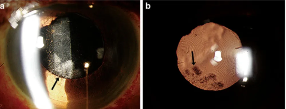
The pupil was pharmacologically dilated and unre- sponsive to light. A posterior chamber intraocular lens was present, and a fluffy grayish white growth was noted on the posterior capsule (Fig.1a, b).
Indirect ophthalmoscopy of the right eye was within normal limits, but in the left eye moderate vitritis was observed. The patient underwent vitreous tap with a 24-gauge needle inserted into the central vitreous cavity along with intravitreal injections of moxifloxacin (500lg/0.1 ml; Vigamox, Alcon Labo- ratories, USA) and voriconazole (100 lg/0.1 ml;
Aurolab, India). He was placed on topical moxi- floxacin (0.5% (w/v); Vigamox, Alcon, India) and fortified tobramycin (14 mg/ml; Toba, Sun Pharma
Laboratories Ltd., India) eight times a day, plus voriconazole (1% (w/v); Vozole, Aurolab, India) eye drops six times a day and homatropine (2% (w/v);
Homide, Warren, India) eye drops twice a day. The topical treatment continued till the time of the intraocular lens removal (1 month from the first vitreous tap).
Vitreous biopsies obtained during the procedure were plated on 5% sheep blood agar (SBA; HiMedia Laboratories, India), potato dextrose agar (PDA;
HiMedia Laboratories, India) and thioglycolate broth (HiMedia Laboratories, India). A part of the sample was sent for molecular identification (Xcyton Diag- nostics Limited, Bengaluru, India). Gram stained smears and 10% potassium hydroxide wet mount were also investigated. The smears as well as the cultures proved to be negative for fungal filaments.
Molecular analyses were negative for all the tested bacteria and fungi. Postoperatively the same medica- tions were continued. Prednisolone acetate (1% (w/v);
Predforte, Allergan, India) was added at a frequency of six times a day. Dexamethasone (8 mg in 2 ml;
Dexadran, Searle Labs Pvt. Ltd. India) was adminis- tered intravenously twice a day for 2 days and was subsequently replaced by oral prednisolone (40 mg/day; Omnacortil, MacLeods Pharmaceuticals Ltd., India), tapered over a period of 15 days.
On subsequent follow-ups, the visual acuity was gradually improving (i.e., VA 6/9 after 1 week post- vitreous tap), but the anterior chamber inflammation persisted, and fluffy deposits on posterior surface of the intraocular lens persisted. Oral steroids were discontinued, and the topical steroid dose (6 times per day; Predforte, Allergan, India) was tapered to four times. A repeated vitreous biopsy was performed on
Fig. 1 Slit lamp biomicroscopy (a) with retro illumination (b) showing the fungal colonies on the posterior surface of the intraocular lens
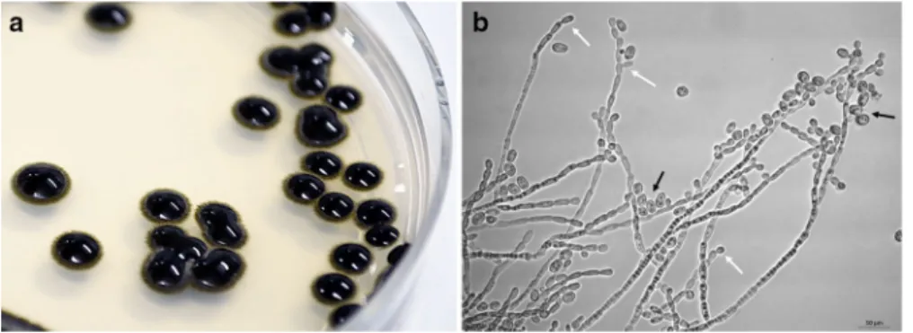
the 9th day following the first vitreous tap under local anesthesia using a 25-gauge vitreous cutter through the pars plana route. Care was taken to take the sample at the location of the fluffy growth with localized posterior capsulectomy and anterior vitrectomy. Spec- imens thus taken from the posterior capsule and anterior vitreous were plated on SBA, chocolate agar (CA; HiMedia Laboratories, India), PDA and Lowen- stein Jensen medium (HiMedia Laboratories, India). A direct microscopic examination of Gram’s smears clearly revealed fungal filaments. The topical steroid therapy was stopped, and the frequency of topically applied voriconazole (1% (w/v)) was increased to hourly intervals. The therapy was supplemented with oral fluconazole (150 mg; Zocon, FDC Limited, India) twice a day. On the 5th day of incubation, a moderate- sized black, mucoid and yeast-like growth was noted on SBA, CA and PDA plates. The fungus was initially identified as E. dermatitidis based on colony mor- phology (Fig.2a) and microscopic features (Fig.2b) and was further subjected to molecular identification.
Medical treatment was continued for the next 10 days.
The infiltrate reappeared in the inferonasal quadrant in between the lens and the iris. Intraocular lens explan- tation and capsular bag removal with anterior vitrec- tomy were performed followed by an intravitreal voriconazole injection (100 lg/0.1 ml). Postopera- tively, corneal edema and keratic precipitates were noted which resolved after 2 weeks. Intravitreal injections of voriconazole (100 lg/0.1 ml) were repeated twice in the ensuing week for persistent exudates in the inferior vitreous. While topical voriconazole (1% (w/v)) was continued and slowly tapered over 2 months, systemic fluconazole (150 mg twice daily) was maintained for a month. After a
month, topical steroids (Predforte, Allergan, India) were started with a very low dose twice a day, and tapered very slowly over the next 4 months. The best corrected Snellen’s visual acuity with aphakic correc- tion was 6/9.
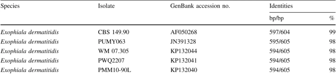
For the molecular identification, genomic DNA was extracted using theMasterPure Yeast DNA Purifica- tion Kit(Epicentre Biotechnologies, USA) following the instructions of the manufacturer. PCR amplifica- tion was carried out by targeting the internal tran- scribed spacer (ITS) region using ITS1 and ITS4 primers [19]. The sequences were determined by LGC Genomics GmbH (Germany), and the consensus of the two reads was assembled by using the Staden package (Pregap4 v.1.5 and Gap4 v4.10) [20] and manually edited in BioEdit [21]. The obtained 592 bp long fragment was compared with the available sequences in the GenBank using the Basic Local Alignment Search Tool (BLAST) [22]. The isolate was finally identified as E. dermatitidis, as its ITS sequence displayed a 99% sequence homology with the ITS region ofE. dermatitidisCBS 149.90 (Table2). While the isolate itself was deposited in the Szeged Micro- biological Collection (SZMC, Szeged, Hungary;
http://szmc.hu/) under the strain number of SZMC 21989, its ITS sequence was deposited in the EMBL Nucleotide Sequence Database under the accession number of LN809939.
The antifungal susceptibility profile of the case isolate was determined to 12 clinically relevant antifungal agents. The Etest method (BioMe´rieux, France) was used to determine the susceptibility to echinocandins (anidulafungin, caspofungin, mica- fungin), ketoconazole and posaconazole according to the instructions of the manufacturer. Minimum
Fig. 2 Colony (a) and microscopic morphology (b) ofExophiala dermatitidisSZMC 21989 cultured on Sabouraud’s agar for 7 days.
White arrows indicate terminal and intercalary phialides, while black arrows indicate the yeast-like cells
inhibitory concentration (MIC) values of the remain- ing seven antifungals were determined in accordance with the instructions of the CLSI (Clinical and Laboratory Standards Institute) M38-A2 broth microdilution method [23], with the involvement of the following commercially available standard pow- ders and eye drops: amphotericin B, (Amphocare injection, BPRL Pvt ltd, Bangalore, India), clotrima- zole (Auroclot, 1% (w/v) suspension, Aurolab, Madu- rai, India), econazole (Aurozole, 2% (w/v) suspension, Aurolab, Madurai, India), fluconazole (0.3% (w/v), Zocon, FDC ltd, Aurangabad, India), itraconazole (Itral, 1% (w/v) suspension, Jawa Pharmaceuticals, Guargon, India), natamycin (Natamet, 5% (w/v) suspension, Sun Pharmaceuticals Ind. Ltd., India) and voriconazole (Vfend, Pfizer Inc., India). Three replicates were involved in all the susceptibility tests.
The obtained MIC values are summarized in Table3.
MIC was defined as the lowest antifungal concentra- tion, which was required for the total growth inhibition of the test isolate. Susceptibility testing revealed that the MICs of amphotericin B, clotrimazole, econazole, fluconazole, itraconazole, ketoconazole, posaconazole and voriconazole were[0.5lg/ml, whereas MICs for natamycin and echinocandins were 8lg/ml and[32lg/ml, respectively.
Discussion and Conclusions
According to the available literature in the PubMed database (http://www.ncbi.nlm.nih.gov/pubmed), only three Exophialaspecies, E. jeanselmei,E. der- matitidisandE. phaeomuriformishave been identified from various human eye infections such as keratitis, endophthalmitis and subconjunctival mycetoma in the last decades (Table1). Until now, six Exophiala endophthalmitis cases have been reported from dif- ferent parts of the world. The first case of endoph- thalmitis caused byE. dermatitidiswas described by Margo et al. in 1990 [14]. Since then, two other cases were reported by Benaoudia et al. in 1999 [15] and Clamp et al. in 2014 [17]. Medical therapy included surgical intervention supplemented with either intravitreal (2.5–5.0lg) or intravenous (0.2–0.8 mg/
kg) injections of amphotericin B in all cases.
Benaoudia et al. [15] also used oral itraconazole (200 mg b.i.d.) and amphotericin B eye drops (0.15%
(w/v) eight times a day). A failed therapeutic approach and consequent enucleation of the blind eyes were reported in the cases of Margo et al. [14] and Clamp et al. [17]. Endophthalmitis cases due to another spe- cies, E. jeanselmei, were also treated with systemic and intraocular amphotericin B, but ocular atrophy Table 2 Top five results of nucleotide–nucleotide BLAST search performed with the ITS sequence of the case isolate in the National Center for Biotechnology Information database (NCBI,http://www.ncbi.nlm.nih.gov/)
Species Isolate GenBank accession no. Identities
bp/bp %
Exophiala dermatitidis CBS 149.90 AF050268 597/604 99
Exophiala dermatitidis PUMY063 JN391328 595/605 98
Exophiala dermatitidis WM 07.305 KP132044 594/605 98
Exophiala dermatitidis PWQ2207 KP132041 594/605 98
Exophiala dermatitidis PMM10-90L KP132040 594/605 98
Table 3 Antifungal susceptibility profile ofExophiala dermatitidisisolate SZMC 21989
Antifungal agent Amphotericin B Anidulafungin Caspofungin Clotrimazole Econazole Fluconazole
MIC (lg/ml) 0.33 [32 [32 0.33 0.83 2.67
Antifungal agent Itraconazole Ketoconazole Micafungin Natamycin Posaconazole Voriconazole
MIC (lg/ml) 0.25 0.25 [32 8 0.38 0.25
developed in both cases [16]. These five, above men- tioned patients, suffered from different underlying disorders such as diabetes, Crohn’s disease and pur- pura. Based on these prior reports of Exophialaeye infections (Table1), underlying conditions seem to increase the chances of a failed therapy. Infections were eradicated in almost all seven cases of keratitis.
Conversely, subconjunctival mycetoma and endoph- thalmitis cases usually had poor visual outcome. In comparison to previous reports, our patient had no other complaints, and after vitrectomy he was suc- cessfully treated with intravitreal injections and eye drops of voriconazole and oral fluconazole. Based on the experience, authors recommend prompt removal of the intraocular lens and capsular bag along with vitrectomy to eradicate the infection. Also in eyes with localized growth (like in our case), care should be taken to reach the area of growth when taking speci- mens for culture and staining to increase the chance of getting a positive yield unlike in routine endoph- thalmitis cases where the specimen is usually taken from the center of the vitreous cavity. In the presented case, the surgeon took utmost care to get the repeat specimen from the suspected area of the exudate as the first specimen proved to be negative.
While in the 1990s mainly amphotericin B was chosen for the treatment ofExophialaeye infections, it was replaced in the last few years by new azole compounds, like voriconazole (Table1). Unfortu- nately, limited data are available about the in vitro antifungal activity of these new agents against Exophialaisolates derived from human eye infections.
Based on the in vitro antifungal susceptibility tests, the case isolate had variable susceptibility to different antifungal drugs (Table3). Basically, the obtained results agreed with the previous report of Chowdhary et al. [5], except from the finding that echinocandins (i.e., anidulafungin, caspofungin, micafungin) proved to be ineffective against the tested isolate in the investigated concentration range (MIC[32lg/ml), while Chowdhary et al. [5] found a much lower MIC range (0.25–8lg/ml) forE. dermatitidisstrains.
In conclusion, endophthalmitis due to E. dermati- tidis is a rare but serious infection of the eye. Its management requires a radical surgical intervention for a successful outcome, and also utmost care is necessary in collecting vitreous sample in eyes with localized infection to achieve higher culture positivity rate. The subsequent application of a carefully chosen,
effective antifungal therapy is critical for a successful outcome. This case obviously demonstrates that besidesAspergillus[24],Curvularia[25] andFusar- ium [26,27] species, Exophiala dermatitidis should also be taken into consideration as a causative agent of eye infections in South India.
Acknowledgements This study was supported by the
‘‘Lendu¨let’’ Grant of the Hungarian Academy of Sciences (LP2016-8/2016) and the Project GINOP-2.3.2-15-2016-00035 (Sze´chenyi 2020 Programme). LG is supported by the Postdoctoral Excellence Programme (PD 120808) of the Hungarian National Research, Development and Innovation Office (NKFI Office). PM would like to thank Deanship of Scientific Research at Majmaah University, Kingdom of Saudi Arabia, for supporting this work under Project No. 37/106.
Compliance with Ethical Standards
Conflict of interest The authors declare that they have no conflict of interest.
Ethical Approval This article does not contain any studies with animals performed by any of the authors.
Informed Consent For this type of study formal consent is not required.
References
1. Bharathi MJ, Ramakrishnan R, Meenakshi R, Padmavathy S, Shivakumar C, Srinivasan M. Microbial keratitis in South India: influence of risk factors, climate, and geographical variation. Ophthalmic Epidemiol. 2007;14:61–9.
2. Chakrabarti A, Shivaprakash MR, Singh R, Tarai B, George VK, Fomda BA, Gupta A. Fungal endophthalmitis: fourteen years’ experience from a center in India. Retina.
2008;28:1400–7.
3. Zeng JS, Sutton DA, Fothergill AW, Rinaldi MG, Harrak MJ, de Hoog GS. Spectrum of clinically relevantExophiala species in the United States. J Clin Microbiol.
2007;45:3713–20.
4. Kondori N, Gilljam M, Lindblad A, Jo¨nsson B, Moore ERB, Wennera˚s C. High rate ofExophiala dermatitidisrecovery in the airways of patients with cystic fibrosis is associated with pancreatic insufficiency. J Clin Microbiol.
2011;49:1004–9.
5. Chowdhary A, Meis JF, Guarro J, de Hoog GS, Kathuria S, Arendrup MC, Arikan-Akdagli S, Akova M, Boekhout T, Caira M, Guinea J, Chakrabarti A, Dannaoui E, van Diepeningen A, Freiberger T, Groll AH, Hope WW, John- son E, Lackner M, Lagrou K, Lanternier F, Lass-Flo¨rl C, Lortholary O, Meletiadis J, Mun˜oz P, Pagano L, Petrikkos G, Richardson MD, Roilides E, Skiada A, Tortorano AM, Ullmann AJ, Verweij PE, Cornely OA, Cuenca-Estrella M, European Society of Clinical Microbiology and Infectious Diseases Fungal Infection Study Group.; European
Confederation of Medical Mycology. ESCMID and ECMM joint clinical guidelines for the diagnosis and management of systemic phaeohyphomycosis: diseases caused by black fungi. Clin Microbiol Infect. 2014;20:47–75.
6. Ben-Simon GJ, Barequet IS, Grinbaum A. More than tears in your eyes (Exophiala jeanselmei keratitis). Cornea.
2002;21:230–1.
7. Pospı´sil L, Skorkovska´ S, Moster M. Corneal phaeohy- phomycosis caused by Wangiella dermatitidis. Ophthal- mologica. 1990;201:128–32.
8. Al-Hedaithy SS, Al-Kaff AS.Exophiala jeanselmeikerati- tis. Mycoses. 1993;36:97–100.
9. Patel SR, Hammersmith KM, Rapuano CJ, Cohen EJ.Ex- ophiala dermatitidis keratitis after laser in situ ker- atomileusis. J Cataract Refract Surg. 2006;32:681–4.
10. Leung EH, Moskalewicz R, Parada JP, Kovach KJ, Bou- chard C.Exophiala jeanselmeikeratitis after laser in situ keratomileusis. J Cataract Refract Surg. 2008;34:1809–11.
11. Saeedi OJ, Iyer SA, Mohiuddin AZ, Hogan RN.Exophiala jeanselmei keratitis: case report and review of literature.
Eye Contact Lens. 2013;39:410–2.
12. Aggarwal S, Yamaguchi T, Dana R, Hamrah P.Exophiala phaeomuriformisfungal keratitis: case report and in vivo confocal microscopy findings. Eye Contact Lens. 2015.
https://doi.org/10.1097/ICL.0000000000000193.
13. Li EY, Yuen HK, Lung DC. Subconjunctival mycetoma as an unusual cause of tears with black deposits. Arch Oph- thalmol. 2010;128:1371–2.
14. Margo CE, Fitzgerald CR. Postoperative endophthalmitis caused by Wangiella dermatitidis. Am J Ophthalmol.
1990;110:322–33.
15. Benaoudia F, Assouline M, Pouliquen Y, Bouvet A, Gue´ho E.Exophiala (Wangiella)dermatitidis keratitis after ker- atoplasty. Med Mycol. 1999;37:53–6.
16. Hofling-Lima AL, Freitas D, Fischman O, Yu CZ, Roizenblatt R, Belfort R Jr.Exophiala jeanselmeicausing late endophthalmitis after cataract surgery. Am J Ophthal- mol. 1999;128:512–4.
17. Clamp MF, Jumper JM, Ku CW, McDonald HR, Johnson RN, Fu AD, Lujan BJ, Cunningham ET Jr. Chronic exogenous Exophiala dermatitidisendophthalmitis. Retin Cases Brief Rep. 2014;8:265–8.
18. Quintero-Estades JA, Walter S, Valenzuela F, Amescua G.
Delayed-onset postoperative endophthalmitis secondary to
Exophiala. BMJ Case Rep. 2015.https://doi.org/10.1136/
bcr-2014-208680.
19. White TJ, Burns S, Lee S, Taylor J. Amplification and direct sequencing of fungal ribosomal RNA genes for phyloge- netics. In: Innis MA, Gelfand DH, Sninsky JJ, White TJ, editors. PCR protocols: a guide to methods and applications.
New York: Academic Press Inc; 1990. p. 315–22.
20. Bonfield JK, Smith KF, Staden R. A new DNA sequence assembly program. Nucleic Acids Res. 1995;23:4992–9.
21. Hall TA. BioEdit: a user-friendly biological sequence alignment editor and analysis program for Windows 95/98/
NT. Nucleic Acids Symp Ser. 1999;41:95–8.
22. Altschul SF, Gish W, Miller W, Myers EW, Lipman DJ.
Basic local alignment search tool. J Mol Biol.
1990;215:403–10.
23. Clinical and Laboratory Standards Institute. Reference method for broth dilution antifungal susceptibility testing of filamentous fungi. In: Approved standard CLSI document M38-A2. Wayne: CLSI; 2008.
24. Manikandan P, Varga J, Kocsube´ S, Anita R, Revathi R, Ne´meth TM, Narendran V, Va´gvo¨lgyi C, Panneer Selvam K, Shobana CS, Babu Singh YR, Kredics L. Epidemiology ofAspergillus keratitis at a tertiary care eye hospital in South India and antifungal susceptibilities of the causative agents. Mycoses. 2013;56:26–33.
25. Krizsa´n K, To´th E, Nagy LG, Galgo´czy L, Manikandan P, Chandrasekaran M, Kadaikunnan S, Alharbi NS, Va´gvo¨lgyi C, Papp T. Molecular identification and antifungal suscep- tibility ofCurvularia australiensis,C. hawaiiensisandC.
spicifera isolated from human eye infections. Mycoses.
2015;58:603–9.
26. Homa M, Shobana CS, Singh YR, Manikandan P, Selvam KP, Kredics L, Narendran V, Va´gvo¨lgyi C, Galgo´czy L.
Fusarium keratitis in South India: causative agents, their antifungal susceptibilities and a rapid identification method for the Fusarium solani species complex. Mycoses.
2013;56:501–11.
27. Hassan AS, Al-Hatmi AM, Shobana CS, van Diepeningen AD, Kredics L, Va´gvo¨lgyi C, Homa M, Meis JF, de Hoog GS, Narendran V, Manikandan P, IHFK Working Group.
Antifungal susceptibility and phylogeny of opportunistic members of the genus Fusarium causing human kerato- mycosis in South India. Med Mycol. 2016;54:287–94.