JRM JRM J
ournal ofR
ehabilitationM
edicineJRM J
ournal ofR
ehabilitationM
edicineORIGINAL REPORT
DOES INSPIRATION EFFICIENCY INFLUENCE THE STABILITY LIMITS OF THE TRUNK IN PATIENTS WITH CHRONIC LOW BACK PAIN?
Regina FINTA, PhD1 , Krisztina BODA, PhD2, Edit NAGY, PhD3#and Tamás BENDER, PhD, DSc4#
From the 1Department of Physiotherapy, Faculty of Health Sciences, University of Szeged, 2Department of Medical Physics and Informatics, Faculty of Medicine, University of Szeged, 3Department of Physiotherapy, Faculty of Health Sciences, University of Szeged and 4Department of Orthopedics, Faculty of Medicine, University of Szeged; Doctoral School of Clinical Medicine, University of Szeged, Hungary
#Both these authors charied senior authorship
LAY ABSTRACT
The stability limit of the trunk is reduced in patients with low back pain. The thoracic diaphragm is one of the sta- bilizer muscles of the trunk. The aim of this study was to assess the effect of diaphragm-strengthening training on inspiratory function and stability limits of the trunk.
Fifty-two people with chronic low back pain were inclu- ded in the study and divided into 2 groups. One group participated in a conventional training programme to- gether with diaphragm-strengthening training, and the other group participated in conventional training only.
Both groups underwent 8 weeks of training, 60-min du- ration, 2 times/week. The results imply that conven- tional training together with diaphragm-strengthening training may be superior to conventional exercises alone in improving inspiratory function and stability limits of the trunk in patients with chronic lumbar pain.
Objective: To determine the effects of diaphragm- strengthening training on the stability limits of the trunk and inspiratory function in patients with low back pain.
Design: A randomized comparative trial including a diaphragm training group that took part in conven- tional training together with diaphragm strengthe- ning, and a control group that took part in conven- tional training only. Both groups participated in an 8-week training, 2 times/week. All subjects under- went the same measurement protocol before and af- ter the intervention.
Patients: The study included 52 subjects with chro- nic low back pain.
Methods: The inspiratory functions (chest excursion, maximal inspiratory pressure, peak inspiratory flow, and volume of inspired air) and stability limits of the trunk with the subject in the sitting position (modi- fied functional and lateral reach test) were assessed.
Results: Maximal inspiratory pressure and stability limit tests showed a statistically significant impro- vement only in the diaphragm training group. Statis- tically significant improvements in chest excursion and peak expiratory flow tests were found in both groups; however, the improvement was more grea- ter in the diaphragm training group.
Conclusion: Conventional exercises together with diaphragm training result in a greater improvement than conventional exercises alone in patients with chronic low back pain.
Key words: low back pain; diaphragm; inspiration; stability limit; postural function.
Accepted Jan 14, 2020; Epub ahead of print Jan 24, 2020 J Rehabil Med 2020; 52: jrm00038
Correspondence address: Regina Finta, Department of Physiotherapy, Faculty of Health Sciences, University of Szeged, Szeged, Hungary. E- mail: fintaregina@etszk.u-szeged.hu
T
he diaphragm muscle is a respiratory muscle with postural function (one of the muscles of the local stabilizers of the lumbar spine) and the deep abdominal muscles are postural muscles with respiratory function (1). Synergistic functioning of the abdominal muscles and the diaphragm is needed for postural stability and intra- abdominal pressure, in normal breathing (1). Intra-abdo- minal pressure can be increased by breath control during a postural task, by controlling the inspired volume of air.Increased abdominal pressure correlates with increased lumbar stability (2, 3). Therefore, segmental instability of the lumbar spine may occur if the respiratory or stabilizer function of the diaphragm or the other stabilizer muscles is reduced, and coordination between the function of the respiratory and postural muscular systems is inaccurate (4). A study by Hagins & Lamberg (4) showed that people with chronic low back pain (CLBP) have different natural breath control from that of healthy individuals. People with CLBP have a higher diaphragm position, a smaller diaphragm excursion (5), and their diaphragm muscle is characterized by greater fatigability (6).
Chronic lumbar pain causes an increased presynaptic inhibition of muscle input (7) and may be associated with diminishing proprioception in muscle spindles (8) causing prolonged latency due to decreased muscle spindle feedback and trunk muscle strength (9). Pos- tural control in healthy subjects is therefore different from that in subjects with lumbar pain (10–12). During prolonged standing, patients with CLBP sway less than healthy subjects in both the anterior–posterior and medial–lateral directions (13). This strategy may be related to the lack of mobility, and might indicate decreased proprioception and lead to a stiffened pos- ture (13). Individuals with low back pain prefer using the ankle strategy to maintain the body in a vertical position (10). A study of individuals with low back pain undergoing inspiratory muscle training showed that, as an effect of the applied training, their postural
JRM JRM J
ournal ofR
ehabilitationM
edicineJRM J
ournal ofR
ehabilitationM
edicinecontrol changed to a normal, multi-segmental postural strategy (10). The above-mentioned studies assessed postural strategy during standing; several regions of the body (ankles, knees, hips, and trunk) contribute to the values of the measurements in the standing posi- tion (14). Reach tests are frequently used to assess dynamic balance and indirectly measure the limits of stability (LOS) (15). However, there is no study of LOS in a sitting position in subjects with CLBP to assess their stability limit. There is a seated version of the reach tests, which is a viable screening tool for seated postural control (16–18) avoiding the ankle strategy.
In an earlier paper we demonstrated that an 8-week diaphragm-strengthening training programme is a viable way to increase the thickness not only of the diaphragm, but also of other stabilizer muscles of the lumbar spine (19). The results suggest that diaphragm- strengthening training may improve trunk stability.
The aim of the current study is to evaluate the effect of diaphragm-strengthening training on the parameters of inspiration and to determine whether such training might improve the stability limits of the trunk in pa- tients with non-specific CLBP.
MATERIAL AND METHODS
This study is part of a larger research project (registered ID number: NCT03600207). The results describing the effect of diaphragm training on the thickness of the stabilizer muscles have been published previously (19). The current study pre- sents the results related to inspiratory functions and stability limit regarding the 2 main functions of the diaphragm muscle.
Therefore, the subjects, the study design, and the training protocol are the same as in the recently published article (19). The new aspects presented in this secondary analysis are described in the
“Measurement protocol” section below.
Subjects
Fifty-two subjects with a history of non-specific CLBP were recruited to the study. The inclusion criterion was a history of low back pain for at least 3 months. The exclusion criteria were: diagnosis of a specific cause of low back pain, balance problems with neurological origin, malignant tumour, severe organ diseases, respiratory diseases, a previous sur- gical intervention that affected the trunk or the ex- tremities, and being an uncooperative subject. Based on these exclusion criteria, 5 subjects were excluded.
All participants took part in the study voluntarily, and their written informed consent was gained before the beginning of the first measurement. The study was based on the principles of the Declaration of Helsinki and was approved by the National Medical Research Council (21416-2/2017/EKU). The trial is registered at www.clinicaltrials.gov (identity number NCT03600207). The baseline characteristics of the groups are shown in Table I (19).
Study design
In this randomized controlled trial participants were divided randomly (randomizer.org) into 2 groups. The diaphragm training (DT) group (n = 26) participated in a complex training programme that included conventional exercises together with diaphragm- strengthening training. The control (C) group (n = 21), partici- pated in conventional exercises only. Both groups participated in an 8-week training, 2 times per week, of 60-min duration.
Measurements were conducted before and after the intervention period. The study flowchart is shown in Fig. 1 (17). The mean age of the DT group was 22 years (SD 5.2), and the mean age of group C was 21 years (SD 4.7). Mean body mass index (BMI) in group DT was 24.9 kg/m2 and in group C 22.1 kg/m2. There were no sstatistically significant differences between the 2 groups regarding age, BMI, or duration of low back pain (19).
Conventional exercise programme
The conventional exercise programme included strengthening, mobilizing and stretching exercises of the trunk muscles. In addition, balance exercises were applied to improve propriocep- tion. The training was divided into 3 parts: warm-up (10 min;
breathing and light dynamic exercises for the whole body), main part (40 min; circuit training with 5 sections with 3 min for each
Table I. Baseline characteristics of the 2 groups (19)
Characteristics C group DT group
Age, year, mean (SD) 21 (4.7) 22 (5.2)
BMI, kg/m2, mean (SD) 22.1 (3.7) 24.9 (6.0) Duration of low back pain (categories), %
>3 months 4.76 11.54
>6 months 4.76 7.69
>1 year 61.90 50.00
>2 years 28.57 30.77
DT: diaphragm training; C: control; BMI: body mass index; SD: standard deviation.
Fig. 1. Study flowchart (19). DT: diaphragm training; C: control; VAS: visual analogue scale; US: ultrasonography; LOS: limits of stability.
Randomisation
Conventional training Diaphragm training +
Conventional training
Statistical analysis Measurements (VAS, US, inspiratory functions, LOS)
Measurements (VAS, US, inspiratory functions, LOS)
Subjects (n=52)
Group DT
(n=26) Group C
(n=21)
8 weeks
Group C
(n=26) - 2 subjects (wish to withdraw)
- 3 subjects (exclusion criteria)
JRM JRM J
ournal ofR
ehabilitationM
edicineJRM J
ournal ofR
ehabilitationM
edicinethe distance between the start and end points. In the mLRT, both the left and the right sides were assessed (17).
Data analysis
Data were analysed in a blinded fashion by comparing group A with group B, without knowing which group was DT and which was C. STATISTICA 13.1 and IBM SPSS Statistics 24 software were used to assess the effect of the intervention and the differences between groups. A Shapiro-Wilk test was used as the normality test. A 2-way repeated measurement analysis of variance (ANOVA) was performed by using the general linear model (GLM) method. There was a within-subject effect (change between before and after training) comparison. For the mean difference of the change, the 95% confidence interval (95% CI) was also calculated. The level of significance was set at p < 0.05.
RESULTS
MIP and LOS tests (mFRT, mLRT) showed statistically significant improvement only in the DT group (p < 0.05).
Chest excursion and peak inspiratory flow (PIF) tests show statistically significant improvements in both groups (p < 0.05); however, the improvement was more greater in the DT group. In VOLUME (the mean volume of inhaled air/breath) levels, some improvement was found in both groups (DT and C), but no statistically significant change was found in either group. The results are summarized in Table II, with descriptive statistics and p-values of the pairwise comparisons.
Chest excursion
Mean chest excursion increased by 42.1% in the DT group and by 20.2% in group C as a result of the 8-week intervention (data not shown). The mean difference of the change between the groups was 1.0 cm (95% CI –0.3 to 2.3 cm). The change was statistically significant in both groups (p < 0.05).
Maximal inspiratory pressure
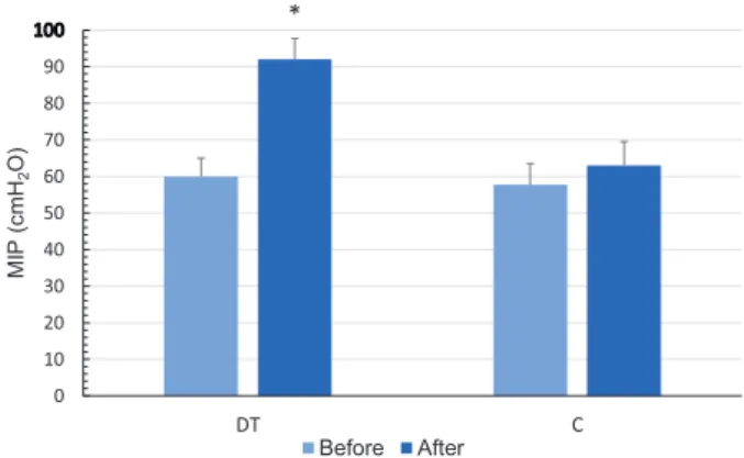
MIP increased in both groups; by 53% in group DT and by 8% in group C. The mean difference of the change between the groups was 27 cmH2O (95% CI 15.5–38.2 cmH2O). The change was statistically significant in the DT group (p < 0.0001), but no statistically significant change was found in group C (Table II, Fig. 2).
Peak inspiratory flow
Improvement in PIF was recorded in both groups; PIF increased by 23.0% in the DT group and by 12.0% in group C. The mean difference of the change between the groups was 0.5 L/s (95% CI 0.1–0.9 L/s). The change was statistically significant in both groups (p < 0.001) (Table II, Fig. 3).
+1 min for changing the sections) and cool-down sections (10 min; light dynamic, stretching, and breathing exercises). In the main part static and dynamic balance exercises and combined static and dynamic strengthening exercises for the flexor and ex- tensor muscles of the trunk were applied. Unstable training tools, elastic bands, balls and dumbbells were also used during the training. The exercises were precisely explained and presented, and their implementation was supervised by a physiotherapist.
The programme is described in detail in our earlier paper (19).
Inspiratory training
The DT group used an inspiratory muscle strengthening device, POWERbreathe Medic Plus (POWERbreathe Ltd, Warwick- shire, UK), which provides resistance during inhalation. The device was used twice a day, in the subject’s own homes, under conditions like 30 inhales per occasion at a speed of 15 inhales/
min (10). The device was also used for training sessions during trunk muscle strengthening exercises. The expected inhalation technique for home-based exercises was abdominal breathing in a comfortable positon. To define the amount of resistance during training, the maximal inspiratory pressure (MIP) (in cmH2O) was measured by the POWERbreathe KH2 (POWERbreathe Ltd) device. The resistance was set individually at 60% of MIP (10). The subject wore a nose clip to prevent nasal breathing. At the first meeting the subjects were trained in the correct use of the POWERbreathe Medic Plus device, with the main purpose of the inspiratory load being to strengthen the diaphragm muscle.
Measurement protocol
Measurements were conducted before and after the intervention period. Chest excursion was measured with an inelastic tape at nipple height. The difference between the measurements at the end of inspiration and at the end of expiration was recorded as chest excursion in cm (20).
Inspiratory functions were assessed with the measurement protocols of the POWERbreathe KH2 device. During MIP testing (cmH2O), the patient inhaled maximally against a closed airway from residual volume. The MIP test provides information on the strength of the inspiratory muscles (21, 22). The peak inspiratory flow (PIF) (L/s) reflects the ability of the inspiratory muscles to contract rapidly and to overcome the inherent resistance and elastance of the respiration (22). Functioning of the diaphragm muscle correlates with MIP and PIF values (22). VOLUME (L) is the mean in an upright standing position, and verbal encoura- gement was given to help the subjects perform maximally. For each patient, the highest value of the inhalation, out of 3 repeat inhalations, was selected for analysis (24, 25).
The stability limit of the trunk was measured with the sitting/
modified Functional Reach Test (mFRT) and the modified Late- ral Reach Test (mLRT). These tests are reliable measurements to quantify sitting balance and LOS (16). The participant was sitting on a table, with the hips and knees flexed at 90°, and the feet hip-distance apart. The feet were placed on the floor and the back was not supported. Initial reach was measured with the arms flexed at 90°. In the mFRT, the participant was sitting next to a wall (on which a tape measure was fixed) and was asked to reach as far forward as possible. In the mLRT, the measurement protocol was similar, but the participant was sitting with their back against the wall, and was asked to reach as far to the left and right sides as possible. Participants were not allowed to take a step forward or to either side, or to raise their buttocks from the table. Measurement of reach was assessed in cm by
JRM JRM J
ournal ofR
ehabilitationM
edicineJRM J
ournal ofR
ehabilitationM
edicineMean volume of air inhaled per breath (VOLUME) Improvement was found in both groups; the VOLUME level increased by 8.2% in the DT group and by 7.3% in group C. The mean difference of the change between the groups was 0.0 L (95% CI –0.28 to 0.35 L). Statistically significant changes were not detected in the DT group (p = 0.07) or group C (p = 0.19) (Table II, Fig. 4).
Stability limits of the trunk
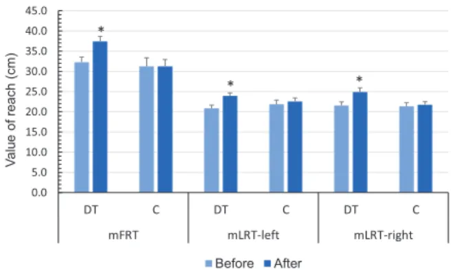
In mFRT, the DT group improved by 15.8%, while no change was found in group C. The mean difference of
the change between the groups was 5.1 cm (95% CI 0.1–9.3 cm). The improvement was statistically signi- ficant in the DT group (p < 0.0001) (Table II, Fig. 5).
Regarding the left-sided mLRT, the output impro- ved in both groups; by 14.6% in the DT group and by 3.2% in group C. The mean difference of the change between the groups was 2.3 cm (95% CI –0.004 to 4.7 cm). The change was statistically significant in group DT (p < 0.0001), but not in group C (p = 0.43) (Table II, Fig. 5).
In the values of the right-sided mLRT the DT group improved by 15.6%, whereas group C improved by
Table II. Results of the statistical comparison Group
Before After
p-value (before vs. after)
Difference
Mean SE Mean SE Mean SE
Chest excursion, cm DT 5.3 0.4 7.5 0.5 < 0.0001 2.2 0.4
C 6.2 0.6 7.5 0.5 0.01 1.3 0.6
Difference (DT-C) –1.0 0.7 0.0 0.8 1.0 0.6
p = 0.14
MIP, cmH2O DT 60 5 92 6 < 0.0001 32 4
C 58 6 63 7 0.23 5 3
Difference (DT-C) 2 8 29 9 27 6
p < 0.0001
PIF (l/s) DT 4.4 0.3 5.4 0.3 < 0.0001 1.0 0.1
C 4.3 0.3 4.8 0.3 0.003 0.5 0.2
Difference (DT-C) 0.2 0.4 0.7 0.5 0.5 0.2
p = 0.025
Volume (l) DT 2.3 0.2 2.5 0.2 0.07 0.2 0.1
C 2.1 0.2 2.2 0.2 0.19 0.2 0.1
Difference (DT-C) 0.3 0.3 0.3 0.3 0.0 0.2
p = 0.81
mFRT, cm DT 32.3 1.3 37.4 1.3 < 0.0001 5.1 1.0
C 31.3 2.1 31.3 1.7 1.00 0.0 2.0
Difference (DT-C) 1.0 2.4 6.1 2.1 5.1 2.1
p = 0.017
mLRT (left-sided), cm DT 20.9 0.8 23.9 0.8 < 0.0001 3.0 0.8
C 21.9 1.0 22.6 0.8 0.43 0.7 0.9
Difference (DT-C) 1.0 1.3 1.4 1.2 2.3 1.2
p = 0.054
mLRT (right-sided), cm DT 21.5 0.9 24.9 1.1 < 0.0001 3.4 0.8
C 21.3 0.9 21.8 0.7 0.62 0.4 0.8
Difference (DT-C) 0.2 1.3 3.1 1.4 2.9 1.1
p = 0.013
DT: diaphragm training; C: control; MIP: maximal inspiratory pressure; PIF: peak inspiratory flow; volume; mean volume of air inhaled per breath; mFRT: modified Functional Reach Test; mLRT: modified Lateral Reach Test (cm); SE: standard error.
Fig. 2. The values of MIP before and after the intervention (mean±SE).
*p <0.05. C: control group; DT: diaphragm training group; MIP: maximal inspiratory pressure (cmH2O); SE: standard error.
MIP (cmH2O)
Before After
Fig. 3. Peak inspiratory flow (PIF) values (L/s) before and after the intervention (mean±SE). *p < 0.05. C: control group; DT: diaphragm training group; SE: standard error.
PIF (l/s)
Before After
JRM JRM J
ournal ofR
ehabilitationM
edicineJRM J
ournal ofR
ehabilitationM
edicine2.0%. The mean difference of the change between the groups was 2.9 cm (95% CI 0.7–5.2 cm). The posi- tive change in group DT was statistically significant (p < 0.0001); however, in group C no statistically signi- ficant change was detected (p = 0.62) (Table II, Fig. 5).
DISCUSSION
MIP and stability limit tests (mFTR, mLRT) showed a statistically significant improvement only in the DT group. Chest excursion and peak inspiratory flow (PIF) tests show statistically significant improvements in both groups; however, the improvement was more greater in the DT group. In VOLUME (the mean volume of inha- led air/breath) levels, some improvement can be seen in both DT and C groups, but statistically significant change is not detectable either group. The main finding of this study is that conventional exercises together with a diaphragm-strengthening protocol (DT group) results in better functional capacity in patients with lumbar pain than conventional exercises alone (group C).
The diaphragm is an essential muscle for breathing;
however, it also has a role in preserving the segmental stability of the lumbar spine by maintaining and increa- sing the intra-abdominal pressure during postural tasks (4). Individuals with low back pain have a disturbed proprioceptive input from the low back area; hence they achieve worse results in the stability limit tests (such as functional and lateral reach tests) than healthy individuals (27). The function of the diaphragm muscle deteriorates if non-specific low back pain occurs (6).
In these cases, the diaphragm has a higher position, decreased mobility, and greater fatigability (6). The lumbar range of movement of patients with CLBP with minimal or no pain at the time of measurement is the same as that of the healthy population (28). The mobility of the lumbar area is an influencing factor in the perfor- mance of LOS tests (27). It is notable that, despite the more larger decrease in pain in group C (19), group DT showed greater improvement in LOS tests. These results suggest that, as an effect of the diaphragm-strengthening training, both the breathing and the postural functions of the diaphragm muscle have improved. By exercising the diaphragm all the aforementioned dysfunctions may be decreased; a stronger, more mobile muscle (29) may be more effective in increasing the intra-abdominal pres- sure, and therefore maintaining lumbar stability.
In our previous study of the severity of pain measu- red by the visual analogue scale (VAS), both training methods resulted in statistically significant improve- ment (19). Considering the evidence that pain reduces proprioception (30), our results may indicate that the positive change in the severity of pain, together with the increased LOS values, might be a sign of improved proprioception from the lumbar area, represented by better mobility. The deteriorated proprioceptive input might cause postural changes in vertical positions in people with low back pain; individuals tend to lean forward more if low back pain develops (12), and they prefer ankle strategy to the normal multi-segmental strategy for postural control (10, 31). The increased stability limits of the trunk might indicate a more complex postural strategy applied by the DT group.
These results are in line with the findings of a former study, describing that improved postural function with the significantly decreased severity of pain may contribute to the normal, multi-segmental strategy in patients with non-specific CLBP (10).
The mobility of the lumbar spine and having strong, well-functioning extensor muscles, are essential to perform the mFRT and mLRT (27). The synergistic function of the global stabilizers (superficial exten- sors) and local stabilizer muscles (lumbar multifidus, transversus abdominis, pelvic floor muscles, and diaphragm) has a major role during postural tasks
Fig. 4. VOLUME values before and after the intervention (mean±SE).
*p <0.05. C: control group; DT: diaphragm training group; VOLUME:
mean of air inhaled per breath; SE: standard error.
VOLUME (l)
Before After
)mc( hcaer fo eulaV
Before After
Fig. 5. Results of trunk stability tests before and after the intervention (mean±SE). *p <0.05. C: control group; DT: diaphragm training group;
mFRT: modified functional reach test (cm); mLRT-left: left-sided modified lateral reach test (cm); mLRT-right: right-sided modified lateral reach test (cm); SE: standard error.
JRM JRM J
ournal ofR
ehabilitationM
edicineJRM J
ournal ofR
ehabilitationM
edicinethese patients to improve their functional level. Mo- reover, diaphragm-strengthening training might be a favourable additional method for people dealing with the consequences of low back pain, and may also be beneficial in the prevention of non-specific CLBP.
Conflicts of interest.
The authors have a signed loan agreement with the manufactu- rer of POWERbreathe device (POWERbreathe Ltd.), which is constricted only to the temporary use of the device. No financial support was provided. This research did not receive any specific grant from funding agencies in the public, commercial, or not- for-profit sectors.
REFERENCES
1. Lewit K. Relation of faulty respiration to posture, with clinical implications. J Am Osteopath Assoc 1980; 79: 525–529.
2. Cholewicki J, Juluru K, Radebold A, Panjabi MM, McGill SM. Lumbar spine stability can be augmented with an abdominal belt and/or increased intra-abdominal pressure.
Eur Spine J 1999; 8: 388–395.
3. Shirley D, Hodges PW, Eriksson AE, Gandevia SC. Spinal stiffness changes throughout the respiratory cycle. J Appl Physiol (1985) 2003; 95: 1467–1475.
4. Hagins M, Lamberg EM. Individuals with low back pain breathe differently than healthy individuals during a lifting task. J Orthop Sport Phys Ther 2011; 41: 141–148.
5. Kolar P, Sulc J, Kyncl M, Sanda J, Cakrt O, Andel R et al.
Postural function of the diaphragm in persons with and without chronic low back pain. J Orthop Sport Phys Ther 2012; 42: 352–362.
6. Janssens L, Brumagne S, McConnell AK, Hermans G, Troos- ters T Gayan-Ramirez G. Greater diaphragm fatigability in individuals with recurrent low back pain. Respir Physiol Neurobiol 2013; 188: 119–123.
7. Sibley KM, Carpenter MG, Perry JC, Frank JS. Effects of postural anxiety on the soleus H-reflex. Hum Mov Sci 2007; 26: 103–112.
8. Capra NF, Ro JY. Experimental muscle pain produces cen- tral modulation of proprioceptive signals arising from jaw muscle spindles. Pain 2000; 86: 151–162.
9. Ruhe A, Fejer R, Walker B. Is there a relationship bet- ween pain intensity and postural sway in patients with non-specific low back pain? BMC Musculoskelet Disord 2011; 12: 162.
10. Janssens L, McConnell AK, Pijnenburg M, Claeys K, Goos- sens N, Lysens R et al. Inspiratory muscle training affects proprioceptive use and low back pain. Med Sci Sports Exerc 2015; 47: 12–19.
11. Ringheim I, Austein H, Indahl A, Roeleveld K. Postural strategy and trunk muscle activation during prolonged standing in chronic low back pain patients. Gait Posture 2015; 42: 584–589.
12. Brumagne S, Janssens L, Janssens E, Goddyn L. Altered postural control in anticipation of postural instability in persons with recurrent low back pain. Gait Posture 2008;
28: 657–662.
13. Lafond D, Champagne A, Descarreaux M, Dubois JD, Prado JM, Duarte M. Postural control during prolonged standing in persons with chronic low back pain. Gait Posture 2009;
29: 421–427.
14. Liao CF, Lin SI. Effects of different movement strategies on forward reach distance. Gait Posture 2008; 28: 16–23.
15. Tantisuwat A, Chamonchant D, Boonyong S. Multi-direc- tional reach test: an investigation of the limits of stability of people aged between 20–79 years. J Phys Ther Sci
(32); for example, during the stability limit tests in our study. It has been demonstrated that reduction in the function of even 1 muscle of the active stabilizer system results in an increased demand on the other subsystems to maintain stability (33); thus, the mem- bers of the stabilizer systems influence each other.
Our results concerning the inspiratory tests show that the inspiratory function of the diaphragm improved after the 8-week diaphragm-strengthening training.
Our previous paper described that the diaphragm- strengthening training might have a statistically significant effect on the stabilizer muscles other than the diaphragm; that is, increased thickness of the muscle belly of the transversus abdominis and lumbar multifidus muscles (19). The significant improvement in functional capacity of the local stabilizer muscles (increased thickness (19) and improved inspiratory function) may result in increased stability limits of the trunk. This finding implies that, in case of non- specific CLBP, an 8-week programme of diaphragm training together with conventional exercises may be superior to conventional exercises alone in improving the functional capacity of the trunk.
Study limitations
A limitation of this study is that the inspiratory mano- euvre applied during MIP and PIF tests is highly effort-dependent (34, 35). However, it has been proven that a co-operative subject can activate the diaphragm maximally during voluntary inspiratory efforts (36).
Diaphragm activity was not measured directly in the current study; therefore the accurate value of contrac- tion of the diaphragm was not known when the MIP tests were performed, although verbal encouragement was provided to help the subjects to perform maxi- mally.
Conclusion
As a result of the 8-week diaphragm-strengthening training, the functions of the diaphragm muscle, such as the thicknesses of the stabilizer muscles of the trunk (19), improved significantly, which may result in better postural stability of the lumbar spine and better functioning in people with non-specific CLBP.
The importance of non-pharmacological treatments, such as physical exercise programmes, is well-known in reducing the intensity of low back pain (37). Ne- vertheless, some patients with low back pain are not capable of performing conventional exercises, due to the intensity of the pain or as a result of other medical conditions. Since stability limit and appropriate pos- tural control are the bases of functional capacity (38), diaphragm training may be an appropriate option for
JRM JRM J
ournal ofR
ehabilitationM
edicineJRM J
ournal ofR
ehabilitationM
edicine2014; 26: 877–880.
16. Lynch SM, Leahy P, Barker SP. Reliability of measurements obtained with a modified functional reach test in subjects with spinal cord injury. Phys Ther 1998; 78: 128–133.
17. Field-Fote EC, Ray SS. Seated reach distance and trunk excursion accurately reflect dynamic postural control in individuals with motor-incomplete spinal cord injury. Spinal Cord 2010; 48: 745–749.
18. Thompson M, Medley A. Forward and lateral sitting fun- ctional reach in younger, middle-aged, and older adults.
J Geriatr Phys Ther 2007; 30: 43–48.
19. Finta R, Nagy E, Bender T. The effect of diaphragm training on lumbar stabilizer muscles: a new concept for improving segmental stability in the case of low back pain. J Pain Res 2018; 11: 3031–3045.
20. Sclauser Pessoa IMB, Franco Parreira V, Fregonezi GA, Sheel AW, Chung F, Reid WD. Reference values for maximal inspiratory pressure: a systematic review. Can Respir J 2014; 21: 43–50.
21. Tudorache V, Oancea C, Mlădinescu OF. Clinical relevance of maximal inspiratory pressure: determination in COPD exacerbation. Int J Chron Obstruct Pulmon Dis 2010; 5:
119–123.
22. Kim M, Lee K, Cho J, Lee W. Diaphragm thickness and inspiratory muscle functions in chronic stroke patients.
Med Sci Monit 2017; 23: 1247–1253.
23. Pupišová Z, Pupiš M, Jančoková Ľ, Pivovarniček P. Changes of inspiratory parameters and swimming performance by influence of PowerBreathe Plus Level 3. Sport Science 2014; 7: 12–15.
24. Volianitis S., McConnell AK., Jones, DA. Assessment of maximum inspiratory pressure. Respiration 2001; 68:
22–27.
25. Altman P, Wehbe L, Dederichs J, Guerin T, Ament B, Moronta MC et al. Comparison of peak inspiratory flow rate via the Breezhaler®, Ellipta® and HandiHaler® dry powder inhalers in patients with moderate to very severe COPD: a randomized cross-over trial. BMC Pulm Med 2018; 18: 100.
26. Robinson EP, Kjeldgaard JM. Improvement in ventilatory muscle function with running. J Appl Physiol 1982; 52:
1400–1406.
27. Silfies SP, Bhattacharya A, Biely S, Smith SS, Giszter S.
Trunk control during standing reach: A dynamical system analysis of movement strategies in patients with mecha- nical low back pain. Gait Posture 2009; 29: 370–376.
28. Ng JK, Richardson CA, Kippers V, Parnianpour M. Compa- rison of lumbar range of movement and lumbar lordosis in back pain patients and matched controls. JRM 2002;
34: 109–213.
29. Kodric M, Trevisan R, Torregiani C, Cifaldi R, Longo C, Can- tarutti F et al. Inspiratory muscle training for diaphragm dysfunction after cardiac surgery. J Thorac Cardiovasc Surg 2013; 145: 819–823.
30. Brumagne S, Cordo P, Verschueren S. Proprioceptive weighting changes in persons with low back pain and elderly persons during upright standing. Neurosci Lett 2004; 366: 63–66.
31. McCaskey MA, Schuster-Amft C, Wirth B, Suica Z, de Bruin ED. Effects of proprioceptive exercises on pain and function in chronic neck- and low back pain rehabilitation:
a systematic literature review. BMC Musculoskelet Disord 2014; 15: 382.
32. Panjabi MM. The stabilizing system of the spine. Part II.
Neutral zone and instability hypothesis. J Spinal Disord 1992; 5: 390–396.
33. Fritz JM, Erhard RE, Hagen BF. Segmental instability of the lumbar spine. Phys Ther 1998; 78: 889–896.
34. Volianitis S, McConnell AK, Jones DA. Assessment of maxi- mum inspiratory pressure. Respiration 2001; 68: 22–27.
35. Kainu A, Timonen KL, Vanninen E, Sovijärvi AR. Reference values of inspiratory spirometry for Finnish adults Scand J Clin Lab Investig 2018; 78: 245–252.
36. Gandevia SC, McKenzie DK.J. Activation of the human diaphragm during maximal static efforts. Physiol 1985;
367: 45–56.
37. Foster NE, Anema JR, Cherkin D, Chou R, Cohen SP, Gross DP et al. Prevention and treatment of low back pain:
evidence, challenges, and promising directions. Lancet 2018; 391: 2368–2383.
38. Shumway-Cook A, Woollacott MH. Motor control: transla- ting research into clinical practice. Philadelphia: Lippincott Williams & Wilkins; 2012.