Élő sejtek és kopolimer bevonatok kezelése zöld tea polifenollal (EGCg):
dinamikai vizsgálatok jelölésmentes bioszenzorokkal
Péter Beatrix Éva - Doktori (PhD) értekezés -
Pannon Egyetem – Molekuláris- és Nanotechnológiák Doktori Iskola Magyar Tudományos Akadémia Műszaki Fizikai és Anyagtudományi Intézet
Témavezető:
Dr. Horváth Róbert
Budapest, 2016
DOI:10.18136/PE.2016.623
Living cells and copolymer coatings exposed to green tea polyphenol (EGCg):
dynamic investigations using label-free optical biosensors
Beatrix Éva Péter - Ph.D. thesis-
University of Pannonia– Doctoral School of Molecular- and Nanotechnology Hungarian Academy of Sciences, Institute of Technical Physics and Materials Science
Supervisor:
Dr. Róbert Horváth
Budapest, 2016
Living cells and copolymer coatings exposed to green tea polyphenol (EGCg):
dynamic investigations using label-free optical biosensors
Értekezés doktori (PhD) fokozat elnyerése érdekében Írta:
Péter Beatrix Éva
Készült a Pannon Egyetem – Molekuláris- és Nanotechnológiák Doktori Iskolájában Témavezető: Dr. Horváth Róbert
Az értekezést témavezetőként elfogadásra javaslom:
Dr. Horváth Róbert: igen / nem ………..
(aláírás) A jelölt a doktori szigorlaton ……… %-ot ért el.
Az értekezést bírálóként elfogadásra javaslom:
Bíráló neve: ………. igen / nem ………..
(aláírás)
Bíráló neve: ………. igen / nem ………
(aláírás) A jelölt az értekezés nyilvános vitáján ……… %-ot ért el.
………
Bíráló Bizottság elnöke A doktori (PhD) oklevél minősítése ………
………
EDT elnöke Veszprém,………..
5 TABLE OF CONTENTS
PhD értekezés kivonata ... 6
Abstract (English) ... 7
Estratto (Italiano) ... 8
1. INTRODUCTION ... 9
1.1. Epigallocatechin-gallate (EGCg), the green tea polyphenol ... 9
1.2. Molecular scale properties and interactions of EGCg ... 11
1.3. Impacts of EGCg at cellular level ... 17
1.4. Observed actions of EGCg using tissues and animal models and results of clinical trials ... 27
1.5. Perspectives: Investigation and application possibilities in the near future ... 30
1.6. Emerging label-free techniques in polyphenol research ... 34
2. MOTIVATION AND OBJECTIVES ... 43
3. MATERIALS AND METHODS ... 44
3.1. Employed solutions and their prepartions ... 44
3.2. Cell culture and cell staining ... 45
3.3. MTT cell viability assay ... 46
3.3. Holomonitor M4 ... 48
3.4. Optical Waveguide Lightmode Spectroscopy (OWLS) ... 49
3.5. The Epic Benchtop resonant waveguide grating biosensor... 51
3.6. Atomic Force Microsopy (AFM) ... 53
4. RESULTS AND DISCUSSION ... 54
4.1. Cell morphological changes during EGCg exposure investigated by Holomonitor M4 .. 54
4.2. Cell repellent and cell adhesive polymer coatings exposed to EGCg: multicomponent model systems using biosensors ... 58
4.3. Whole cells exposed to EGCg: comparison of classical cell viability assay with label-free biosensor data and fluorescent imaging of the treated cells ... 69
4.4. Nanoparticle clusters assembled from oppositely charged nanoparticles ... 77
5. SUMMARY AND OUTLOOK ... 83
6. THESIS HIGHLIGHTS ... 86
TÉZISPONTOK ... 88
7. ACKNOWLEDGEMENTS ... 90
8. LIST OF PUBLICATIONS ... 91
8.1. Papers used to compose the thesis highlights ... 91
8.2. Other papers ... 92
8.3. Participation in conferences ... 93
9. REFERENCES ... 96
6
PhD értekezés kivonata
A zöld tea egyik hatóanyaga, az epigallokatekin-gallát (EGCg) hatásait már régóta vizsgálják, és számos esetben derült fény egészséget megőrző tulajdonságaira, többek között arra, hogy csökkenti a rák kialakulásának, az áttétek képződésének, illetve a szív, -és érrendszeri betegségek kockázatát, csökkenti a vérnyomást, koleszterinszintet és vércukrot, gyorsítja az anyagcserét, továbbá egyes tanulmányok szerint segít a vírusok és baktériumok elleni védekezésben is. Pozitív hatásairól rengeteg publikáció beszámol, azonban a szakirodalomban ellentmondásos adatok, eredmények is fellelhetők. Ennek oka valószínűleg az, hogy az EGCg kis molekulatömegű és rendkívül instabil anyag. Ezen tulajdonságai miatt jelöléses módszerekkel problémás, nehézkes lehet e polifenol vizsgálata. A szakirodalomban az esetek túlnyomó részében jelöléses módszereket alkalmaztak, így tehát hiánypótló munkának is nevezhető ezen különleges hatóanyag jelölésmentes technnikákkal történő vizsgálata.
A jelölésmentes bioszenzorok és képalkotó technikák robbanásszerű fejlődésen mentek keresztül az utóbbi években; a biológiai alapkutatásokban csak most kezdődött a kiaknázásuk, a műszerek egyre érzékenyebbek, alkalmazásuk új utakat nyithat meg a biológiai alapkutatásokban és biotechnológiai alkalamzásokban. Az említett technikák fontos előnye, hogy a méréseket valós időben, jelölő anyagok, festékek nélkül tudjuk megvalósítani, így ezek nem befolyásolják a vizsgált mintákat.
Kutatásaim során modern jelölésmentes módszereket alkalmaztam az EGCg vizsgálatára, hogy megfigyeljem a hatásait élő sejtekre (HeLa sejtvonal), és kopolimer bevonatokra, mindezt valós időben. Különös figyelmet fordítottam a szakirodalomban eddig meglehetősen hiányosan taglalt oxidált formájára is. Megvizsgáltam, hogy az oxidált forma hogyan befolyásolja a bevonatokat, illetve a sejtek életfolyamatait, adhézióját, morfológiáját.
PhD munkám során Holomonitor képalkotó technikával bebizonyítottam, hogy az EGCg gátolja a HeLa rákos sejtek mozgását. Epic BT bioszenzorral mért eredmények alapján megállapítottam, hogy az EGCg hozzákötődik az alkalmazott PLL-g-PEG (sejttaszító) és PLL-g-PEG:PLL-g-PEG-RGD (sejtek adhézióját segítő) szintetikus kopolimer bevonatokhoz, és multiréteget alkot. Az EGCg molekulák kötődése a bevonatokhoz elhangolja azok tulajdonságait, és az alkalmazott koncentrációk függvényében befolyásolják a HeLa sejtek adhézióját az említett felületekre. A bioszenzorral rögzített adhéziós kinetikai görbék tekintetében felfedeztem egy határ koncentrációt (27,3 ± 10 µg/ml), mely alatt szigmoid jellegű adhéziós kinetikai görbéket kapunk (élő, aktív folyamatra jellemző), míg ennél nagyobb koncentrációnál adszorpciós görbéket rögzítettem (élettelen folyamat jellemzője). Ezen eredmények egyezést mutatnak a sejtek életképességét vizsgáló MTT teszttel. Megfigyeltem, hogy az oxidált formának még erőteljesebb a hatása; több réteg képződik a bevonatokon, és a sejtadhézióra gyakorolt befolyása is jelentősebb. Az orvostudomány többek között nanoméretű részecskék segítségével igyekszik megoldást nyújtani a hatóanyagok kizárólag a célsejtekbe történő bejuttatására. Ezen irányvonalat felvéve kísérletet tettem olyan ellentétesen töltött, arany nanorészecskékből álló klaszterek (AuMUA és AuTMA) előállítására, melyek a jövőben esetlegesen alkalmassá válhatnak az EGCg célsejtekbe történő szállítására, és így betegségek gyógyítására.
7
Abstract (English)
Scientists examine the effects of epigallocatechin-gallate (EGCg), one of the active substances of green tea, for a long while. Beneficitial impacts on health are revealed in a lot of cases, for example, it reduces the risk of the formation and metastasis of cancer, it reduces the risk of cardiovascular diseases, blood pressure, level of cholesterol and blood sugar, it intensifies the metabolism and helps to fight against bacteria and viruses. Numerous papers have been published about its positive effects, however, there are contradictory data and results in the literature as well.
The reason is probably that the EGCg has small molecular weight and it is a very unstable, sensitive material. Because of these properties, its examinations by labeling techniques can be difficult and problematic. In the literature the authors mostly uses labeling methods in general, thus, examination of this specific material EGCg by label-free techniques may supply the incompletions.
Label-free biosensors and imaging techniques underwent enormous progress in the past years; their utilization in biological basic researches has just begun, the appliances are more and more sensitive, their application may open new ways in biological basic research and biotechnology. The advantages of these methods are that they are real-time, and do not use labels, such as dyes, that may affect the samples. In my experiments, I applied these novel techniques to investigate EGCg and its effects on living cells (HeLa cell line), and on copolymer coatings in a real-time way. I paid attention to the EGCg’s oxidized form, which is a pretty untended topic in the literature, and I examined that how can the oxidized EGCg affect the coatings and the action, adhesion, morphology of the cells.
In my PhD work, I proved by Holomonitor M4 imaging technique that EGCg inhibit the movement of the HeLa cervical cancer cells. I revealed by Epic BT biosensor that EGCg binds to the PLL-g- PEG (cell repellent) and PLL-g-PEG: PLL-g-PEG-RGD (cell attractive) synthetic copolymer coatings and the EGCg molecules form multilayers. The EGCg binding modifies the original properties of the coatings and inhibits HeLa cell adhesion to them in a concentration dependent manner. In the case of kinetic curves, I discovered a border concentration (27.3 ± 10 µg/ml µg/ml). At lower concentration of EGCg than this value, I recorded sigmoid type cell adhesion curves (active, living process), and at higher concentrations, adsorption type curves (dead process) were obtained. I verified with MTT assay, that the cell viability of HeLa cells start to decrease at around 27.3 ± 10 µg/ml EGCg concentration, thus I got the same result by MTT as well. The oxidized form has more powerful effect; more layers formed than in the case of freshly created EGCg solutions, and its inhibitory impact on cellular adhesion is more robust as well.
Medical sciences try to cure illnesses by transporting active substances in nanoparticles to reach only the targeted cells. To follow this trend, I attempted to create clusters from oppositely charged gold nanoparticles (AuMUA and AuTMA), which may be able to transport EGCg to targeted cells and thus cure illnesses in the future.
8
Estratto (Italiano)
Gli scienziati esaminano gli effetti di epigallocatechina gallato (EGCg), uno dei principi attivi del tè verde, per lungo tempo. Effetti caritatevoli sulla salute sono rivelati in molti casi, per esempio riduce il rischio della formazione e la metastasi del cancro, riduce il rischio di malattie cardiovascolari, pressione sanguigna, livelli di colesterolo e di zucchero del sangue, si intensifica il metabolismo e aiuta a combattere contro i batteri e virus. Esso è stato pubblicato numerosi articoli di suoi effetti positivi, tuttavia, ci sono dati ed i risultati opposti nella letteratura. La ragione è probabilmente che EGCg ha piccolo peso molecolare ed è molto instabile. Grazie a queste proprietà, i suoi esami di tecniche di etichettatura può essere difficile e problematico.
Nella letteratura gli autori utilizza prevalentemente i metodi di etichettatura in generale, in tal modo, l'esame di questo materiale specifico, EGCg con tecniche senza etichette possono fornire le carenze. Biosensori senza etichette e tecniche di imaging ha subito enormi progressi negli ultimi anni; il loro utilizzo in ricerche di base biologica è appena iniziata, gli apparecchi sono sempre più sensibili, la loro applicazione potrebbe aprire nuove strade nel campo della ricerca di base biologica e biotecnologia. I vantaggi di questi metodi è che sono in tempo reale, e non utilizzano etichette o coloranti che possono influenzare i campioni. Nei miei esperimenti, ho applicato queste nuove tecniche per indagare EGCg e il suo impatto sulle cellule viventi (linea cellulare HeLa), e sui rivestimenti copolimero in un modo in tempo reale. Ho preso in considerazione la forma ossidata della EGCg, che è un argomento incustodito abbondanza nella letteratura, e ho esaminato che come può il EGCg ossidato influenzare i rivestimenti e l'azione, l'adesione, morfologiche delle cellule. Nel mio lavoro di dottorato, ho dimostrato da Holomonitor M4 tecnica di imaging che EGCg inibisce il movimento delle cellule del cancro del collo dell'utero HeLa. Ho studiato da Epic BT biosensore che l'EGCg si lega al PLL-g-PEG (repellenti cellulare) e PLL-g-PEG: PLL-g-PEG-RGD (cellule attraente) rivestimenti copolimeri sintetici e le molecole EGCg formano multistrati su di loro. EGCg vincolante modifica le proprietà originali dei rivestimenti e inibisce l'adesione delle cellule HeLa a loro in un modo dipendente dalla concentrazione. Nel caso di curve cinetiche, ho scoperto una concentrazione confine (27.3 ± 10 µg/ml). A valori inferiori a 27.3 ± 10 µg/ml di EGCg ho ricevuto diffusione curve (attivi, processo vivente), e al di sopra di questo valore, le curve di assorbimento (processo morto). Ho verificato con test MTT, che la vitalità cellulare di cellule HeLa inizia a diminuire a 27.3 µg ± 10 µg/ml concentrazione d’EGCg, quindi ho ottenuto lo stesso risultato MTT pure. La forma ossidata ha un effetto più potente; più strati possono essere formati che nel caso di appena creato EGCg, e il suo impatto inibitorio sulla adesione cellulare è più robusto pure. Scienze mediche cerca di curare le malattie da trasporto di sostanze attive in nanoparticelle di raggiungere solo le cellule bersaglio. Per seguire questa tendenza, ho cercato di creare grappoli da nanoparticelle d'oro di carica opposta (AuMUA e AuTMA), che possono essere in grado di trasportare EGCg alle cellule mirati e quindi curare le malattie in futuro.
9
1. INTRODUCTION
Herbs and traditional medicines have been applied for thousands of years, but researchers started to study their mode of action at the molecular, cellular and tissue levels only recently. Nowadays, just like in the ancient ages, natural compounds are still determining factors in remedy. To support this statement, the recently won Nobel Prize in 2015 could be mentioned for an anti- malaria agent from the plant sweet wormwood which had been used to effectively treat the disease. Among natural compounds and traditional Chinese medicines, the green tea polyphenol epigallocatechin gallate (EGCg) is one of the most studied active substance. However, in general scientists apply labeling techniques to investigate its effects on cell adhesion, migration, motility, apoptosis, etc. However, label-free biosensors can be emerging platforms to investigate the mode of action of small molecules and are highly relevant for EGCg research and development [1]. My first authored “Biophysical characteristics of proteins and living cells exposed to the green tea polyphenol epigallocatecin-3-gallate (EGCg): Review of recent advances from molecular mechanisms to nanomedicine and clinical trials” titled review paper was mainly used for the introduction of EGCg (chapters 1.1-1.6) [1].
1.1. Epigallocatechin-gallate (EGCg), the green tea polyphenol
Drinking tea is an ancient habit; legends from India and China indicate that it started about 5000 years ago [2]. Traditionally, it was drunk to eliminate toxins and improve blood flow and resistance to illnesses [2], so its habitual consumption has long been associated with health benefits [3]. Infusion of the unfermented leaves of Camellia sinensis (L.) Kuntze, known as green tea, is a very popular drink worldwide nowadays as well. Green tea beverage contains polyphenolic compounds, including phenolic acids, flavanols, flavonoids and flavandiols [4]. In a typical green tea infusion, prepared in a portion of 1 g leaves to 100 ml water in a few minutes brew, most of the polyphenols are flavanols, known as catechins, and account for about 30–42 % of the dry weight of the solids [4][5]. Among green tea catechins, epigallocatechin gallate (EGCg) is the most abundant, around 16.5 wt% of the water extractable fraction of green tea leaves [5]. This component is regarded as a constituent characterizing green tea, because it is not found in any plants except C. sinensis (L.) Kuntze [5]. Tea catechins, especially EGCg, have been shown and reviewed to have various health benefits, for example, anti-metastasis, anti- cardiovascular, anti-cancer, antiinflammatory and antioxidant effects [4][6][7].
EGCg is one of the most studied active substance, and a lot of studies observed its effects on several cancer and normal cell lines and on animal models (Fig.1.1) [1][6][8].
10
Fig.1.1 Diversified effects of EGCg. This polyphenol affect proteins: EGCg causes aggregation, association, sedimentation, precipitation and gelation, furthermore, increases viscosity, and act as a cross-linker in the case of mucins. It is suggested that EGCg interacts with phospholipids and plasma membrane proteins. It inhibits the amyloid-fibril formation of α-synuclein, amyloid-β and huntingtin. Cancer cell adhesion, movement, proliferation are decreased, while apoptosis is increased in the presence of EGCg via gene expression and biological pathways in in vitro tests. In vivo studies also showed reduced tumor weight and suppression of tumor growth in animal models and tissues, as well as clinical trials also showed its beneficial effects on the human health. Experiments proved that it inhibited the viability of microbes as well. Due to EGCg’s special properties, curing and other utilizations, experiments can be put in perspective [1].
11
1.2. Molecular scale properties and interactions of EGCg
Epigallocatechin gallate (EGCg)1, is the ester of epigallocatechin and gallic acid. EGCg is an odorless white, (sometimes faint pink) crystals (or powder). It is soluble in ethanol, dimethyl- formamide, dimethyl sulfoxide (DMSO) and water2. This compound ([(2R,3R)-5,7-dihydroxy-2- (3,4,5-trihydroxyphenyl)-3,4-dihydro-2H-chromen-3-yl] 3,4,5-trihydroxybenzoate*) consists of a meta-5,7-dihydroxyl substituted A ring and trihydroxy phenol structures on both the B and D rings. The polyphenolic structure of tea polyphenols makes them good donors for hydrogen bonding [8]. For instance, hydrogen bonding of water molecules to EGCg forms a big hydration shell, which reduces the absorption of EGCg [8].The hydrogen bonding capacity enables these compounds to bind strongly to nucleic acids and proteins [1][8].
Bioavailability of EGCg
Poor bioavailability of tea catechin compounds needs to be considered when we extrapolate in vitro results to in vivo situations [5][7][9]. Cool and dry storage, fasting conditions, vitamin C, fish oil, albumin, piperine and soft water enhance the bioavailability of EGCg, while air contact oxidation, gastrointestinal inactivation, glucuronidation, metal ions (e.g., Ca2+, Mg2+), sulfation and COMT polymorphisms decrease its bioavailability [7]. In living organisms, EGCg absorption takes places mostly in the small intestine, and it passes to the large intestine where it is broken down to phenolic acids by the action of colonic microflora [7]. EGCg is principally excreted through the bile to the colon [5]. In human volunteers, the time to reach the maximal concentration (326 ng/ml (=0.326 µg/ml)) in the plasma was between 1.5 and 2.5 h after consumption of decaffeinated green tea solids (4.5 g dissolved in 500 ml water) [5][10].
According to publications, its level in blood after consuming the equivalent of 2-3 cups of green
1 (CAS Registry number: 989-51-5, PubChem CID: 65064; IUPAC Name*: [(2R,3R)-5,7-dihydroxy- 2-(3,4,5-trihydroxyphenyl)-3,4-dihydro-2H-chromen-3-yl] 3,4,5-trihydroxybenzoate; alternative and trade names: (−)-cis-3,3′,4′,5,5′,7-Hexahydroxy-flavane-3-gallate, (−)-cis-2-(3,4,5-Trihydroxyphenyl)- 3,4-dihydro-1(2H)-benzopyran-3,5,7-triol 3-gallate (Sigma-Aldrich) (2R,3R)-2-(3,4,5-
Trihydroxyphenyl)-3,4-dihydro-1[2H]-benzopyran-3,5,7-triol-3-(3,4,5-trihydroxybenzoate), DNA Methyltransferase Inhibitor IV, HAT Inhibitor X, Histone Acetyltransferase Inhibitor X, p300/CBP Inhibitor VIII, PCAF Inhibitor V, DNA MTase Inhibitor IV (Merck), (2R,3R)-2-(3,4,5-
Trihydroxyphenyl)-3,4-dihydro-1[2H]-benzopyran-3,5,7-triol 3-(3,4,5-trihydroxybenzoate), inhibitor of NOS, telomerase and Dnmt (Enzo Life Sciences), molecular Formula: C22H18O11, molecular weight: 458.40 Da)
2according to substance records available from Sigma-Aldrich; Merck and Alexis Biochemicals (now fully integrated into Enzo Life Sciences)
12
tea was 0.1-0.6 µM and for an equivalent of 7-9 cups was still lower than 1 µM (=0,458 µg/ml) [5].The catechin levels were not detectable by 24 h, and the half-life of EGCg was 5.0–5.5 h [5][10].
Antioxidant activity and stability
Tea polyphenols are well known for their antioxidant activities [11]. Among them, EGCg is the most effective in reacting with the reactive oxygen species (ROS) [5]. Thus, some studies have reported that EGCg is an antioxidant; however, on the other hand, Kim et. al reviewed that it exerts pro-oxidant actions as well [12]. Its antioxidant activities are due to the presence of phenolic groups that are sensitive to oxidation and can generate quinone, an oxidized derivative of aromatic compounds [3]. The antioxidant potential of EGCg is reported to be greater than those of vitamin C and E [13]. Tea polyphenols are also strong chelators [5]. They have a significant interaction with transition metal ions and form insoluble complexes with iron [2].
Thus, this binding in the gastrointestinal tract inhibits iron absorption; however, it only affects non-heme iron and can be overcome by the presence of ascorbic acid [2]. As pointed out by Tachibana, the chelation of free metal ions prevents the formation of ROS from auto-oxidation of many components [5]. It has been reported by several authors that EGCg and other catechins are unstable at high temperature and under alkaline as well as neutral conditions, at pH above 7, EGCg dimerizes and oxidizes easily [11][14]. It changes from non-colored at around pH 7 to yellow at higher pH regions in aqueous solution, and the absorption is in the UV range [15]. This observation indicated that the oxidation is an irreversible reaction, and Mizooku et al. reported that the oxidation species was found to correspond to Mw + 14 (where Mw is the molecular weight of EGCg), which has two hydrogen atoms removed and addition of one oxygen atom to the gallyl moiety in the B-ring of EGCg [15]. EGCg is stable in saline or Ringer’s solutions at low temperature, unless there are particular metal ion contaminants present [16]. According to a study of Zhou and co-workers, there are clear advantages to stabilizing EGCg solutions by using an antioxidant (ascorbic acid), metal scavenger (EDTA), keeping the pH somewhat below neutral and keeping the temperature low during storage and sampling of EGCg [16]. EGCg is known to be unstable under cell culture conditions, and both autooxidation and H2O2 generation are induced [15][17][18]; however, formation of H2O2 was observed in aqueous solutions as well [15]. According to a proposed mechanism by Hou and co-workers, the reaction in cell culture medium is probably catalyzed by metal ions such as Cu2+ and produces EGCg radicals and superoxide [19]. The unpaired electron may delocalize around the B ring [19]. The superoxide radical can further react with another EGCg molecule to produce EGCg radical and H2O2 [19].
Two EGCg radical molecules probably collide to form a dimer, or it can be possible that the EGCg radical may attack the B ring of another EGCg molecule, which is more abundant, to form a dimer radical [19]. It can react with molecular oxygen to form the EGCg dimer and regenerate the superoxide radical [see Fig. 2(a)] [19]. An alternative mechanism by the same authors is that
13
the EGCg radical is oxidized by an oxygen molecule to form ·O2− and EGCg quinone (oxidized derivative), and the quinone reacts with another molecule of EGCg to form the dimer [see Fig.
2(b)] [19]. In both cases, the process is propagated by the reaction of superoxide with EGCg, generating H2O2 and EGCg dimers [19]. Then these dimers can be further transformed to other compounds, supposedly polymers, in a similar manner of oxidation (see Fig. 1.2) [19].
Fig.1.2 Proposed mechanisms of EGCg auto-oxidation under cell culture conditions by Hou and co-workers [1][19].
(a) The reaction in cell culture medium is probably catalyzed by metal ions such as Cu2+, and produces EGCg radicals and superoxide radicals [19]. The unpaired electron may delocalize around the B ring [19]. The superoxide radical can further react with another EGCg molecule to produce EGCg radical and H2O2 [19]. Two EGCg radical molecules probably collide to form a dimer, or it can be possible that the EGCg radical may attack the B ring of another EGCg molecule, which is more abundant, to form a dimer radical [19]. It can react with oxygen molecule to form the EGCg dimer and regenerate the superoxide radical [19]. (b) An alternative mechanism by the same authors is that the EGCg radical is oxidized by an oxygen molecule to form ·O2− and EGCg quinone (oxidized derivative), and the quinone mayreact with another molecule of EGCg to form the dimer [19].
The major oxidation products are theasinensin A (molecular weight 914 Da) and another dimer with a molecular weight of 884 Da [11][19]. Both are dimers of EGCg, which have been reported to be formed in mild alkaline fluids or after radical reaction with 1,1-diphenyl-2-picryl-hydrazyl radical, and show a brown-yellow color (Fig.1.3) [11].
14
Fig.1.3 Freshly created EGCg suspensions on the left, and their oxidized form on the right. The solutions became brown in a concentration dependent manner when left at ambient temperatures.
It remains to be determined whether the auto-oxidation of EGCg occurs in vivo, but there is no evidence for auto-oxidation in the mouse: the EGCg is rather stable, and dimers have not been detected in the blood [19]. Furthermore, the O-methylated form of EGCg, the (−)- epigallocatechin 3-O-(3-O-methyl) gallate (EGCg3″Me), is more stable than EGCg in animal and human plasma [18]. However, in a previous study of Hong et al., EGCg was unstable in McCoy’s 5A culture media with a half-life of <30 min, and it increased to 130 min in the presence of cells:
addition of 50 μM (= 22.9 µg/ml ) EGCg to the culture media caused the production of H2O2, but the amount was lower and decreased in the presence of cells [11]. Addition of superoxide dismutase (SOD) and catalase is proposed to avoid artifacts and stabilize itself. Since EGCg degradation is intensified by the higher temperature and humidity, capsules should be stored in dry, cool and hermetically closed places as well, to avoid enhanced oxidation with air contact [7].
However, human serum albumin participates in the stabilization of EGCg in the blood circulation (as an in vivo carrier) [20]. According to a study of Ishii and co-workers, EGCg is stable in human plasma or in aqueous solutions with human serum albumin [20]. The authors suggest that the reversible covalent modification of EGCg via a Schiff base formation and immobilization of the polyphenol to human serum albumin through the formation of a stable complex prevent its decomposition and polymerization [20].
Role of EGCg in protein layer structures and its effect on protein misfolding associated conditions
Saliva is the human and animal organism’s first line of defense against ingested insults [21].
Aberrant saliva can result in signs of xerostomia (“dry mouth”) and it can interfere with oral defense too [21].The cure of some of these symptoms by treatment with mucin-based saliva substitutes highlights the significance of the mucin-rich network in saliva [21]. Among tea polyphenols, EGCg has the highest affinity to interact with mucins, causing gelation and
15
precipitation, whereas epicatechin and non-galloylated epicatechins do not [22]. Thus, it is suggested that the presence of the galloyl moiety [D-ring (gallyl)] in the EGCg molecule allows it to bind to mucin, which leads to association and aggregation [22]. EGCg-mucin mixtures indicate that discrete particles are formed whose size increases with the ratio of EGCg to mucin [23]. A study of Zhao et al. suggests that the EGCg-mucin binding process happens by single and/or clusters of EGCg molecules driven to the surface of the mucin’s hydrophobic globules by the hydrophobic interaction followed by hydrogen bonding between mucin and EGCg [24].
Multilayer formation by the adsorbing EGCg molecules onto already bound EGCg molecules can occur as well [24]. The adsorption properties of EGCg-mucin mixtures on metal-oxide and biomimetic surfaces were also investigated [23]. In the experiment by optical waveguide lightmode spectroscopy (OWLS), the authors proved that the EGCg triggers a massive adsorption-desorption hysteresis, reflecting a complicated structurally sequential adsorption- desorption behavior [23]. Furthermore, the enhancement of this phenomena with an increasing excess of EGCg mimics the increase of mucin concentration in an EGCg-free system as well [23][25]. Furthermore, it is suggested that EGCg directly interacts with phospholipids and plasma membrane proteins which stimulates intracellular signaling pathways as well [12].EGCg can bind to fibronectin and histidinerich glycoprotein and to the α, β and γ subunit chains of fibrinogen [6][26]. Furthermore, it can bind to keratin as well, and the process is governed by both hydrophobic interactions and hydrogen bonding [27].
Protein-misfolding diseases associated with the failure of a specific peptide/polypeptide or protein to adopt or remain in their native functional form cause diseases such as cataract, type II diabetes, Alzheimer’s and Parkinson’s diseases [28]. The majority of misfolding diseases are associated with amyloidosis, which involves the aggregation of specific polypeptides into highly stable and cytotoxic amyloid fibrils [28]. Recent studies indicate that EGCg has the ability to inhibit protein aggregation and efficiently inhibits the amyloid-fibril formation of α-synuclein, amyloid-β and huntingtin, the amyloidogenic proteins involved in Parkinson’s, Alzheimer’s and Huntington’s diseases [28][29]. The current understanding of the non-covalent interactions between amyloidogenic polypeptides and EGCg (or other polyphenols) is based on the physicochemical properties and structural similarities of polyphenols [28]. It is conjectured that EGCg and similar polyphenols interact with fibril-forming proteins with aromatic π–π interactions between the polyphenolic rings and the aromatic residues common to most fibril- forming sequences and hydrogen bonding to the polypeptide main chain via the phenolic hydroxyls [28]. In vitro data and results on in vivo animal models suggest that tea consumption may decrease the incidence of dementia [29]. In particular, EGCg showed neuroprotective/
neurorescue activities in various cellular and animal models of neurological disorders [29].
16
Interaction with laminin, laminin receptor and regulation of signaling
A receptor for the extracellular matrix glycoprotein laminin was isolated and identified by three different laboratories in 1983 [30]. Isolation of the protein from either normal or cancerous cells results in a product with an approximate molecular mass of 67 kDa [30]. The so-called 67LR (67- kDa laminin receptor) is a cell surface receptor with high affinity for its primary ligand [30]. Its role as a laminin receptor makes it an important molecule in both cell adhesion and signal transduction following this binding event [30]. At the cell surface, the laminin receptor exists as both a monomer (37 kDa) and a dimer (67 kDa), and the association with the cell surface is mediated by fatty acid acylation [31]. The 67LR receptor has been implicated in laminin-induced tumor cell migration, attachment, metastasis, angiogenesis, and invasion, too [5]. It is possible that the receptor plays a role in the regulation of cell adherence via the basement membrane laminin [5]. The biological activities of EGCg are mediated through the binding to the cell surface 67LR [32]. Experiments of Tachibana and co-workers using surface plasmon resonance demonstrated the binding of EGCg to the 67LR with a 39.9 nM Kd value [33]. Furthermore, affinity chromatography revealed the affinity between laminin and EGCg [34]. In an experiment of Suzuki and Isemura, spreading of the cells was markedly inhibited on laminin pretreated with EGCg at 6.25 μM compared to the untreated laminin [34]. These results also suggest the direct binding of EGCg to laminin [34]. Furthermore, it was demonstrated by the authors that gallatecontaining catechins inhibit the adhesion of B16 melanoma cells to laminin [34]. 67LR is a cancer metastasis-associated protein expressed in of tumor cells [31]. The anticancer action of EGCg is due to the lipid raft-associated laminin receptor, which hijacks EGCg from the surface of the cell [31]. According to a study of Fujimura et al., the ten-amino-acid sequence IPCNNKGAHS might be a functional domain responsible for the EGCg’s anticancer activity [32]. Expression of the 67-kDa laminin receptor confers EGCg responsiveness to cancer cells at physiologically relevant concentrations (0.1–1 μM (=0.0458-0.458 µg/ml)) [33]. Through 67LR, EGCg can activate myosin phosphatase by reducing MYPT1 phosphorylation and can be involved in EGCg-induced cell growth inhibition [35].
Eukaryotic translation elongation factor 1A (eEF1A) is identified as a component responsible for the anticancer activity of EGCg, through both eEF1A and 67LR; EGCg induces the dephosphorylation of myosin phosphatase-targeting subunit 1 (MYPT1) at Thr-696 and activates myosin phosphatase and silencing of 67LR, eEF1A or MYPT1 in tumor cells, which results in abrogation of EGCg-induced tumor growth inhibition in vivo [35]. eEF1A is upregulated by EGCg through 67LR [35]. These findings of Umeda et al. implicate both eEF1A and MYPT1 in EGCg signaling for cancer prevention through 67LR [36]. 67LR is a critical sensor molecule to respond to EGCg and mediate the beneficial activities of this polyphenol [5]. MYPT1 and eEF1A are EGCg-sensing molecules for EGCg-elicited tumor prevention at physiological concentrations [5]. According to Byun and co-workers, silencing of signaling of Toll-interacting protein (Tollip), a negative regulator of the Toll-like receptor (TLR), impaired the TLR2 (one of the TLRs) signaling inhibitory activity of EGCg, suggesting that TLR2 response can be inhibited by EGCg
17
via Tollip and 67LR [37]. Thus, EGCg action through the cell surface 67LR can negatively regulate TLR2 signaling [37]. The obstructive effect of EGCg on tumor cell proliferation is exerted through its binding to the 67LR as a cell surface receptor [37]. This green tea polyphenol inhibits cell growth by reducing the myosin regulatory light chain phosphorylation mediated through 67LR [37]. As pointed out by the study of Byun et al., Tollip is a mediator responsible for the anti-TLR2 signaling action of EGCg and provides new perspective into the understanding of negative regulatory mechanisms for the TLR2 signaling pathway [37]. Silencing of 67LR or anti- 67LR antibody treatment resulted in repeal of the inhibitory action of EGCg on peptidoglycan (PGN)-induced output of proinflammatory mediators and activation of mitogen- activated protein kinases [37]. Downregulation of 67LR expression induced a reduction in activity of galloylated catechins [38]. The B-ring hydroxylation pattern and galloyl moiety also participate in the exertion of biological activities of tea catechins and their 67LR-dependencies [38]. 67LR not only mediates the anticancer action of EGCg, but it has also been shown to be responsible for the antiallergic effects of EGCg [38]. Fujimura and co-workers found that the binding of catechins to the cell surface 67LR is responsible for the inhibition of histamine release [38]. 67LR mediated the EGCG3″Me-induced suppression of the high-affinity IgE receptor FcεRI expression by decreasing ERK1/2 (MAP kinases) phosphorylation [18]. According to these findings, the anti-allergic effects of EGCG3″Me may be generated by the inhibition of ERK1/2 or myosin regulatory light chain (MRLC) phosphorylation mediated through the cell surface 67LR [18]. EGCg suppresses the expression of FcεRI and histamine
release by binding to 67LR [37].
1.3. Impacts of EGCg at cellular level
Much of the information on the biological activities of green tea polyphenols originated from in vitro studies on cell cultures. The cellular uptake of EGCg is concentration dependent and does not reach a plateau, suggesting a passive diffusion process [11][39]. It is metabolized in the cell, and the metabolites and ECGg are pumped out by multidrug-resistant proteins (MRPs) or Pgp [9][11]. The temperature is important in the uptake process [11]. For example, in the case of HT- 29 cells, EGCg accumulation is significantly higher at 4 °C than at 37 °C by over 40 % [11]. In the studied cell lines, this compound has a significant effect on cell adhesion and movement, apoptosis and proliferation generally by altering gene expression [13][40][41][42][43][44][45].
18
Inhibition of cellular adhesion and spreading in the presence of EGCg
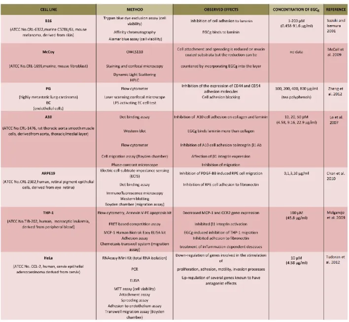
Monitoring of the cellular adhesion and spreading procedures are substantial because these processes take part in maintaining the multicellular structure of the cells, migration, survival, proliferation, differentiation, gene expression, cell-cell communication and immunity, and in cancer metastasis as well. Tea polyphenols, especially EGCg, have become more popular in cell adhesion studies, and many experiments have proved its inhibitory effect [26][34][40][41][46][47][48][49]. EGCg blocks retinal pigment epithelium (RPE) cell adhesion to fibronectin, and it changes the actin cytoskeleton organization during the adhesion process [47]. Fibroblast cell adhesion is greatly reduced on mucin-coated substrata when compared with PLL, but the reduction can be countered by incorporating EGCg into the layer [46]. It is possible that the substratum’s viscoelastic properties are the leading influence on initial cell spreading [46]. Tea polyphenols inhibited the expression of CD44 and CD54 adhesion molecules in PG cells (highly metastatic lung carcinoma cell lines) in a dose-dependent manner and significantly blocked the adhesion of these cells to endothelial cells not only in a state of rest but also when active [40]. EGCg influenced CD44 and CD54 expression during the adhesion process of PG cells to endothelial cells [40]. Lo and co-workers reported that EGCg and also epicatechin gallate (ECG) were able to inhibit smooth muscle cell (SMC) adhesion on laminin and collagen (ECM proteins) expressed in physiological and pathological conditions, and the authors found that EGCg can bind more strongly to laminin than collagen [41]. EGCg could inhibit smooth muscle cell adhesion to integrin β1 Ab and affect their β1 integrin expression; proposing it also affects the cellular components [41]. Suzuki and Isemura reported that EGCg in the culture medium was found to inhibit B16 melanoma cell adhesion, furthermore, approximately 50 % adhesion inhibition occurred when the laminin coating was incubated with 4.8 μM (= 2.19 µg/ml) EGCg [34] Table 1.1 summarizes the results and details of the listed references [1].
19
Table 1.1 Summary of the listed references about the experiments of cellular adhesion and spreading3. Cancerous and normal cell cultures were examined by different methods and techniques to reveal the effect of EGCg (concentration between 0 and 200 µM) on adhesion and spreading. EGCg inhibited the cell adhesion and down- regulated the genes evolved in the stimulation of proliferation, adhesion, motility, and invasion processes [1].
3(ATCC: American Type Culture Collection, CCR2: chemokine (C-C Motif) receptor 2,CD54: cluster of differentiation protein 54 (also known as ICAM-1, intercellular adhesion molecule 1), ELISA: enzyme-linked immunosorbent assay, FRET: fluorescence resonance energy transfer, HPLC: high performance liquid chromatography, LPS:
lipopolysaccharide, MCP-1: monocyte chemoattractant protein-1, MTT: 3-(4,5-dimethylthiazol-2-yl)-2,5- diphenyltetrazolium bromide, OWLS: optical waveguide lightmode spectroscopy, PCR: polimerase chain reaction, PDGF: platelet-derivated growth Factor, RPE: retinal pigment epithelium).
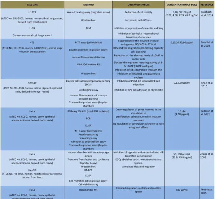
20 Impact on cell motility, migration and stiffness
Observation of the movement of the tumor cells is an important topic: the formation of tumors and metastasis arises when cells are moving [50]. Highly tumorigenic cells move faster than non- tumorigenic cells [51]. Developing drugs that inhibit migration are the focus of many research projects [50]. Traditional herbal extracts have become increasingly popular in the cure of these illnesses. Some studies showed that EGCg is effective against many cancer cell lines [2][3][4][52]. The action of EGCg on cell motility and migration is an actively researched topic today [42][49][52][53][54]. Cell stiffness and motility are related to metastatic activity in tumor cells [42]. An earlier experiment of Takahashi and co-workers also showed that EGCg treatment dose dependently inhibited cell motility and increased cell stiffness in human lung cancer cells and inhibited the expression of vimentin protein and Slug transcription factor as well [42]. EGCg has inhibitory effects on chemoattractant- and hypoxia-stimulated migration of HeLa cells [52]
and blocks plateletderived growth factor-BB-induced (PDGF-BB) RPE cell migration [47].
EGCg inhibits the migration of 4T1 cells through the inhibition of nitric oxide production, reduces the levels of cGMP, and inhibits mammary cancer cell migration through the inhibition of NO/NOS and guanylate cyclase [43]. EGCg dose dependently inhibits cell motility and induces rigid elasticity of cell membranes [42]. According to a study of Takahashi et al., treatment with 50 and 100 μM (= 22.9 and 45.8 µg/ml) EGCg can block H1299 cell migration from 100 to 67.5 and 43.7 % [42]. Furthermore, addition of 10, 50 and 100 μM (= 4.58, 22.9 and 45.8 µg/ml) EGCg inhibited Lu99 cell migration to 81.3, 71.7 and 31.5 % as well [42]. In an experiment of Tudoran et al., EGCg caused an average of 48 and 68 % reduction of migration capability after 24 and 48 h [49]. In a novel study, the inhibitory effect on HeLa cell migration, motility and motility speed was also monitored by an incubator proof miniaturized Holomonitor [54]. Table 1.2 summarizes the results and details of the listed references.
21
Table 1.2 Summary of the listed references about the experiments of motility and migration4. Different cancer cell cultures were examined by different methods and techniques to reveal the effect of EGCg (concentration between 0 and 500 µM) on migration, motility and stiffness. EGCg reduced cell movement (migration, motility), increased stiffness and affected the genes evolved in the stimulation of movement of the cells [1].
4(AFM: atomic force microscopy, ATCC: American Type Culture Collection, ELISA: enzyme-linked immunosorbent assay, HPLC: high performance liquid chromatography, MTT: 3-(4,5-dimethylthiazol-2-yl)-2,5-diphenyltetrazolium bromide, NO/NOS: nitrogen monoxide, nitrous oxide, PDGF: platelet-derivated growth factor, RT-PCR:reverse transcripton polimerase chain reaction, RPE: retinal pigment epithelium).
22
Reduced proliferation and induced apoptosis of cancer cells
A study of Hou and co-workers showed that in cell culture conditions, the auto-oxidation of EGCg result in epidermal growth factor receptor (EGFR) inactivation, however, the inhibition of cell growth is due to other mechanisms [19]. EGCg itself induces chromosomal damage in WIL2- NS cells at 100 μmol/l (=45.8 µg/ml), and this damage is due to the production of H2O2 [55]. But EGCg at <10 μmol/l (=4.58 µg/ml) does not induce chromosomal damage and does not produce H2O2 [55]. Hypothesis of Song et al. presents that EGCg exerts no direct effect on cancer cells in culture; instead, any anticancer effect observed is an indirect result of the generation of H2O2
during the oxidation of EGCg [17]. However, it blocked the expression of HSP70 and HSP90 and demonstrated that this effect was not induced by H2O2 [17][56]. As pointed out by Hayakawa and co-workers, EGCg elevates the caspase8 activity and its fragmentation in U937 cells [57].
According to this study, DNA ladder formation caused by EGCg treatment was inhibited by the caspase 8 inhibitor [57]. These findings of the authors suggested the involvement of the Fas- mediated cascade in the EGCg-induced apoptosis in U937 cells, and thus EGCg binding to Fas presumably on the surface of the cells, triggers the Fas-mediated apoptosis in these cells [57].
EGCg treatment not only results in the downregulation of genes involved in the stimulation of proliferation, motility, adhesion, and invasion processes, but also leads to the upregulation of several genes known to have antagonist effects [49]. EGCg exposure decreases the HeLa cell proliferation rate and invasion potential; thus, this compound might be an important anti- angiogenic therapeutic approach in cervical cancers [49]. EGCg treatment resulted in a dose- dependent inhibition of cell growth, induction of apoptosis, and G0/G1-phase arrest of the cell cycle in human epidermoid carcinoma A431 cells, furthermore, itwas found that 10–80 μM (=
4.58-36.64 µg/ml) EGCg treatment results in loweringof nuclear factor κB NF-κB levels in both the cytoplasm and nucleus in a dose-dependent manner [13]. EGCg exposure was found to result in a dosebased differential inhibition of TNF-α- and LPS-mediated activation of (NF-κB) in the previously mentioned cells in the study of Ahmad and co-workers [13]. EGCg at 50 μg/ml inhibited the activity of NF-κB and hypoxia-inducible factor 1-α (HIF-1α) as well as vascular endothelial growth factor (VEGF) expression in cultured E0771 cells compared to control, thus, EGCg suppresses breast tumor angiogenesis and growth [44]. The hypothesis of Gu et al. is that EGCg directly targets both cancer cells and also their vasculature, thereby reducing tumor growth, proliferation, migration and angiogenesis of breast cancer, which is mediated by the inhibition of NF-κB and HIF-1α activation as well as VEGF expression [44].In a previous study of Weber and co-workers, it was demonstrated that EGCg blocked PDFG-BB isoform-mediated signal transduction pathways and cell proliferation [45]. In the experiments of Siddiqui et al., EGCg decreased the growth and proliferation of prostate cancer cells (PCa), accompanied by the reduction of prostate-specific antigen [58] . EGCg inhibits the activity of the protein phosphatase- 1 (PP1) recombinant δ-isoform of the PP1 catalytic subunit (PP1c), and the galloyl group of the polyphenol may play a role in phosphatase inhibition [59] . An experiment by D’Agostino et al.
demonstrated that the mucus gel layer on HT29 human colonic adenocarcinoma cells probably
23
protects the cells against EGCg toxicity [60]. Cytotoxic impacts of high polyphenol levels may be associated with the ability of polyphenolic compounds to interact with cellular mucins and proteins [60]. According to the authors, food-related ingredients also can influence the toxicity of EGCg, for instance, β-casein is effective in protecting cells against the toxicity effect; however, maltodextrin is less efficient [60]. The speculation that certain carcinogenetic processes are initiated and maintained by cancer stem cells (CSCs) has been partially verified [61]. It has been reviewed by Scarpa and Ninfali that phytochemicals and herbal extracts are able to target and kill CSCs, therefore, EGCg and other phytochemicals, for example, β-carotene, genistein curcumin, piperine, sulforaphane and whole extracts of some plants can inhibit CSCs [61]. Most of these active substances act by interfering with the canonical Wnt [β-catenin/T cell factorlymphoid enhancer factor (TCF–LEF)] pathway implicated in the pathogenesis of some tumors [61]. Table 1.3 summarizes the results and details of the listed references.
Table 1.3 Summary of the listed references about the experiments of apoptosis and proliferation5. Different cancer cell lines were examined by different methods and techniques to reveal the effect of EGCg (concentration between 0 and 4000 µM) on migration, motility and stiffness. EGCg reduced proliferation and induced apoptosis via different signaling pathways [1].
5(ATCC: American Type Culture Collection, CBMN: cytokinesis-block micronucleus, EGFR: epidermal growth factor receptor, FLICE: FADD-like interleukin-1 beta-converting enzyme, ERK: extracellelar-signal regulated kinases, FRET:
fluorescence resonance energy transfer, HPLC: high performance liquid chromatography, HSP: heat shock protein, MTT: 3-(4,5- dimethylthiazol-2-yl)-2,5-diphenyltetrazolium bromide, NK: natural killer cells, NMR: Nuclear Magnetic Resonance, RT- PCR:reverse transcripton polimerase chain reaction, SPR: surface plasmon resonance, TEM: transmission electron
microscopy, TNF: tumor necrosis factor).
24
25
Differences in effects on healthy (normal) and cancer cells
It is very important to know the effects of a prospective anticancer agent on healthy cells as well.
In an experiment by Tao and co-workers, EGCg induced mitochondrialocalized ROS in human oral squamous carcinoma cells (SCC-9, SCC-25) and in premalignant leukoplakia cells (MSK- Leuk-1) as well, but not in normal human gingivalfibroblast cells (HGF-1) [62]. EGCg suppressed mitochondrial redox modulator SIRT3 in SCC-25 cells, whereas it increased SIRT3 activity in HGF-1 cells [62]. The polyphenol selectively decreased the nuclear localization of the estrogen-related receptor α (ERRα), the transcription factor regulating SIRT3 expression in SCC- 25 cells, and this indicates that EGCg may regulate SIRT3 transcription in oral tumor cells via ERRα [62]. In the mentioned cells, SIRT3 activity and expression are inhibited by EGCg, resulting in the accumulation of mtROS, as well as mitochondrial dysfunction and death [62]. In normal oral cells, EGCg activates SIRT3 and related downstream antioxidant responsive genes (AOX genes); thus, this process protects them cells from oxidative injuries [62]. In another study, EGCg exposure resulted in a dose-dependent inhibition of cell growth, G0/G1-phase arrest of the cell cycle and induction of apoptosis as well human epidermoid carcinoma A431 cells, but not in normal human epidermal keratinocytes (NHEK) [13]. The inhibition of NF-κB expression and activation in NHEK was monitored only at high concentrations [13]. The authors found that EGCg imparts differential dose-based NF-κB inhibitory responses in tumor cells versus normal cells [13]. EGCg caused inhibition of NF-κB expression, and activation was found to happen at much higher doses of the polyphenol in NHEK as compared to A431 cells [13]. However, another result showed that EGCg is much more toxic against normal cells, while at low concentrations (<100 μM=45.8 µg/ml) increased the lung cancer cell viability slightly [63].
EGCg induced DNA double-strand breaks and apoptosis in normal cells and intensified their mutation frequency, too [63]. EGCg did not induce chromosomal damage at <10 μmol/l (=4.58 µg/ml) concentrations; however, 100 μmol/l EGCg triggered chromosomal injury in WIL2-NS cells (human B lymphoblastoid cell line) [55]. EGCg at physiological concentrations (<1 μmol/l=
0.458 µg/ml) are not genotoxic, but rather can anticipate ROS-induced chromosomal damage [55]. Experiments showed that EGCg inhibited the growth and proliferation of various prostate cancer cells through multiple mechanisms, with minimal impacts on normal human prostate epithelial cells [58].
26 Inhibitory properties on microbes
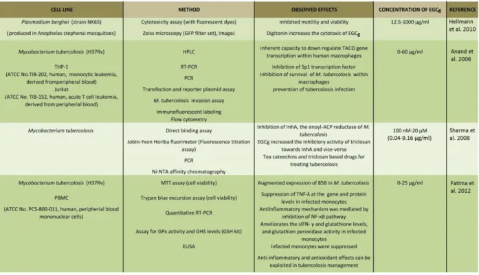
EGCg has a broad antimicrobial (antifungal, antibacterial, antiviral) spectrum as reviewed earlier [64]. EGCg and ECG showed inhibitory effects on the growth and adherence of carcinogenic bacteria on the tooth surface and on the glucan synthesis of streptococci [2]. EGCg also blocks the adherence and growth of bacterium Porphyromonas gingivalis, responsible for periodontal disease [2]. Furthermore, other compounds of tea, delta-cadinene and indole have a synergistic effect on the inhibition of the growth of Streptococcus mutans [2]. It has been reviewed that tea extracts have inhibitory effects against several other bacteria, for example, Vibrio cholerae, Salmonella typhi, Helicobacter pylori, Campylobacter coli, Salmonella, Shigella, Clostridium, Pseudomonas and Candida as well [2]. Tea can exhibit a beneficial effect against viral infections, too: tea polyphenols significantly inhibit influenza A virus in animal cell culture and rotavirus in monkey cell culture [2][64]. Recent studies showed that tea polyphenols inhibit the survival of Mycobacterium tuberculosis in macrophages [65]. EGCg blocks the NADH-dependent enoyl- ACP reductase encoded by the Mycobacterium gene inhA with an IC50 of 17.4 μM (= 7.96 µg/ml ) and interferes with the binding of NADH [66]. The enoyl acyl carrier protein reductase (ACP, ENR) is one of the most important enzymes involved in the type II fatty acid biosynthesis pathway of the intracellular pathogen and a relevant factor for their survival within host cells [66]. EGCg has anti-inflammatory and antioxidant effects on infected host cells by suppressing the augmented expression of MTB 85B and proinflammatory TNF-α of human monocytes [67].
A study of Hellmann et al. also showed that EGCg inhibits even the motility of the Plasmodium sporozoite, the causative agent of malaria [53]. It happens because the EGCg may bind to the adhesion of molecules of the parasite surface, for example circumsporozoite protein (CSP) and thrombospondin- related adhesive protein (TRAP), which are important for their motility [53].
The surface antigen PfEMP of Plasmodium falciparum is involved in the cytoadherence of the pathogen-infected erythrocytes to a range of receptors on the host endothelium, e.g., ICAM-1, which is related to cerebral malaria [68]. EGCg can be an effective inhibitor of ICAM-1-based cytoadherence of P. falciparum-infected erythrocytes [68]. EGCg is able to inhibit the formation of merozoite surface protein 2 (MSP2) fibrils and can alter the β-sheet-like structure of the fibril and disaggregate pre-formed fibrils of MSP2 into soluble oligomers as well [69] . The summary of the results and details of the listed references is shown in Table 1.4.
27
Table 1.4 Summary of the listed references about the experiments with microbes6
.
Protozoons and bacteria and human cells were examined by different methods and techniques. The treatment inhibited the viability of the microbes. Measurements with Mycobacterium tuberculosis showed that EGCg maybe can be used in tuberculosis infection or prevention in the future [1].1.4 . Observed action of EGCg using tissues and animal models and results of clinical trials
Drug pharmacokinetic profile determines the onset, duration, and intensity of a compound’s effect. Understanding the pharmacokinetics (the uptake of the compound into, through, and out of the human body, the time course of its absorption, bioavailability, distribution, metabolism, excretion, receptor binding, postreceptor effects, and chemical interactions) of EGCg is urgently needed to interpret epidemiological, experimental data and to extrapolate in vitro or in vivo animal data to humans [1].
6 (ATCC: American Type Culture Collection, ELISA: enzyme-linked immunosorbent assay, HPLC: high performance liquid chromatography, GFP: green fluorescent protein, MTT: 3-(4,5-dimethylthiazol-2-yl)-2,5-diphenyltetrazolium bromide, Ni-NTA: nickel-charged affinity resin (nitrilotriaceticacid), NK: ntural killer cells, PMBC: peripheral blood mononuclear cell, RT-PCR:reverse transcripton polimerase chain reaction, TNF: tumor necrosis factor).
28 Experiments with tissues and animal models
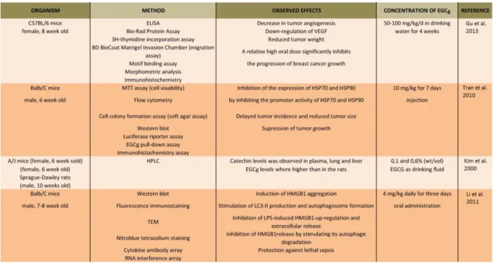
In CF-1 mice, EGCg exhibited a linear dose relationship in their plasma, prostate and liver [70].
In the colon and small intestine the levels of EGCg plateaued between 500 and 2,000 mg/kg [70].
These results of Lambert and co-workers suggest that absorption of EGCg from small intestine is largely via passive diffusion; however, at higher concentrations, the colonic and small intestinal tissues become saturated [70]. EGCg treatment reduced tumor weight in mice; nevertheless, it had no effects on body weight, heart weight, angiogenesis and VEGF expression in the heart and skeletal muscle of these animals [44]. Another study also showed that administration of EGCg caused a 70 % decrease in tumor volume in BALB/c mice, and the cure did not alter body weight compared with PBS-treated controls [56]. Furthermore, the expression levels of HSP70 and HSP90 stress-inducible proteins were decreased in EGCg-treated mice [56]. In rats, epicatechin (EC) and epigallocatechin (EGC) in plasma increased over time and reached peak values [71].
Among tea catechins, the plasma concentrations of EGCg were much lower than those of EC and EGC [71]. Kim and co-workers found high levels of EC and EGC in urine, while high levels of EGCg were found in feces [71]. When purified EGCg or green tea were administrated to mice in drinking water, traversed angiogenesis in vivo and restrained Kaposi’s sarcoma tumor growth [31]. Consumption of tea by rodents could induce adaptive responses influencing tissue and blood levels of catechins with time, and an investigation of a similar phenomenon in humans can be predicted [71]. In an experiment of Li and co-authors, it has been suggested that EGCg inhibits high-mobility group box 1 protein (HMGB1, late proinflammatory mediator of lethal sepsis) release by stimulating its aggregation and autophagic (“self-eating”) degradation [72].
EGCg blocked bacterial endotoxin-induced HMGB1 release in vitro and saved mice from lethal sepsis when EGCg was given to them intraperitoneally, even when the first dose was given orally at 24 hours after onset of the illness [72]. The authors validated the therapeutic potential of EGCg in animal models of sepsis by administering it through a clinically practicable process [72]. The summary of the results and details of the listed references are presented in Table 1.5.
29
Table 1.5 Summary of the listed references about the experiments with animal models7. Male and female mice and rats were treated by EGCg for days (oral administration or injection). The tumor growth was suppressed and the treatment reduced tumor size in the animals [1].
Clinical trials and beneficial influences of EGCg
The effects of tea consumption on the risk of human cancer have been investigated in many studies; however, the results have been inconclusive. Tachibana reviewed in 2011, that some experiments suggested a cancer-preventive effect of tea, while other studies did not show an association [5]. The inconsistent results of the studies were probably due to, for example, different disturbing factors, difficulties in quantifying tea consumption, population heterogeneity and varied cancer etiology in different populations [5]. Traditional cancer drugs often destroy some healthy cells along with tumor cells, but EGCg appears to target functions unique to tumor cells, thus, this green tea compound has a very acceptable safety profile [3]. For example, in previous a clinical trial reviewed by Singh et al, EGCg capsules (200 mg p.o.) for 12 weeks was reported to be effective in human papilloma virus-infected persons [3]. Epidemiological studies identified an inverse association between tea consumption and the frequency of rectal and colon cancer as well as esophageal and gastric tumors [9]. Other epidemiological data also show that
7(ATCC: American Type Culture Collection, ELISA: enzyme-linked immunosorbent assay, HMGB: high-mobility group box proteins, HPLC: high performance liquid chromatography, MTT: 3-(4,5-dimethylthiazol-2-yl)-2,5-
diphenyltetrazolium bromide, LC3: microtubule-associated protein light chain 3, LPS: lipopolysaccharide, TEM:
transmission electron microscopy, VEGF: vascular endothelial growth factor).
30
prostate cancer is sharply rising in the west, but its incidence has been especially low in East Asian countries where green tea is a popular drink [58]. Fujiki and co authors reviewed that ten Japanese-size cups of green tea daily, supplemented with tablets of green tea extract, reduced the recurrence of colorectal adenoma by 51.6 % in persons after polypectomy according to a trial in 2008 [73]. Furthermore, a study also revealed that green tea most significantly prevented lung cancer (the relative risk of 0.33 with at least 10 cups of green tea per/day), and at high consumption prevented tumors of liver, colorectum, and stomach as well [73]. Unfortunately, the antimicrobial activity of tea in humans is not well documented and should be investigated in a multidimensional way because certain mechanisms are implicated, for example, the immune system, heredity, microbial ecology, etc. [2]. Mereles and Hunstein summarized in their review article that providing EGCg is a pharmacon, side effects should also be expected, for example, effects on the anxiolytic and hypoglycemic activity, hypochromic anemia, and liver and kidney failure [7]. However, 800 mg caffeine-free EGCg per day for 4 weeks was shown to be well tolerated and safe in healthy persons [7].
1.5. Perspectives: Investigation and application possibilities in the near future
Recently, several novel directions appeared concerning the research and development and future applications of EGCg.
Nanocarriers and nanocomposite systems for targeted drug delivery are also emerging and may involve the application of EGCg.
Combination of EGCg and anticancer compounds
As reviwed by Fujiki and co-authors, some studies showed that the combination of green tea catechins and anticancer drugs has the potential to enhance the efficacy of the drugs [73]. The combination of these components induced similar synergistic anticancer effects for both in vivo and in vitro experiments and showed an average reduction in tumor volume of 70.3 % [73].
Furthermore, among others, in in vivo mouse xenograft models, the combinations of paclitaxel and EGCg as well as docetaxel and EGCg completely eliminated tumors of human prostate cancer cell line PC-3ML, but other combinations also reduced the tumor volumes in the case of various cancer cell lines [73]. The amount of EGCg for the elimination of tumors in mice varied from 4.56 to 6.84 mg/day/mouse [73].Converted these numbers to humans, it would be 1.37–2.05 g/day/ person (6–9 Japanese-size cups of green tea) [73]. The results suggest that the combinations of nonsteroidal anti-inflammatory drugs and EGCg activate the GADD153-DR5- TRAIL apoptotic pathway [73]. There are also experiments involving EGCg alone or in