Data Descriptor: Transcriptome- wide survey of pseudorabies virus using next- and third-generation sequencing platforms
Dóra Tombácz1, Donald Sharon2, Attila Szűcs1, Norbert Moldován1, Michael Snyder2
& Zsolt Boldogkői1
Pseudorabies virus (PRV) is an alphaherpesvirus of swine. PRV has a large double-stranded DNA genome and, as the latest investigations have revealed, a very complex transcriptome. Here, we present a large RNA-Seq dataset, derived from both short- and long-read sequencing. The dataset contains1.3million100 bp paired-end reads that were obtained from the Illumina random-primed libraries, as well as10million50 bp single-end reads generated by the Illumina polyA-seq. The Pacific Biosciences RSII non-amplified method yielded57,021reads of inserts (ROIs) aligned to the viral genome, the amplified method resulted in 158,396PRV-specific ROIs, while we obtained12,555ROIs using the Sequel platform. The Oxford Nanopore’s MinION device generated44,006reads using their regular cDNA-sequencing method, whereas 29,832and120,394reads were produced by using the direct RNA-sequencing and the Cap-selection protocols, respectively. The raw reads were aligned to the PRV reference genome (KJ717942.1). Our provided dataset can be used to compare different sequencing approaches, library preparation methods, as well as for validation and testing bioinformatic pipelines.
Design Type(s) transcription profiling by high throughput sequencing design • parallel group design
Measurement Type(s) messenger RNA • total RNA • Coding Region Technology Type(s) RNA sequencing
Factor Type(s)
temporal_interval • assay material selection • cap analysis of gene expression • enzymatic amplification • size fractionation • nucleic acid library construction protocol • Technology Platform
Sample Characteristic(s) Suid herpesvirus1strain Kaplan
1Department of Medical Biology, Faculty of Medicine, University of Szeged, Szeged6720, Hungary.2Department of Genetics, School of Medicine, Stanford University, Stanford, California 94305, USA. Correspondence and requests for materials should be addressed to Z.B. (email: boldogkoi.zsolt@med.u-szeged.hu).
OPEN
Received:30January2018 Accepted:28March2018 Published:19June2018
Background & Summary
Pseudorabies virus (PRV) is a causative agent of Aujeszky’s disease (AD)1in pigs. PRV has a double- stranded DNA genome with a size of approximately 143 kbp. PRV is often employed in laboratories to study the molecular pathomechanism of the herpesviruses2. It is also a suitable tool as concerns gene and tumour therapy3, as well as for mapping of neuronal circuits4–8. This virus has also been used as live vaccines against AD9–11.
Here, we provide a large dataset derived from RNA-Seq experiments including different next- generation sequencing (NGS) – and third-generation sequencing (3rdGS) techniques (Fig. 1). Our aim with this study was to provide a dataset that can be used for comparison of the different sequencing platforms and library preparation methods using PRV as a model organism. In addition, these data are also applicable for identifying novel coding and non-coding transcripts, transcript isoforms, splice variants of PRV, and for defining full-length transcripts by using a combination of sequencing platforms.
One of the most popular NGS platforms, the Illumina HiScanSQ was used to generate high quality short-reads and extremely high coverage throughout the entire PRV genome. Random-primed cDNA library was prepared from viral RNAs. Paired-end RNA sequencing was carried out to characterize novel splice isoforms, as well as to obtain general information on the transcription activity of PRV12. PolyA- sequencing was used to determine the 3′-ends of RNA molecules. With this technique, we were able to detect alternative polyadenylation events in the PRV transcripts. Both libraries were run on a singleflow cell resulting in 1.3 million 100 bp paired-end reads from the random hexamer-primed libraries, and 10 million 50 bp single-end reads from the poly(A)-enriched RNA-Seq, aligning to the viral reference13 (KJ717942.1).
Although the error rate of 3rdGS techniques is higher than those of NGS’s14, they are able to identify novel full-length transcripts15–17and are therefore more applicable for global transcriptome profiling and RNA isoform detection compared to short-read techniques.
The Real-Time Sequencer II (RSII) and the Sequel 3rdGS platforms from Pacific Biosciences (PacBio) and the Oxford Nanopore Technologies (ONT) MinION 3rdGS device were used to characterize the static18,19and dynamic20PRV transcriptome. These sequencing techniques, with the library preparation methods [e.g. non-amplified SMRT method and amplified, Iso-seq protocol from the PacBio; full-length cDNA-sequencing, direct RNA-sequencing, and cDNA-sequencing on 5′Cap-selected samples from ONT, (Fig. 1,2)] used in these studies made it possible to identify several hundreds of novel transcript isoforms (including 3′- and 5′UTR variants, and splice isoforms), as well as dozens of protein-coding and non-coding RNAs and numerous complex transcripts of PRV.
Seventy-one SMRT Cells were run on RSII system. P5-C3 chemistry and 180-minute data collection mode was used for the non-amplified samples, while P6-C4 enzymes were applied and 240 or 360 min movies were recorded for the amplified samples. cDNAs were sequenced on a single Sequel SMRT Cell with P6-C4 reagents; 10 h run-time was applied. Altogether seven MinIONflow cells were used for the different ONT approaches.
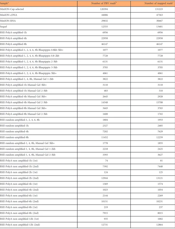
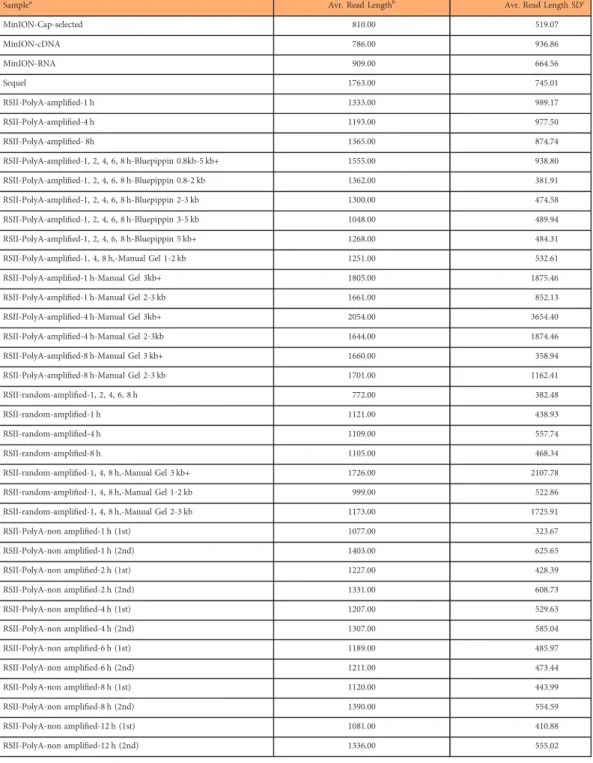
The raw sequencing reads were mapped to the above-mentioned reference genome. Sequencing on the RSII platform resulted in 215,417 reads of inserts (ROIs), while the utilized nanopore sequencing methods generated altogether 194,232 PRV specific reads (Table 1). The average read lengths aligning to the PRV genome were 1,326 bp for PacBio RSII, 1,763 bp for the Sequel and 827 bp for ONT. It should be noted that the library preparation and size-selection methods resulted in different samples in length (Table 2).
Figure 1. Dataflow diagram shows the detailed overview of the study design.
This dataset can help explore the advantages and disadvantages associated with each sequencing method used in this work. This approach can be used for the analysis of multiple features of the sequencing platforms, including read length, base-calling error rate, coverage and mappability. The application of the various sequencing techniques can be evaluated by the analysis of the identified transcript isoforms, and the quantification of the transcriptome comparing the performance of Illumina, PacBio and ONT. This dataset is also useful for the analysis of the transcriptome complexity of PRV. Our data include a sub-dataset which can be used for the transcriptome analysis of PRV during an infection period including six different time-points.
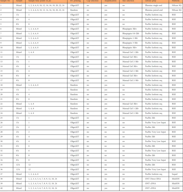
Here we provide a detailed overview of the library preparation techniques and a description of the data (Table 3, Figs. 1 and 2).
Methods
Schematic overviews of the methodological workflow are shown in theflowchart of Figs. 1 and 2. The applied reagents and utilized approaches are listed in Table 4.
Cells, viruses and infection conditions
Immortalized porcine kidney-15 (PK-15; ATCC® CCL-33™) cells were used for the propagation of pseudorabies virus strain Kaplan (PRV-Ka) at 37 °C and 5% CO2 in Dulbecco’s modified Eagle medium (DMEM, Gibco Invitrogen) supplemented with 5% foetal bovine serum (FBS; Gibco Invitrogen). The virus stock was originally obtained from the Kaplan Lab (Department of Microbiology, Vanderbilt University School of Medicine, Nashville, Tennessee)21, but Vanderbilt University received it from Dr. Richard F. Haff in a suspension of infected mouse brain22. Gentamycin (80μg/ml) was also added to the cell culture medium. The virus stock was prepared as follows: the medium was removed from the rapidly-growing semi-confluent PK-15 cells then it was infected with the Kaplan strain of PRV (a multiplicity of infection of 0.1 plaque-forming unit (pfu)/cell). Infected cells were incubated until complete cytopathic effect was observed. Samples were taken through three times freeze-thaw cycles, followed by centrifugation at 10,000 g for 15 min. The titre of the virus stock was determined in PK-15 cells. For all experiments, cells were infected with a high MOI (10 pfu/cell) and incubated for 1 h, followed by removal of the virus suspension and washing of the cells with phosphate-buffered saline (PBS). The number of cells in a cultureflask was 5 × 10⁶. After the addition of new medium to the cells, they were incubated for 1, 2, 4, 6, 8, 10, 12, 14, 16, 18, 20, 22 or 24 h pi and they both were mixed for Illumina sequencing (Table 3). One, 2, 4, 6, 8 and 12 h of incubation were used for the non-amplified PacBio sequencing, while the 1, 2, 4, 6 and 8 h pi samples were utilized for the PacBio amplified, Iso-Seq protocol. Samples from different time points were individually sequenced on the RSII, but they were also mixed for PacBio sequencing (Table 3). The incubation time was 1, 2, 3, 4, 5, 6, 7, 8, 9, 12, 18 and 24 h and a mixture from them was used for all types of ONT sequencing (Table 3).
RNA purification
Isolation of total RNAsThe NucleoSpin®RNA II kit (Macherey-Nagel) was used to isolate RNA from samples for Illumina sequencing, while the new version, the NucleoSpin®RNA kit (Macherey-Nagel) was used for all the other samples, as was described earlier12,18,20. Briefly, cells were collected by centrifugation and lysed by incubation in a solution containing large amounts of chaotropic ions. This buffer inactivates the RNase. Nucleic acid molecules bind to the silica membrane. All samples were handled with DNase I solution (provided by the kit) to remove residual DNA contaminations. Total RNAs were eluted from the membrane in RNase-free water. To eliminate the potential remaining DNA contamination, samples were Figure 2. Dataflow diagram shows the detailed overview of the wet lab experiments and bioinformatics pipelines.
treated by Ambion® TURBO DNA-free™ Kit. The final concentrations of the RNA samples were determined by Qubit®. 2.0 Fluorometer using Qubit RNA BR Assay Kit (Life Technologies).
RNA quality was assessed with the Agilent Bioanalyzer 2100 and RIN scores above 9.6 were used for cDNA production. RNA samples were stored at −80 °C until further use. Samples were made as follows: The Illumina oligodT- and random-primed sequencing reactions were carried out from the same RNA mixture. The libraries for the kinetic analysis and for the mixed sequencing using PacBio RSII (non-amplified method) were all prepared from different cell culture flasks (containing 5 × 10⁶ cells/
flask), but the same virus stock was used for these infections. For the amplified RSII sequencing, samples were prepared from separate flasks (each containing 5 × 10⁶ cells and infected with the same
Samplea Number of PRV readsb Number of mapped readsc
MinION-Cap-selected 120394 131223
MinION-cDNA 44006 47363
MinION-RNA 29832 30667
Sequel 12555 13481
RSII-PolyA-amplified-1h 6956 6956
RSII-PolyA-amplified-4h 22958 22958
RSII-PolyA-amplified-8h 46147 46147
RSII-PolyA-amplified-1, 2, 4, 6, 8h-Bluepippin 0.8kb-5kb+ 1077 1077
RSII-PolyA-amplified-1, 2, 4, 6, 8h-Bluepippin 0.8-2kb 7728 7728
RSII-PolyA-amplified-1, 2, 4, 6, 8h-Bluepippin 2-3kb 6131 6131
RSII-PolyA-amplified-1, 2, 4, 6, 8h-Bluepippin 3-5kb 3705 3705
RSII-PolyA-amplified-1, 2, 4, 6, 8h-Bluepippin 5kb+ 4061 4061
RSII-PolyA-amplified-1, 4, 8h,-Manual Gel 1-2kb 3822 3822
RSII-PolyA-amplified-1h-Manual Gel 3kb+ 3110 3110
RSII-PolyA-amplified-1h-Manual Gel 2-3kb 483 510
RSII-PolyA-amplified-4h-Manual Gel 3kb+ 2645 2928
RSII-PolyA-amplified-4h-Manual Gel 2-3kb 14348 15708
RSII-PolyA-amplified-8h-Manual Gel 3kb+ 3443 3765
RSII-PolyA-amplified-8h-Manual Gel 2-3kb 1608 1765
RSII-random-amplified-1, 2, 4, 6, 8h 1804 1953
RSII-random-amplified-1h 2327 2485
RSII-random-amplified-4h 7202 7629
RSII-random-amplified-8h 11452 12259
RSII-random-amplified-1, 4, 8h,-Manual Gel 3kb+ 1778 1855
RSII-random-amplified-1, 4, 8h,-Manual Gel 1-2kb 2218 2425
RSII-random-amplified-1, 4, 8h,-Manual Gel 2-3kb 3393 3627
RSII-PolyA-non amplified-1h (1st) 74 81
RSII-PolyA-non amplified-1h (2nd) 7392 7440
RSII-PolyA-non amplified-2h (1st) 124 125
RSII-PolyA-non amplified-2h (2nd) 12944 13121
RSII-PolyA-non amplified-4h (1st) 1369 1574
RSII-PolyA-non amplified-4h (2nd) 1023 1054
RSII-PolyA-non amplified-6h (1st) 2144 2269
RSII-PolyA-non amplified-6h (2nd) 10151 10231
RSII-PolyA-non amplified-8h (1st) 219 237
RSII-PolyA-non amplified-8h (2nd) 7915 8015
RSII-PolyA-non amplified-12h (1st) 935 1002
RSII-PolyA-non amplified-12h (2nd) 12731 12864
Table 1. Summary of the obtained read counts from long-read sequencing aligned to the PRV genome.aType of the samples.bTotal number of PRV-specific reads.cTotal number of reads mapped to the PRV genome. (There is an approximately 15 kb-long inverted repeat sequence region in the PRV genome, therefore those reads which map to this location occur in duplicate in rowc).
virus stock). The Sequel and the various MinION libraries have been prepared using the same RNA mixture.
Ribosomal RNA depletion For the Illumina sequencing and for the PacBio random-primed sequencing, the total RNA samples were depleted from rRNA using the Epicentre Ribo-Zero™Magnetic Kit H/M/R (Illumina).
Selection of PolyA(+) RNAFor the PacBio and MinION polyA sequencing, the polyA(+) fraction of the RNA samples were isolated using the Qiagen Oligotex mRNA Mini Kit, following the “Spin Columns”protocol.
PolyA purified and rRNA depleted RNA samples were quantified through use of the Qubit RNA HS Assay Kit (Life Technologies) and then subjected to cDNA synthesis according to the downstream applications.
Samplea Avr. Read Lengthb Avr. Read Length SDc
MinION-Cap-selected 810.00 519.07
MinION-cDNA 786.00 936.86
MinION-RNA 909.00 664.56
Sequel 1763.00 745.01
RSII-PolyA-amplified-1 h 1333.00 989.17
RSII-PolyA-amplified-4 h 1193.00 977.50
RSII-PolyA-amplified- 8h 1365.00 874.74
RSII-PolyA-amplified-1, 2, 4, 6, 8 h-Bluepippin 0.8kb-5 kb+ 1555.00 938.80
RSII-PolyA-amplified-1, 2, 4, 6, 8 h-Bluepippin 0.8-2 kb 1362.00 381.91
RSII-PolyA-amplified-1, 2, 4, 6, 8 h-Bluepippin 2-3 kb 1300.00 474.58
RSII-PolyA-amplified-1, 2, 4, 6, 8 h-Bluepippin 3-5 kb 1048.00 489.94
RSII-PolyA-amplified-1, 2, 4, 6, 8 h-Bluepippin 5 kb+ 1268.00 484.31
RSII-PolyA-amplified-1, 4, 8 h,-Manual Gel 1-2 kb 1251.00 532.61
RSII-PolyA-amplified-1 h-Manual Gel 3kb+ 1805.00 1875.46
RSII-PolyA-amplified-1 h-Manual Gel 2-3 kb 1661.00 852.13
RSII-PolyA-amplified-4 h-Manual Gel 3kb+ 2054.00 3654.40
RSII-PolyA-amplified-4 h-Manual Gel 2-3kb 1644.00 1874.46
RSII-PolyA-amplified-8 h-Manual Gel 3 kb+ 1660.00 358.94
RSII-PolyA-amplified-8 h-Manual Gel 2-3 kb 1701.00 1162.41
RSII-random-amplified-1, 2, 4, 6, 8 h 772.00 382.48
RSII-random-amplified-1 h 1121.00 438.93
RSII-random-amplified-4 h 1109.00 557.74
RSII-random-amplified-8 h 1105.00 468.34
RSII-random-amplified-1, 4, 8 h,-Manual Gel 3 kb+ 1726.00 2107.78
RSII-random-amplified-1, 4, 8 h,-Manual Gel 1-2 kb 999.00 522.86
RSII-random-amplified-1, 4, 8 h,-Manual Gel 2-3 kb 1173.00 1725.91
RSII-PolyA-non amplified-1 h (1st) 1077.00 323.67
RSII-PolyA-non amplified-1 h (2nd) 1403.00 625.65
RSII-PolyA-non amplified-2 h (1st) 1227.00 428.39
RSII-PolyA-non amplified-2 h (2nd) 1331.00 608.73
RSII-PolyA-non amplified-4 h (1st) 1207.00 529.63
RSII-PolyA-non amplified-4 h (2nd) 1307.00 585.04
RSII-PolyA-non amplified-6 h (1st) 1189.00 485.97
RSII-PolyA-non amplified-6 h (2nd) 1211.00 473.44
RSII-PolyA-non amplified-8 h (1st) 1120.00 443.99
RSII-PolyA-non amplified-8 h (2nd) 1390.00 554.59
RSII-PolyA-non amplified-12 h (1st) 1081.00 410.88
RSII-PolyA-non amplified-12 h (2nd) 1336.00 555.02
Table 2. Summary of the obtained read lengths from long-read sequencing.aType of the samples.
bAverage read lengths of the different library preparation and long-read sequencing approaches.cStandard deviation (SD) values.
cDNA synthesis, library preparation and sequencing Illumina sequencing
Total RNA was purified from PK-15 cells in various stages of PRV infection from 1 to 24 h pi and then, the samples were mixed together to uncover an extensive variety of viral transcripts. Libraries were prepared from ribo-depleted samples using the ScriptSeq v2 RNA-Seq Library Preparation Kit (Epicentre/Illumina) according to the manufacturer’s recommendations. The kit uses a random-primed (random hexamer with tagging sequence) cDNA synthesis reaction; this“original”protocol was used to construct a paired-end library, but for PolyA sequencing (PA-seq), a single-end library was prepared through the use of custom anchored adaptor-primer oligonucleotides with an oligo(VN)T20 primer sequence (Table 5). Briefly, the rRNA-depleted RNA samples were mixed with the primer (random or oligo(d)T) and the RNA Fragmentation Solution (part of the ScriptSeq Kit), and the mixtures were
Sample No Sample Sample time points (h pi) RT priming Cap-selection Amplification Size selection Library prep Platform
1 Mixed 1, 2, 4, 6, 8, 10, 12, 14, 16, 18, 20, 22, 24 Oligo(d)T no yes no Illumina single-end HiScan SQ
2 Mixed 1, 2, 4, 6, 8, 10, 12, 14, 16, 18, 20, 22, 24 Random no yes no Illumina paired-end HiScan SQ
3 1 h 1 Oligo(d)T no yes no PacBio Isoform seq RSII
4 4 h 4 Oligo(d)T no yes no PacBio Isoform seq RSII
5 8 h 8 Oligo(d)T no yes no PacBio Isoform seq RSII
6 Mixed 1, 2, 4, 6, 8 Oligo(d)T no yes Bluepippin 5kb+ PacBio Isoform seq RSII
7 Mixed 1, 2, 4, 6, 8 Oligo(d)T no yes Bluepippin 0.8-2kb PacBio Isoform seq RSII
8 Mixed 1, 2, 4, 6, 8 Oligo(d)T no yes Bluepippin 2-3kb PacBio Isoform seq RSII
9 Mixed 1, 2, 4, 6, 8 Oligo(d)T no yes Bluepippin 3-5kb PacBio Isoform seq RSII
10 Mixed 1, 2, 4, 6, 8 Oligo(d)T no yes Bluepippin 5kb+ PacBio Isoform seq RSII
11 Mixed 1, 4, 8 Oligo(d)T no yes Manual Gel 1-2kb PacBio Isoform seq RSII
12 1 h 1 Oligo(d)T no yes Manual Gel 3kb+ PacBio Isoform seq RSII
13 1 h 1 Oligo(d)T no yes Manual Gel 2-3kb PacBio Isoform seq RSII
14 4 h 4 Oligo(d)T no yes Manual Gel 3kb+ PacBio Isoform seq RSII
15 4 h 4 Oligo(d)T no yes Manual Gel 2-3kb PacBio Isoform seq RSII
16 8 h 8 Oligo(d)T no yes Manual Gel 3kb+ PacBio Isoform seq RSII
17 8 h 8 Oligo(d)T no yes Manual Gel 2-3kb PacBio Isoform seq RSII
18 Mixed 1, 2, 4, 6, 8 Random no yes no PacBio Isoform seq RSII
19 1 h 1 Random no yes no PacBio Isoform seq RSII
20 4 h 4 Random no yes no PacBio Isoform seq RSII
21 8 h 8 Random no yes no PacBio Isoform seq RSII
22 Mixed 1, 4, 8 Random no yes Manual Gel 3kb+ PacBio Isoform seq RSII
23 Mixed 1, 4, 8 Random no yes Manual Gel 1-2kb PacBio Isoform seq RSII
24 Mixed 1, 4, 8 Random no yes Manual Gel 2-3kb PacBio Isoform seq RSII
25 1 h 1 Oligo(d)T no no no PacBio 2kb RSII
26 1 h 1 Oligo(d)T no no no PacBio Very Low Input RSII
27 2 h 2 Oligo(d)T no no no PacBio 2kb RSII
28 2 h 2 Oligo(d)T no no no PacBio Very Low Input RSII
29 4 h 4 Oligo(d)T no no no PacBio 2kb RSII
30 4 h 4 Oligo(d)T no no no PacBio Very Low Input RSII
31 6 h 6 Oligo(d)T no no no PacBio 2kb RSII
32 6 h 6 Oligo(d)T no no no PacBio Very Low Input RSII
33 8 h 8 Oligo(d)T no no no PacBio 2kb RSII
34 8 h 8 Oligo(d)T no no no PacBio Very Low Input RSII
35 12 h 12 Oligo(d)T no no no PacBio 2kb RSII
36 12 h 12 Oligo(d)T no no no PacBio Very Low Input RSII
37 Mixed 1, 2, 4, 6, 8 Oligo(d)T no yes no PacBio Isoform seq Sequel
38 Mixed 1, 2, 3, 4, 5, 6, 7, 8, 9, 12, 18, 24 Oligo(d)T no no no ONT Direct RNA MinION
39 Mixed 1, 2, 3, 4, 5, 6, 7, 8, 9, 12, 18, 24 Oligo(d)T yes yes no ONT cDNA MinION
40 Mixed 1, 2, 3, 4, 5, 6, 7, 8, 9, 12, 18, 24 Oligo(d)T no yes no ONT cDNA MinION
Table 3. Summary table of the various wet lab approaches used in this study.
incubated at 85 °C for 5 min. The following kit components were mixed together: cDNA Synthesis Premix, DTT and StarScript AMV Reverse Transcriptase. This reagent mix was added to the pre-heated RNA-mixtures and they were incubated at 25 °C for 5 min and then 42 °C for 20 min. Reactions were cooled down to 37 °C. Finishing solution was added to the samples and incubation was continued at 37 ° C for an additional 10 min, then at 95 °C for 3 min. Samples were cooled down to 25 °C, and Terminal Tagging Premix, as well as DNA Polymerase were added. The incubation was continued at 25 °C for 15 min and then at 95 °C for 3 min. The di-tagged cDNA samples were purified by using the AMPure XP beads (Beckman Coulter). The purified samples were amplified by PCR using the FailSafe PCR Premix E, primers and FailSafe PCR enzyme (Lucigen, Epicentre). The PCR conditions were as follows: initial denaturation at 95 °C for 1 min, followed by 15 cycles at 95 °C for 30 sec 55 °C for 30 sec and 68 °C for 3 min. Thefinal incubation step was carried out at 68 °C for 7 min. The PCR amplicons were purified with AMPure beads. The quantity and the quality of the samples were checked using Qubitfluorometer (Life Technologies) and Agilent 2100 Bioanalyzer, respectively.
PacBio SMRTbell library preparation & sequencing –the non-amplified method & RSII sequencing
Generation of cDNAs. The SuperScript Double-Stranded cDNA Synthesis Kit (Life Technologies) was used to prepare cDNAs from the polyA(+) RNA samples. These samples were used to quantify the PRV transcriptome during the infection period between 1-12 h. The enzyme-which was included in the kit-was changed to SuperScript III Reverse Transcriptase. Thefirst-strand synthesis reactions were primed with Anchored Oligo(dT)20 primers (Life Technologies, Table 5). The obtained cDNAs were measured by using Qubit HS dsDNA Assay Kit (Life Technologies). The total amount of cDNA synthesized at each time point was used to prepare SMRTbell templates.
Preparation of SMRTbell libraries-“Barcoding method”
The cDNA samples (~500 ng/sample) were used to prepare SMRTbell templates by using the PacBio DNA Template Prep Kit 1.0 following the Pacific Biosciences’2 kb Template Preparation and Sequencing protocol.
Repairing the cDNA ends. Template Prep Buffer, ATP High, dNTP and End Repair Mix (PacBio) were added to the samples and then they were incubated at 25 °C for 15 min.
Sample purification0.6x volume. AMPure PB bead was added to the samples. They were mixed using VWR vortex mixer for 10 min at 2000 rpm and room temperature. Tubes were placed in a magnetic bead rack for 3 min. After the bead pellets were formed, the supernatant were discarded. Beads were 2-times washed with freshly prepared ethanol (70%). Samples were dried and then they were eluted in 30μl Elution Buffer (PacBio).
Adapter ligation. This step was carried out at 25 °C for 15 min with the addition of specific bar-coded adapters (Table 6), Template Prep Buffer, ATP Low and Ligase (PacBio). The enzyme was inactivated at 65 °C (10 min).
Total RNA isolation PolyA selection Ribodepletion Reverse transcription & dscDNA production
cDNA synthesis by PCR
Library preparation kit Sequencing chemistry
Instrument
Macherey-Nagel RNA
Qiagen Oligotex mRNA mini Kit –
StarScript AMV Reverse Transcriptase FailSafe PCR PreMix Selection Kit
ScriptSeq v2 RNA-Seq
Library Preparation Kit NA HiScan 2500
– Epicentre Ribo-Zero™
Magnetic Kit H/M/R
Qiagen Oligotex mRNA mini Kit –
SuperScript III & SuperScript
double-stranded cDNA Synthesis Kit –
PacBio Template Preparation Kit
P5-C3
RSII
Clontech SMARTer PCR
cDNA Synthesis Kit KAPA HiFi PCR Kit P6-C4
– Epicentre Ribo-Zero™
Magnetic Kit H/M/R
Qiagen Oligotex mRNA mini Kit –
Sequel
SuperScript III – Direct RNA
Sequencing Kit NA
MinION SuperScript IV KAPA HiFi PCR Kit Ligation Sequencing
Kit 1D
NA
Lexogen Teloprime Kit
enzymes & reagents Lexogen Teloprime
PCR mix Ligation Sequencing
Kit 1D NA
Table 4. Summary table of the reagents and chemistries used for the sequencing.
Exonuclease treatment. ExoIII (50U) and ExoVII (5U) enzymes were added to the carrier DNA- cDNA mixture, then they were incubated at 37 °C for 1 h, then the reactions were returned to 4 °C.
Sample purification. SMRTbell Templates were purified using 0.6 × AMPure PB beads, as was described above. Two purification steps were applied after one other. Thefinal elution volume was 10μl.
Qubitfluorometer was used for quantitation.
SMRTbell templates were bound to the PacBio’s P5 DNA polymerase. These complexes were bound to MagBeads using the Pacific Biosciences MagBead Binding Kit. The concentrations of the SMRTbell libraries were measured by Qubit and they were also qualified by Agilent 2100 Bioanalyzer. The PacBio RSII platform and C3 sequencing chemistry was used for sequencing. 180 min movies were applied for each SMRT Cell.
Annealing of the sequencing primers to the template DNA and the DNA polymerase binding. The PacBio Calculator v.2.3.0.0. was used to set the annealing and binding reactions. 2000 bp insert size and 1 ng/μl concentration was set. The Sequencing Primer (5000 nM) was diluted to 150 nM with the PacBio Elution Buffer (EB). Oneμl from the diluted primer and 10x Primer Buffer were added to the template DNA. Annealing was carried out at 80 °C for 2 min, and then the temperature was ramp to 25 °C at a rate of 0.1 °C/sec. The total volume of annealed template was bound to the Polymerase.
For this, 2μl dNTP, 2μl DTT, 2μl Binding Buffer (BB) and 2μl diluted Polymerase were added to the samples. Mixtures were incubated at 30 °C for 4 h and then they were heated to 37 °C for 30 min. 2μl from the complexes were used for MagBead binding. Cleaned MagBeads (74μl) were added to the samples and they were incubated at 4 °C for 2 h on a HulaMixer rotator (Invitrogen). After the incubation, samples were washed with 19μl BB, then 19μl Wash Buffer (WB), andfinally they were resuspended in 19μl BB. The total amount of the MagBead-bound complex was loaded onto the RSII sequencer.
Preparation of SMRTbell libraries-“Carrier DNA method”
The total amount of cDNA synthesized at each time point was used to prepare SMRTbell templates by using the PacBio DNA Template Prep Kit 1.0 and following the Pacific Biosciences template preparation and sequencing. protocol for Very Low (10 ng) Input 2 kb libraries with carrier DNA (pBR322, Thermo Scientific).
Preparing the carrier DNA. The concentration of the pBR322 plasmid DNA was measured by Nanodrop. A 100ng/μl stock solution was prepared from the plasmid using the PacBio Elution Buffer (EB). The DNA was exonuclease treated with the PacBio ExoIII (200U) and ExoVII (20U) enzymes and the Template Prep buffer (10 × ). The mixture was incubated at 37 °C for 1 h, then it was cooled down to 4 °C. The DNA was purified and concentrated by using 0.6 × AMPure®PB beads and it was eluted in 50 μL EB. The exonuclease-treated carrier DNA was quantified by Qubitfluorometer.
Repairing the DNA damage. cDNA samples were mixed with DNA Damage Repair Buffer, NAD+, ATP High, dNTP and DNA Damage Repair Mix (all from the PacBio DNA Template Prep Kit), and then were incubated at 37 °C for 20 min. Samples were cooled to 4 °C.
Repairing the cDNA ends. End Repair Mix (PacBio) was added to the samples and then they were incubated at 25 °C for 5 min.
Sample purification0.6x volume. AMPure PB bead was added to the samples and they were purified as in case of the barcoded samples.
Adapter ligation. This step was carried out at 25 °C for 60 min with the addition of Blunt Adapter, Template Prep Buffer, ATP Low and Ligase (PacBio). The enzyme was inactivated at 65 °C (10 min).
After this step, the ExoIII and ExoVII-treated carrier DNA (5μl; 100 ng/μl) was mixed with the adapter-ligated cDNA samples (40μl).
Exonuclease treatment. ExoIII (50U) and ExoVII (5U) enzymes were added to the carrier DNA- cDNA mixture, then they were incubated at 37 °C for 1 hour, then the reactions were returned to 4 °C.
Sample purification. Two purification steps were carried out successively, as was described earlier.
SMRTbell libraries were bound to DNA polymerases by using the DNA polymerase binding kit P5 and v2 sequencing primers. The DNA polymerase/template complexes were bound to MagBeads using the MagBead Binding Kit. The concentrations of the SMRTbell libraries were measured by Qubit and they were further analysed by Agilent 2100 Bioanalyzer. The cDNA sequencing reactions were carried out on the PacBio RSII platform with C3 sequencing chemistry with 180 min movies.
Annealing of the sequencing primers to the template DNA and the DNA polymerase binding. Conditioning and annealing of the Sequencing Primer, the binding of the Polymerase to the libraries, as well as Polymerase-template complex binding to the magnetic beads was done exactly as
indicated by the PacBio Very Low Input protocol. The total amounts of prepared libraries (10μl) were used for the binding. The DNA concentrations were set to 0.1μl in the Calculator version 2.3.0.0. The
“small-scale”preparation protocol and the“non-standard”protocol were chosen. The Sequencing Primer (5000 nM) was diluted to 150 nM in EB. One μl from the diluted primer and 10x Primer Buffer were added to the template DNA. Annealing was carried out at 80 °C for 2 min then the temperature was ramp to 25 °C at a rate of 0.1 °C/sec. The total volume of annealed template was bound to the Polymerase. For this, 2μl dNTP, 2μl DTT, 2μl BB and 2μl diluted Polymerase were added to the samples. Mixtures were incubated at 30 °C for 4 h and then they were heated to 37 °C for 30 min. The total volume from the polymerase binding step was used for MagBead binding. The salt molarity was adjusted for optimal binding by adding WB (0.3 × volume) to the bound complex instead of BB. Cleaned MagBeads (26μl) were added to the samples and they were incubated at 4 °C for 30 min on a HulaMixer rotator (Invitrogen). After the incubation, samples were washed with 26μl BB, then 26μl BW, andfinally they were resuspended in 19μl BB. The total amount of the MagBead-bound complex was loaded onto the PacBio machine.
PacBio SMRTbell library preparation-Iso-Seq method/the amplified protocol & sequencing on RSII as well as Sequel platforms
Full-length cDNAs were generated using the Clontech SMARTer PCR cDNA Synthesis Kit based on the PacBio Isoform Sequencing (Iso-Seq) protocol. No Size Selection method was carried out for the analysis of short viral transcripts, while Manual Agarose-gel Size Selection, as well as SageELF™and BluePippin™
Size-Selection Systems (Sage Science) were used for the isolation of long RNA molecules. Thefirst-strand cDNAs were generated by using the SMARTer PCR cDNA Synthesis Kit (Clontech), the reactions were primed with oligo(dT) (part of the Clontech Kit) or adapter-linked GC-rich random primers (ordered from IDT DNA). The single-stranded cDNAs were PCR-amplified using KAPA HiFi Enzyme (Kapa Biosystems), in accordance with recommendations provided by PacBio, as follows: initial denaturation was carried at−95 °C for 2 min, followed by 16 cycles for PA-seq, 20 or 30 cycles for random-primed samples (the optimal cycle was determined in the optimization step) at−98 °C for 20 s (denaturation),
−65 °C for 15 s (annealing)−72 °C for 4 min (extension). Thefinal extension was carried out at−72 °C for 5 min. (n: 16 cycles was ideal for the No size-selection protocol. For the agarose size-selection, 12 cycles and 1:45 min extension was set for the amplification of transcripts between 2–3 kb and 15 cycles and 3 min extension was used for the longer transcripts. Sixteen cycles were set for the SageELF and BluePippin samples. PCR products were pooled then size selected manually by using 0.8% agarose gel or with the SageELF™System according to the PacBio's protocol. Size-selected samples were amplified with KAPA enzyme using the conditions as above. The fraction of cDNAs with a size over 5 kb was run on BluePippin™System to eliminate the short SMRTbell libraries. Five-hundred ng of each non-size-selected cDNA sample was applied for the SMRTbell template preparation, using the PacBio DNA Template Prep Kit 1.0. The amount of cDNAs from the size-selected samples used in the library preparation reaction were based on the following PacBio protocols: Procedure & Checklist–Isoform Sequencing (Iso-Seq™) using the Clontech SMARTer PCR cDNA Synthesis Kit and (a) Manual Agarose-gel Size Selection; (b) SageELF™ Size Selection System; and (c) BluePippin™ Size-Selection System. SMRTbell sequencing libraries were bound to polymerases by using the DNA/Polymerase Binding Kit P6 and v2 primers. The polymerase-template complexes were bound to MagBeads with the PacBio MagBead Binding Kit. The qualities of the samples were checked on the Agilent 2100 Bioanalyzer. Sequencing reactions were performed by using the PacBio RS II sequencer with DNA Sequencing Reagent 4.0. Movie lengths were 240 min or 360 min (one movie was recorded for each SMRT Cell).
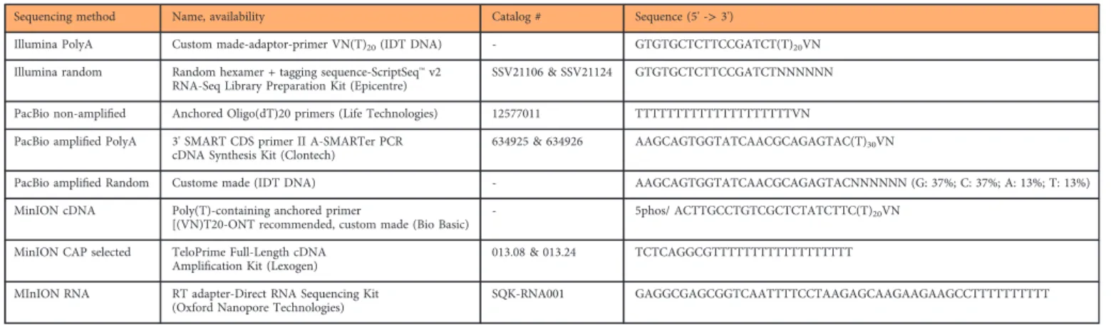
Sequencing method Name, availability Catalog # Sequence (5' ->3')
Illumina PolyA Custom made-adaptor-primer VN(T)20(IDT DNA) - GTGTGCTCTTCCGATCT(T)20VN
Illumina random Random hexamer + tagging sequence-ScriptSeq™v2 RNA-Seq Library Preparation Kit (Epicentre)
SSV21106 & SSV21124 GTGTGCTCTTCCGATCTNNNNNN
PacBio non-amplified Anchored Oligo(dT)20 primers (Life Technologies) 12577011 TTTTTTTTTTTTTTTTTTTTVN PacBio amplified PolyA 3' SMART CDS primer II A-SMARTer PCR
cDNA Synthesis Kit (Clontech)
634925 & 634926 AAGCAGTGGTATCAACGCAGAGTAC(T)30VN
PacBio amplified Random Custome made (IDT DNA) - AAGCAGTGGTATCAACGCAGAGTACNNNNNN (G: 37%; C: 37%; A: 13%; T: 13%)
MinION cDNA Poly(T)-containing anchored primer
[(VN)T20-ONT recommended, custom made (Bio Basic) - 5phos/ ACTTGCCTGTCGCTCTATCTTC(T)20VN MinION CAP selected TeloPrime Full-Length cDNA
Amplification Kit (Lexogen) 013.08 & 013.24 TCTCAGGCGTTTTTTTTTTTTTTTTTT
MInION RNA RT adapter-Direct RNA Sequencing Kit (Oxford Nanopore Technologies)
SQK-RNA001 GAGGCGAGCGGTCAATTTTCCTAAGAGCAAGAAGAAGCCTTTTTTTTTT
Table 5. The list of primers used in this study for the reverse transcription reactions.
The volume of the sequencing primer for the annealing, and the polymerase (P5 or P6) for the binding was determined using the PacBio Calculator version 2.3.1.1., by adding the concentrations and the average insert sizes of SMRTbell templates.
The polymerase-template complexes were bound to MagBeads, loaded onto SMRT Cells and sequenced on the RSII instrument.
The PacBio’s Binding Calculator was used to prepare the library for sequencing using the MagBead one-cell per well (OCPW) protocol, and binding kit P6v2 was used with an on-plate concentration of 0.05 nM. The insert sizes were set according to the size-selections which were applied: 1000, 2500 and 6000 bp sizes were chosen.
In short, the sequencing primer was diluted in PacBio EB to 150 nM. The annealing step was performed with 1μl template DNA (cc: ~20 ng/μl), the diluted sequencing primer and primer buffer (10x). Thefinal concentration of this mixture was 0.8333 nM. Annealing was carried out at 20 °C for 30 min then the DNA polymerase enzyme was diluted to afinal concentration of 50 nM in PacBio BB v2, and then it was bound to the annealed template followed by the addition of DTT, dNTP and BB. The complex (0.5 nMfinal concentration) was incubated at 30 °C for 4 h. The sample complex (0.5μl) was mixed with and 18.5μl MagBead Binding Buffer (0.0125 nM final concentration). MagBeads were prepared as follows: 73.9μl MagBeads were washed with 73.9μl MagBead WB, then 73.9μl MagBead BB was added. The sample complex was bound to the washed, prepared MagBeads for loading to the RSII machine: 19μl sample complex was added to the beads, and then it was placed at 4 °C for 30 min in a HulaMixer. After incubation, the MagBead-bound complex was washed with 19μl BB, then with 19μl WB andfinally, it was resuspended in 19μl BB. The total amount of the MagBead-bound complex was loaded onto the instrument. The MagBead One Cell Per Well protocol was used. One SMRT Cell was also run on Sequel instrument.
Oxford Nanopore cDNA sequencing. PRV transcripts were sequenced on MinION device using the 1D Strand switching cDNA by ligation method (Version: SSE_9011_v108_revS_18Oct2016) and the ONT Ligation Sequencing Kit 1D (SQK-LSK108). For this, PolyA(+)-selected RNAs were used. 50ng from the samples were subjected to reverse transcription. Poly(T)-containing anchored primer [(VN)T20; ordered from Bio Basic, Canada, (Table 5)] and dNTPs (10 mM, Thermo Scientific) was added to the RNA samples and then the mixture was incubated at 65 °C for 5 min. Buffer and DTT from SuperScipt IV Reverse Transcriptase kit (Life Technologies), RNase OUT (Life Technologies) and strand-switching oligo with three O-methyl-guanine RNA bases (PCR_Sw_mod_3G; ordered from Bio Basic, Canada) were added and the sample was incubated at 42 °C for 2 min. 200U SuperScript IV Reverse Transcriptase enzyme was measured into the mix. First-strand cDNA synthesis was carried out at 50 °C for 10 min; it was followed by the strand switching step at 42 °C for 10 min. Enzymes were inactivated at 80 °C for 10 min. Five μl from the prepared double-stranded cDNA was amplified in a single PCR reaction using KAPA HiFi DNA Polymerase (Kapa Biosystems) and Ligation Sequencing Kit Primer Mix (provided by the 1D Kit). The Veriti Thermal Cycler (Applied Biosystems) was set as the 1D Kit’s protocol recommended: initial denaturation for 30 sec at 95 °C (1 cycle); denaturation for 15 sec at 95 °C (15 cycles); annealing for 15 sec at 62 °C (15 cycles); elongation for 4 min at 65 °C (15cycles);final extension 10 min at 65 °C. NEBNext End repair / dA-tailing Module (New England Biolabs) was used for end repair, while NEB Blunt/TA Ligase Master Mix (New England Biolabs) was applied for adapter ligations.
The adapter sequences were supplied by the kit. Agencourt AMPure XP magnetic beads (Beckman Coulter) were used for purification following each enzymatic step. The Qubit Fluorometer (Life Technologies Qubit 2.0) and the Qubit (ds)DNA HS Assay Kit were used to quantify the concentration of the libraries. Samples were loaded on R9.4 SpotON Flow Cells, and base calling was performed using Albacore v1.2.6.
Oxford Nanopore sequencing on Cap-selected samples. To obtain full-length transcripts with the exact 5′-ends, Cap selection was carried out. For this, the TeloPrime Full-Length cDNA Amplification Kit (Lexogen) was used, which has an exceptional specificity for 5′-Cap. The starting material was 2μg total
Name Sequence
PBBC adapter #1 [Phos]TATGCTAATCTCTCTCTTTTCCTCCTCCTCCGTTGTTGTTGTTGAGAGAGATTAGCATA
PBBC adapter #2 [Phos]GACAGTGATCTCTCTCTTTTCCTCCTCCTCCGTTGTTGTTGTTGAGAGAGATCACTGTC
PBBC adapter #3 [Phos]GATCTCGATCTCTCTCTTTTCCTCCTCCTCCGTTGTTGTTGTTGAGAGAGATCGAGATC
PBBC adapter #4 [Phos]TACACGTATCTCTCTCTTTTCCTCCTCCTCCGTTGTTGTTGTTGAGAGAGATACGTGTA
PBBC adapter #5 [Phos]GAGCTCAATCTCTCTCTTTTCCTCCTCCTCCGTTGTTGTTGTTGAGAGAGATTGAGCTC
PBBC adapter #6 [Phos]TCTGCAGATCTCTCTCTTTTCCTCCTCCTCCGTTGTTGTTGTTGAGAGAGATCTGCAGA
Table 6. Barcode sequences utilized for PacBio sequencing.This table contains the modified adapter sequences with unique barcodes which were used for multiplex sequencing. The barcode sequences are labelled by blue colour.
RNA diluted in 12μl water, from a mixed PRV sample (containing RNA from 1, 2, 3, 4, 5, 6, 7, 8, 12, 18 and 24 h post-infection). The method based on cDNA generation. Reverse transcription (RT) was carried out according to the kit’s manual. Briefly, the diluted RNA was mixed with RT buffer, primer (both are supplied by the kit). The RT primer contains an“oligodT”sequence (Table 5) to select the polyadenylated transcripts. The mixture was preheated at 70 °C for 30 sec, then it was cooled down to 37 °C for 1 min. RT enzyme and reagents (part of the kit) were added and the reaction was contain at 37 °C for 2 min.
Temperature was increased to 46 °C for 50 min. The RNA-cDNA hybrid was purified using silica columns (kit’s component). A specific adapter was ligated to the cDNA by base-pairing of the 5’C to the cap structure of the RNA. This step was carried out by the double-strand specific ligase of the kit. Ligation was performed at 25 °C, overnight. The sample was purified after ligation using the silica columns. The cDNA was converted to dscDNA using the Second-Strand Mix and the Enzyme Mix from the Teloprime kit. The reaction was carried out in a Veriti Cycler with the following protocol: 98 °C for 90 sec, 62 °C for 60 sec, 72 °C for 5 min, hold at 25 °C.
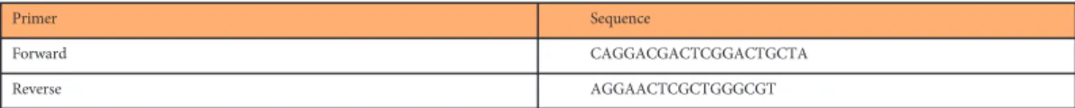
Sample concentration was measured using Qubit dsDNA HS Assay Kit (Life Technologies). Specificity of the obtained cDNA was checked by qPCR (Rotor-Gene Q) using a gene specific primer (us9, 10μM each; Table 7), cDNA and ABsolute qPCR SYBR Green Mix (Thermo Fisher Scientific) in 20μlfinal volume. The initial denaturation was 94 °C 15 min, and it was followed by 35 cycles of 94 °C for 25 sec, 60 °C 25 sec and 72 °C 6 sec.
The PolyA(+)-CAP-selected samples were also sequenced on MinION using the 1D Strand switching cDNA by ligation method. These samples were subjected to the end repair and adapter ligation steps, and then they were loaded on the ONT Flow Cells.
Oxford Nanopore direct RNA sequencing. Three flow cells were used for sequencing PRV samples following the Direct RNA sequencing (DRS) protocol from the ONT (Version: DRS_9026_v1_revM_15- Dec2016). Total RNAs from 12 different time points were mixed together, and then polyA selection was carried out. RNA from the PolyA(+) fraction in 9μl was used as a template for sequencing. RNA was mixed with the RT (oligodT-containing T10) adapter (supplied by the ONT Direct RNA Sequencing Kit;
SQK-RNA001; Oxford Nanopore Technologies) and T4 DNA ligase (2M U/ml; New England BioLabs).
The mixture was incubated at room temperature for 10 min. First-strand cDNA synthesis was carried out in 40μlfinal volume with SuperScript III Reverse Transcriptase (Life Technologies), according to the DRS protocol, at 50 °C for 50 min, then 70 °C for 10 min in a Veriti Thermal Cycler. Samples were washed with Agencourt AMPure XP Beads (Beckman Coulter). XP Beads were treated before usage with RNase OUT (40 U/μl; Life Technologies); 2U enzyme was added to 1μl bead. Purified RNA-cDNA hybrids were eluted in 20μl Ambion Nuclease-Free Water (Thermo Fisher Scientific). RMX sequencing adapter was ligated to the eluted samples with T4 DNA ligase and NEBNext Quick Ligation Reaction Buffer (New England BiceoLabs) at room temperature for 10 min. Samples were purified with RNase OUT-treated XP beads using Wash Buffer (part of the DRS Kit) and then eluted in 21μl Elution Buffer (provided by the DRS Kit). The concentration of the reverse-transcribed and adapted RNA was measured by using the Qubit 2.0 Fluorometer and Qubit dsDNA HS Assay Kit (Life Technologies). Samples were loaded onto the R9.4 SpotON Flow Cell.
Data on the quality of PacBio RSII, Sequel, and ONT MinION reads including insertions, deletions, and mismatches, as well as the coverages are summarized in Table 8 (available online only).
Read processing
Raw reads from the random-primed Illumina sequencing were aligned to the PRV genome (KJ717942.1), using Tophat v2.09 (ref. 23); ambiguous reads were discarded. For PA-Seq, mapping was carried out with Bowtie v2 (ref. 24).
The PacBio RSII and Sequel consensus reads were generated following the RS_ReadsOfInsert protocol of the SMRT Analysis (v2.3.0 and v5.0.0) (Fig. 2), with the following settings: Minimum Full Passes=1, Minimum Predicted Accuracy=90, Minimum Length of Reads of Insert=1, Maximum Length of Reads of Insert=No Limit. These consensus reads were mapped using GMAP25, with the following settings:
gmap -d Genome.fa --nofails -f samse File.fastq>Mapped_file.sam.
The ONT's Albacore software (v.2. 0.1) was used for base calling. This basecaller identify the nucleotide sequences directly from raw data. The sequencing reads were mapped with GMAP using the same setting as was described above.
Custom routines were used to acquire the quality information presented in this data descriptor. The codes have been archived on Github (https://doi.org/10.5281/zenodo.1034511).
Primer Sequence
Forward CAGGACGACTCGGACTGCTA
Reverse AGGAACTCGCTGGGCGT
Table 7. The gene-specific primers used for the amplification ofus9 gene of PRV.
Data Records
All sequencing data have been uploaded to the European Nucleotide Archive under the project accession PRJEB24593 (Data Citation 1)-contains BAM files-and PRJEB9526 (Data Citation 2) – containing FASTQfiles -. All sequencing reads were mapped to the KJ717942.1 genome build. All data can be used without restrictions.
Technical Validation
The quantity of the isolated total RNAs, the polyA-selected RNAs, the rRNA-depleted samples, as well as the synthesized cDNA fractions and sequencing-ready libraries were measured by Qubit 2.0 (Life Technologies)fluorometer using the Qubit RNA, HS RNA and HS dsDNA Assay Kits. The conditions for primer annealing and binding of the polymerase to the templates were determined by PacBio’s Binding Calculator in RS Remote. The libraries were measured by Agilent 2100 Bioanalyzer using the Agilent High Sensitivity DNA Kit.
Usage Notes
The provided dataset was primarily produced to discover and determine the complexity and expression dynamic properties of PRV transcriptome. The uploaded binary alignment (BAM)files contain reads already mapped to the KJ717942.1 reference. These aligned files can be further analysed using various bioinformatics program packages, such as bedtools26, samtools27, or visualized using e.g. IGV28, Geneious29 or Artemis30. The uploaded Illumina, PacBio and ONT files have not been trimmed, they contain terminal poly(A) sequences as well as the 5′and 3′ adapter sequences, which can be used to determine the orientations of the reads.
References
1. Aujeszky, A. A contagious disease, not readily distinguishable from rabies, with unknown origin.Veterinarius12,387–396 (1902).
2. Pomeranz, L., Reynolds, A. & Hengartner, C. Molecular biology of pseudorabies virus: Impact on neurovirology and veterinary medicine.Microbiol Mol Biol Rev.69,462 (2005).
3. Boldogkői, Z. & Nógrádi, A. Gene and cancer therapy--pseudorabies virus: a novel research and therapeutic tool?Curr Gene Ther.
3,155–182 (2003).
4. Yang, M.et al.Retrograde, transneuronal spread of pseudorabies virus in defined neuronal circuitry of the rat brain is facilitated by gE mutations that reduce virulence.J. Virol.73,4350–4359 (1999).
5. Song, C. K., Enquist, L. W. & Bartness, T. J. New developments in tracing neural circuits with herpesviruses.Virus Res.111, 235–249 (2005).
6. Boldogkői, Z.et al.Genetically timed, activity-sensor and rainbow transsynaptic viral tools.Nat. Methods6,127–130 (2009).
7. Granstedt, A. E., Kuhn, B., Wang, S. S. & Enquist, L. W. Calcium imaging of neuronal circuits in vivo using a circuit-tracing pseudorabies virus.Cold Spring. Harb. Protoc2010,pdb.prot5410 (2010).
8. Card, J. P.et al.A dual infection pseudorabies virus conditional reporter approach to identify projections to collateralized neurons in complex neural circuits.PLoS ONE6,e21141 (2011).
9. Zhu, L.et al.Growth, physicochemical properties, and morphogenesis of Chinese wild-type PRV Fa and its gene-deleted mutant strain PRV SA215.Virol. J.8,272 (2011).
10. Maresch, C.et al.Oral immunization of wild boar and domestic pigs with attenuated live vaccine protects against pseudorabies virus infection.Vet. Microbiol.161,20–25 (2012).
11. Klingbeil, K.et al.Immunization of pigs with an attenuated pseudorabies virus recombinant expressing the hemagglutinin of pandemic swine origin H1N1 influenza A virus.J. Gen. Virol.95,948–959 (2014).
12. Oláh, P.et al.Characterization of pseudorabies virus transcriptome by Illumina sequencing.BMC Microbiol.15,130 (2015).
13. Tombácz, D.et al.Strain Kaplan of Pseudorabies Virus Genome Sequenced by PacBio Single-Molecule Real-Time Sequencing Technology.Genome Announc2,14–15 (2014).
14. Rhoads, A. & Fai, K. PacBio Sequencing and Its Applications. Genomics, Proteomics &.Bioinformatics13,278–289 (2015).
15. Sharon, D., Tilgner, H., Grubert, F. & Snyder, M. A single-molecule long-read survey of the human transcriptome.Nat.
Biotechnol.31,1009–1014 (2013).
16. Tilgner, H., Grubert, F., Sharon, D. & Snyder, M. P. Defining a personal, allele-specific, and single-molecule long-read tran- scriptome.Proc. Natl. Acad. Sci. USA111,9869–9874 (2014).
17. Chen, S. Y., Deng, F., Jia, X., Li, C. & Lai, S. J. A transcriptome atlas of rabbit revealed by PacBio single-molecule long-read sequencing.Sci. Rep7,7648 (2017).
18. Tombácz, D.et al.Full-Length Isoform Sequencing Reveals Novel Transcripts and Substantial Transcriptional Overlaps in a Herpesvirus.PLoS ONE11,e0162868 (2016).
19. Moldován, N.et al.Multi-Platform Sequencing Approach Reveals a Novel Transcriptome Profile in Pseudorabies Virus.Front.
Microbiol8,2708 (2018).
20. Tombácz, D.et al.Characterization of the Dynamic Transcriptome of a Herpesvirus with Long-read Single Molecule Real-Time Sequencing.Sci. Rep7,43751 (2017).
21. Lomniczi, B., Watanabe, S., Ben-Porat, T. & Kaplan, A. S. Genetic basis of the neurovirulence of pseudorabies virus.J. Virol.52, 198–205 (1984).
22. Kaplan, A. S. & Vatter, A. E. A comparison of herpes simplex and pseudorabies viruses.Virology7,394–407 (1959).
23. Trapnell, C., Pachter, L. & Salzberg, S. L. TopHat: discovering splice junctions with RNA-Seq.Bioinformatics25, 1105–1111 (2009).
24. Langmead, B. & Salzberg, S. Fast gapped-read alignment with Bowtie 2.Nat. Methods.9,357–359 (2012).
25. Wu, T. D. & Watanabe, C. K. GMAP: a genomic mapping and alignment program for mRNA and EST sequences.Bioinformatics 21,1859–1875 (2005).
26. Quinlan, A. R. & Hall, I. M. BEDTools: aflexible suite of utilities for comparing genomic features.Bioinformatics26, 841–842 (2010).
27. Li, H.et al.The Sequence Alignment/Map format and SAMtools.Bioinformatics25,2078–2079 (2009).
28. Robinson, J. T.et al.Integrative genomics viewer.Nat. Biotechnol.29,24–26 (2011).
29. Kearse, M.et al.Geneious Basic: an integrated and extendable desktop software platform for the organization and analysis of sequence data.Bioinformatics28,1647–1649 (2012).
30. Rutherford, K.et al.Artemis: sequence visualization and annotation.Bioinformatic16,944–945 (2000).
31. Weirather, J. L.et al.Comprehensive comparison of Pacific Biosciences and Oxford Nanopore Technologies and their appli- cations to transcriptome analysis.F1000Research6,100 (2017).
32. Schirmer, M., D’Amore, R., Ijaz, U. Z., Hall, N. & Quince, C. Illumina error profiles: resolvingfine-scale variation in metagenomic sequencing data.BMC Bioinformatics.17,125 (2016).
Data Citations
1.European Nucleotide ArchivePRJEB24593 (2018).
2.European Nucleotide ArchivePRJEB9526 (2015).
Acknowledgements
This work was supported by the Swiss-Hungarian Cooperation Programme: SH/7/2/8 to ZBo. The work was also supported by the Bolyai János Scholarship of the Hungarian Academy of Sciences to DT and by the NIH Centers of Excellence in Genomic Science (CEGS) Center for Personal Dynamic Regulomes:
5P50HG00773502 to MS.
Author Contributions
D.T. performed the Illumina, PacBio and ONT sequencing, analyzed the data and drafted the manuscript.
D.S. performed the PacBio sequencing. A.S. conducted bioinformatics analysis. N.M. took part in ONT sequencing experiments. M.S. conceived and designed the experiments. Z.B. conceived and designed the experiments, coordinated the project and wrote thefinal manuscript. All authors read and approved the final paper.
Additional information
Table 8 is available only in the online version of this paper.
Competing interests: The authors declare no competing interests.
How to cite this article: Tombácz, D.et al. Transcriptome-wide survey of pseudorabies virus using next- and third-generation sequencing platforms. Sci. Data5:180119 doi: 10.1038/sdata.2018.119 (2018).
Publisher’s note: Springer Nature remains neutral with regard to jurisdictional claims in published maps and institutional affiliations.
Open AccessThis article is licensed under a Creative Commons Attribution 4.0 Interna- tional License, which permits use, sharing, adaptation, distribution and reproduction in any medium or format, as long as you give appropriate credit to the original author(s) and the source, provide a link to the Creative Commons license, and indicate if changes were made. The images or other third party material in this article are included in the article’s Creative Commons license, unless indicated otherwise in a credit line to the material. If material is not included in the article’s Creative Commons license and your intended use is not permitted by statutory regulation or exceeds the permitted use, you will need to obtain permission directly from the copyright holder. To view a copy of this license, visit http://creativecommons.
org/licenses/by/4.0/
The Creative Commons Public Domain Dedication waiver http://creativecommons.org/publicdomain/
zero/1.0/ applies to the metadatafiles made available in this article.
© The Author(s) 2018