0139–3006 © 2018 Akadémiai Kiadó, Budapest DOI: 10.1556/066.2018.47.3.13
Preliminary communication
THE EFFECTS OF ENVIRONMENTAL FACTORS ON PLANKTONIC GROWTH AND BIOFILM FORMATION OF SERRATIA ODORIFERA AND SERRATIA MARCESCENS ISOLATED FROM TRADITIONALLY
MADE CHEESE
K.G. MLADENOVIĆ*, M.Ž. MURUZOVIĆ and L.R. ČOMIĆ
Department of Biology and Ecology, Faculty of Science, University of Kragujevac, Radoja Domanovića 12, 34000 Kragujevac. Republic of Serbia
(Received: 23 August 2017; accepted: 15 November 2017)
In this study, the effects of different temperature, pH, salt and glucose concentrations on the planktonic growth, biofi lm formation, and formed biofi lm of Serratia odorifera and Serratia marcescens, isolated from traditionally made cheese, were investigated using spectrophotometric method. The investigated strains demonstrated best planktonic growth and biofi lm formation in Tryptic soy broth. The limiting factors for the planktonic growth and biofi lm formation were temperature below 4 °C and salt concentration above 4%. Temperature of 37 °C and 44 °C, as well as various concentrations of glucose, stimulated the planktonic growth of bacteria. Moderate infl uence on biofi lm formation was demonstrated at 37 °C as well as at various concentrations of glucose. These results were in accordance with the origin of bacteria, since the isolates were obtained from cheese.
Keywords: biofi lm formation, environmental factors, planktonic growth, Serratia
Bacteria from the Enterobacteriaceae family may affect the quality of food (usually milk and cheese) by their metabolism, and they might propagate during maturity process (CHAVES- LOPEZ et al., 2005). The ability of enterobacteria (Enterobacter, Serratia, Escherichia, Hafnia, Citrobacter, and Klebsiella), isolated from cheese, to produce acid and biogenic amines was examined by a few researchers. MARINO and co-workers (2000) found a positive correlation between the concentration of cadaverine and the number of enterobacteria.
Serratia marcescens is Gram-negative bacterium, which is able to populate a wide variety of ecological niches (GRIMONT et al., 1977). According to AUCKEN and PITT (1998), it is an opportunistic human pathogen responsible for many infections and resistant to antibiotics. Serratia odorifera was identifi ed in a local Italian cheese (CHAVES-LOPEZ et al., 2005).
Environmental factors (temperature, sugar, salt, pH, and nutrients) present in foods and food-processing environments play signifi cant role in adhesion and biofi lm formation (MIRKAR et al., 2016). According to KHANGHOLI and JAMALLI (2016), one way of bacterial adaptation to the environmental conditions is the ability to form biofi lm. The capability of S.
marcescens to cause infections and survive in the environment is attributed to its ability to form biofi lm (KALIVODA et al., 2010). Bacteria regulate gene expression in response to different environmental signals, such as temperature, oxygen and carbon dioxide concentrations, pH, and nutrient availability (HARJAI et al., 2005).
* To whom correspondence should be addressed.
Phone: +34 336 223; fax: +34 335 040; e-mail: katarinamladenovic90@gmail.com
Acta Alimentaria 47, 2018
The aims of this study were investigation of the planktonic growth and ability to form biofi lm of S. odorifera and S. marcescens biogp 1 in two different broths, under the infl uence of different temperatures, pH, concentrations of NaCl and glucose, as well as the impact of the mentioned environmental factors on the formed biofi lm.
1. Materials and methods
1.1. Strains and growth conditions
For the tests in this study, strains S. odorifera KGPMF 18 and S. marcescens biogp 1 KGPMF 19 were used. The bacteria were previously isolated from traditionally made Serbian cheese (Sokobanja region) and identifi ed at the Laboratory for Microbiology at the Faculty of Science, University of Kragujevac (KGPMF) (MLADENOVIĆ et al., 2018). The collection of identifi ed bacterial species was stored in a 20% glycerol/medium mixture at –80 °C.
1.2. Infl uence of temperature, pH, salt and glucose concentrations on planktonic growth in TSB and MH media
The examination of the effect of temperature (4 °C, 37 °C, 44 °C) on the growth of Serratia sp. was conducted on Tryptic soy broth (TSB) and Muller-Hinton broth (MH), standard or modifi ed compositions.
To study the effect of pH, media with different pH values (5.5, 6.5, 7, 7.5, 8.5) were prepared. The pH was set to 5.5, 6.5, and 7 with addition of HCl and to 7.5 and 8.5 with addition of NaOH. For TSB growth pH 7.5 was used as control, while for growth in MH, control was set to pH 7.
Both tested media were modifi ed with the addition of NaCl (4%, 6.5%, and 8%) in order to investigate the effects of different salt concentrations. Growth in TSB with 4% NaCl and in pure MH served as controls.
The effect of different concentrations of glucose (0.5%, 1.5%, 2.5%, and 3.5%) was investigated in modifi ed TSB and MH media. Growth in TSB with 0.25% of glucose and in pure MH served as controls.
Initial bacterial suspension of 10 μl (108–109 CFU ml–1) was added to 3 ml of each type of medium. Samples were incubated for 24 h. Sterility controls were pure TSB and MH media. Results were determined with spectrophotometer at 600 nm. Each experiment was performed in triplicate.
1.3. The determination of antibiofi lm activity
1.3.1. Pellicle test. The ability to form a biofi lm phenotype or pellicle formation on the air- liquid interphase was determined using pellicle assay according to the method of VESTBY and co-workers (2009), with modifi cations. TSB and MH media of 1.8 ml were inoculated with 0.2 ml of initial bacterial suspension (108–109 CFU ml–1) and then incubated for 96 h at 37
°C. Categorization of isolates and their ability to produce biofi lm were based on the production of pellicle on the surface of the liquid phase according to the following scheme: solid fat formed pellicle (+++) – good biofi lm producer; thin pellicle formed (++) – moderate biofi lm producer; very thin pellicle (+) – weak biofi lm producer; complete absence of pellicle (-) – no biofi lm production. Pellicle test was repeated three times for each strain.
Acta Alimentaria 47, 2018
1.3.2. Biofi lm formation assay and quantifi cation. The ability of S. odorifera KGPMF 1 and S. marcescens biogp 1 KGPMF 19 to form biofi lm was assayed according to O’TOOLE
and KOLTER (1998), with some modifi cations.
In sterile 96-well tissue culture plates (Sarstedt, Germany) 100 μl TSB or MH broth (with different pH, salt and glucose concentrations) and 10 μl of fresh bacterial suspension (1.0 McFarland) was added to each well. After incubation at 4 °C, 37 °C, and 44 °C for 48 h, the content of each well was gently removed by tapping the plates. The rest of the experiment was done as described by MURUZOVIĆ and co-workers (2016).
1.3.3. The effect of temperature, pH, salt and glucose concentrations on formed biofi lm.
The tissue culture 96-well microtiter plates (Sarstedt, Germany) were prepared by adding 100 μl TSB or MH broth and 10 μl of fresh bacterial suspension (1.0 McFarland) to each well. The inoculated microtiter plates were incubated at 37 °C for 24 hours. After incubation, the content of each well was gently pulled out. Then, 100 μl TSB or MH broth with different pH, salt and glucose concentrations was added to each well, and the microtiter plates were incubated at 4 °C, 37 °C, and 44 °C for 24 hours. After incubation, the content of each well was gently removed by tapping the microtiter plates. After that, experiment was carried out as described by MURUZOVIĆ and co-workers (2016).
1.4. Data analysis
All data were presented as means ± standard deviations using Microsoft Excel (Redmond, Washington, DC, USA). Paired t-test was used for statistical analysis of the results via IBM SPSS Statistics 20.
2. Results and discussion
Tested bacteria were incubated in different media at three temperatures. After incubation, there was no growth at 4 °C. Planktonic growth at 37 °C in TSB was statistically signifi cantly higher compared to growth in MH (P<0.05). The differences between planktonic growth in TSB and MH at 44 °C were statistically not signifi cant (P>0.05) (Tables 1, 2, and 3).
2.1. Infl uence of different temperature, pH, and different concentrations of salt and glucose on the planktonic growth in TSB and MH media
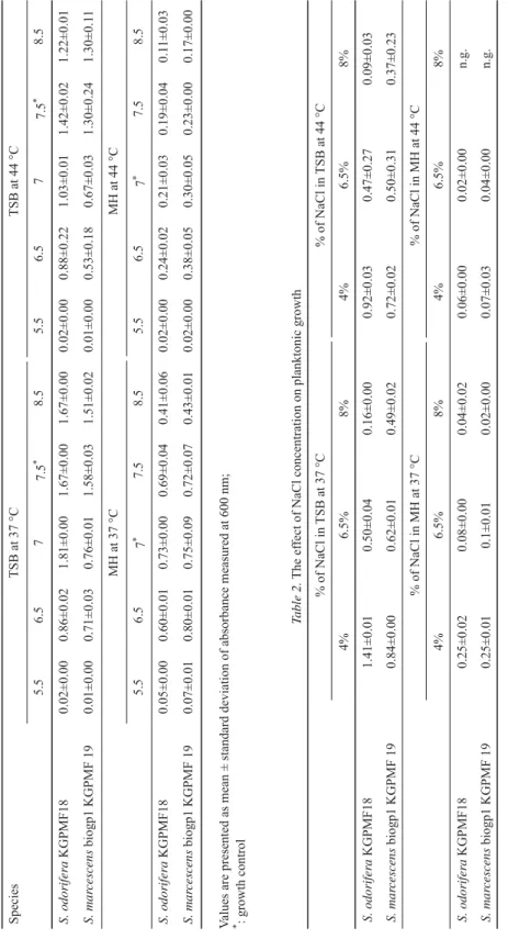
All tested pH values were limiting factor for planktonic growth of S. odorifera and S.
marcescens in TSB at 37 °C and 44 °C, except for pH 8.5.
All tested pH values were limiting factor for planktonic growth of S. odorifera and S.
marcescens in MH at 37 °C, except for pH 6.5 in case of S. marcescens, where the growth was stimulated. At 44 °C, the results were similar. Based on the results, it can be concluded that the bacterial growth was better at the 37 °C than at 44 °C at all tested pH (Table 1).
All concentrations of salts reduced the growth of S. odorifera and S. marcescens in both media at 37 °C and 44 °C (Table 2).
In TSB with different concentrations of glucose at 37 °C and 44 °C, both species demonstrated lower growth compared to control. In MH with glucose at 37 °C, S. odorifera showed better growth than in control. S. marcescens growth was equal that of the control for each glucose concentrations, except for 2.5%, where growth was lower. At 44 °C, both species demonstrated better growth in the presence of glucose than in control (Table 3).
Acta Alimentaria 47, 2018
Table 1. The effect of pH on planktonic growth SpeciesTSB at 37 °CTSB at 44 °C 5.56.577.5*8.55.56.577.5*8.5 S. odorifera KGPMF180.02±0.000.86±0.021.81±0.001.67±0.001.67±0.000.02±0.000.88±0.221.03±0.011.42±0.021.22±0.01 S. marcescens biogp1 KGPMF 190.01±0.000.71±0.030.76±0.011.58±0.031.51±0.020.01±0.000.53±0.180.67±0.031.30±0.241.30±0.11 MH at 37 °C MH at 44 °C 5.56.57*7.58.55.56.57*7.58.5 S. odorifera KGPMF180.05±0.000.60±0.010.73±0.000.69±0.040.41±0.060.02±0.000.24±0.020.21±0.030.19±0.040.11±0.03 S. marcescens biogp1 KGPMF 190.07±0.010.80±0.010.75±0.090.72±0.070.43±0.010.02±0.000.38±0.050.30±0.050.23±0.000.17±0.00 Values are presented as mean ± standard deviation of absorbance measured at 600 nm; *: growth control Table 2. The effect of NaCl concentration on planktonic growth % of NaCl in TSB at 37 °C% of NaCl in TSB at 44 °C 4%6.5%8%4%6.5%8% S. odorifera KGPMF181.41±0.010.50±0.040.16±0.000.92±0.030.47±0.270.09±0.03 S. marcescens biogp1 KGPMF 190.84±0.000.62±0.010.49±0.020.72±0.020.50±0.310.37±0.23 % of NaCl in MH at 37 °C% of NaCl in MH at 44 °C 4%6.5%8%4%6.5%8% S. odorifera KGPMF180.25±0.020.08±0.000.04±0.020.06±0.000.02±0.00n.g. S. marcescens biogp1 KGPMF 190.25±0.010.1±0.010.02±0.000.07±0.030.04±0.00n.g. Values are presented as mean ± standard deviation of absorbance measured at 600 nm n.g.: no growth
Acta Alimentaria 47, 2018
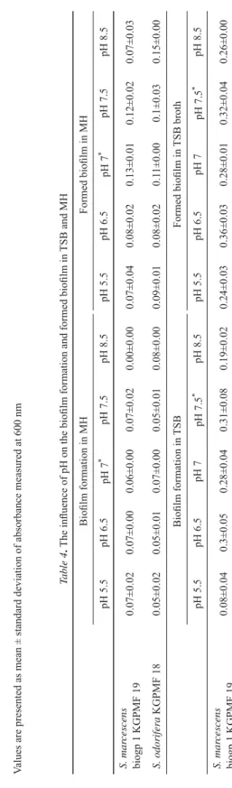
Table 3. The effect of glucose concentration on planktonic growth Species% of glucose in TSB at 37 °C% of glucose in TSB at 44 °C Growth control0.5%1.5%2.5%3.5%Growth control0.5%1.5%2.5%*3.5% S. odorifera KGPMF181.67±0.001.31±0.041.07±0.111.09±0.040.96±0.141.42±0.021.25±0.091.15±0.291.08±0.040.97±0.09 S. marcescens biogp1 KGPMF 191.58±0.030.90±0.230.86±0.220.77±0.010.65±0.011.30±0.240.84±0.070.68±0.040.57±0.060.47±0.00 % of glucose in MH at 37 °C% of glucose in MH at 44 °C Growth control0.5%1.5%2.5%3.5%Growth control0.5%1.5%2.5%3.5% S. odorifera KGPMF180.73±0.000.81±0.020.72±0.020.87±0.010.81±0.050.21±0.030.60±0.010.60±0.030.61±0.030.63±0.02 S. marcescens biogp1 KGPMF 190.75±0.090.76±0.040.73±0.020.48±0.050.74±0.020.30±0.050.61±0.140.65±0.100.49±0.010.69±0.09 Values are presented as mean ± standard deviation of absorbance measured at 600 nm Table 4. The infl uence of pH on the biofi lm formation and formed biofi lm in TSB and MH Biofi lm formation in MHFormed biofi lm in MH pH 5.5pH 6.5pH 7*pH 7.5pH 8.5pH 5.5pH 6.5pH 7*pH 7.5pH 8.5 S. marcescens biogp 1 KGPMF 190.07±0.020.07±0.000.06±0.000.07±0.020.00±0.000.07±0.040.08±0.020.13±0.010.12±0.020.07±0.03 S. odorifera KGPMF 180.05±0.020.05±0.010.07±0.000.05±0.010.08±0.000.09±0.010.08±0.020.11±0.000.1±0.030.15±0.00 Biofi lm formation in TSBFormed biofi lm in TSB broth pH 5.5pH 6.5pH 7pH 7.5*pH 8.5pH 5.5pH 6.5pH 7pH 7.5*pH 8.5 S. marcescens biogp 1 KGPMF 190.08±0.040.3±0.050.28±0.040.31±0.080.19±0.020.24±0.030.36±0.030.28±0.010.32±0.040.26±0.00 Values are presented as mean ± standard deviation of absorbance measured at 630 nm * growth control
Acta Alimentaria 47, 2018
S. odorifera was identifi ed in semi-hard cheese of Portuguese origin (KONGO et al., 2008). Serratia sp. reaches food incidentally, during the process of production. In our study, it was noticed that temperature at 4 °C, low pH, and all concentrations of salt showed inhibitory effect on the planktonic growth of Serratia species in TSB and MH media.
2.2. Determination of antibiofi lm activity
2.2.1. Pellicle test. According to the results, neither species were able to form pellicle in TSB or MH at 37 °C.
2.2.2. The infl uence of different temperature, pH, and different concentrations of salt and glucose on biofi lm formation and on biofi lm formed in TSB and MH. The biofi lm formation ability of the strains was tested in different media at three temperatures. After incubation, it was noticed that none of them formed biofi lm at 4 °C and 44 °C. It was also observed that S. odorifera had no ability to form biofi lm in TSB.
According to the results, all tested pH values, except pH 7.5, were limiting factors for biofi lm formation and on formed biofi lm of S. marcescens grown in TSB (Table 4.).
In MH pH 8.5, biofi lm formation of S. odorifera was stimulated, but biofi lm formation of S. marcescens was reduced. The infl uence of pH on formed biofi lm was manifested in the reduction of formed biofi lm (Table 4).
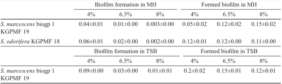
TSB with different concentrations of salt showed reducing effect on biofi lm formation of S. marcescens. The size of formed biofi lm was reduced, too (Table 5).
MH with different concentrations of salts had reducing effect on the biofi lm formation of both species. In case of S. odorifera 4% and 6.5% of salt showed stimulating effect on biofi lm formation. The same concentrations of salt reduced the formed biofi lm of S.
marcescens, while concentration of 8% stimulated the further growth of biofi lm (Table 5).
Table 5. Infl uence of NaCl on the biofi lm formation and formed biofi lm in TSB and MH
Biofi lm formation in MH Formed biofi lm in MH
4% 6.5% 8% 4% 6.5% 8%
S. marcescens biogp 1 KGPMF 19
0.04±0.01 0.01±0.00 0.003±0.00 0.05±0.02 0.12±0.02 0.15±0.02 S. odorifera KGPMF 18 0.06±0.01 0.02±0.00 0.002±0.00 0.12±0.01 0.12±0.00 0.11±0.00
Biofi lm formation in TSB Formed biofi lm in TSB
4% 6.5% 8% 4% 6.5% 8%
S. marcescens biogp 1 KGPMF 19
0.09±0.00 0.03±0.00 0.01±0.01 0.2±0.02 0.15±0.01 0.12±0.01
Values are presented as mean ± standard deviation of absorbance measured at 630 nm
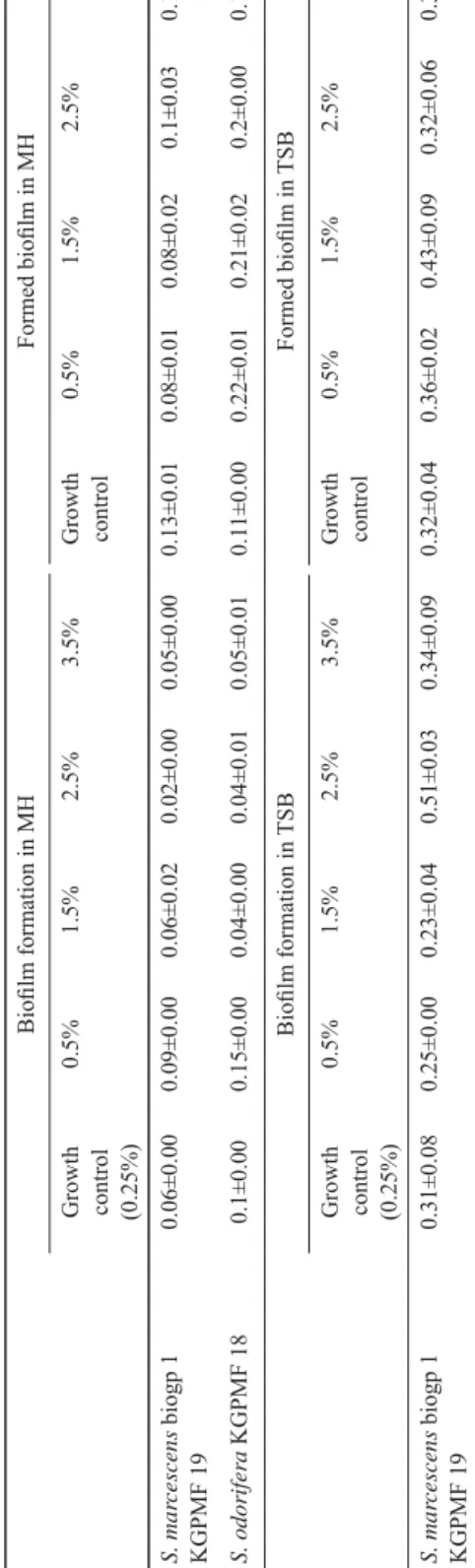
TSB with 0.5% and 1.5% of glucose reduced the biofi lm formation, while 2.5% and 3.5% showed stimulating effect on the biofi lm formation of S. marcescens. Further growth of formed biofi lm of S. marcescens was stimulated by 0.5% and 1.5% of glucose (Table 6).
MH with 0.5% of glucose stimulated biofi lm formation of both bacteria, while other tested concentrations showed reducing effect. All tested concentrations of glucose demonstrated stimulating effect on already formed biofi lm of S. odorifera, but on formed biofi lm of S. marcescens they showed reducing effect. The only exception was the concentration of 3.5%, where further growth of formed biofi lm was stimulated (Table 6).
Acta Alimentaria 47, 2018
Table 6. The infl uence of different concentrations of glucose on biofi lm formation and formed biofi lm in TSB and MH Biofi lm formation in MHFormed biofi lm in MH Growth control (0.25%)
0.5%1.5%2.5%3.5%Growth control0.5%1.5%2.5%3.5% S. marcescens biogp 1 KGPMF 190.06±0.000.09±0.000.06±0.020.02±0.000.05±0.000.13±0.010.08±0.010.08±0.020.1±0.030.16±0.04 S. odorifera KGPMF180.1±0.000.15±0.000.04±0.000.04±0.010.05±0.010.11±0.000.22±0.010.21±0.020.2±0.000.19±0.02 Biofi lm formation in TSBFormed biofi lm in TSB Growth control (0.25%)
0.5%1.5%2.5%3.5%Growth control0.5%1.5%2.5%3.5% S. marcescens biogp 1 KGPMF 190.31±0.080.25±0.000.23±0.040.51±0.030.34±0.090.32±0.040.36±0.020.43±0.090.32±0.060.33±0.04 Values are presented as mean ± standard deviation of absorbance measured at 630 nm
Acta Alimentaria 47, 2018
S. odorifera and S. marcescens showed no ability of pellicle formation in TSB and MH, but demonstrated ability of biofi lm formation at 37 °C. According to NANDHAGOPAL and SUBASHKUMAR (2016), biofi lm formation of S. marescens at refrigeration temperature reached its maximum, compared to room temperature and 37 ºC. It has been confi rmed that a temperature of 37 ºC can induce further biofi lm development of Enterococcus species (TENDOLKAR et al., 2004). WHITE-ZIEGLE and co-workers (2008) showed that biofi lm was formed by S. marescens isolates at 23 ºC. According to NANDHAGOPAL and SUBASHKUMAR
(2016), biofi lm formation of Serratia sp. at low temperature may have a role in the contamination of refrigerated foods. They also found that 1% NaCl decreased the biofi lm formation, but 2% NaCl decreased the biofi lm density. Studies showed that the increase of salt concentration, led to decrease of biofi lm formation, up to a point when a higher concentration of salt did not affect further the growth of biofi lm formed by enterococci (ASHA
et al., 2013). Our research showed that all concentrations of salt in TSB reduced the biofi lm formation and also reduced the already formed biofi lm of the Serratia species investigated.
In MH, formed biofi lm was stimulated at 8% of salt. Various concentrations of glucose showed stimulating or reducing effects on biofi lm formation and formed biofi lm. These results indicated that the presence of lactic acid bacteria in cheese affecting pH, can contribute to the control of number and presence of enterobacteria in cheese. Also, adding salt to cheese can prevent their growth. These are the possible mechanisms of cheese preservation.
3. Conclusions
The effects of pH, salt, and glucose at different temperatures on planktonic growth of S.
odorifera and S. marcescens isolated from Sokobanja cheese were investigated for the fi rst time in this paper. It was noticed that temperature at 4 °C, low pH, and all concentrations of salt showed inhibitory effect on the planktonic growth of Serratia species in both tested media. Glucose in TSB and MH showed stimulating effect on planktonic growth. Biofi lm formation was possible only at 37 °C. These results contribute to better understanding the infl uence of environmental factors on the growth and development of bacteria. Further studies should be conducted to investigate other factors that could be used as preservatives in traditionally made cheeses.
*
This investigation was supported by the Ministry of Education, Science and Technological Development of the Republic of Serbia (Grant No. 41010).
References
ASHA, P., JYOTHIS, M. & SHINI, Z. (2013): A comparative study of virulence factors in enterococci from clinical and nonclinical sources. J. Pharm. Biomed. Sci., 29, 745–752.
AUCKEN, H.M. & PITT, T.L. (1998): Antibiotic resistance and putitive virulence factors of Serratia marcescens with respect to O and K serotype. J. Med. Microbiol., 47, 1105–1113.
CHAVES-LOPEZ, C., ANGELIS, DE M., MARTUSCELLI, M., SERIO, A., PAPARELLA, A. & SUZZI, G. (2005): Characterization of the Enterobacteriaceae isolated from an artisanal Italian ewe’s cheese (Pecorino Abruzzese). J. Appl.
Microbiol., 101, 353–360.
GRIMONT, P.A., GRIMONT, F. & DE ROSNAY, H.L. (1977): Taxonomy of the genus Serratia. J. Gen. Microbiol., 98, 39–66.
Acta Alimentaria 47, 2018
HARJAI, K., KHANDWAHA, R.K., MITTAL, R., YADAV, V., GUPTA, V. & SHARMA, S. (2005): Effect of pH on production of virulence factors by biofi lm cells of Pseudomonas aeruginosa. Folia Microbiol., 50, 99–102.
KALIVODA, E.J., STELLA, N.A., ASTON, M.A., FENDER, J.E., THOMPSON, P. P., KOWALSKI, R.P. & SHANKS, R.M. (2010):
Cyclic AMP negatively regulates prodigios in production by Serratia marcescens. Res. Microbiol., 161, 158–
167.
KHANGHOLI, M. & JAMALLI, A. (2016): The effects of sugars on the biofi lm formation of Escherichia coli 185p on stainless steel and polyethylene terephthalate surfaces in a laboratory model. Jundishapur J. Microbiol., 9, e40137.
KONGO, J.M., GOMES, A.P. & MALCATA, F.X. (2008): Monitoring and identifi cation of bacteria associated with safety concerns in the manufacture of São Jorge, a Portuguese traditional cheese from raw cow’s milk. J. Food Protect., 71, 986–992.
MARINO, M., MAIFRENI, M., MORET, S. & RONDININI, G. (2000): The capacity of Enterobacteriaceae species to produce biogenic amines in cheese. Lett. Appl. Microbiol., 31, 169–173.
MIRKAR, K., RAWAT, A. & SATISH, R. (2016): Effect of environmental factors on biofi lm formation. Indian J. Life Sci., 5, 53–64.
MLADENOVIĆ, K.G., MURUZOVIĆ, M., ZUGIĆ-PETROVIĆ, T., STEFANOVIĆ, O.D. & COMIĆ, L.R. (2018): Isolation and identifi cation of Enterobacteriaceae from traditional Serbian cheese and their physiological characteristics. J.
Food Saf., e12387.
MURUZOVIĆ, M.Z., MLADENOVIĆ, K.G., STEFANOVIĆ, O.D., VASIĆ, S.M. & COMIĆ, L.R. (2016): Extracts of Agrimonia eupatoria L. as sources of biologically active compounds and evaluation of their antioxidant, antimicrobial and antibiofi lm activities. J. Food Drug Anal., 24, 539–547.
NANDHAGOPAL, G. & SUBASHKUMAR, R. (2016): Effect of environmental factors on biofi lm formation by Serratia marcescens isolates. IJRAMR, 3, 1601–1604.
O’TOOLE, G. & KOLTER, R. (1998): Initiation of biofi lm formation in Pseudomonas fl uorescens WCS365 proceeds via multiple, convergent signalling pathways: A genetic analysis. Mol. Microbiol, 28, 449–461.
TENDOLKAR, P.M., BAGHDAYAN, A.S., GILMORE, M.S. & SHANKAR, N. (2004): Enterococcus surface protein, Esp, enhances biofi lm formation by Enterococcus faecalis. Infect. Immun., 72, 6032–6039.
VESTBY, L., MORETRO, T., LANGSRUD, S., HEIR, E. & NESSE, L.L. (2009): Biofi lm forming abilities of Salmonella are correlated with persistence in fi sh meal-and feed factories. BMC Vet. Res., 27, 1–20.
WHITE-ZIEGLE, C.A., UM, S., PEREZ, N.M., BERNS, A.L., MALHOWSKI, A.J. & YOUNG, S. (2008): Low temperature (23 degrees C) increases expression of biofi lm- cold-shock and RhoS-dependent genes in Escherichia coli K-12.
Microbiology, 154, 148–166.