The Mechanochemical System behind Streaming in Physarum
HIROMICHI NAKAJIMA
Department of Biology, Faculty of Science, and Institute for Protein Research, Osaka University, Osaka, Japan, and Department of Biology,
Princeton University, Princeton, New Jersey
Plasmodia of the myxomycete, Physarum polycephalum, exhibit vigor- ous streaming of endoplasm which rhythmically reverses its direction.
The streaming of endoplasm is also responsible for changes in the form of the organism and causes locomotion in general.
Just as in the case of muscle, streaming protoplasm is a mechano- chemical system where the chemical energy acquired by metabolism is converted into the mechanical work of streaming. The nature of the mechanochemical system behind streaming, therefore, is certainly one of the essential problems in considering the mechanism of streaming.
In our earlier studies we showed that the immediate energy source for the flow in Physarum is adenosine triphosphate (ATP) provided by a fermentation process (Kamiya et al., 1957). T h e first problem we shall consider is the nature of the mechanochemical system which responds to ATP. What draws our attention to this subject is a contractile protein like actomyosin.
Actomyosin-Like Protein in Plasmodia of Physarum Loewy (1952) first discovered an actomyosin-like protein system in Physarum plasmodia. Later, Ts'o et al. (1956a, b, 1957a, b) obtained an ATP-sensitive protein in this organism. This they purified by successive salt fractionation and differential centrifugation and named it "myxo- myosin." They studied especially its physicochemical properties. Recently, detailed examinations of the so-called "contractile protein" of Physarum have been made by Hatano (1962) and Rebhun (1962).
We (Nakajima, 1956, 1960) also isolated a structural protein fraction from the same source by the procedure used for the preparation of muscle myosin B. Thus, the protein fraction was extracted with Weber- Edsall solution (0.6 M K C l , 0.01 M N a
2C 0
3, 0.04 M N a H C 0
3) and was precipitated by dilution. It was yellowish-white and very turbid. We have called this preparation "plasmodial myosin B " to denote its method of preparation. Some of its properties will now be discussed.
I l l
Viscous PROPERTIES
Figure 1 shows the change in viscosity of plasmodial myosin Β on the addition of A T P . When A T P is added, the viscosity drops rapidly at first, then slowly increases to the original level. T h e time course of this change in viscosity is practically identical with that of muscle myosin B.
ι ,ΑΤΡ
—' ' 1 1 1 l_
Ο 5 10 /S 20 25
Time in minuits
FIG. 1. Effect of A T P on the viscosity of plasmodial myosin B; pH 6.6, 15°C; K+, 0.55 M. T h e final concentrations of A T P are indicated on the figure (Nakajima, 1960).
I—I 1 1 I I
0 5 10 /5 20
Time in minutes
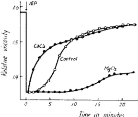
FIG. 2. Effect of CaCl9 and MgCl2 on the viscosity of plasmodial myosin B; pH 6.6, 15°C; K + , 0.55 M; A T P , 0.56 χ 1 0 - 3 M; CaCL,, 1 0 - 2 M; MgCl0, 1 0 - 2 M (Nakajima, 1960).
The degree of change in viscosity, i.e., A T P sensitivity as expressed by Portzehl et al. (1950), is 40-60%. These values are smaller than those for myosin Β from skeletal muscle but similar to myosin Β from smooth muscle (Portzehl et al. 1950; Needham and Cawkwell, 1956; Tonomura et al., 1955; Tonomura and Sasaki, 1957).
In Fig. 2 is shown the effect of CaCl
2and MgCl
2on the viscosity of
plasmodial myosin B. When added along with A T P , CaCl
2shortens the
duration of the viscosity drop, whereas MgCl
2lengthens it. On the other
Me chemo chemical System in Physarum 113 hand, the magnitude of the ATP-induced viscosity drop is not affected by these cations.
We have obtained the following additional results:
1. Adenosine monophosphate (AMP) produces no viscosity change such as does A T P in plasmodial myosin B.
2. Inorganic pyrophosphate does cause a decrease in viscosity and its effect is considerably enhanced by MgCl
2. However, unlike the case of MgCl
2, CaCl
2nearly returns the pyrophosphate-induced viscosity drop to the control value.
3. MgCl
2or CaCl
2alone hardly causes any change in viscosity.
4. When added with ATP, ethylenediaminetetraacetic acid (EDTA) inhibits both the initial drop and subsequent return to the original viscosity.
Comparing the viscous properties and enzymatic activity which will be discussed later, it is inferred that, as with muscle myosin B, the initial drop in viscosity induced by A T P is a nonenzymatic process which is un- related to the dephosphorylation of ATP, whereas the recovery process is enzymatic and associated with the hydrolysis of A T P .
SUPERPRECIPITATION
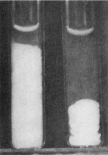
We have also observed the superprecipitation of plasmodial myosin Β gels on the addition of A T P (Fig. 3). Superprecipitation of this protein, however, proceeds rather slowly under the present experimental con- ditions.
FIG. 3 . Superprecipitation of plasmodial myosin B ; pH 6 . 6 , 1 9 ° C ; K+, 0 . 1 1 M;
ATP, 0 . 4 7 χ 1 0 - 3 M; plasmodial myosin B , 1 . 3 1 mg/ml. Left, control; right, A T P treatment. Photographed 7 0 min after addition of A T P (Nakajima, 1 9 6 0 ) .
ENZYMATIC PROPERTIES
As mentioned previously, plasmodial myosin Β either shows viscosity changes or superprecipitation on the addition of ATP. Which of these changes takes place depends on whether the plasmodial myosin Β is in the sol or gel state; this in turn depends of the K+ concentration. There- fore, we studied the enzymatic properties on varying the concentration of K + .
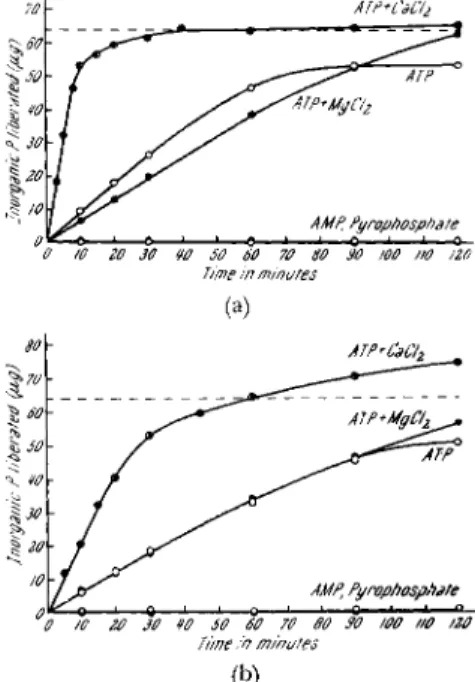
Figures 4a and b show the time courses of orthophosphate liberation when ATP, AMP, and inorganic pyrophosphate were added to plasmodial myosin B. Figure 4a is the case at a high K+ concentration (0.5 M) and Fig. 4b at a low K+ concentration (0.1 M). The broken line indicates the
ATP+CaC'h
/0 20 30 ¥0 50 60 70 60 30 100 »0 IZO Time in minutes
80 ATP+CaCJz
~%70 r
SO
SO JV^^^ATP*
\so - /
§ 20 'S
10 /IMP, Pyrophosphate
"θ 7C 20 30 tO SO 60 70 SO SO WO ttO /20 Time in minutes
(b)
FIG. 4a. Time courses for hydrolysis of A T P , AMP, and inorganic pyrophosphate at a high K+ concentration; pH 6.6, 28°C; K + , 0.5 M; A T P , 0.69 χ 1 0 - 3 M; AMP, 0.83 χ 1 0 - 3 M; pyrophosphate, 0.85 χ 1 0 - 3 M; CaCl2, 3.3 χ 1 0 - 3 M; MgCl0, 3.3 χ 10 — 3 M; plasmodial myosin B; 0.141 mg of N (Nakajima, 1960).
FIG. 4b. Time courses for hydrolysis of A T P , AMP, and inorganic pyrophosphate at a low K+ concentration. T h e conditions are the same as in Fig. 4a, except that the K+ concentration is 0.1 M (Nakajima, 1960).
level corresponding to half the labile phosphate of ATP. It is clearly
shown in these figures that plasmodial myosin Β has ATPase activity,
and that it hydrolyzes neither AMP nor inorganic pyrophosphate. In the
presence of a high K+ concentration, the ATPase activity is stimulated
Mechanochemical System in Physarum 115 by CaClo, whereas it is inhibited by MgCl
2(see also Table I). At a low K+
concentration, CaCl
2accelerates the activity, whereas MgCl
2has no effect on it. The specific activities calculated from these figures are 1.44 and 0.50 μιτιο^ of P/mg of N/min at K+ concentrations of 0.5 and 0.1 M, respectively (in the presence of 3.3 χ 1 0 ~
3M CaCl
2). These values are smaller than those of skeletal muscle myosin Β but higher than those of smooth muscle myosin Β (Needham and Cawkwell, 1956, 1958).
As shown in these figures, when the K+ concentration is low, or when MgClo is present in the reaction mixture, more than half the labile phos- phate of A T P is split during longer incubation periods. This may be due to myokinase which may be present in this preparation.
Table I summarizes the effect of the K+ concentration on the ATPase activity with and without CaCl
2and MgCl
2. The ATPase activity is
T A B L E I
E F F E C T OF K+ CONCENTRATION ON A T P A S E ACTIVITY WITH AND WITHOUT CaCI2 AND MgCl2
-m f ι t . umoles of P / m g of N/min Molar concentration "
K+ None CaCl2 MgCl2
0.08 0.11 0.24 0.15
0.15 0.21 0.76 0.18
0.30 0.23 1.46 0.14
0.45 0.25 1.46 0.12
0.60 0.25 1.34 0.10
0.80 0.24 1.34 0.09
α p H 6.6, 28°C; A T P , 0.74 χ 1 0 - 3 M; CaCl2, 3.3 χ 1 0 - 3 M; MgCl2, 3.3 χ 1 0 - 3 M;
plasmodial myosin Β; 0.101 mg of Ν (Nakajima, 1960).
stimulated by an appropriate amount of K + . CaCl
2accelerates the activ- ity over a wide range of K + . On the other hand, MgCl
2increases ATPase activity at low K+ concentrations of less than 0.1 M but inhibits it at high K+ concentrations.
In addition to these facts, we found the following enzymatic properties:
1. There is antagonism between Ca++ and Mg++ both at high and low K+ concentrations.
12. The pH optima are at about 6.2 and 5.4 in the presence of high and low K+ concentrations, respectively. On the alkaline side, the activity increases with pH.
3. 2,4-Dinitrophenol (DNP) stimulates the enzymatic activity at both high and low K + concentrations.
ι High and low K+ concentrations correspond to around 0.5 and 0.1 M, respec- tively.
4. Parachloromercuribenzoic acid (PCMB) inhibits the ATPase activ- ity. This inhibition is partially reversed by cysteine.
5. Monoiodoacetic acid (MIA) has no effect on the ATPase activity at either high or low K+ concentration.
6. E D T A inhibits the enzyme both with high and low K+ concen- trations.
These results demonstrate that the major part of the enzymatic prop- erties of plasmodial myosin Β coincide with the distinctive features of myosin Β in muscle.
As mentioned previously, the A T P sensitivity of the viscosity drop is small in plasmodial myosin B. Besides, the ATPase activity is stimulated by DNP at low K+ concentrations. These facts suggest a low actin content in this protein preparation (cf. Szent-Györgyi, 1951; Chappell and Perry, 1955; Greville and Needham, 1955; Greville, 1956; Yagi, 1957).
T o sum up, data represented here indicate that there is in myxomycete plasmodia a protein fraction which has many physicochemical and enzy- matic similarities to the properties of muscle myosin Β (or actomyosin).
On the other hand, we have demonstrated already that when A T P is added externally to plasmodia of Physarum (Kamiya et al., 1957), or when A T P is injected into the organism (Takata, 1957), the motive force of streaming is strongly increased. In addition, it is known that Physarum plasmodia contain a large amount of A T P (Hatano and Takeuchi, 1960).
As the protein in question reacts specifically with A T P in vitro, we may assume that this protein is the active principle that responds to A T P in a characteristic way in vivo. It is possible to say that plasmodial myosin Β is a substance playing a crucial part in mechanochemical phenomena underlying protoplasmic streaming in Physarum.
Birefringence in Plasmodia
If actomyosin-like protein is a substance corresponding to the mech- anochemical system behind streaming in Physarum, it would be expected that the deployment of molecules or micelles of this protein could be studied in the living plasmodia by means of the polarizing microscope.
A study of the pattern of birefringence in Physarum plasmodia has been in progress with Dr. Allen (Princeton University); the analysis is so far incomplete, but we shall present a brief progress report at this time, pending a more complete report to be published later. The photographs (Figs. 5-11) are of flattened portions of plasmodia suspended in a sucrose solution (0.1 and 0.05 M) to prevent osmotic damage.
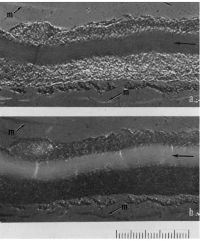
One of the most striking features of plasmodial ultrastructure visible
with polarized light is shown in Fig. 5. The arrow in the pictures indi-
Mechanochemical System in Physarum 117
FIG. 5. Birefringence in a plasmodial strand. Angle settings of mica compensator (λ/23): (a) 3°; (b) — 2 ° . m, Mucus secreted by plasmodia. Sucrose, 0.05 M. Scale, 10 μ interval.
cates the streaming endoplasm and its direction of flow. Portions sur- rounding endoplasm are stationary ectoplasm. The upper and lower pictures are taken at opposite compensator settings to show reversal of contrast in the two pictures. The compensator settings indicate that the signs of birefringence with respect to the axis of streaming are opposite for the endoplasm and ectoplasm; the endoplasm is negative, whereas the ectoplasm is positive.
If proteins are responsible for birefringence, positive axial birefrin- gence in the ectoplasm is interpreted as orientation of proteins at the
micellar or filament level along the axis of the strand. In the case of the
endoplasm, which presumably contains the same molecular species as the
ectoplasm, and differs supposedly only in physical state, the sign is nega-
tive, indicating that the alignment of proteins would have to be perpendi- cular to the axis of streaming. However, we cannot say with any assurance what kind of molecules and what kind of deployment might be respon- sible for these birefringence effects.
Concerning the endoplasmic birefringence, we further observed that a substantial part of it remains not only when the flow is stopped during the streaming cycle, but even after fixation and preservation in 5 0 % glycerol.
2These facts suggest that the endoplasmic birefringence is not simple flow birefringence alone.
It is also noticed in these pictures that there are biréfringent structures either in or around the endoplasm and oriented orthogonally to the streaming. T h e mucus secreted by plasmodia has positive birefringence (designated by m in the pictures).
Figures 6-8 show very clearly the presence of biréfringent fibrillar structures in the cytoplasm as well as diffuse birefringence surrounding them. In these specimens, scarcely any endoplasmic streaming occurs. In the top pictures are shown the advancing front; in the middle frames, the expanded inner portion; and in the lower pictures, a portion of an area with ramifying strand. In the case of active plasmodia, the endoplasm usually streams through such a strand with rhythmical changes in direc- tion, then diverges into smaller and smaller branches through the ex- panded portion, and finally into very small streamlets at the advancing front.
These pictures show that in the expanded portion (Figs. 6 and 7), the biréfringent structures are arranged orthogonally, with one almost perpendicular to the edge of the spreading tip. On the ramifying strand (Fig. 8), the fibrillar structures are arranged along the axis of the strand.
These fibrillar structures have positive birefringence.
In general, biréfringent fibrillar structures are usually not visible in many plasmodia in which the endoplasm exhibits the most active stream- ing. In other words, they are induced structures, which may be thought of as having crystallized out of pre-existing organization of lower order.
We can also observe the belt-shape biréfringent structures (Figs. 9-11).
They are clearly visible in this specimen, in which no streaming was dis- cernible. T h e orientations of belt-shape structures are very similar to those of the fibrillar structures; in the expanded portion (Figs. 9 and 11) the biréfringent structures are arranged orthogonally, and in the ramify- ing strand (Fig. 10) they are oriented parallel to the axis of the strand.
Therefore, both types of structures may have originated from the same source.
2 T h e specimen was washed three times with water and suspended in water before observation.
FIGS. 6-8. Fibrillar biréfringent structures in plasmodia. Angle settings of mica compensator (λ/23), Figs. 6a, 7a, 8a; 3°; Figs. 6b, 7b, 8b; — 3 ° . Sucrose, 0.1 M. Scale,
10 μ interval.
119
T o summarize the polarized-light results obtained so far, it can be said that the endoplasm shows negative birefringence, whereas the ecto- plasm shows positive birefringence owing both to visible fibrils and to unresolved structural elements. At the present time, we are unable to give a detailed interpretation at either the submicroscopic or molecular levels of this birefringence.
We have shown the beginnings of two different experimental ap- proaches to the molecular basis of protoplasmic streaming in Physarum.
The first is chemical; it is obviously necessary to learn what molecules are present in plasmodia and how they interact under well-defined in vitro conditions. The second method is physical; without identifying the mole- cules, it attempts to delineate their deployment and alignment within the living plasmodia. Living motile systems are obviously dynamically or- ganized societies of molecules. Therefore, both approaches are required if we are to understand the whole process.
REFERENCES
Chappell, J . B., and Perry, S. V. (1955). Biochim. Biophys. Acta 16, 285.
Greville, G. D. (1956). Biochim. Biophys. Acta 20, 440.
Greville, G. D., and Needham, D. M. (1955). Biochim. Biophys. Acta 16, 284.
Hatano, S. (1962). Personal communication.
Hatano, S., and Takeuchi, I . (1960). Protoplasma 52, 169.
Kamiya, N., Nakajima, H., and Abé, S. (1957). Protoplasma 48, 94.
Loewy, A. G. (1952). ./. Cellular Comp. Physiol. 40, 127.
Nakajima, H. (1956). Seitat no Kagaku 7, 256 (in Japanese).
Nakajima, H. (1960). Protoplasma 52, 413.
Needham, D. M., and Cawkwell, J . M. (1956). Biochem. J. 63, 337.
Needham, D. M., and Cawkwell, J . M. (1958). Biochem. J. 68, 31p.
Portzehl, H., Schramm, G., and Weber, H. H. (1950). Z. Naturforsch. 5b, 61.
Rebhun, L. (1962). Personal communication.
Szent-Györgyi, A. (1951). / . Biol. Chem. 192, 361.
Takata, M. (1957). 22nd Ann. Meeting Botan. Soc. Japan.
Tonomura, Y., and Sasaki, A. T . (1957). Enzymologia 18, 111.
Tonomura, Y., Yagi, Κ., and Matsumiya, Η. (1955). Arch. Biochem. Biophys. 59, 76.
Ts'o, P. O. P., Bonner, J . , Eggman, L., and Vinograd, J . (1956a). / . Gen. Physiol. 39, 325.
Ts'o, P. O. P., Eggman, L., and Vinograd, J . (1956b). / . Gen. Physiol. 39, 801.
Ts'o, P. O. P., Eggman, L., and Vinograd, J . (1957a). Arch. Biochem. Biophys. 66, 64.
Ts'o, P. O. P., Eggman, L., and Vinograd, J . (1957b). Biochim. Biophys. Acta 25, 532.
Yagi, K. (1957). / . Biochem. 44, 337.
DISCUSSION
DR. REBHUN: I recently had an opportunity to examine the protein that has been extracted by Dr. Nakajima's technique. I think it is rather fortunate you use different terms for your protein than Dr. Ts'o and his collaborators used for theirs. I think the evidence is they are different proteins. Specifically, we started out using Dr. Nakajima's techniques on frozen plasmodia. W e were unsuccessful after six or seven tries in ex-
FIGS. 9-11. Belt-shape biréfringent structures in plasmodia. Angle settings of mica compensator (λ/23): Fig. 9a, 8°; Fig. 9b, — 6 ° ; Fig. 10a, 9°; Fig. 10b, — 1 0 ° ; Fig. 11a, 6°;
Fig. lib, — 7 ° . Sucrose, 0.1 M. Scale, 10 μ interval.
121
trading the so-called plasmodial myosin B , using Dr. Nakajima's technique on the frozen mold. However, we went to fresh mold and it came out perfectly well.
We also found that if we varied the pH of the extracting solution—and we used a variety of solutions—if we got down below approximately pH 8.0, we were not able to extract the protein. Similarly, in diluting the extract, the pH had to be below pH 6.6 or a precipitate would not form.
Thus, myxomyosin (Dr. Ts'o) and plasmodial myosin Β (Dr. Nakajima) differ on several counts: first, plasmodial myosin Β is not extractable, under any conditions that we have tried, from frozen mold, whereas myxomyosin is; second, plasmodial myosin Β is not extractable at pH's below approximately 8 , whereas myxomyosin is extractable in KCl in distilled water, presumably below pH 7.0; and last, plasmodial myosin Β precipitates at low ionic strength, whereas myxomyosin not only is soluble but appears to dissociate irreversibly into smaller units. These proteins are undoubtedly different.
They may very well be related, however, and it may be that myxomyosin is a degenera- tion product of plasmodial myosin B . At least I presume degeneration goes in that direction. I also presume that fresh mold is a better material to use for extraction than slowly frozen mold.
I think it is rather interesting that one can prepare at least two proteins, both of which have mechanochemical properties in the sense of an ATPase which shows reversi- ble change in viscosity on addition of adenosine triphosphate (ATP).
CHAIRMAN THIMANN: Do you want to make comment on that? Have you ever ex- tracted frozen mold?
DR. NAKAJIMA: No. I have used only fresh mold.
DR. HOFFMANN-BERLING: Judging from Dr. Rebhun's comment, the contractile protein of slime mold plasmodia behaves somewhat like the contractile protein from smooth muscle. T h e contractile complex can be extracted only from fresh muscle. With the onset of rigor mortis, the actin component becomes inextractable, and the muscle delivers a myosin which is devoid of actin and which, of course, is noncontractile.
Furthermore, actin can be extracted more completely from fresh muscle, if the pH is kept well above 8. I would guess that plasmodial myosin Β (Dr. Nakajima) corresponds to the contractile complex protein of the slime mold plasmodia, whereas myxomyosin (Dr. Ts'o) represents the plasmodial myosin deficient in actin.
In Dr. Nakajima's paper it struck me that the rate of A T P dephosphorylation is enhanced at ionic strengths of 0.2. T h e ATP-splitting activity of muscle actomyosin decreases sharply in concentrated salt solutions. However if activation is brought about, not in the usual way by 10 — 3 mole liter M g+ + but by adding C a+ + , there is an augmentation of enzymatic dephosphorylation of muscular actomyosin at high salt concentrations. Have your splitting experiments been done in the presence of high concentrations of C a+ +?
DR. NAKAJIMA: Yes. These are the values obtained in the presence of 3 . 3 χ 10 — 3 M C a C l2 (28°C, pH 6.6). As you pointed out, in the case of skeletal muscle myosin B , the ATPase activity is maximal when potassium is at the physiological concentration, that is, at about 0.1 M, in the presence of CaCl2 and decreases with increasing potassium concentration. However, in the case of smooth muscle myosin B , the activity increases with increasing potassium concentration until the maximum at about 0.6 M is reached. Therefore, plasmodial myosin Β ATPase is affected by the K+ concentration in a manner more like smooth muscle than skeletal muscle.
DR. HOFFMANN-BERLING: Since the actin component of smooth muscle actomyosin and of fibroblast cell actomyosin is much less soluble in concentrated salt solutions than the myosin component, it is a problem to get out with the myosin an appropriate
Mechanochemical System in Physarum 123
amount of actin. Since the addition of C a+ + activates the ATP-splitting activity only of free muscular myosin (L-myosin) but not of the actomyosin complex, and since C a+ + increases the ATP-splitting rate in your preparations, I would guess that your prepara- tions contain an excess of free myosin and are deficient in actin. A deficiency in actin can roughly be estimated by measuring the mechanical activity of the protein, i.e., by centrifuging the gel in graduated tubes and measuring the amount of volume shrink- age after the incubation of the gel with A T P . Can you say anything about this?
DR. NAKAJIMA: I did not measure the volume shrinkage. However, the superprecipi- tation of plasmodial myosin Β proceeds rather slowly under the present experimental conditions. In addition, the A T P sensitivity of the viscosity drop is small and the ATPase activity is stimulated by DNP at low K+ concentrations. From these facts, I suggest that actin content in plasmodial myosin Β is low.
DR. INOUÉ: May I speak concerning this negative birefringence of the endoplasm?
Did I notice that the contrast of the endoplasm was similar to that portion of the plasma surface which was parallel to the endoplasm?
DR. NAKAJIMA: In the case of the mucus layer, surface membrane, and ectoplasm, the slow axis is parallel to the strand axis. In the case of the endoplasm, the slow axis is perpendicular to the strand axis.
DR. INOUÉ: In cell division when asters first develop, they appear with a positive birefringence. When the fibers are about to disappear in telophase, they sometimes show negative birefringence. W e do not know the explanation, but it may be related to your observation.
DR. SATO: Considering the negative biréfringent nature of endoplasmic streaming, which I believe would be based on both flow and form birefringence, did you notice any changes of the sign and the order of magnitude of birefringence during the whole process of flow reversal?
DR. NAKAJIMA: W e have not yet seen such changes.
DR. ALLEN: I think it is probably safe to assume that the fibers Dr. Nakajima has been looking at in polarized light are either similar to or identical with the structures that Dr. Wohlfarth-Bottermann described. W e have been rather encouraged to find that he found evidence of the ATPase system being there, and I am sure he is some- what relieved to find we see the same thing in the living state. I think these studies complement one another well.