Original Article
Accuracy of digitally planned, guided apicoectomy with a conventional trephine and a custom-made endodontic trephine: An in vitro
comparative study
Eszter Nagy
a, G abor Braunitzer
b, D aniel Gerhard Gryschka
a, Ibrahim Barrak
c, Mark Adam Antal
a,d,*
aDepartment of Operative and Esthetic Dentistry, Faculty of Dentistry, University of Szeged, Szeged, Hungary
bdicomLAB Dental, Ltd, Szeged, Hungary
cFaculty of Medicine, Department of Oral and Maxillofacial Surgery, University of Szeged, Szeged, Hungary
dDepartment of Preventive and Restorative Dentistry, College of Dental Medicine, University of Sharjah, Sharjah, UAE
A R T I C L E I N F O
Article History:
Received 20 July 2021 Accepted 27 September 2021 Available online xxx
A B S T R A C T
Purpose:Static guided trephine apicoectomy has been developed as a less invasive and more accurate alter- native to conventional freehand apicoectomy with drills. Overpenetration is a frequent issue with this proce- dure, which deteriorates accuracy and raises safety concerns. A safety improvement to address this problem is presented.
Materials and Methods:Guided apicoectomies were performed in porcine mandibles with either a conven- tional bone trephine or a custom-made endo-trephine with built-in depth control. The deviation of the apical endpoint of the trephine from the digital surgical plan was analyzed. Overpenetration frequency was recorded.
Results:Procedures performed with the custom trephine were significantly more accurate both along thex- axis and globally, but no significant difference was found for theyandzaxes. Overpenetration frequency was 70% in the conventional trephine group versus 38% in the stop trephine group.
Conclusion:The results indicate that the lack of physical depth control can interfere with the accuracy (and safety) of these procedures to a significant extent, as visual cues (such as the depth markings on a conven- tional trephine) are insufficient to prevent overpenetration. Our results show that custom-made trephines with a built-in stop offer an optimal solution for this problem.
© 2021 Elsevier Masson SAS. All rights reserved.
Keywords:
Endodontic microsurgery Apicoectomy
3D printing Static surgical guide
1. Introduction
Apicoectomy is a routine endodontic surgical procedure in which the apical 3 mm of the root tip is removed. This method allows the elimination of>90% of the ramifications and lateral canals [1,2], and is used when the conventional treatment of an endodontically com- promised tooth is not possible (for instance because of the presence of ramifications that cannot be accessed with conventional ortho- grade instrumentation). Success rates have been reported from a range as wide as 17 to 96% [3,4]. This high variability is most probably related to the fact that success with this approach presupposes high accuracy. There is agreement in the literature that to eliminate the internal structures that interfere with the success of root canal treat- ment, the terminal 3 mm of the apical third must be removed [5,6].
Besides, it is recommended that the section be performed
perpendicularly to the root axis to minimize the chance of bacterial leakage via the dentinal tubuli during and after the surgery[7]. When these principles are observed, elimination rates may be as high as 98% for apical ramifications and 93% for accessory canals[8]. These requirements are quite difficult to meet with the conventional tech- nique, which is freehand drilling based on mental navigation, thus being highly dependent on the operator’s skills and experience.
Furthermore, the intervention may be quite invasive and carries the risk of damaging anatomical structures in the vicinity of the target area[9].
Cone-beam computed tomographic imaging (CBCT) is of consider- able help in these situations, so much so that by today it is considered essential before periapical surgical interventions [10,11]. Indeed, CBCT provides valuable information about the three-dimensional relations of the infected area and the neighboring anatomical struc- tures, but without any further aid, the accuracy of the procedure still depends on the operator.
*Corresponding author.
E-mail address:antal.mark@szte.hu(M.A. Antal).
https://doi.org/10.1016/j.jormas.2021.09.014
2468-7855/© 2021 Elsevier Masson SAS. All rights reserved.
ARTICLE IN PRESS
JID: JORMAS [m5G;October 7, 2021;12:18]
Please cite this article as: E. Nagy, G. Braunitzer, D.G. Gryschka et al., Accuracy of digitally planned, guided apicoectomy with a conventional trephine and a custom-made endodontic trephine: Anin vitrocomparative study, Journal of Stomatology oral and Maxillofacial Surgery (2021),https://doi.org/10.1016/j.jormas.2021.09.014
J Stomatol Oral Maxillofac Surg 000 (2021) 1−7
Available online at
ScienceDirect
www.sciencedirect.com
The advantages of using CAD/CAM templates as apicoectomy guides werefirst pointed out by Pinsky and co-workers in 2007, who demonstrated that guided apicoectomy was significantly more pre- cise than the conventional freehand approach[12]. The idea caught on only about a decade later, most probably because it was in this period that stereolithographic techniques became widely available.
Liu and colleagues used guides to aid osteotomy and localization of the apex[13]. The group of Patel used 3D printed devices as soft tis- sue retractors to aid apicoectomy[14]. With the development of 3D surgical planning software, it became possible to plan the depth and angle of the resection and print a 3D template from the plan to guide the operation: this was the birth of the“pilot-drill”protocol where the template was used for apex localization[15−17]. Note, however, that in these studies the template did not physically guide the drill or bur; it served merely as an optical reference to support mental navi- gation. The most efficient combination of these novel methods was publishedfirst by Giacomino and co-workers[18]who used a tre- phine bur to perform a fully guided osteotomy, apicoectomy, and biopsy at the same time. Since then, this method has appeared in var- ious case studies and case series with generally favorable results,[19
−21]but still little is known about its accuracy.
Our research group has dealt with guided endodontic surgery for almost a decade, including the trephine approach. In one of our previ- ous publications, we showed that the accuracy of guided apicoec- tomy performed with a commercially available trephine was similar to the accuracy of guided implant surgery. However, we also found overpenetration to be a regularly occurring (and potentially hazard- ous) issue as commercially available bone trephines have no physical stop on them to prevent this, only depth markings [22]. We con- cluded that this might be a major limitation of the method and a con- siderable source of inaccuracy. This observation prompted us to design a set of trephines especially for endodontic purposes and inte- grate the new instrument in an existing surgical planning software to enable use in a fully digital workflow. [20] Among other
modifications, a key feature of the new instrument is that it has a built-in physical stop to prevent overpenetration.
In the presentin vitrostudy, we performed guided apicoectomies with commercially available trephines and our instrument. The pur- pose was,first of all, to determine if an overpenetration tendency is an inherent weakness of trephine apicoectomy. Thus, we sought to test if it could be reproduced under controlled conditions. Related to this, we sought to test if the addition of the stop can effectively pre- vent overpenetration and, if so, to what extent it improves the accu- racy of trephine apicoectomy, especially in the buccolingual dimension. We hypothesized that overpenetration would be a fre- quent finding with a conventional trephine, and less frequent or absent with the trephine equipped with a stop. We also hypothesized that the accuracy of the procedures performed with the stop trephine would be higher. Given the general paucity of data on the accuracy of this type of intervention in the literature, it was also a goal to gener- ate and publish such data.
2. Methods 2.1. Specimens
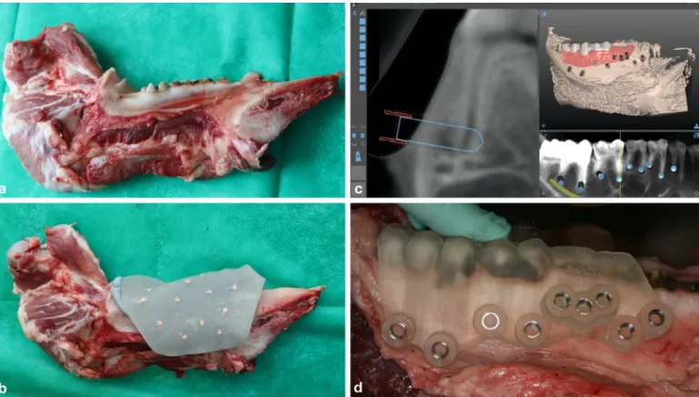
For the experiments, we used porcine mandible, an accepted model in the literature of dental implantology,[23−25]because its density and mineral content is similar to that of the human mandible[26−29]. Fresh porcine mandibles with teeth were obtained from the local meat proc- essing facility (Tisza-Maros Hus Kft., Szeged, Hungary,Fig. 1a). The ani- mals were not sacrificed for the sake of the experiments. Altogether 8 mandibles were used, 4 in the conventional trephine group (control group) and 4 in the stop trephine group (study group). After the experi- ments, the remnants were disposed of according to the pertinent regu- lations regarding the treatment of organic waste.
Twenty-one procedures were performed in the study group, and twenty-three in the control group (5−9 apicoectomies per mandible,
Fig. 1.Preparations for the apicoectomies. (a) a porcine mandible with teeth; (b) impression taking in an individualized tray with gutta-percha markers; (c) digital planning; (d) the 3D- printed, tooth-supported surgical guide on the porcine mandible.
ARTICLE IN PRESS
JID: JORMAS [m5G;October 7, 2021;12:18]
E. Nagy, G. Braunitzer, D.G. Gryschka et al. Journal of Stomatology oral and Maxillofacial Surgery 00 (2021) 1−7
depending on the anatomy of the given specimen as judged from an initial CBCT scan).
2.2. Imaging and surgical planning
To provide imaging input for the digital planning, we followed a double CBCT scan protocol with an individualized impression tray with gutta-percha markers made from Photopolymerizable Resin Trays (Elite LC Tray, Zhermack, Italy,Fig. 1b). The markers served the purpose of digital image registration. First, we took a silicone impres- sion into the individualized tray (Zetaplus, Zhermack, Italy). Thefirst CBCT scan was taken of the mandible with the impression on (in the tray) and the second scan was taken of the impression in the tray (without the mandible). For the CBCT scans, an i-CAT Next Generation device was used (Imaging Sciences- Kavo, Hatfield, PA, USA) with standard settings (tube voltage: 120 kV, tube current: 5 mA, exposi- tion time: 14.7 s, voxel size: 250
m
m, FOV: 160£130 mm).Preparation of the input images for planning, the process of surgi- cal planning itself, and 3D printing of the surgical templates hap- pened entirely according to the surgical guide production of dicomLAB Dental Ltd. (Szeged, Hungary) [30]. Specifically, surgical planning was done in SMARTGuide 1.26 (dicomLAB Dental, Szeged, Hungary,Fig. 1c), and the surgical templates were 3D printed with a multijet technology printer (ProJet MD3510, 3D Systems). Note that all procedures were planned with the help of a cylindrical implant model (as shown inFig. 1c), regardless of whether the given proce- dure was to be performed in the study group or the control group.
This way, it was possible to avoid the bias caused by the method of planning (i.e. we did not use the digital model of the new instrument for planning, as no such model was available for the conventional tre- phine). In any given case, the physical dimensions of the cylindrical model corresponded to those of the body of the trephine to be used.
This was possible because the“universal”module of the software is not implant brand-specific and it allows the user to define the length and diameter of the model used for planning.
2.3. Instruments and instrumentation
In both groups, trephines of 20 mm working length were used and always through a surgical template (Fig. 1d).
In the control group, commercially available, 4.21 mm diameter bone trephines were used (Hager & Meisinger, Neuss, Germany) through a 4.25 mm guiding sleeve embedded in the surgical tem- plate.Fig. 2a shows one of these trephines.
In the study group, we used the 4.46 mm endo-trephine from our set[20](Fig. 2b) through a 4.50 mm guiding sleeve embedded in the surgical template. This way, the difference between the diameter of the sleeve and that of the trephine’s body was the same as in the con- trol group. This difference is large enough to allow frictionless rota- tion but small enough not to risk efficient guidance. All trephines of the set are made of sulfur-alloyed martensitic stainless steel with 13% chromium content and high corrosion resistance (W.nr. 1.4197) and have a stop at 20 mm above the working end. The bits are manu- factured by a local company specializing in metal surgical devices (Lajos D€ome EV, Szeged, Hungary).Figs. 3a and3b demonstrate how the endo-trephine fits in the sleeve of the surgical guide how the built-in stop works.Figs. 3c and3d show how the conventional tre- phinefits in the sleeve of the surgical guide and that without a built- in stop, it is possible to insert the instrument beyond its working length. In both groups, the trephines were used in a surgical hand- piece (WS-75 L, W&H, Austria) connected to an implant motor (Implantmed, W&H, Austria). The drilling speed was 800 RPM. The implant motor provided constant external irrigation throughout the procedures (Fig. 3e). As for the depth of the osteotomy, in the control group, the operator relied on the depth markings of the instrument, and in the study group, the instrument was pushed in until the stop
prevented further penetration. Most of the time, the apex was removed along with the bone when the trephine was removed from the canal, but if it did not happen or bone fragments remained in the canal, debridement was performed with a periotome.
2.4. Analysis
Control CBCT scans for the analysis were taken with the same set- tings as described before. For the scans, metal cylinders of the same length and diameter as the body of the trephine used were inserted in the canals formed by the osteotomies. This was necessary because the analysis was segmentation-based, that is, it used grayscale values to differentiate between objects. Without the insertion of these cylin- ders, the inside of the canals would have appeared continuous with the surrounding air in the analytical software, rendering analysis impossible. By applying a metal cylinder of the same dimensions as the trephine, we reproduced the end-position of the trephine inside the bone, which could then be digitally compared to the planned end-position.
Analyses were conducted in Amira 5.4 (ThermoFisher Scientific, USA), with dedicated algorithms (dicomLAB Dental, Szeged, Hun- gary). First, the pre-and postoperative CBCT scans were registered so that they were in the same coordinate system. Next, the metal cylin- ders placed for the postoperative scans were segmented (Fig. 4a) and transformed into virtual bodies (Fig. 4b). It was in this step that we obtained the virtual reconstruction of the trephines’ end-position inside the bone. This was followed by the reconstruction of the planned positions. For this, virtual cylinders corresponding to the dimensions of the given trephine’s body were used. The spatial coor- dinates of the plan were applied to the virtual cylinder. This step yielded the end-position of the trephine as planned. After this step, it was possible to compare the end- positions of the cylinder as planned and as actually placed in three dimensions (Fig. 4c).
The analysis concentrated on the imaginary apical (distal) central endpoint of the trephine conceptualized as a solid cylinder. The basis of comparison was the spatial position of this point according to the surgical plan. Deviation along three axes (x,y,z) and global deviation were calculated. Global deviation (GD) was defined as the square root of the summed squares of the deviations along the three axes. As for the axes,xrepresented the horizontal (bucco-lingual) dimension, yrepresented depth (mesio-distal deviation) andzrepresented the vertical (cranio-caudal) dimension. The analysis paid special atten- tion to the bucco-lingual dimension (x), as this dimension allowed the assessment of over/underpenetration.
Fig. 2.The trephines used in the study. (a) the conventional trephine used in the con- trol group; (b) the endo-trephine used in the study group. Note that the body of the instrument widens at the proximal end to form a shoulder. When this shoulder (the stop) meets the outer rim of the guiding tunnel of the surgical template, it prevents the instrument from being introduced any deeper (see alsoFig. 3b).
ARTICLE IN PRESS
JID: JORMAS [m5G;October 7, 2021;12:18]
E. Nagy, G. Braunitzer, D.G. Gryschka et al. Journal of Stomatology oral and Maxillofacial Surgery 00 (2021) 1−7
The statistical analyses were performed in SPSS 23.0 (IBM, USA).
First, the frequency and magnitude of over-and underpenetration were calculated for both groups. These were characterized descrip- tively. Then we used linear regression analysis for GD (as a summed measure of deviations along the three axes) with group and surgical template ID as factors to exclude the possibility that the poorfit or some manufacturing issue of the surgical templates interfered with the results. Having excluded the confounding effect of the templates themselves, the variables (GD,x, y, z) were compared with one-way ANOVA by group. Descriptive statistics (means and standard devia- tions) were also calculated by group.
Accuracy was defined as the closeness between the planned and the actual spatial position of the apical endpoint.
3. Results
3.1. Over- and underpenetration
The observations regarding the frequency and magnitude of over- and underpenetration by group are given inTable 1. Both over- and underpenetration occurred in both groups, but there was a vast dif- ference in their occurrence: in the study group, underpenetration was more frequent (in 62% of the procedures), while in the control group, the situation was just the opposite: overpenetration was the predominantfinding (in 70% of the procedures). The degree of under- penetration was quite similar in the two groups, approximately 0.7 mm, while the degree overpenetration differed vastly. In the study group, the mean overpenetration was 0.36§0.31 mm, while the control group overpentrated by a mean of 2.45§1.88 mm.
3.2. Accuracy
The results of the linear regression analysis indicated that the model was a significant predictor of GD (F (2,41) = 7.21,P= 0.002, R2= 0.26. Group contributed significantly to the model (
b
= 0.53,P= 0.040), but surgical template did not (
b
= 0.02,P= 0.951). Con- sidering this result, all further analyses were done by group.As for GD, ANOVA indicated a significant difference between the groups (F= 14.77, df=1, P = 0.0004, two-tailed). According to the descriptive analysis, the significant difference stemmed from the higher global accuracy of the study group: the mean global deviation
in the study group was 0.92§0.60 mm [95% CI: 0.64−1.18 mm], in contrast to 2.45§1.88 mm [95% CI: 1.66−3.05 mm] in the control group.
ANOVA also indicated significant difference in deviation along the x-axis (F= 12.01, df=1,P= 0.001, two-tailed). This, again, reflected the higher accuracy of the study group. In contrast to a mean deviation of 1.47§2.22 mm [95% CI: 0.51−2.43 mm] in the control group, the study group exhibited a mean deviation of only 0.28§0.72 mm [95% CI: 0.61−0.5 mm] (the negative value indicates that most of the procedures were characterized by slight underpenetration, see above).
As for the x and y axes, no significant difference was found between the groups.Table 2provides the descriptive statistics for all studied parameters by group, with the significance levels for the between-groups comparisons.
4. Discussion
Guided trephine apicoectomy is an emerging endodontic surgical technique to enhance the accuracy of apicoectomy. While there is already some evidence to suggest that guided apicoectomy, in gen- eral, is indeed superior to the freehand approach in terms of accuracy, [20,22,31,32] but data are scarcely available on guided interventions performed with a trephine. In this study, we sought to compare the accuracy of digitally planned, guided apicoectomy performed with a commercially available and a custom-made trephine,in vitro. The rationale for this comparison was an earlier observation of our research group, which suggested that trephine apicoectomy might carry the risk of (even considerable) overpenetration in the bucco- lingual direction. The endo-trephine used in this study was designed to prevent such overpenetration. It must be noted that bone tre- phines of varying working lengths and diameters are available. Our endo-kit, for instance, contains altogether six pieces (2 diameters and 3 working lengths)[20]. It can be hypothesized that a shorter work- ing length (especially with a wider diameter) allows more accurate outcomes. In this study, we used only one working length (20 mm) and two negligibly different diameters (4.21 and 4.46 mm). The con- crete, numerical accuracy results, therefore, are best interpreted as characterizing instruments of similar dimensions, which is a limita- tion of this study. Further research is necessary to clarify how the Fig. 3.The trephines in use. Panels (a) to (d) illustrate how the instrumentsfit in the surgical guide. Note that the endo-trephine cannot be introduced into the guide any further than what the shoulder of the trephine allows, so the degree of insertion cannot exceed the instrument’s working length (a) and (b). In contrast, there is nothing to prevent the con- ventional trephine from being introduced beyond its working length (c) and (d). This is what makes overpenetration possible. Panel (e) shows guided trephine apicoectomy being carried out on a porcine mandible.
ARTICLE IN PRESS
JID: JORMAS [m5G;October 7, 2021;12:18]
E. Nagy, G. Braunitzer, D.G. Gryschka et al. Journal of Stomatology oral and Maxillofacial Surgery 00 (2021) 1−7
working length and the diameter of the applied instrument influence the accuracy of these procedures.
We hypothesized that overpenetration would be a frequentfind- ing with a conventional trephine, and less frequent or absent with the trephine equipped with a stop. The results confirmed this hypothesis: while overpenetration did occur in the study group, it occurred less frequently, and its degree was well within a safe 1 mm margin. In contrast, overpenetration was seen in 70% of the interven- tions performed with the conventional trephine, and its degree was considerably higher. The explanation for this tendency of overpene- tration is probably quite simple: as the operator uses the instrument,
he or she applies pressure on the handpiece to push the trephine for- ward in the bone. Even if the pressure is not excessive and is applied evenly, the hardness and structure of the bone are not completely
Table 1
Over- and underpenetration (deviation along thex-axis) in the study and control groups. Means and standard deviations are given in millimeters.
S (N= 21) C (N= 23)
OVER UNDER OVER UNDER
N (%) 8 (38%) 13 (62%) 16 (70%) 7 (30%)
Mean 0.36 0.76 2.45 0.75
SD 0.31 0.54 1.88 0.97
Fig. 4.Accuracy analysis in Amira. (a) segmentation is based on the difference in radiodensity between the tissues and the inserted metal cylinders; (b) the segmented cylinders are transformed into three-dimensional bodies that correspond to the cylinders in the bone; (c) a cylindrical model of the same dimensions as the body of the trephine is aligned with the segmented cylinder in the position to be analyzed (blue) and compared to an identical model positioned according to the surgical plan (red) with a custom algorithm. The algo- rithm returns the global apical deviation and deviation along the axesx,yandzin millimeters.
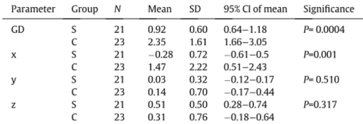
Table 2
Descriptive statistics of the studied parameters by group and the significance levels of the between-groups comparisons (one-way ANOVA). S: study group (stop tre- phine); C: control group (conventional trephine); GD: global deviation of the apical endpoint (millimeters);x: deviation of the apical endpoint along the bucco-lingual axis;y: deviation of the apical endpoint along the mesio-distal axis;z: deviation of the apical endpoint along the cranio-caudal axis. Means and standard deviations are given in millimeters.
Parameter Group N Mean SD 95% CI of mean Significance
GD S 21 0.92 0.60 0.64−1.18 P= 0.0004
C 23 2.35 1.61 1.66−3.05
x S 21 0.28 0.72 0.61−0.5 P=0.001
C 23 1.47 2.22 0.51−2.43
y S 21 0.03 0.32 0.12−0.17 P= 0.510
C 23 0.14 0.70 0.17−0.44
z S 21 0.51 0.50 0.28−0.74 P=0.317
C 23 0.31 0.76 0.18−0.64
ARTICLE IN PRESS
JID: JORMAS [m5G;October 7, 2021;12:18]
E. Nagy, G. Braunitzer, D.G. Gryschka et al. Journal of Stomatology oral and Maxillofacial Surgery 00 (2021) 1−7
homogeneous. If the working end of the instrument meets an area of lesser resistance (which the operator cannot see and thus applies the same amount of pressure), it might cause the instrument to slip for- ward in the bone. This cannot be prevented by observing the depth markings on the trephine, while the addition of a physical stop does efficiently prevent major overpenetration.
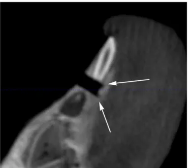
Underpenetration was present in both groups and to a notably similar degree. We have concluded that this is not a real problem, but a consequence of a minorflaw of our measurement method. When we planned the apicoectomies, we planned how deep the trephine should penetrate the bone, and we used solid cylindrical models for planning. We conceptualized the trephines as solid cylinders and used the distal endpoint of these cylinders for our measurements.
However, the trephines are, in fact, not solid but hollow and they have a cutting working end. This way, thefinal depth of penetration is defined by how deep the working end cuts, not where the endpoint of the body conceptualized as a solid cylinder is. Following from this, if a solid body of the same length and diameter as the trephine is introduced in the tunnel (like we did for the postoperative control scans), it cannot penetrate just as deep as the trephine, as it will be blocked by the small piece of unreduced bone left behind by the non- working inside of the trephine (Fig. 5). To put it simply, it is safe to assume that in most cases, the trephine penetrated slightly deeper than what we could measure because the metal cylinders we used as segmentation aids were solid. The difference is obviously negligible, and it probably has no clinical relevance, but it is both a reasonable explanation to part of thefindings and a limitation we need to men- tion.
We also hypothesized that the accuracy of the procedures per- formed with the stop trephine would be higher. This hypothesis was also confirmed. Significant difference between the two groups was found in global apical deviation and in deviation along the x axis (bucco-lingual depth, as discussed above). Note that the two instru- ments were characterized by almost identical accuracy along they andzaxes, which means that considerable deviation along thexaxis in the control group had been the source of error that resulted in the
significant global difference between the two instruments. This sug- gests that the control of penetration depth is indeed the key issue of the accuracy (and safety) of trephine apicoectomy. It is also notewor- thy that the standard deviations for GD and x in the control group are much higher than in the study group, indicating that the lack of appropriate depth control resulted not only in poorer accuracy but also in poorer precision.
How accurate and satisfactory is the global apical deviation of 0.92§0.60 mm [95% CI: 0.64−1.18 mm] achieved with the endo-tre- phine? Given the novelty of the approach, a direct comparison with the literature is not possible. However, it makes sense to compare the results against those of studies that examined the accuracy of digi- tally planned guided implant placement through a tooth-supported surgical template, because a goal of key importance in both proce- dures is to position a cylindrical body in the human mandible or max- illa as accurately as possible. Global apical deviation is a frequently reported measure of accuracy in the literature of implant surgery.
[30,33−37] In their latest systematic review and meta-analysis, based on the analysis of 20 studies, Tahmaseb and co-workers concluded that the mean error of apical position for partially edentulous cases (i.e., cases where a tooth-supported template could be used) was 1.2 mm [95% CI: 1.11−1.20 mm]. In a previous randomized controlled clinical trial of our research group[30], where we used the same soft- ware and digital workflow for implant placement as here for apicoec- tomy, we found a mean of 1.59 mm global apical deviation for both partial and full guidance (the two comparable study arms of the four, where the surgical guide was used for the entire process of osteot- omy). For this discussion, we re-analyzed the old dataset for partial and full guidance only. As the analysis did not indicate significant dif- ference in global apical deviation between the two groups, we treated them as a single group and found a mean global apical deviation of 1.66§0.84 mm [95% CI: 1.44−1.88 mm]. The results may make the impression that the accuracy of guided trephine apicoectomy is somewhat higher than that of guided implant placement. It must be taken into consideration, though, thatin vitro results tend to show higher accuracy than what is clinically achievable. It appears more proper to conclude that the guided use of the endo-trephine allows results approximately of the same accuracy as guided implant sur- gery. Clearly, it is not possible to reach the same level of accuracy when a conventional trephine is used.
5. Conclusions
Within the limitations of the study, we conclude that the lack of depth control is a major source of error if guided apicoectomy is car- ried out with a conventional trephine. The use of a custom-made endo-trephine with a stop reduced this error to a safe and acceptable level. With proper depth control, the accuracy of apicoectomy was similar to that of guided implant surgery. Without such control, major overpenetration was a frequentfinding. Obviously, this surgi- cal approach is a new one, and much testing is yet to be done before firm conclusions may be drawn. Other safety issues, such as intraoss- eous heat generation during these interventions, also remain to be clarified. It does not seem to be far-fetched, however, to assume that protection against overpenetration increases both the accuracy and the safety of these interventions. Visual cues (such as the depth markings on a conventional trephine) are insufficient to prevent overpenetration. Our results show that custom-made trephines with a built-in stop offer an optimal solution for this problem.
Funding None.
Fig. 5.The characteristic mark that the trephine leaves in the bone. The serrated work- ing end makes a circular cut that surrounds a small piece of unreduced bone. Thefigure also illustrates the problem of overpenetration: a shallower penetration would have been enough for a successful procedure, but there was no stop to prevent overpenetra- tion and the instrument stopped in the cortical bone on the opposite side. Any further penetration could have ended up in perforation, which is not a desirable outcome in a clinical setting.
ARTICLE IN PRESS
JID: JORMAS [m5G;October 7, 2021;12:18]
E. Nagy, G. Braunitzer, D.G. Gryschka et al. Journal of Stomatology oral and Maxillofacial Surgery 00 (2021) 1−7
Declaration of Competing Interest
Gabor Braunitzer is chief researcher at dicomLAB Dental, Ltd.
References
[1]Pop I. Oral surgery: part 2. Endodontic surgery. Br Dent J 2013;215(6):279–86.
[2]Gilheany PA, Figdor D, Tyas MJ. Apical dentin permeability and microleakage associ- ated with root end resection and retrogradefilling. J Endod 1994;20(1):22–6.
[3]Setzer FC, Kohli MR, Shah SB, Karabucak B, Kim S. Outcome of endodontic sur- gery: a meta-analysis of the literature−Part 2: comparison of endodontic micro- surgical techniques with and without the use of higher magnification. J Endod 2012;38(1):1–10.
[4]Rahbaran S, Gilthorpe MS, Harrison SD, Gulabivala K. Comparison of clinical out- come of periapical surgery in endodontic and oral surgery units of a teaching den- tal hospital: a retrospective study. Oral Surg Oral Med Oral Pathol Oral Radiol Endod 2001;91(6):700–9.
[5]Kim S, Kratchman S. Modern endodontic surgery concepts and practice: a review.
J Endod 2006;32(7):601–23.
[6]Morfis A, Sylaras SN, Georgopoulou M, Kernani M, Prountzos F. Study of the apices of human permanent teeth with the use of a scanning electron microscope. Oral Surg Oral Med Oral Pathol 1994;77(2):172–6.
[7]Mjor IA, Nordahl I. The density and branching of dentinal tubules in human teeth.
Arch Oral Biol 1996;41(5):401–12.
[8]Kim S, Pecora G, Rubinstein R, Dorscher-Kim J. Color atlas of microsurgery in end- odontics. Philadelphia: BW Saunders; 2001.
[9]Kim D, Ku H, Nam T, Yoon TC, Lee CY, Kim E. Influence of size and volume of periapical lesions on the outcome of endodontic microsurgery: 3-Dimen- sional analysis using cone-beam computed tomography. J Endod 2016;42 (8):1196–201.
[10]European Society of E, Patel S, Durack C, Abella F, Roig M, Shemesh H, et al. Euro- pean Society of Endodontology position statement: the use of CBCT in endodon- tics. Int Endod J 2014;47(6):502–4.
[11]Patel S, Brown J, Semper M, Abella F, Mannocci F. European Society of Endodon- tology position statement: use of cone beam computed tomography in Endodon- tics: European Society of Endodontology (ESE) developed by. Int Endod J 2019;52 (12):1675–8.
[12]Pinsky HM, Champleboux G, Sarment DP. Periapical surgery using CAD/CAM guidance: preclinical results. J Endod 2007;33(2):148–51.
[13]Yunfeng L, Guangsheng J, Quanming Y, Wei P. Additive manufacturing and digital design assisted precise apicoectomy: a case study. Rapid Prototyp J 2014;20 (1):33–40.
[14]Patel S, Aldowaisan A, Dawood A. A novel method for soft tissue retraction during periapical surgery using 3D technology: a case report. Int Endod J 2017;50 (8):813–22.
[15]Ye S, Zhao S, Wang W, Jiang Q, Yang X. A novel method for periapical microsurgery with the aid of 3D technology: a case report. BMC Oral Health 2018;18(1):85.
[16]Kim JE, Shim JS, Shin Y. A new minimally invasive guided endodontic microsur- gery by cone beam computed tomography and 3-dimensional printing technol- ogy. Restor Dent Endod 2019;44(3):e29.
[17]Lai PT, Yang SF, Lin YM, Ho YC. Computer-aided design-guided endodontic micro- surgery for a mandibular molar with hypercementosis. J Formos Med Assoc 2019;118(10):1471–2.
[18]Giacomino CM, Ray JJ, Wealleans JA. Targeted endodontic microsurgery: a novel approach to anatomically challenging scenarios using 3-dimensional-printed guides and Trephine Burs-a report of 3 cases. J Endod 2018;44(4):671–7.
[19]Ahn SY, Kim NH, Kim S, Karabucak B, Kim E. Computer-aided design/computer- aided manufacturing-guided endodontic surgery: guided osteotomy and apex localization in a mandibular molar with a thick buccal bone plate. J Endod 2018;44(4):665–70.
[20]Antal M, Nagy E, Sanyo L, Braunitzer G. Digitally planned root end surgery with static guide and custom trephine burs: A case report. Int J Med Robot 2020;16(4):e2115.
[21]Popowicz W, Palatynska-Ulatowska A, Kohli MR. Targeted endodontic microsur- gery: computed tomography-based guided stent approach with platelet-rich fibrin graft: a report of 2 cases. J Endod 2019;45(12):1535–42.
[22]Antal M, Nagy E, Braunitzer G, Frater M, Piffko J. Accuracy and clinical safety of guided root end resection with a trephine: a case series. Head Face Med 2019;15 (1):30.
[23]Quaranta A, Andreana S, Spazzafumo L, Piemontese M. Anin vitroevaluation of heat production during osteotomy preparation for dental implants with compres- sive osteotomes. Implant Dent 2013;22(2):161–4.
[24]Pandey RK, Panda SS. Drilling of bone: a comprehensive review. J Clin Orthop Trauma 2013;4(1):15–30.
[25]Mishra SK, Chowdhary R. Heat generated by dental implant drills during osteot- omy-a review: heat generated by dental implant drills. J Indian Prosthodont Soc 2014;14(2):131–43.
[26]Mohlhenrich SC, Modabber A, Steiner T, Mitchell DA, Holzle F. Heat generation and drill wear during dental implant site preparation: systematic review. Br J Oral Maxillofac Surg 2015;53(8):679–89.
[27]Atkinson PJ, Powell K, Woodhead C. Cortical structure of the pig mandible after the insertion of metallic implants into alveolar bone. Arch Oral Biol 1977;22 (6):383–91.
[28]Powell K, Atkinson PJ, Woodhead C. Cortical bone structure of the pig mandible.
Arch Oral Biol 1973;18(2):171–80.
[29]Stembirek J, Kyllar M, Putnova I, Stehlik L, Buchtova M. The pig as an experi- mental model for clinical craniofacial research. Lab Anim 2012;46(4):269–
79.
[30]Varga Jr. E, Antal M, Major L, Kiscsatari R, Braunitzer G, Piffko J. Guidance means accuracy: a randomized clinical trial on freehand versus guided dental implanta- tion. Clin Oral Implants Res 2020;31(5):417–30.
[31]Ackerman S, Aguilera FC, Buie JM, Glickman GN, Umorin M, Wang Q, et al. Accu- racy of 3-dimensional-printed endodontic surgical guide: a human Cadaver study. J Endod 2019;45(5):615–8.
[32]Fan Y, Glickman GN, Umorin M, Nair MK, Jalali P. A novel prefabricated grid for guided endodontic microsurgery. J Endod 2019;45(5):606–10.
[33]Schnutenhaus S, Edelmann C, Rudolph H, Luthardt RG. Retrospective study to determine the accuracy of template-guided implant placement using a novel nonradiologic evaluation method. Oral Surg Oral Med Oral Pathol Oral Radiol 2016;121(4):e72–9.
[34]Cassetta M, Di Mambro A, Di Giorgio G, Stefanelli LV, Barbato E. The influence of the tolerance between mechanical components on the accuracy of implants inserted with a stereolithographic surgical guide: a retrospective clinical study.
Clin Implant Dent Relat Res 2015;17(3):580–8.
[35]Cassetta M, Di Mambro A, Giansanti M, Stefanelli LV, Barbato E. Is it possible to improve the accuracy of implants inserted with a stereolithographic surgical guide by reducing the tolerance between mechanical components? Int J Oral Maxillofac Surg 2013;42(7):887–90.
[36]Cassetta M, Di Mambro A, Giansanti M, Stefanelli LV, Cavallini C. The intrinsic error of a stereolithographic surgical template in implant guided surgery. Int J Oral Maxillofac Surg 2013;42(2):264–75.
[37]Cassetta M, Stefanelli LV, Giansanti M, Calasso S. Accuracy of implant placement with a stereolithographic surgical template. Int J Oral Maxillofac Implants 2012;27(3):655–63.
ARTICLE IN PRESS
JID: JORMAS [m5G;October 7, 2021;12:18]
E. Nagy, G. Braunitzer, D.G. Gryschka et al. Journal of Stomatology oral and Maxillofacial Surgery 00 (2021) 1−7