water
Article
Physiological, Developmental, and Biomarker Responses of Zebrafish Embryos to Sub-Lethal Exposure of Bendiocarb
Gyöngyi Gazsi1,2, Zsolt Czimmerer3, Bence Ivánovics1,2, Izabella Roberta Berta1,2, Béla Urbányi1,2, Zsolt Csenki-Bakos1,2and AndrásÁcs1,2,*
Citation: Gazsi, G.; Czimmerer, Z.;
Ivánovics, B.; Berta, I.R.; Urbányi, B.;
Csenki-Bakos, Z.; Ács, A.
Physiological, Developmental, and Biomarker Responses of Zebrafish Embryos to Sub-Lethal Exposure of Bendiocarb.Water2021,13, 204.
https://doi.org/10.3390/w13020204
Received: 15 September 2020 Accepted: 14 January 2021 Published: 16 January 2021
Publisher’s Note: MDPI stays neu- tral with regard to jurisdictional clai- ms in published maps and institutio- nal affiliations.
Copyright:© 2021 by the authors. Li- censee MDPI, Basel, Switzerland.
This article is an open access article distributed under the terms and con- ditions of the Creative Commons At- tribution (CC BY) license (https://
creativecommons.org/licenses/by/
4.0/).
1 Department of Aquaculture, Institute for Conservation of Natural Resources, Faculty of Agricultural and Environmental Sciences, Szent István University, H-2100 Gödöll˝o, Hungary; gazsi.gyongyi@szie.hu (G.G.);
ivanovics.bence@phd.uni-szie.hu (B.I.); berta.izabella.roberta@szie.hu (I.R.B.); urbanyi.bela@szie.hu (B.U.);
csenki-bakos.zsolt.imre@szie.hu (Z.C.-B.)
2 Institute of Aquaculture and Environmental Safety, Hungarian University of Agriculture and Life Sciences, H-2100 Gödöll˝o, Hungary
3 Department of Biochemistry and Molecular Biology, Faculty of Medicine, University of Debrecen, H-4032 Debrecen, Hungary; czimmerer.zsolt@med.unideb.hu
* Correspondence: acs.andras@szie.hu
Abstract:Bendiocarb is a broad-spectrum insecticide recommended for malaria control by the World Health Organization (WHO). Still, bendiocarb poses a toxic risk to populations of nontargeted aquatic organisms. Thus, our study was aimed to evaluate the sub-lethal effects of bendiocarb exposure on zebrafish (Danio rerio) embryos by assessing of physiological, developmental, and biochemical parameters. Bendiocarb-induced adverse effects on embryonic development, larval growth, heart rate, changes in phase II detoxifying enzyme glutathione-S-transferase (GST) activity, oxidative stress-related enzyme activities (superoxide dismutase (SOD), catalase (CAT)), and the damage- linked biomarker lipid peroxidation (LPO) in early life stage zebrafish were investigated. Our results highlight that the selected nonlethal concentrations (96 h median lethal concentration in this study was 32.52 mg/L−1) of bendiocarb inflicted adverse effects resulting in embryo deformities (96 h EC50
= 2.30 mg L−1), reduced body- and notochord length (above 0.75 and 0.39 mg L−1 bendiocarb concentrations at 96 hpf, respectively), oxidative stress, and altered heart rate (above 0.4 mg L−1at 48 hpf) in the studied model system.
Keywords:bendiocarb; zebrafish; sub-lethal
1. Introduction
Insecticides are natural or synthetic compounds that are widely used to kill one or more species of insects. Based on their chemical structure, insecticides can be classified into several groups including carbamates, pyrethroids, and organophosphates [1]. These chemicals have high biological activity and contribute to the increased productivity of agriculture [2] by reducing loss and are an integral part of most modern practices.
Carbamates are one of the most frequently applied pesticides, commonly used in household and agricultural pest control [2–4]. WHO also recommends carbamates for malaria control [5–8], although various carbamate compounds are more toxic to verte- brates than other groups of pesticides [9]. Bendiocarb (2,2-dimethyL,3-benzodioxol-4-yl-N- methylcarbamate) belongs to the widely used carbamate compounds. This broad-spectrum insecticide can be extensively absorbed, widely distributed, completely metabolized in mammalian cells, and then excreted quickly reducing its bioaccumulation [5,6]. In the last two decades, bendiocarb was detected in pregnant women, who used some form of household pest control during pregnancy. It was also shown that bendiocarb can transfer through the placenta exposing the developing fetus to high risk [10–12]. According to many previous studies, bendiocarb induces various morphological changes in mammalian tissues
Water2021,13, 204. https://doi.org/10.3390/w13020204 https://www.mdpi.com/journal/water
Water2021,13, 204 2 of 16
and organs [2,5,8,13–18]. It has also been described that the reversible inhibition of acetyl- cholinesterase (AChE) activity has an impact on heart rate in different vertebrates [19,20].
Finally, bendiocarb reversibly inhibits cholinesterase activity and affects antioxidant and detoxifying enzyme activities in mammals [11,14,21].
In field trials, bendiocarb did not cause significant reductions in the populations ofAnisopsspp., orStreptocephalusspp., and in the number of macroinvertebrates. How- ever, bendiocarb killed surface-dwelling insects and caused a decrease in cladocerans density [22]. Recent studies have assessed the acute effects of bendiocarb on some fish species. Hayes and Lawes [8] have found that the acute oral toxicity (LD50) falls in a range of 0.7–1.8 mg kg−1in fish. The acute LC50value reported for sheepshead minnow (Cyprinodon variegatus) was 0.86 mg/L. For rainbow trout (Oncorhynchus mykiss), LC50
ranged from 0.87 to 1.55 mg/L. For bluegills (Lepomis macrochirus) LC50values reported ranges were 0.47 to 1.65 mg/L [23]. Tatarazako and Iguchi [24] found that the no observed effect concentration (NOEC) was 12.5 mg/L in chronic fish toxicity test (FET) on sac-fry stage zebrafish.
Pesticide runoff from agricultural pollutes rivers, ponds, lakes, and other impound- ments inclined to receive and accumulate contaminants, with pesticides [25,26]. Due to its widespread usage as a malaria vector control and pest control agent, bendiocarb can be found in various waters worldwide. In 2009, bendiocarb was detected in Japanese surface waters in concentrations between 0.16 and 5.68µg/L [24]. In a study, El-Saeid et al. [27]
selected 15 regions in Central East, North, and South of Saudi Arabia to perform a survey of pesticide residues in groundwater. Analysis of groundwater samples revealed that bendio- carb was present at 0.181 mg/L concentration in water samples from Abha. Lahr et al. [22]
have investigated the ecological impact of fenitrothion, diflubenzuron, deltamethrin, and bendiocarb in field trials in natural temporary ponds in a cultivated savannah area of Senegal, West Africa. Sixteen ponds were selected in the area from which 10 ponds were treated by the experimental insecticides. They found that the pseudo–first-order half-life of bendiocarb was 17 days.
Pesticides in different water bodies [28,29] can pose serious toxicological risks to resident vertebrates, including fish [23,30]. Further information is needed on account of intensive bendiocarb usage regarding its effect on non-target freshwater organisms. Apart from some acute or chronic toxicity tests, the effect of bendiocarb in aquatic organisms is less studied. Thus, we aimed to investigate the sublethal, environmentally relevant bendiocarb doses-induced adverse effects on embryonic development, larval growth, and heart rate in early life stage zebrafish. Furthermore, we followed the changes in oxidative stress and damage-linked biomarkers, including superoxide dismutase (SOD), catalase (CAT), glutathione-S-transferase (GST), and peroxidized lipid content (LPO).
2. Materials and Methods 2.1. Chemicals
Bendiocarb (PESTANAL®, analytical standard, CAS no.: 22781-23-3) was purchased from Sigma-Aldrich. Stock solutions of bendiocarb were prepared by dissolving it in dimethyl sulfoxide (DMSO,≥99.5% Gas Chromatography, plant cell culture tested; CAS no.: 67-68-5), purchased from Sigma-Aldrich, Darmstadt, Germany.
2.2. Fish Husbandry
AB wild-type and 2.2shh:gfp:ABC#15 transgenic zebrafish lines were supplied by the zebrafish research facility of the Department of Aquaculture, Szent István University (Gödöll˝o, Hungary). The 2.2shh:gfp:ABC#15 transgenic line contains a transgene that har- bors the green fluorescent protein (gfp) under the control of sonic hedgehog (ssh) regulatory elements, responsible for shh expression in the floor plate and notochord [31–33]. Fishes were maintained at constant water quality parameters (25±0.5◦C, pH 7.0±0.2, conduc- tivity 500±50µS, alkalinity < MDL, 0 mM CO32−, 0.4 mM HCO32−; hardness < 0.5◦dH;
DOC > 90%; system water) in a Tecniplast ZebTec (Buguggiate, Italy) recirculating zebrafish
Water2021,13, 204 3 of 16
housing system. The photoperiod was set to 14 h light/10 h dark cycle. The fishes were fed twice a day with ZEBRAFEED (Sparos, 400–600µm) and twice a week with brine shrimp (Ocean Nutrition > 230000NPG).
Zebrafish embryos were obtained from the spawning adults (adult male and female ra- tio 2/1), placed in breeding chambers the day before embryos were needed. The spawning was induced in the morning, by turning the lights on. Non-fertilized eggs were separated from the fertilized ones using a pipette under a stereomicroscope (Leica M 205 FA, Wet- zlar, Germany). Incubation was carried out at ambient temperature (25.5±0.5◦C) with 14 h light/10 h dark cycles in a constant temperature-light incubator (Memmert HPP IPP 110 PLUS with AtmoCONTROL software).
The Animal Protocol was approved under the Hungarian Animal Welfare Law and all studies were completed before the treated individuals would have reached the free-feeding stage.
2.3. Zebrafish Embryo Toxicity Tests
The 90-h acute toxicity tests were carried out according to the OECD guideline 236 (fish embryo acute toxicity test (FET), [34]). The FET tests were performed using fertilized and healthy zebrafish eggs. Before the embryos reached the 8-cell stage, three replicates were set up for each test concentration. All experiments were carried out in plastic 24-well plates equipped with covers. Preliminary sensitivity tests were conducted to ensure no difference appears in the sensitivity of the two different genetic lines. The test was repeated twice in triplicates, and each replicate referring to one plate. Embryos were placed individually in the plate cells (20 individuals per plate), containing 2 mL test or control solution. The following test concentrations were used: 200, 100, 50, 41.60, 34.60, 28.80, 25, 24, 20, 16.60, 13.80, 12.50, 11.50, 9.58, 7.98, 6.65, 6.25, 3.12, 1.56, 1.30, 1.08, 0.78, 0.39 mg/L for bendiocarb.
Maintenance system water and vehicle control (0.026% DMSO in system water) were used as negative controls. The 96 h FET test’s endpoints were mortality, embryonic malformation (present or absent), and hatching delay. Melanocyte number and size were compared to controls visually, and differences were documented. Photomicrographs were made for embryos at 72 hpf, using a Leica M205FA stereomicroscope (software LAS X, camera: DFC 7000T, bright field, zoom: 3X exp. time: 4.0 ms). Ninety-six-hour LC/EC1,10,50,90values were calculated for mortality and embryonic deformities (tail malformations and reduced body length) using probit analysis, according to the OECD guideline [34].
2.4. Heart Rate, Total Body and Notochord Length Determinations
Based on the results of the FET tests, sub-lethal bendiocarb concentrations of 3, 1.5, 0.75, 0.4, and 0.07 mg/L were selected to determine the non-lethal functional consequences of exposure to bendiocarb. Maintenance water and vehicle control (DMSO in maintenance water) were used as negative controls. DMSO exposure was below the limit (0.01% OECD 236 [31]). All of the exposure experiments were conducted in plastic Petri dishes equipped with covers in this study. Tests were repeated twice in triplicates for all concentrations and controls (50 embryos were placed in 50 mL test or control solution for each replicate) and the exposure solutions were changed daily. Selected sub-lethal test endpoints (heart rates, body, and notochord lengths) were recorded and larvae for biochemical marker level measurements were collected. Heart rates were monitored using a Leica M 205 FA microscope (software: LAS V 3.8, camera: DFC 425C, bright field, zoom: 9.5 exp. time:
40.1 ms) at 48 hpf. Ten larval fish in each treatment replicate were video recorded (20 s) for analysis. Heart rates of embryos were counted in each recording three times and the average among the counts was calculated. At 96 hpf, the total body length and notochord length of hatched larvae were measured using Image J software [35] (10 larvae/replicates) performed on photomicrographs taken with a Leica M 205 FA microscope (software LAS X, camera: DFC 7000T, zoom: 3X, bright field, exp. time: 5.67 ms, GFP2 ET exp. time:
800.534 ms).
Water2021,13, 204 4 of 16
2.5. Tissue Preparation and Biochemical Marker Measurements
Twenty fish larvae were pooled together in Eppendorf tubes (two subsamples for every replicate) at each sampling time (48, 72, 96 hpf) and homogenized for biochemical marker assessments. Homogenization was performed in a small bead mill (TissueLyser LT, Qiagen, Germantown, MD, USA). Enzymatic activities were evaluated in triplicates for each replicate, at 25◦C, using a Thermo Varioskan™ LUX multimode microplate reader (Thermo Fisher Scientific, Waltham, MA, USA).
One batch of subsamples of the collected whole fish larvae was homogenized in a general buffer (25 mM Hepes-NaOH, 130 mM NaCl, 1 mM EDTA, 1 mM dithiothreitol, pH 7.4) at a weight to volume ratio of 1:5. Homogenates were frozen at−80◦C for analysis of lipid peroxidation (LPO) and total protein content. Remaining subsamples were homog- enized in 100 mM of phosphate buffer (pH = 7.4) containing KCl 100 mM, EDTA 1 mM, dithiothreitol (DTT), 0.5 M sucrose, and 40µg/mL aprotinin, and centrifuged at 12,000×g for 30 min at 4◦C. Supernatants were collected and aliquots were kept at−80◦C until the measurement of the activity of acetylcholine esterase (AChE) glutathione peroxidase (GPx) glutathione-S-transferase (GST), catalase (CAT), and superoxide dismutase (SOD). The pro- tein concentration of the samples was determined in triplicate by the Bradford method [36], adapted to microplate, using bovine serum albumin as standard. The absorbance was recorded at 595 nm after an incubation period of 15 min.
2.5.1. Lipid Peroxidation Measurement
Lipid peroxidation was evaluated based on the formation of malonaldehyde in tis- sue homogenates by the thiobarbituric acid method elaborated by Wills [37]. A 150µL homogenate was mixed with 300µL of 10% TCA containing 1 mM FeSO4and 150µL of 0.67% thiobarbituric acid. The mixture was heated to 80◦C for 10 min, then precipitates were removed by centrifugation (10,000×gfor 10 s). The supernatant was subjected to fluorescence measurement at 516 excitation/600 nm emission. Blanks and standards of tetramethoxypropane were prepared in homogenization buffer. Results were expressed as µmoles of thiobarbituric acid reactants (TBARS) per milligram of homogenate protein.
2.5.2. AChE Activity Measurement
The determination of AChE activity was carried out according to the method of Ellman et al. [38] adapted to microplate [39]. A 96-well microplate was loaded with 3 replicates of 50µL of homogenate supernatant and 250µL of a solution made with 0.075 M acetylthiocholine iodide and 10 mM 5,5 dithio-bis (2-nitrobenzoic acid) in phosphate buffer (0.1 M, pH 7.2). In assay, blank samples were substituted with phosphate buffer and electric eel acetylcholine esterase was used as positive control. Absorbance was measured at 414 nm for 15 min at every min. Enzymatic activity was calculated from the slope of the absorbance curve and was expressed in Units (U) per mg of protein content (1 U being 1µmol of substrate hydrolyzed/min).
2.5.3. CAT Activity Measurement
CAT activity was measured in triplicates following the method of Aebi [40]. Decreases in the absorbance of a 50 mM H2O2solution (ε=−0.0436 mM−1cm−1) in 50 mM phosphate buffer (pH 7.8) and 10µL of tissue supernatant were continuously recorded at 240 nm at 10 s intervals for 1 min. The results were expressed as U/mg protein; a unit of CAT was defined as the amount of enzyme that catalyzed the dismutation of 1 mmol of H2O2/min.
2.5.4. GST Activity Measurement
GST activity was determined by the method of Habig et al. [41] adapted to microplate, according to the following procedure: A solution of glutathione (GSH) 100 mM in phos- phate buffer (pH = 6.5), and a second solution of 60 mM 1-chloro-2,4-dinitrobenzene (CDNB,ε= 9.6 mM−1cm−1) in ethanol was prepared just before the assay. The reaction mixture consisted of phosphate buffer, GSH solution, and CDNB solution in a proportion
Water2021,13, 204 5 of 16
of 4.95 mL (phosphate buffer): 0.9 mL (GSH): 0.15 mL (CDNB). In the microplate, 0.2 mL of the reaction mixture was added to 0.1 mL of the sample and the GST activity was measured immediately at every 20 s, at 340 nm, during the first 5 min. GST from equine liver was used as positive control. Enzymatic activity was calculated from the slope of the absorbance curve and was expressed in Units (U) per mg of protein content (1 U being 1µmol of substrate hydrolyzed/min).
2.5.5. Glutathione Peroxide Activity Measurement
Glutathione peroxide activities were measured according to Paglia and Valentine [42]
modified by Lawrence and Burk [43] and adapted to a 96-well microplate [44]. The reac- tion mixture contained 100 mM phosphate buffer (pH 7.5), 2 mM GSH, 2 U glutathione reductase, 0.12 mM NADPH, sodium azide (0.5 mM), 0.2 mM H2O2, or 3 mM cumene hydroperoxide (CHP). GPX activity was monitored by following the decrease in NADPH concentration (at 340 nm), which is consumed during the generation of GSH from oxidized glutathione (ε= 6.2 cm−1M−1), using H2O2(Se dependent activity), or cumene hydroper- oxide (total GPX) as substrate. GPx activity was expressed as U per mg of protein. (a U corresponding to 1µM NADPH hydrolyzed/min).
2.5.6. SOD Activity Measurement
Total SOD activity was measured in triplicates using the xanthine oxidase/cytochrome c method proposed by Crapo et al. [45]. Cytochrome c reduction by superoxide anions generated by the xanthine oxidase/hypoxanthine reaction was detected at 550 nm at room temperature. The reaction mixture contained 46.5 mM KH2PO4/K2HPO4(pH 8.6), 0.1 mM EDTA, 195 mM hypoxanthine, 16 mM cytochrome c, and 2.5 mU xanthine oxidase. The enzyme activity was calculated from the slope of the absorbance curve and was expressed in Units (U) per mg of protein (1 U causing 50% inhibition of the rate of cytochrome c reduction).
2.6. Statistical Analyses
All FET test data were analyzed using the statistical software package Prism 6.0 (version 6.01) for Windows (GraphPad Software Inc., La Jolla, CA, USA). In the FET test, nonlinear regression was used. A 95% confidence interval was used in the analyses. The remaining results are expressed as mean±SD. Data normality were checked using the Shapiro–Wilk and Kolmogorov–Smirnov, respectively. As the ANOVA assumptions were satisfied, the parametric Ordinary One-Way ANOVA test was used, followed by a post- hoc Tukey’s test in order to delimit the groups in which significant differences occurred.
Similarly, the non-parametric Kruskal-Wallis test was used, followed by a post-hoc Dunn’s test for pairwise comparisons between group. Ifpvalues were less than 0.05, the results were regarded as statistically significant.
Biochemical marker data were analyzed using OriginPro (version 2019, OriginLab Cor- poration, Northampton, MA, USA) software package. To detect concentration dependency of biochemical marker activities, nonlinear regression (dose-response curve fit with variable Hill slope given by parameter ‘p’, the confidence level for curves: 95%) was applied. To examine the interactive effects of different bendiocarb concentrations and exposure time on biochemical markers, a two-way analysis of variance (ANOVA) was used, where time (t = 48; t = 72 and t = 96 hpf), treatment (control, vehicle control, and 3, 1.5, 0.75, 0.4 and 0.07 mg/L), and their interaction were categorical predictor factors, while the measured biomarkers were considered as dependent variables. When the interaction of bendiocarb concentration and the exposure time was detected, a one-way ANOVA was conducted to examine the effects of one main factor at a specific level of the other main factor. Factors detected to be significant were further analyzed using a post-hoc Tukey test for multiple comparisons at the significance level of 0.05 (p< 0.05). Before statistical analyses, raw data were diagnosed for normality of distribution and homogeneity of variance with the Kolmogorov-Smirnov test and Levene’s test, respectively.
Water2021,13, 204 6 of 16
3. Results
3.1. Zebrafish Embryotoxicity Induced by Bendiocarb Exposure
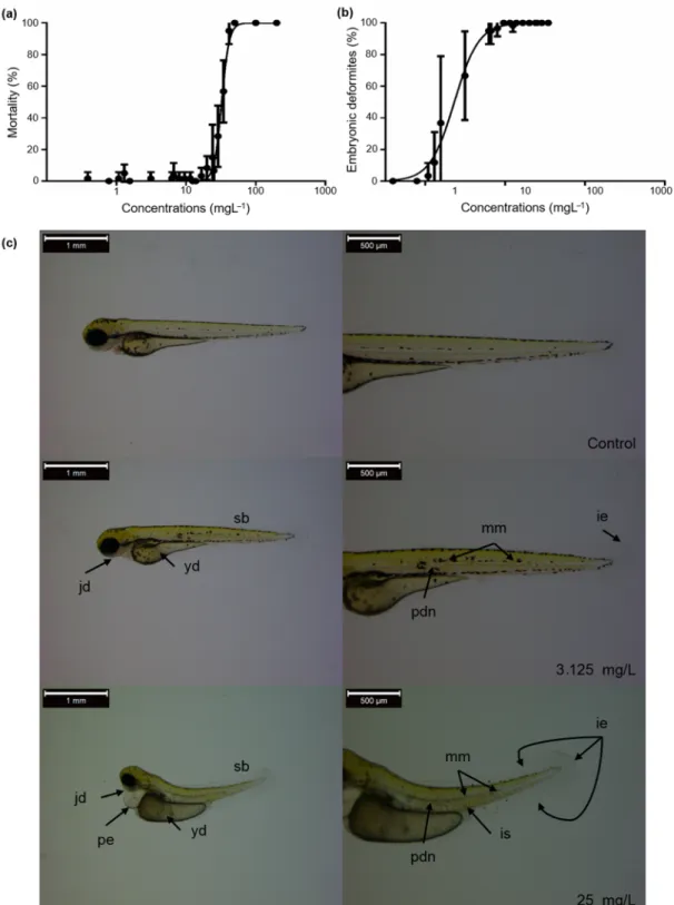
The LC and EC values were calculated for mortality and developmental abnormalities to characterize the bendiocarb exposure-caused adverse effects. LC50 value proved to be 32.52 mg/L, while EC50 value of embryonic deformities was 2.30 mg/L at 96 hpf (Figure1a,b, and Table 1). Tail malformations (above 3.12 mg/L) and reduced body length (above 0.39 mg/L) were observed in the bendiocarb-exposed groups. In addition, bendiocarb-induced shape deformities of the yolk sac, as well as abnormal melanocyte migration, number, and size were also identified in 96 hpf zebrafish larvae. The abnormal presence of melanocytes was detected in the notochord line and in the extra notochordal area after bendiocarb treatment. Decreased melanocyte number and size were also found at the higher bendiocarb concentrations, and melanocytes were not visible above 25 mg/L bendiocarb concentration. These deformities were not observed in the control and vehicle control groups (Figure1c).
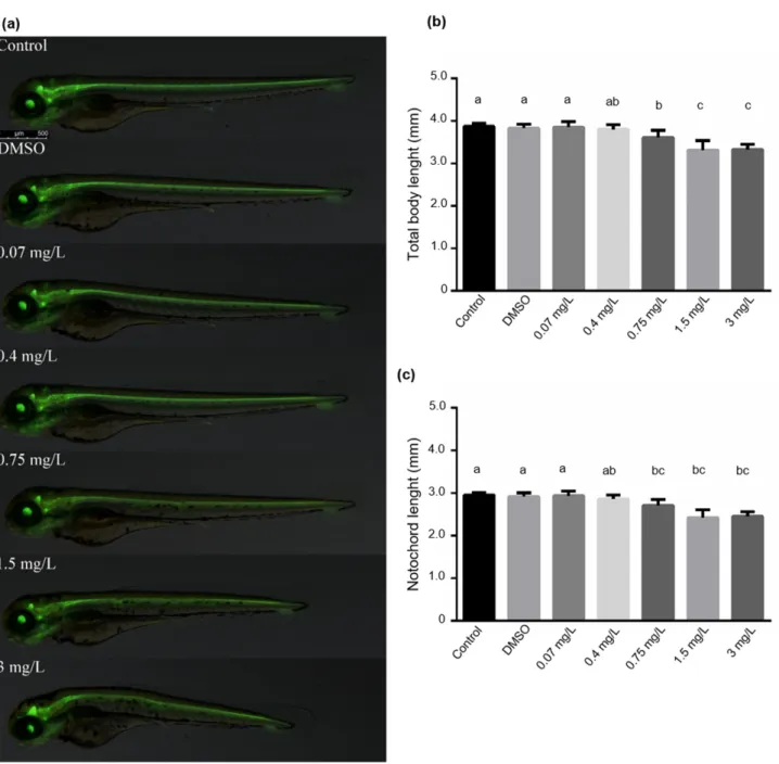
The results of zebrafish embryo acute toxicity tests prompted us to study the bendio- carb’s effects on the total body and notochord length. Since the zebrafish transgenic line Tg (shh:GFP) is useful for investigating the floor plate and the nervous system, we applied it to determine the total body- and notochord length. As expected, both body- and notochord length were significantly decreased above 0.75 mg/L bendiocarb concentration compared to the control groups (p< 0.05;N= 60). However, the development of the floor plate did not show any alterations (Figure2a–c).
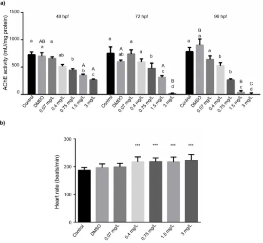
3.2. Acetylcholinesterase Activity and Heart Rate Assessments
In line with the previous studies, we detected significantly reduced AChE activ- ity at all of the three developmental stages including 48, 72, and 96 hpf, already at 0.75 mg/L bendiocarb concentration. The bendiocarb-induced inhibition proved to be concentration-dependent at every time points (48 h (p= 0.0086) EC10= 0.22±0.026 mg/L, EC50= 5.9±0.23 mg/L; 72 h (p= 0.0087) EC10= 0.084±0.011 mg/L, EC50= 1.1±0.099 mg/L; 96 h (p= 0.0019) EC10 = 0.076±0.081 mg/L, EC50 = 0.9±0.057 mg/L;N= 18) (Figure3a, Supplementary Table S1). We could also observe time dependency in bendiocarb exposure-induced inhibition of AChE activity (p= 0.02495, two-way ANOVA).
In contrast to the expected attenuation, we found significantly increased heart rate above 0.4 mg/L (0.75 mg/L, 1.5 mg/L and 3 mg/L) bendiocarb concentrations compared to the control groups (p< 0.05, Figure3b).
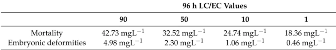
Table 1. Lethal concentration (LC) values for mortality and effective concentration (EC) values for embryonic deformities (tail malformations, reduced body length) induced by bendiocarb on zebrafish embryos exposed to various concentrations. Values are given in milligram per liter at 96 h post-fertilization for embryos.
96 h LC/EC Values
90 50 10 1
Mortality 42.73 mgL−1 32.52 mgL−1 24.74 mgL−1 18.36 mgL−1 Embryonic deformities 4.98 mgL−1 2.30 mgL−1 1.06 mgL−1 0.46 mgL−1
Water2021,13, 204 7 of 16
Water 2021, 13, x FOR PEER REVIEW 8 of 23
Figure 1. (a) Mortality and (b) embryonic deformities of zebrafish embryos exposed to various concentrations of
bendiocarb during a 96-h test. Error bars indicate the standard deviation of the actual endpoint. (c) Some of the detected abnormalities (jd: Not well identifiable jaws; yd: The shape of the yolk is deformed; sb: Shorter body; pe:
Pericardial edema; mm: The number and size of the irregular melanocytes; ie: Ventral and caudal fin with moderate, irregular edges; pdn: A portion of the notochord has a poorly defined structure; is: Somite boundaries
are slightly irregular) in larval zebrafish exposed to various concentrations of bendiocarb.
Figure 1.(a) Mortality and (b) embryonic deformities of zebrafish embryos exposed to various concentrations of bendiocarb during a 96-h test. Error bars indicate the standard deviation of the actual endpoint. (c) Some of the detected abnormalities (jd: Not well identifiable jaws; yd: The shape of the yolk is deformed; sb: Shorter body; pe: Pericardial edema; mm: The number and size of the irregular melanocytes; ie: Ventral and caudal fin with moderate, irregular edges; pdn: A portion of the notochord has a poorly defined structure; is: Somite boundaries are slightly irregular) in larval zebrafish exposed to various concentrations of bendiocarb.
Water2021,13, 204 8 of 16
Water 2021, 13, x FOR PEER REVIEW 9 of 23
The results of zebrafish embryo acute toxicity tests prompted us to study the bendiocarb’s effects on the total body and notochord length. Since the zebrafish transgenic line Tg (shh:GFP) is useful for investigating the floor plate and the nervous system, we applied it to determine the total body- and notochord length. As expected, both body- and notochord length were significantly decreased above 0.75 mg/L bendiocarb concentration compared to the control groups (p < 0.05; N = 60). However, the development of the floor plate did not show any alterations (Figure 2a–c).
Figure 2. (a) Photomicrographs taken at 72 hpf. (b) Total body length and (c) notochord lengths of zebrafish embryos exposed to various concentrations of bendiocarb during a 96-h test. Data represent the mean ± SD of two repeated tests. Columns superscripted with different letters are significantly different (One-way ANOVA followed by post-
hoc Tukey’s test; N = 30; p < 0.05).
3.2. Acetylcholinesterase Activity and Heart Rate Assessments
In line with the previous studies, we detected significantly reduced AChE activity at all of the three developmental stages including 48, 72, and 96 hpf, already at 0.75 mg/L bendiocarb concentration. The bendiocarb-induced inhibition proved to be concentration-dependent at every time points (48 h (p = 0.0086) EC
10= 0.22 ± 0.026 mg/L,
Figure 2. (a) Photomicrographs taken at 72 hpf. (b) Total body length and (c) notochord lengths of zebrafish embryos exposed to various concentrations of bendiocarb during a 96-h test. Data represent the mean±SD of two repeated tests.
Columns superscripted with different letters are significantly different (One-way ANOVA followed by post-hoc Tukey’s test;N= 30;p< 0.05).
Water2021,13, 204 9 of 16
Water 2021, 13, x FOR PEER REVIEW 10 of 23
EC50 = 5.9 ± 0.23 mg/L; 72 h (p = 0.0087) EC10 = 0.084 ± 0.011 mg/L, EC50 = 1.1 ± 0.099 mg/L; 96 h (p = 0.0019) EC10 = 0.076 ± 0.081 mg/L, EC50 = 0.9 ± 0.057 mg/L; N = 18) (Figure 3a, Supplementary Table S1). We could also observe time dependency in bendiocarb exposure-induced inhibition of AChE activity (p = 0.02495, two-way ANOVA).
In contrast to the expected attenuation, we found significantly increased heart rate above 0.4 mg/L (0.75 mg/L, 1.5 mg/L and 3 mg/L) bendiocarb concentrations compared to the control groups (p < 0.05, Figure 3b).
Figure 3. (a) Effects of bendiocarb exposure on AChE activity at 48, 72, and 96-hpf. Different lower-case letters indicate significant differences between concentrations within each time point (One-way ANOVA followed by post-hoc Tukey’s test; p < 0.05, N = 42). Different upper-case letters indicate significant differences between time
points within each concentration (Tukey-test after one-way ANOVA; p < 0.05). (b) The average heart rate of zebrafish exposed to various bendiocarb concentrations in a 48- h test. Data represent the mean ± SD of two repeated tests. Asterisks represent significant differences to control values (determined by Tukey-test after one-
way ANOVA; * p < 0.05, ** p < 0.01, and *** p < 0.001).
3.3. Determination of Biochemical Markers in Larval Fish
Figure 3.(a) Effects of bendiocarb exposure on AChE activity at 48, 72, and 96-hpf. Different lower-case letters indicate significant differences between concentrations within each time point (One-way ANOVA followed by post-hoc Tukey’s test;
p< 0.05,N= 42). Different upper-case letters indicate significant differences between time points within each concentration (Tukey-test after one-way ANOVA;p< 0.05). (b) The average heart rate of zebrafish exposed to various bendiocarb concentrations in a 48- h test. Data represent the mean±SD of two repeated tests. Asterisks represent significant differences to control values (determined by Tukey-test after one-way ANOVA; *p< 0.05, **p< 0.01, and ***p< 0.001).
3.3. Determination of Biochemical Markers in Larval Fish
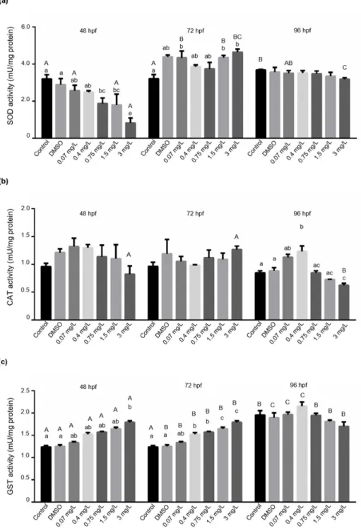
To further characterize the bendiocarb-induced harmful effects during zebrafish em- bryogenesis, we measured some enzyme activities, including SOD, CAT, GST, GPxSE, GPxTOT, and LPO participating in the biotransformation of various xenobiotics. Our analysis showed significant concentration-dependent inhibition of SOD activity at 48 hpf in bendiocarb-exposed zebrafish embryos (48 h (p= 0.03605) EC10= 0.62±0.094 mg/L, EC50= 0.79±0.084 mg/L). In contrast, significantly elevated levels of SOD activity were detected in embryos exposed to 0.07, 1.5, and 3 mg/L bendiocarb at 72 hpf. However, significant elevation of SOD activity was also detected in the DMSO exposed group com- pared to the control group at 72 hpf, suggesting that bendiocarb has a marginal effect on SOD activity at this time point. SOD activity values did not show any significant changes compared to controls at 96 hpf (Figure4a), but it was significantly increased in control group (p< 0.05) compared to 48 and 72 hpf controls.
CAT activity did not show any significant changes in bendiocarb exposed embryos at 48 and 72 hpf. However, CAT activity was significantly increased in larvae exposed to 0.4 mg/L bendiocarb and decreased in larvae subjected to 3 mg/L bendiocarb at 96 hpf (Figure4b). We found that the effects of bendiocarb administration on GST activity showed concentration and time dependency at 48 and 72 hpf (48 h (p< 0.04317) EC10= 6.76±0.36 mg/L, EC50= 6.92±0.13 mg/L; 72 h (p< 0.001) EC10= 2.38±0.088 mg/L, EC50= 2.7±0.046 mg/L). We observed a significant increase in GST activity in larvae exposed to 3 mg/L bendiocarb at
Water2021,13, 204 10 of 16
48 hpf. GST activity was also significantly (p< 0.05) increased above 0.4 mg/L bendiocarb concentrations at 72 hpf (Figure4c). Interestingly, both CAT and GST a reached the highest activity at 0.4 mg/L bendiocarb concentration, but these enzyme activities continuously dropped above 0.4 mg/L bendiocarb at 96 hpf. Nevertheless, despite the visible trends, significant (p< 0.05) differences and concentration dependence were only detectable in the case of CAT (Figure4b,c). Similar to 96 hpf SOD control levels, an elevated GST activity was detected in the case of 96 hpf controls (p< 0.001).
Water 2021, 13, x FOR PEER REVIEW 12 of 23
Figure 4. Effects of bendiocarb exposure on (a) superoxide dismutase (SOD) activity, (b) catalase (CAT) activity, and (c) Glutathione-S-transferase (GST) activity in a 48, 72 and 96-h test. Data represent the mean ± SD of two repeated experiments. Different lower-case letters indicate significant differences between concentrations within
each time point (Tukey-test after one-way ANOVA; p < 0.05; N = 42). Different upper-case letters indicate significant differences between time points within each concentration (Tukey-test after one-way ANOVA; p <
0.05).
Figure 4.Effects of bendiocarb exposure on (a) superoxide dismutase (SOD) activity, (b) catalase (CAT) activity, and (c) Glutathione-S-transferase (GST) activity in a 48, 72 and 96-h test. Data represent the mean±SD of two repeated experiments. Different lower-case letters indicate significant differences between concentrations within each time point (Tukey-test after one-way ANOVA;
p< 0.05;N= 42). Different upper-case letters indicate significant differences between time points within each concentration (Tukey-test after one-way ANOVA;p< 0.05).
Water2021,13, 204 11 of 16
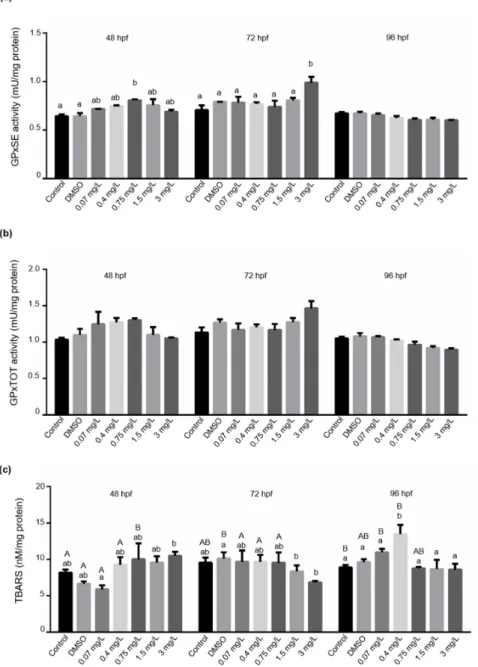
Both total glutathione peroxidase (GPx Tot) and Se-dependent glutathione peroxidase (GPx-Se) activities showed very similar patterns. Significantly increased GPx-Se activities were found in embryos only at 48 hpf after exposure to 0.75 mg/L bendiocarb and at 72 hpf after 3 mg/L exposure compared to control groups (Figure5a,b). Both, GPxTot and GPx-Se activities progressively diminished at 96 hpf, but the statistical analysis did not support this visible concentration-dependent trend.
Water 2021, 13, x FOR PEER REVIEW 14 of 23
Figure 5. Effects of bendiocarb exposure on (a) GPx-SE activity, (b) GPx-TOT activity, and (c) LPO levels expressed as TBARS after 48, 72, and 96 h. Data represent the mean ± SD of two repeated test series. Different lower-case
letters indicate significant differences between concentrations within each time point (Tukey-test after one-way ANOVA; p < 0.05; N = 42). Different upper-case letters indicate significant differences between time points
within each concentration (Tukey-test after one-way ANOVA; p < 0.05).
LPO levels did not show any significant changes at 48 and 72 hpf after the exposure to bendiocarb. Lipid
peroxidation was elevated significantly (p < 0.05) at 0.4 mg/L bendiocarb concentration at 96 hpf, but we could not detect any significant changes at higher concentrations. This peak coincides with the highest activity levels of GST and CAT at 96 hpf.
Figure 5.Effects of bendiocarb exposure on (a) GPx-SE activity, (b) GPx-TOT activity, and (c) LPO levels expressed as TBARS after 48, 72, and 96 h. Data represent the mean±SD of two repeated test series. Different lower-case letters indicate significant differences between concentrations within each time point (Tukey-test after one-way ANOVA;p< 0.05;N= 42). Different upper-case letters indicate significant differences between time points within each concentration (Tukey-test after one-way ANOVA;p< 0.05).
LPO levels did not show any significant changes at 48 and 72 hpf after the exposure to bendiocarb. Lipid peroxidation was elevated significantly (p< 0.05) at 0.4 mg/L ben- diocarb concentration at 96 hpf, but we could not detect any significant changes at higher concentrations. This peak coincides with the highest activity levels of GST and CAT at 96 hpf.
Water2021,13, 204 12 of 16
4. Discussion
Bendiocarb is a widely used broad-spectrum insecticide in agriculture and it is also recommended for malaria control by WHO [5–8]. However, the bendiocarb contamination- induced harmful effects are not completely investigated in non-target freshwater verte- brates. Therefore, we studied the adverse effects of bendiocarb in the zebrafish embryo model system, applying classical toxicological and biochemical approaches. In our exper- iments, LC50proved to be 32.52 mg/L for zebrafish embryos. This value is significantly higher than the previously identified LD50values for other fish species. [8,22]. Interestingly, these values fall in the range reported for small planktonic crustaceanDaphnia magnaLC50
ranging from 32 to 160 mg/L [46].
We found that bendiocarb induced various embryonal deformities, including tail malformations, reduced body length, and shape deformities of the yolk sac. In addition, bendiocarb exposure also influenced melanocyte migration, number, and size. Namely, de- creased melanocyte number and size were also found in fish exposed to higher bendiocarb concentrations, and melanocytes were not visible above 25 mg/L. Similar to bendiocarb- induced pigmentation defect, other carbamates, such as physostigmine and non-carbamate AChE inhibitors, like organophosphate metabolites could also modulate the melanocyte patterning and reduce pigmentation in zebrafish embryos [47]. However, it has been described that the xenobiotics-induced pigmentation defects are not restricted to the AChE inhibitors. Both fungicide boscalid and carrier solvent ethanol could reduce pigmentation, while penthiopyrad induced hyperpigmentation at the head and abdominal region in zebrafish embryos [48]. Overall, these findings indicate that the melanocyte number and distribution are sensitive to AChE inhibitors and other xenobiotics, but the exact molecular background of pigmentation defects remains uncertain.
It has been previously described that various carbamate compounds can reduce the body length in different fish species, including Japanese medaka (Oryzias latipes), zebrafish (Danio rerio) and Nile tilapia (Oreochromis niloticus) [49–51]. We found that both body and notochord lengths were significantly reduced above 0.75 mg/L, 1.5 mg/L, and 3 mg/L bendiocarb concentrations suggesting that bendiocarb similarly affects embryonal development in fish like other carbamates.
As with other carbamates, bendiocarb inhibits AChE activity by reversible carbamyla- tion of the esterase enzyme. This results in the accumulation of acetylcholine inducing mus- carinic, nicotinic, and central nervous system effects [2,5,14]. As expected, we could already detect significant AChE inhibition at 48 hpf above 0.4 mg/L bendiocarb concentration and the bendiocarb-induced AChE inhibition proved to be concentration and time-dependent.
Bendiocarb-inhibited AChE activity may result in pathological changes in heart rate of zebrafish embryos. It is well known that the first heartbeats of developing zebrafish are often irregular and arrhythmic, but it becomes specifically regular at around 36 hpf [52,53].
In zebrafish, the muscarinic M2 Ach receptor plays an essential role in controlling heart rate and is fully functional at this developmental stage. It has been described that the excessive stimulation of M2 Ach receptor by the continuous accumulation of acetylcholine in the synaptic gap leads to bradycardia [54–56]. However, stimulation of nicotinic receptors may result in tachycardia at lower test concentrations before bradycardia appears [57]. In the present study, tachycardia was observed associating with significantly increased heart rate above 0.4 mg/L (0.75 mg/L, 1.5 mg/L, and 3 mg/L) bendiocarb concentrations. Küster and Altenburger [58] reported very similar results studying aldicarb metabolite aldicarb- sulfoxide in zebrafish embryos. Lower concentrations of aldicarb-sulfoxide increased heart rates significantly, and bradycardia was only observed at higher concentrations. Our results suggest that bendiocarb exposure causes the nicotinic receptors’ stimulation in the applied concentration range leading to the observed tachycardia.
The biomarkers are useful tools to investigate the potential interactions between bi- ological, chemical, or physical environmental parameters and a biological system [59]
and play an important role in the evaluation of environmental contamination [60]. Fi- nally, biomarkers help to gain information about impaired organ functions and estimate
Water2021,13, 204 13 of 16
the organism’s damage level. Primary enzymatic markers of oxidative stress including catalase (CAT), superoxide dismutase (SOD), glutathione peroxidase (GPx), and lipid peroxidation (LPO) can reflect on oxidative cell damage. Glutathione-S-transferase (GST) is a phase II biotransformation enzyme, playing an important role in the excretion and detoxification process of xenobiotics, and it is also influenced by the organism’s peroxi- dation state through glutathione. It has been previously demonstrated that carbamates induce oxidative stress impairing mitochondrial functions. In addition, these chemicals also cause pathological changes in the neuronal and hormonal systems [60]. Recent studies demonstrated that bendiocarb modifies antioxidant and detoxifying enzyme activities in mammals [11,17,22,61,62]. Interestingly, we found significantly elevated SOD activity at the later developmental stages of untreated zebrafish larvae [63]. The significant inhibition of SOD activity was also detected at 48 hpf after exposure to 3 mg/L bendiocarb concentra- tion. However, this phenomenon was not observed at 72 and 96 hpf, suggesting that the enhanced enzyme synthesis capacity at the late developmental stages can compensate for the bendiocarb-induced reduction of SOD activity. Meng et al. [64] also detected elevated hepatic SOD activity in Nile tilapia (Oreochromis niloticus) following methomyl treatment indicating that the reduced SOD activity is not limited to bendiocarb among carbamate insecticides.
Here we found that CAT activity showed a specific pattern in bendiocarb-exposed zebrafish larvae at 96 hpf. We measured the maximally induced CAT activity at 0.4 mg/L bendiocarb exposure, which was significantly reduced at the higher concentrations. Car- bamate insecticide-induced CAT activity was also observed in the liver samples from methomyl-exposed Nile tilapia and carbaryl-exposed juvenile rainbow trout (Oncorhynchus mykiss) [64,65]. In contrast to the short 24 and 48 h carbaryl treatment-induced hepatic CAT activity, 96 h carbaryl exposure could attenuate it [65]. Overall, these findings suggest that carbamates’ effect on CAT activity is complex and may be concentration, cell, and exposure-time dependent.
It has been previously described that bendiocarb can enhance hepatic and renal GST activity in rats [21]. Similar to these findings, we also observed the bendiocarb concentration-dependent induction of GST activity in zebrafish embryos at 48 and 72 hpf.
Carbamate-induced GST activity was also observed in other fish-based model systems, including liver samples from juvenile rainbow trout following 24 h carbaryl treatment or methomyl-exposed Nile tilapia [52,66]. In contrast, 96-h carbaryl exposure could repress hepatic GST activity in juvenile rainbow trout [65]. In agreement with this finding, both, 96-h methomyl exposure in the topmouth gudgeon(Pseudorasbora parva), and 24-h car- bofuran exposure in mosquitofish (Gambusia yucatana) resulted in declined hepatic GST activities [66,67]. Taken together, these contrasting results indicate that carbamates’ effect on GST activity may be compound, fish species, and exposure-time dependent.
We observed that peroxidized lipid content showed a similar pattern as CAT activity in bendiocarb-exposed zebrafish larvae at 96 hpf. Maximal bendiocarb-mediated LPO level was detected at 0.4 mg/L and peroxidized lipid content was decreased at the higher con- centrations. Hernández-Moreno et al. [68,69] found a similar phenomenon in carbofuran exposed sea bass (Dicentrarchus labrax). Following 31, 63, 125, and 250 ug/L carbofuran exposure, LPO levels only showed a significant increase at 63 ug/L concentration. These results suggest that the peroxidized lipid content shows hormetic response to carbamates in various fish species.
Overall, our results show that bendiocarb—like other carbamates—causes adverse effects, including embryo deformities, reducing body- and notochord length, and inducing oxidative stress in aquatic vertebrates. Our lowest applied bendicarb concentrations are in the same range found in surface waters [22,23,25]. These low-dose bendiocarb-induced pathological processes may result in competitive disadvantage and impairing viability of fish larvae under natural conditions. Therefore, further evaluation of the ecotoxicological risks of bendiocarb is necessary and essential.
Water2021,13, 204 14 of 16
Supplementary Materials:The following are available online athttps://www.mdpi.com/2073-4 441/13/2/204/s1, Table S1: Results of Tukey test (after one-way ANOVA) on AChE data. Table S2:
Results of Tukey test (after one-way ANOVA) on GST data. Table S3: Results of Tukey test (after one-way ANOVA) on SOD data. Table S4: Results of Tukey test (after one-way ANOVA) on LPO data. Table S5: Results of Tukey test (after one-way ANOVA) on CAT data. Table S6: Results of Tukey test (after one-way ANOVA) on GPxSe.
Author Contributions:Conceptualization, A.Á., Z.C. and G.G.; investigation, A.Á., I.R.B., B.I., G.G.;
resources, B.I. and I.R.B.; writing—original draft preparation, A.Á. and G.G.; writing—review and editing, A.Á., Z.C.-B. and Z.C.; visualization, G.G. and A.Á.; supervision, B.U.; All authors have read and agreed to the published version of the manuscript.
Funding:This work was supported by the National Research, Development and Innovation Office (NKFIH) from the National Research, Development and Innovation Fund (NKFIA; Grant Agreement:
NVKP_16-1-2016-0023, EFOP-3.6.3-VEKOP-16-2017-00008) project co-financed by the European Union, and the Thematic Excellence Program-National Challenges Sub-program 2020 TKP2020-NKA- 16 of Szent István University, awarded by Ministry for Innovation and Technology. Project no. OTKA FK128705 has been implemented with the support provided by the National Research, Development and Innovation Fund of Hungary, financed under the FK 18 funding scheme. Z.C. is supported by the Premium Postdoc-toral Fellowship Program of the Hungarian Academy of Sciences and the Hungarian Scientific Research Found (OTKA FK132185).
Institutional Review Board Statement:Not applicable.
Informed Consent Statement:Not applicable.
Data Availability Statement:Data available in a publicly accessible repository.
Acknowledgments: The authors gratefully thank Marta Toth (University of Debrecen, Faculty of Medicine, Department of Immunology) for critical reading of the manuscript. Shh:GFP transgenic zebrafish line was (Provider: Uwe Strähle KIT) obtained from the European Zebrafish Resource Center of the Karlsruhe Intsitute of Technology (KIT).
Conflicts of Interest:The authors declare no conflict of interest.
References
1. Sogorb, A.M.; Vilanova, E. Enzymes involved in the detoxification of organophosphorus, carbamate and pyrethroid insecticides through hydrolysis.Toxicol. Lett.2002,128, 215–228. [CrossRef]
2. Krockova, J.; Massányi, P.; Toman, R.; Danko, J.; Roychoudhury, S. In vivo and in vitro effect of bendiocarb on rabbit testicular structure and spermatozoa motility.J. Environ. Sci. Health A2012,47, 1301–1311. [CrossRef] [PubMed]
3. Leong, K.H.; Benjamin Tan, L.L.; Mustafa, A.M. Contamination levels of selected organochlorine and organophosphate pesticides in the Selangor River, Malaysia between 2002 and 2003.Chemosphere2006,66, 1153–1159. [CrossRef] [PubMed]
4. Hallenbeck, W.H.; Cunningham-Burns, K.M.Pesticides and Human Health; Springer: New York, NY, USA, 1985.
5. Holovska, K.; Almasiova, V.; Cigankova, V. Ultrastructural changes in the rabbit liver induced by carbamate insecticide bendiocarb.
J. Environ. Sci. Health B2014,49, 616–623. [CrossRef]
6. Polláková, J.; Kovalkoviˇcová, N.; Csank, T.; Pistl, J.; Koˇcišová, A.; Legáth, J. Evaluation of bendiocarb cytotoxicity in mammalian and insect cell cultures.J. Environ. Sci. Health. B2012,47, 538–543. [CrossRef]
7. Sadasivaiah, S.; Tozan, Y.; Breman, J.G. Dichlorodiphenyltrichloroethane (DDT) for indoor residual spraying in Africa: How can it be used formalaria control?Am. J. Trop. Med. Hyg.2007,77, 249–263. [CrossRef]
8. Hayes, W.J.; Laws, E.R. Classes of pesticides. InHandbook of Pesticide Toxicology; Academic Press: New York, NY, USA, 1990;
Volume 3, p. 1576.
9. Chambers, H.W.; Boone, J.S.; Carr, R.L.; Chanbers, J.E. Chemistry of organophosphorus insecticides. InHandbook of Pesticide Toxicology, 2nd ed.; Robert, I.K., Ed.; Academic Press: San Diego, CA, USA, 2001; pp. 913–917.
10. Berman, T.; Hochner-Celnikier, D.; Barr, D.B.; Needham, L.L.; Amitai, Y.; Wormser, U.; Richter, E. Pesticide exposure among pregnant women in Jerusalem, Israel: Results of a pilot study.Environ. Int.2011,37, 198–203. [CrossRef]
11. Capcarova, M.; Petrovova, E.; Flesarova, S.; Dankova, M.; Massanyi, P.; Danko, J. Bendiocarbamate induced alterations in selected parameters of rabbithomeostasis after experimental peroral administration.Pestic. Biochem. Physiol.2010,98, 213–218. [CrossRef]
12. Whyatt, R.M.; Barr, D.B.; Camann, D.E.; Kinney, P.L.; Barr, J.R.; Andrews, H.F.; Hoepner, L.A.; Garfinkel, R.; Hazi, Y.; Reyes, A.; et al. Contemporary-use pesticides in personal air samples duringpregnancy and blood samples at delivery among urban minority mothers andnewborns.Environ. Health Perspect.2003,111, 749–756. [CrossRef]
13. Almasiova, V.; Holovska, K.; Tarabova, L.; Cigankova, V.; Lukacinova, A.; Nistiar, F. Structural and ultrastructural study of rabbit testes exposed tocarbamate insecticide.J. Environ. Sci. Health Part B2012,47, 1319–1328. [CrossRef]
Water2021,13, 204 15 of 16
14. Apaydin, F.G.; Pandir, D.; Kalender, S.; Ba¸s, H.; Kalender, Y. Hematoprotective effect of vitamins C and E against subchronic toxicity of bendiocarb: Biochemical evidences.J. Food Biochem.2018, 126–159.
15. Petrovova, E.; Massanyi, P.; Capcarova, M.; Zivcak, J.; Stodola, L. Structural alterations in rabbit spleen after bendiocarb administration.J. Environ. Sci. Health B2011,46, 788–792. [PubMed]
16. Petrovova, E.; Purzyc, H.; Mazensky, D.; Luptakova, L.; Torma, N.; Sopoliga, I.; Sedmera, D. Morphometric alterations, steatosis, fibrosis and activecaspase-3 detection in carbamate bendiocarb treated rabbit liver.Environ. Toxicol.2015,30, 212–222. [CrossRef] [PubMed]
17. Flesarova, S.; Lukac, N.; Danko, J.; Massanyi, P. Bendiocarbamate inducedstructural alterations in rabbit thymus after experimental peroraladministration.J. Environ. Sci. Health Part B2007,42, 329–334. [CrossRef]
18. Baron, R.L. Carbamate insecticides. InHandbook of Pesticide Toxicology; Hayes, W.J., Laws, E.R., Eds.; Academic Press: New York, NY, USA, 1991; pp. 3–6. ISBN 978-0123341600.
19. Lazartigues, E.; Freslon, J.L.; Tellioglu, T. Pressor and bradycardic effects of tacrine and other acetylcholinesterase inhibitors in the rat.Eur. J. Pharmacol.1998,361, 61–71. [CrossRef]
20. Masuda, Y.; Kawamura, A. Acetylcholinesterase inhibitor (donepezil hydrochloride) reduces heart rate variability.J. Cardiovasc.
Pharmacol.2003,41(Suppl. 1), S67–S71.
21. Sobeková, A.; Holovská, K.; Lenártová, V.; Flešárová, S.; Javorský, P. The another toxic effect of carbamate insecticides.Acta Biol.
Hung.2009,60, 45–54. [CrossRef]
22. Lahr, J.; Diallo, A.O.; Gadji, B.; Diouf, P.S.; Bedaux, J.J.M.; Badji, A.; Ndour, K.B.; Andreasen, J.E.; Straalen, N.M.V. Ecological effects of experimental insecticide applications on invertebrates in sahelian temporary ponds.Environ. Toxicol. Chem.2000,19, 1278–1289. [CrossRef]
23. Bendiocarb (Technical Fact Sheet) NPIC Technical Fact Sheets. 2002. Available online:http://npic.orst.edu/factsheets/archive/
bendiotech.pdf(accessed on 30 December 2020).
24. Tatarazako, N.; Iguchi, T. Evaluation of toxicities of herbicides using short-term chronic tests of alga, daphnid and fish, herbicides.
InHerbicides—Environmental Impact Studies and Management Approaches; Alvarez-Fernandez, R., Ed.; InTech: Rijeka, Croatia, 2012;
pp. 135–166.
25. Scott, G.I.; Fulton, M.H.; Moore, D.W.Agricultural Insecticide Runoff Effects on Estuarine Organisms: Correlating Laboratory and Field Toxicity Testing with Ecotoxicological Biomonitoring; Report #CR813138-01-1; U.S. National Marine Fisheries Service, Southeast Fisheries Science Center, Charleston Laboratory: Charleston, SC, USA, 1990.
26. Teles, M.; Pacheco, M.; Santos, M.A. Endocrine and metabolic responses of Anguilla anguilla L. caged in a freshwater-wetland (Pateira de Fermentelos-Portugal).Sci. Total Environ.2007,372, 562–570. [CrossRef]
27. El-Saeid, M.H.; Al-Turki, A.M.; Al-Wable, M.I.; Abdel-Nasser, G. Evaluation of pesticide residues in saudi arabia ground water.
Res. J. Environ. Sci.2011,5, 171–178.
28. Spalding, R.F.; Exner, M.E.; Snow, D.D.; Cassada, D.A.; Burbach, M.E.; Monson, S.J. Herbicides in ground water beneath Nebraska’s management systems evaluation area.J. Environ. Qual.2003,32, 92–99. [CrossRef] [PubMed]
29. Leu, C.; Singer, H.; Müller, S.R.; Schwarzenbach, R.P.; Stamm, C. Comparison of atrazine losses in three small headwater catchments.J. Environ. Qual.2005,34, 1873–1882. [CrossRef] [PubMed]
30. Senger, M.R.; Rico, E.P.; de Bem Arizi, M.; Rosemberg, D.B.; Dias, R.D.; Bogo, M.R. Carbofuran and malathion inhibit nucleotide hydrolysis in zebrafish (Danio rerio) brain membranes.Toxicology2005,212, 107–115. [CrossRef] [PubMed]
31. Hadzhiev, Y.; Lele, Z.; Schindler, S.; Wilson, S.W.; Ahlberg, P.; Strähle, U.; Müller, F. Hedgehog signaling patterns the outgrowth of unpaired skeletal appendages in zebrafish.BMC Dev. Biol.2007,27, 7–75. [CrossRef] [PubMed]
32. Müller, F.; Chang, B.; Albert, S.; Fischer, N.; Tora, L.; Strahle, U. Intronic enhancers control expression of zebrafish sonic hedgehog in floor plate and notochord.Development1999,126, 2103–2116.
33. Shkumatava, A.; Fischer, S.; Muller, F.; Straehle, U.; Neumann, C.J. Sonic Hedgehog, secreted by amacrine cells, acts as a short range signal to direct differentiation and lamination in the zebrafish retina.Development2004,131, 3849–3858. [CrossRef]
34. OECD.OECD Guidelines for the Testing of Chemicals, Section 2; Test No. 236: Fish Embryo Acute Toxicity (FET) Test; OECD Publishing: Paris, France, 2013.
35. Rasband, W.S.; ImageJ, U.S. National Institutes of Health, Bethesda, Maryland, USA, 1997–2018. Available online: https:
//imagej.nih.gov/ij/(accessed on 30 December 2020).
36. Bradford, M. A rapid and sensitive assay of protein utilizing the principle of dye binding.Anal. Biochem.1976,772, 242–264.
37. Wills, E.D. Evaluation of lipid peroxidation in lipids and biological membranes. InBiochemical Toxicology: A Practical Approach;
Snell, K., Mullock, B., Eds.; IRL Press: Washington, DC, USA, 1987; pp. 127–152.
38. Ellman, G.L.; Courtney, K.D.; Andres, V.; Featherstone, R.M. A new and rapid colorimetric determination of acetylcholinesterase activity.Biochem. Pharmacol.1961,7, 88–95. [CrossRef]
39. Guilhermino, L.; Lopes, M.C.; Carvalho, A.P.; Soares, A.M.V.M. Inhibition of acetylcholinesterase activity as effect criterion in acute tests with juvenile Daphnia magna.Chemosphere1996,32, 727–738. [CrossRef]
40. Aebi, H. Catalase in Vitro.Meth. Enzymol.1984,105, 121–126.
41. Habig, W.H.; Pabst, M.J.; Jakoby, W.B. Glutathione S-transferases—The first enzymatic step in mercapturic acid formation.J. Biol.
Chem.1974,249, 7130–7139. [CrossRef]
42. Paglia, D.E.; Valentin, W.N. Studies on quantitative and qualitative characterization of erythrocyte glutathione peroxidase.J. Lab.
Clin. Med.1967,70, 158–162. [PubMed]
Water2021,13, 204 16 of 16
43. Lawrence, R.A.; Burk, R.F. Glutathione peroxidase activity in selenium deficient rat liver.Biochem. Biophys. Res. Commun.1976, 71, 952–958. [CrossRef]
44. Faria, M.; Carrasco, L.; Diez, S.; Riva, M.C.; Bayona, J.M.; Barata, C. Multi-biomarker responses in the freshwater mussel Dreissena polymorpha exposed to polychlorobiphenyls and metals.Comp. Biochem. Phys. C2009,149, 281–288. [CrossRef] [PubMed]
45. Crapo, J.D.; McCord, J.M.; Fridovich, I. Preparation and assay of superoxide dismutases.Methods Enzymol.1978,53, 382–393.
[PubMed]
46. Visser, J.T.; Linders, J.Bendiocarb. Milieu-Fiche; Advisory Report 90/670101/007; College voor de Toelating van Bestrijdingsmidde- len: Wageningen, The Netherlands, 1999.
47. Fischer, A.; Wolman, M.; Granato, M.; Parsons, M.; McCallion, A.-S.; Proescher, J.; English, E. Carbamate nerve agent prophylatics exhibit distinct toxicological effects in the zebrafish embryo model.Neurotoxicol. Teratol.2015,50, 1–10. [CrossRef]
48. Qian, L.; Qi, S.; Cao, F.; Zhang, J.; Li, C.; Song, M.; Wang, C. Effects of penthiopyrad on the development and behaviour of zebrafish in early-life stages.Chemosphere2019,214, 184–194. [CrossRef]
49. Kashiwada, S.; Tatsuta, H.; Kameshiro, M.; Sugaya, Y.; Sabo-Attwood, T.; Chandler, G.T.; Ferguson, P.L.; Goka, K. Stage-dependent differences in effects of carbaryl on population growth rate in japanese medaka (Oryzias latipes).Environ. Toxicol. Chem.2008,27, 2397–2402. [CrossRef]
50. Mensah, P.K.; Okuthe, G.H.; Onani, M. Sublethal effects of carbaryl on embryonic and gonadal developments of zebrafishDanio rerio.Afr. J. Aquat. Sci.2012,37, 271–275. [CrossRef]
51. Meng, S.L.; Chen, J.Z.; Hu, G.D.; Song, C.; Fan, L.M.; Qiu, L.P.; Xu, P. Effects of chronic exposure of methomyl on the antioxidant system in liver of Nile tilapia (Oreochromis niloticus).Ecotoxicol. Environ. Saf.2014,101, 1–6. [CrossRef]
52. Kimmel, C.B.; Ballard, W.W.; Kimmel, S.R.; Ullmann, B.; Schilling, T.F. Stages of embryonic development of the zebrafish.Dev.
Dyn.1995,203, 253–310. [CrossRef] [PubMed]
53. Barrionuevo, W.R.; Burggren, W.W. O2consumption and heart rate in developing zebrafish (Danio rerio): Influence of temperature and ambient O2.Am. J. Physiol.1999,276, R505–R513. [CrossRef] [PubMed]
54. Watson, F.L.; Schmidt, H.; Turman, Z.K.; Hole, N.; Garcia, H.; Gregg, J.; Tilghman, J.; Fradinger, E.A. Organophosphate pesticides induce morphological abnormalities and decrease locomotor activity and heart rate inDanio rerioand Xenopus laevis.Environ.
Toxicol. Chem.2014,33, 1337–1345. [CrossRef] [PubMed]
55. Lin, C.C.; Hui, M.N.; Cheng, S.H. Toxicity and cardiac effects of carbaryl in early developing zebrafish (Danio rerio) embryos.
Toxicol. Appl. Pharmacol.2007,222, 159–168. [CrossRef]
56. Hsieh, D.J.; Liao, C.F. Zebrafish M2 muscarinic acetylcholine receptor: Cloning, pharmacological characterization, expression patterns and roles in embryonic bradycardia.Br. J. Pharmacol.2002,137, 782–792. [CrossRef]
57. Ecobichon, D.J. Carbamate insecticides. InHandbook of Pesticide Toxicology-Agents, 2nd ed.; Krieger, R., Ed.; Academic Press: San Diego, CA, USA, 2001; pp. 1087–1106.
58. Küster, E.; Altenburger, R. Suborganismic and organismic effects of aldicarb and its metabolite aldicarb-sulfoxide to the zebrafish embryo (Danio rerio).Chemosphere2007,68, 751–760. [CrossRef]
59. WHO/IPCS.Environmental Health Criteria 155. Biomarkers and Risk Assessment: Concepts and Principles; IPCS/World Health Organization: Geneva, Switzerland, 1993.
60. Filho, D.W. Fish antioxidant defences—A comparative approach.Braz. J. Med. Biol. Res.1996,29, 1735–1742.
61. Apaydin, F.G.; Bas, H.; Kalender, S.; Kalender, Y. Bendiocarb induced histopathological and biochemical alterations inr at liver and preventive role of vitamins C and E.Environ. Toxicol. Pharmacol.2017,49, 148–155. [CrossRef]
62. Karami-Mohajeri, S.; Abdollahi, M. Toxic influence of organophosphate, carbamate, and organochlorine pesticides on cellular metabolism of lipids, proteins, and carbohydrates: A systematic review.Hum. Exp. Toxicol.2011,30, 1119–1140. [CrossRef]
63. Peterman, E.M.; Sullivan, C.; Goody, M.F.; Rodriguez-Nunez, I.; Yoder, J.A.; Kim, C.H. Neutralization of mitochondrial superoxide by superoxide dismutase 2 promotes bacterial clearance and regulates phagocyte numbers in zebrafish.Infect. Immun.2015,83, 430–440. [CrossRef]
64. Meng, S.L.; Chen, J.Z.; Xu, P.; Qu, J.H.; Fan, L.M.; Song, C.; Qiu, L.P. Hepatic antioxidant enzymes SOD and CAT of Nile tilapia (Oreochromis niloticus) in response to pesticide methomyl and recovery pattern.Bull. Environ. Contam. Toxicol.2014,92, 388–392.
[CrossRef] [PubMed]
65. Ferrari, A.; Venturino, A.; Pechén de D’Angelo, A.M. Effects of carbaryl and azinphos methyl on juvenile rainbow trout (Oncorhynchus mykiss) detoxifying enzymes.Pestic. Biochem. Physiol.2007,88, 134–142. [CrossRef]
66. Li, H.; Jiang, H.; Gao, X.; Wang, X.; Qu, W.; Lin, R.; Chen, J. Acute toxicity of the pesticide methomyl on the topmouth gudgeon ((Pseudorasbora parva): Mortality and effects on four biomarkers.Fish. Physiol. Biochem.2008,34, 209–216. [CrossRef] [PubMed]
67. Rendón-von Osten, J.; Ortíz-Arana, A.; Guilhermino, L.; Soares, A.M. In vivo evaluation of three biomarkers in the mosquitofish (Gambusia yucatana) exposed to pesticides.Chemosphere2005,58, 627–636. [CrossRef]
68. Hernández-Moreno, D.; Soler, F.; Míguez, M.P.; Pérez-López, M. Brain acetylcholinesterase, malondialdehyde and reduced glutathione as biomarkers of continuous exposure of tench, Tinca tinca, to carbofuran or deltamethrin.Sci. Total Environ.2010, 408, 4976–4983. [CrossRef]
69. Hernández-Moreno, D.; Pérez-López, M.; Soler, F.; Gravato, C.; Guilhermino, L. Effects of carbofuran on the sea bass (Dicentrarchus labraxL.): Study of biomarkers and behaviour alterations.Ecotoxicol. Environ. Saf.2011,74, 1905–1912. [CrossRef]