Contents lists available atScienceDirect
Journal of Magnetism and Magnetic Materials
journal homepage:www.elsevier.com/locate/jmmm
Research articles
Magnetic properties and heating e ffi cacy of magnesium doped magnetite nanoparticles obtained by co-precipitation method
Vladan Kusigerski
a,⁎, Erzsebet Illes
a,1, Jovan Blanusa
a, Saso Gyergyek
b, Marko Boskovic
a, Marija Perovic
a, Vojislav Spasojevic
aaCondensed Matter Physics Laboratory, The Vinca Institute, University of Belgrade, 11001 Belgrade, Serbia
bDepartment for Materials Synthesis, Jozef Stefan Institute, 1000 Ljubljana, Slovenia
A R T I C L E I N F O
Keywords:
Nanostructured materials Oxide materials Ferrofluids Magnetic properties Magnetic hyperthermia
A B S T R A C T
Ferrofluids based on magnesium substituted magnetite nanoparticles MgxFe3−xO4(x = 0.1; 0.2; 0.4) were synthesised by a chemical co-precipitation method. Their physical properties have been compared with those of the magnetite based ferrofluid obtained by the same synthesis route. Both XRD and TEM studies showed particle size decrease with the increased Mg content while DLS experiments pointed to the more prominent aggregation of Mg-containing nanoparticles. Magnetic properties investigation conducted on the powder (i.e. dried) speci- mens showed decrease of magnetization values with increased Mg content except for the lowest concentration of x = 0.1 where substantial saturation magnetization rise of about 40% was recorded at room temperature.
Heating abilities of the studied ferrofluids under the applied ACfields (SAR values) also showed decreasing trend with the increased Mg content even for x = 0.1 sample despite its elevated magnetization value. This trend has been understood as a consequence of the changed intrinsic nanoparticle properties such as size and magnetic anisotropy, as well as contribution of a collective behaviour due to an increased nanoparticle aggregation in Mg- doped systems.
1. Introduction
Nanoparticle magnetic materials have provoked an enormous re- search interest in the last few decades due to the emergence of new magnetic phenomena compared to their bulk counterparts. Properties like superparamagnetism, increased anisotropy (i.e. high coercivity), exchange bias (intra-particle interactions), and collective phenomena (inter-particle interactions) proved to be of interest regarding both basic science and vast number of technological applications including biomedicine, data storage, magnetic sensing, environmental remedia- tion, etc.[1–3].
Due to the reduction of a particle size down to a typical size of biological entities the application of magnetic nanomaterials in bio- medicine is considered among the most promising ones, compre- hending cell separation, contrast agents, targeted drug delivery, mag- netic hyperthermia, tissue engineering, magnetic biosensors, etc.[4–7].
For these applications stable suspension of magnetic nanoparticles in a liquid medium is required, commonly referred to as magnetic colloid or ferrofluid. Of a critical importance for clinical use in diagnostics and
therapy is material biocompatibility, and so far iron-oxides based ma- terials are the only FDA and EMA approved2magnetic nanomaterials for such purposes [8]. Among all iron-oxides, magnetite Fe3O4 and maghemite (γ-Fe2O3) have been most thoroughly studied so far due to their superior magnetic properties[9,10].
On the other hand, particle nanosizing can also cause a certain drawbacks among which the most important is a magnetization re- duction due to the existence of a disordered nanoparticle surface layer, usually referred to as a particle shell[11]. One possibility to compen- sate for this reduction is to increase intrinsic magnetization of the material by controlling its composition and/or its crystal structure parameters. Well known materials convenient for such manipulations are spinel ferrites with chemical formula (X2+1−δFe3+δ )[X2+δ Fe3+2−δ]O4, where X denotes divalent cation while parentheses and square brackets denote tetrahedral A and octahedral B crystal sites, respectively[12].
Cation inversion degreeδcan take any value in the range 0 <δ< 1, where 0 and 1 denotes normal or inverse spinel, respectively. Mag- netism of spinel ferrites is determined by both cation composition and their distribution over A and B sites (i.e. inversion degreeδ), and thus
https://doi.org/10.1016/j.jmmm.2018.11.127
Received 15 August 2018; Received in revised form 26 October 2018; Accepted 29 November 2018
⁎Corresponding author.
E-mail address:vladank@vinca.rs(V. Kusigerski).
1Present address: Department of Physical Chemistry and Materials Science, University of Szeged, H-6720 Szeged, Hungary.
2FDA - Food and Drug Agency; EMA - European Medicines Agency.
Available online 30 November 2018
0304-8853/ © 2018 Elsevier B.V. All rights reserved.
T
many research studies have been devoted to correlations between these parameters and magnetic properties. In case of a nanoparticle magne- tite, which in ideal case is considered as an inverse spinel (Fe3+) [Fe2+Fe3+]O4, numerous investigations were dealing with partial substitution of iron ions for a variety of other ions: magnetic 3d ions (Ni, Co, Mn, V)[13–18]or 4f ions (Gd, Dy, Ho, Tm, Yb)[19,20], or nonmagnetic (i.e. diamagnetic) ions (Y, Ga, Zn)[21–24]. In addition, for the preparation of the above mentioned compounds many different synthesis techniques have been applied, including co-precipitation, thermal decomposition, mechanochemistry, hydrothermal route, sol- gel, and microemulsion [13–24]. Direct comparison of the obtained results is difficult since the applied synthesis method has a decisive impact on both cation distribution and nanoparticle size, and they both influence magnetic properties. This is especially true in case when magnetic ions are used as dopants since their magnetic moments and exchange interaction affinities become additional parameters that should be taken into account.
In case of nonmagnetic ions doping, it turns out that a notable in- crease of magnetization could be obtained in case of preferential oc- cupation of tetrahedral sites by a substituent cations, and so far the best results have been achieved in case of Zn2+[23,25]and Ga3+[22]ions.
In contrast, magnetization reduction has been observed for Y3+ions which preferentially occupy octahedral sites in the spinel structure [21]. These results could be understand by adopting the simple model of Néel[26]in which magnetic ordering of spinels is governed by A-O- A, A-O-B and B-O-B superexchange interactions only. These interactions force an antiparallel spin orientation of iron ions, so Fe3+atoms in A and B sites of magnetite compensate each other, and consequently the net magnetization originates from uncompensated Fe2+moments. By replacing part of Fe3+ ions by diamagnetic ions number of un- compensated spins increases thus elevating the net magnetization of the system.
Magnesium ion Mg2+is a promising candidate for Fe3O4doping because its ionic radius in tetrahedral surrounding (0.57) is closer to A- site Fe3+radius (0.49) compared to tetrahedral Zn2+radius (0.60)[27]
while biocompatibility reasons further justify its convenience. Studies of the nanoparticle magnesium ferrite MgFe2O4have shown that the degree of inversion depended on the synthesis method, and that it might be influenced by processing parameters [28–30]. At the same time heating performances in the applied ACfields also depended on the synthesis pathway, and in some cases they were superior compared to nanoparticle magnetite. However, despite promising properties of Mg2+ion, to our best knowledge there has been just a single research devoted to magnesium doping of nanosized magnetite with the focus on its applicability as anode material in lithium batteries[31].
In this work we have investigated effects of magnesium substitution for iron in nanoparticle magnetite MgxFe3−xO4obtained by a chemical co-precipitation for three concentrations x = 0.1, 0.2 and 0.4. Research comprehended samples' structure/microstructure, magnetic properties and heating efficacy in AC magnetic fields, with the magnetic hy- perthermia as a targeted application in mind.
2. Experimental
All studied samples (magnetite and Mg doped magnetite with var- ious magnesium content) were synthesized by a chemical reverse co- precipitation method. Analytical grade chemicals, MgSO4*7 H2O (Merck), (NH4)2Fe(SO4)2*6 H2O (Sigma-Aldrich), FeCl3*6 H2O (Sigma- Aldrich) and NaOH (Carlo Erba) were used. Calculated amounts of MgCl2, (NH4)2Fe(SO4)2and FeCl3were dissolved in 10 mL of distilled water in separate beakers, then mixed together and added to tempered 0.4 M NaOH solution during vigorous stirring. Firstly, few drops of the mixture of the metal salts were put to induce the nucleus formation, and then the whole volume of the solution was slowly poured into the balloon. The slurry was kept at 80 °C for 2 h under continuous stirring.
After cooling the mixture down to room temperature, the formed dark
brown product was washed several times with distilled water combined with magnetic separation. Finally the pH of the slurry was set down to pH∼2 and the nanoparticles were dialysed against 0.001 M HCl so- lution to eliminate the excess salt left from the synthesis. The final product was stored in ferrofluid form in a fridge at pH∼4 until further use. Samples were labelled as S0, S10, S20 and S40, according to their Mg-content of 0, 10, 20 and 40%, respectively. The concentration of each suspension was determined by gravimetric method and found to be 8.80, 3.10, 3.13, and 9.20 g/l for samples S0, S10, S20 and S40, respectively. The powder counterparts of the studied samples were obtained by drying the synthesized suspensions.
To investigate crystal structure and phase composition of the ob- tained samples powder X-ray diffraction data were recorded in Bragg- Brentano geometry on a Rigaku Smartlab 3 kW powder diffractometer.
The setup included Cu Kα1/2radiation, passive Ni-filter, 0.5° divergent slit, and DTex Ultra 250 detector. The DTex detector was used in narrow energy gap mode in order to suppress ironfluorescence back- ground originating from the copper radiation. The so-collected X-ray diffraction data were analyzed by using FullProf program[32]in profile matching mode (LeBailfit). For particle size estimation, instrumental broadening (resolution function) was extracted from LaB6 standard diffraction pattern.
Particle size and morphology were investigated by using JEOL‐JEM 2100 high resolution transmission electron microscope operating at 200 KV. In the process of samples preparation for TEM ferrofluids werefirst diluted by 0.001 M HCl solution in order to prevent particle aggregation by keeping the suspension pH in the acidic region. The so-diluted so- lutions were deposited on a nickel supported carbonfilm and dried in air. Besides recording TEM micrographs, SAED images were also made to check a crystal phase purity.
The pH-dependent surface charging, the hydrodynamic size and particle aggregation of the studied samples were characterized by Malvern ZetaSizer NANO ZS90 device which utilizes 90° scattering optics and a 4 mW He-Ne laser source (λ= 633 nm). Hydrodynamic size was determined by dynamic light scattering (DLS) using poly- styrene cuvettes in dilute sols of ferrofluids from pH∼3 up to pH∼10 in the presence of 10 mM NaCl. The pH was adjusted by adding 0.01 M HCl or NaOH solutions. Z-average hydrodynamic diameter and poly- dispersity index (PDI) were obtained by cumulant analysis of the cor- relation function. Electrokinetic potential measurements were per- formed by laser Doppler electrophoresis on the same samples using in DTS 1071 disposable zeta cells. The Zetasizer software automatically calculated the zeta potentials from the measured electrophoretic mo- bility values applying the Smoluchowski equation, assuming, that K·a> > 1 (whereKandaare the Debye-Hückel parameter and particle radius, respectively). The accuracy of the electrokinetic potential de- termination was ± 5 mV.
Magnetic measurements comprehending temperature dependence of magnetization in a zero-field-cooled (ZFC) andfield-cooled (FC) re- gimes as well asfield dependence of isothermal magnetization were done on a commercial SQUID-based magnetometer Quantum Design MPMS XL-5. Research on the heating abilities of the investigated fer- rofluids under the applied AC magneticfields has been performed on a commercial nB nanoScale Biomagnetics DM1 device equipped with the fibre optics temperature sensor. Measurements of the ferrofluid tem- perature change with time were made under non-adiabatic conditions in a wide range of field amplitudes (50–300 Oe) and frequencies (252–808 kHz).
3. Results and discussion
3.1. 1 Crystal structure and morphology
The recorded X-ray diffraction patterns are presented in Fig. 1.a, which confirms that reflections broadening were proportional to Mg content. All diffraction peaks were indexed within magnetite spinel
structure (space group Fd-3 m), and no other phases have been ob- served. As an example, one of the refined patterns (for x = 0.4) is shown separately inFig. 1.b. The obtained cell parameters and particle sizes from LeBailfit are summarized inTable 1.
By inspection of Table 1it could be noticed that the biggest dif- ference in cell parameterais between S0 and S10 samples. The mag- nitude of this change diminishes with Mg content increase and becomes almost negligible for samples S20 and S40. The average domain size D decreases from about 8 nm for S0 sample to below 4 nm for S40 sample.
Such a drop in particle size by iron substitution in magnetite has been already reported in several cases for different dopant ions, such as Zn [23,25], Mg[31], Y[21]and Ni[14]. In addition, the certain degree of anisotropy found over different crystallographic directions suggests that particles slightly deviate from spherical shape, which is also con- firmed by TEM observations (see below).
TEM micrographs of samples S0, S10 and S20 with corresponding selected area electron diffraction (SAED) patterns are shown in Fig. 2(a)–(c), respectively. Reduction of nanoparticle size with in- creasing Mg content is apparent, while the average particle size and size distribution were determined by using the imageJ program. The dia- meter of at least 100 particles per sample was measured andfitted to a log normal distribution (insets in TEM micrographs inFig. 2). The so- obtained average diameters and standard deviations are given in Table 1from which the wide size distribution of particles could be seen.
Bigger particle diameters obtained by TEM compared to XRDfindings is not uncommon, and it is usually ascribed to poorly crystallized layer on the particle surface. It could be also noticed that nanoparticle shapes span from almost spherical to simple polyhedrons, which is in line with the X-rayfindings on the crystalline anisotropy, also given inTable 1.
SAED patterns confirm that crystal structure is of the cubic spinel type for all the samples without presence of other crystal phases.
3.2. Hydrodynamic size, surface charging and particle aggregation Intensity distribution of particle sizes obtained from DLS experi- ments at pH∼4 are shown inFig. 3, while maximums of hydrodynamic diameter values together with Z-average hydrodynamic diameters and PDI values are summarized inTable 2. All Mg-doped samples showed larger average hydrodynamic diameter (above 100 nm) compared to the non-substituted one (∼44 nm), although the PDI values remained constant (∼0.2) thus indicating a moderate polydispersity of the na- noparticles. The largest hydrodynamic diameter was obtained for S20 sample. Literature data is scarce but we could compare the obtained values for Mg substituted samples to the hydrodynamic diameter of approximately 146 nm determined for magnesium ferrite[33].
In aqueous media metal oxide nanoparticles are hydrated, and amphoteric≡Me-OH surface sites may exist on their surface in proto- nated or deprotonated state depending on the pH of the system[34].
This can be represented as:
≡Me−OH+H+ ⇌ ≡Me−OH+2 (1)
≡ − ⇌ ≡ − + ≡ − + ⇌
≡ − +
− + −
−
Me OH Me O H or Me OH OH
Me O H O2 (2)
where Me stands for either iron or magnesium. The pH-dependent surface charging of S0 and S10 ferrofluid samples studied from pH∼3 up to pH∼10 in the presence of 10 mM NaCl can be seen inFig. 4. It can be seen that nanoparticles in both samples have positive charge below pH∼7, while they are negatively charged above pH∼8. At low pHs positively charged≡Me–OH+2 sites form due to the protonation of the surface sites. An increase of pH leads to gradual decrease of the zeta potential and it becomes zero around pH∼8, indicating the isoelectric point (IEP), i.e. the pH value where the charge reversal takes place. At pHs higher than∼8 the zeta potential is negative, because the surface active sites are deprotonated and ≡Me–O−sites are present on the particle’s surface. For undoped magnetite the pHIEPwas∼7.9 which is in good accordance with the published data[34,35], while moderate reduction down to∼7.3 could be observed for S10 sample. Similar reduction of pHIEPwas obtained for Mg-ferrite with approximately 10%
Mg content[36]. The reduced pHIEPcould be explained by the presence of Mg, since for nanoparticle Mg-ferrite a value of pHIEP∼7 was ob- served[33].
The pH-dependent aggregation behaviour of S10 sample follows the change of the zeta potential as shown inFig. 4. Far from the pHIEPvalue (i.e. in the range 3 < pH < 5) the measured size is about 120 nm due to the electrostatic stabilization arising from the strong repulsion Fig. 1.Diffraction patterns for MgxFe3−xO4samples: (a) Broadening of the diffraction peaks with the increasing Mg content; (b) Refined pattern (line) and ex- perimental data (symbols) for x = 0.40 sample: line at the bottom represents the difference between experimental and calculated values.
Table 1
Cell parameter a and average domain size D obtained by refinement of X-ray powder data for MgxFe3−xO4samples. Particle sizes determined from TEM micrographs (see text) are given in the last column.
Sample a (Å) D (nm)–XRD D (nm)–TEM
S0 8.359(1) 7.8(9*) 10 (2)
S10 8.366(1) 6.1(1.7*) 9 (2)
S20 8.368(2) 3.8(9*) 7 (2)
S40 8.369(1) 3.4(7*) 6 (2)
* Represents the degree of anisotropy, not the standard deviation.
between the highly positively or negatively charged particles. Around the IEP (i.e. in the range from pH∼6 to∼9) the absolute value of the electrokinetic potential is small (or zero), which leads to particle ag- gregation resulted in large size values (> 1000 nm) and thus pointing to the compromised ferrofluid stability.
3.3. Static magnetic properties
Magnetic field dependence of isothermal magnetization of the powder specimens recorded at the room temperature is shown in Fig. 5a. Coercivefield values HCof all the samples are less than 10 Oe Fig. 2.TEM micrograph and respective SAED patterns for samples S0 (a), S10 (b) and S20 (c). Insets in TEM micrographs represent particle size distributions while lines show bestfit to a log-normal distribution.
Fig. 3.Intensity distribution of particle hydrodynamic sizes obtained by DLS experiments on ferrofluid samples at pH∼4.
Table 2
Hydrodynamic parameters of investigated samples obtained from DLS experi- ments at pH∼4. HD denotes hydrodynamic diameter maximum with standard deviation given in parentheses.
Sample Z-Average [nm] PDI HD [nm]
S0 44 0.20 52 (24)
S10 112 0.13 122 (37)
S20 149 0.17 162 (59)
S40 134 0.25 124 (41)
which is in the range of a remnantfield of superconducting magnets in QD MPMS device[37]. This points to superparamagnetic nature of all the samples at room temperature. Magnetization of the sample S10 is considerably higher compared to the undoped S0 sample, and the in- crease of its value at the maximumfield of 5 T is about 40% (Table 3).
This value decreases with Mg content, and for S40 sample it drops for 34% in respect to S0.
M(H) dependencies recorded at the temperature of 5 K are shown in
Fig. 5b. Their behaviour at higher field values resembles the room temperature pattern i.e. while the considerable magnetization increase is obtained for S10 sample magnetization values of samples with higher dopant content experience decrease. All magnetization curves display hysteretic behaviour with almost constant coercivefield value HCof about 250 Oe except in the case of S40 sample where HCincreases up to 400 Oe. This result, together with the fact that highfield values of magnetization for S40 show more pronounced departure from satura- tion, point to the increased anisotropy present in this sample. Existence of magnetic hysteresis indicates that all investigated nanoparticle sys- tems are either in the blocked or frozen state at low temperatures.
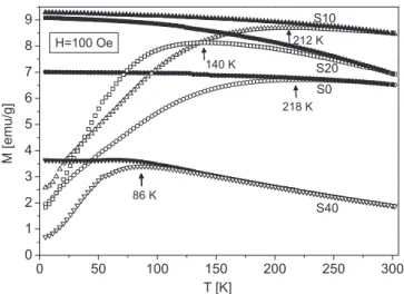
To investigate the temperature dependence of magnetization we have performed magnetization vs. temperature measurements in both ZFC and FC regimes in thefield of 100 Oe. Results are shown onFig. 6.
where it can be seen that all ZFC curves possess maximum (denoted for each curve) whose position Tmaxshifts to lower temperatures with the increased Mg concentration. At the same time, values of mass magne- tization increases for samples S10 and S20 compared to S0 sample while considerable decrease occurs for S40 specimen.
It has been established in many researches that the value of blocking temperature TB diminishes with the nanoparticle size reduction [38,39]. Thus, if we consider position of a maximum in ZFC magneti- zation curve as a TBvalue then its decrease with the Mg content is in line with the concomitant particle size reduction already revealed in previous paragraphs by other experimental techniques. Consequently, the magnetization increase for samples S10 and S20 despite the particle size reduction is a result of the intrinsic magnetization rise due to the Mg doping. This points to the predominant distribution of Mg2+ions in tetrahedral positions in these two samples. In addition, the considerable magnetization reduction obtained for S40 together with the increased anisotropy (as found from M(H) measurements) may be an indication that for this dopant concentration Mg2+ions occupy octahedral posi- tions in an elevated percentage.
Temperature at which bifurcation between ZFC and FC curves Fig. 4.Electrokinetic potential (full symbols) and hydrodynamic particle size
(empty symbols) of S0 (circles) and S10 (triangles) ferrofluid samples as the function of pH.
Fig. 5.Field dependence of the isothermal magnetization for powder MgxFe3−xO4samples: (a) Room temperature data; (b) Data recorded at 5 K temperature - inset shows lowfield behaviour of M(H) curves.
Table 3
Important parameters of the investigated samples obtained from magnetic measurements.
Sample Tmax[K] Tirr[K] M5T(5 K) [emu/g]
HC(5 K) [Oe]
M5T(300 K) [emu/g]
HC(300 K) [Oe]
S0 218 270 58 250 47 ∼0
S10 212 255 76 270 67 ∼0
S20 140 275 59 240 49 ∼0
S40 86 230 46 400 31 ∼0
Fig. 6.Temperature dependence of mass magnetization in 100 Oefield in ZFC (empty symbols) and FC (full symbols) regimes for powder MgxFe3−xO4sam- ples. Position of a maximum in ZFC regime is denoted for each curve.
occurs is usually referred to as irreversibility temperature Tirr, and it is commonly accepted that its value is determined at the point where the condition (MFC-MZFC)/MFC< 1% is fulfilled. Values of Tirrobtained by applying this condition are listed inTable 3, and it can be seen that all of them are below the room temperature. Since Tirris considered as the blocking temperature of the largest nanoparticles in the system[40], we may conclude that the obtained irreversibility temperatures ad- ditionally point to superparamagnetic behaviour of all the samples at room temperature.
FC magnetization curves of all samples show “flatness” i.e. they undergo a very slow increase with the temperature reduction which is a signature of considerable interparticle interactions. This is a common occurrence in a system of“naked”magnetic nanoparticles which are in a close proximity like in powder samples. To check the impact of in- terparticle interaction on the magnetism of powder samples we have performed ZFC-FC measurements on their ferrofluid counterparts in the frozen state i.e. in the temperature range 5–250 K for samples S0 and S10. Results are depicted inFig. 7. where also data for powder S0 and S10 samples are indicated for easier comparison. By inspecting this figure three mainfindings can be drawn: (i) magnetization of ferrofluid samples are considerably higher in respect to dried (powder) counter- parts; (ii) position of ZFC maximum for both ferrofluid samples is shifted to lower temperatures in comparison to powder counterparts, and (iii) ration between magnetizations of S1 and S0 in powder form has been preserved in ferrofluids. Based on the abovefindings we may conclude that interparticle interactions are predominantly of the di- pole-dipole type which tend to antiparallely orient particle moments and reduce the net magnetization. These interactions are considerably stronger in powder samples due to the increased proximity of magnetic nanoparticles, and thus net magnetization of dried samples is lower compared to ferrofluide counterparts. Since thesefindings are the same for both S0 and S10 samples, we can conclude that increase of the magnetization magnitude in S10 sample is not related to interparticle interactions, and so it originates from the Mg doping of S0 sample.
Assuming the homogeneous particle distribution in FF samples and using the mean particle diameters determined by XRD (Table 1) we have estimated the average interparticle distances in investigated fer- rofluids (in these calculations density value of 5.17 g/cm3 for bulk magnetite has been used). The so-obtained values were approximately 60, 80, 60 and 35 nm for samples S0, S10, S20 and S40, respectively. It has been shown in several researches that interparticle distances of even few particle diameters resulted in negligible interparticle inter- actions [40,41]. This implies that in our suspensions distribution of particles is not homogeneous which is in line with the DLSfindings on
the presence of particle aggregates in investigated ferrofluids. As al- ready pointed out in Section 3.2, particle aggregation is more pro- nounced in Mg-doped systems which hydrodynamic radii is about three times bigger compared to undoped S0. Having in mind the above mentioned interparticle distances and decreasing dipole moments due to the particle size decrease with doping, we may conclude that in- creased aggregation in doped systems is not caused by a dipolar in- teractions. It rather originates from the changed surface charge of Mg- doped particles i.e. it is predominantly influenced by electrostatic forces in the suspension.
3.4. Magnetic heating in ACfields
Heating abilities in the applied ACfields of the ferrofluid samples under consideration have been evaluated by calculating the Specific Absorption Rate (SAR) value on the basis of the measured temperature vs. time dependences. Since our measurements have been done in non- adiabatic conditions we have used the following expression:
= =
SAR [C /mps NPs]·|dT/dt|t 0 (3) which utilizes time derivative of the temperature at initial time t = 0 [6]. Cpsdenotes heat capacity of the sample while mNPsrepresents mass of the dispersed nanoparticles. Since we have been dealing with fer- rofluid samples, the value of Cpswas related to the specific heat capa- city of the dispersion medium Cpdand specific heat of nanoparticles CpNPby the expression:
= +
Cps C mpd d CpNPmNP (4)
For the calculation purposes literature values of the specific heat capacities of water Cpd= 4187 J/kgK and of magnetite CpNP= 640 J/
kgK [42] were taken into account. The so-obtained SAR values are presented onFig. 8, where in (a) are shown frequency dependences for the fixed value of field amplitude while (b) represents field de- pendences for thefixed frequency.
In order to compare the heating efficacy of our samples with the available literature data for similar ferrofluids we have also expressed the obtained SAR values by so-called ILP (Intrinsic Loss Power) values defined as:
=
ILP SAR/[f·H ]2 (5)
where f and H denote applied ACfield frequency and amplitude, re- spectively [43]. The ILP has been introduced to allow more direct comparisons of published SAR data by making them independent on AC field parameters. However, the validity of expression (5) also has cer- tain limitations in respect to field parameters and particle size dis- tribution[43]. Consequently, inTable 4we have compared both SAR and ILP values for our samples with the highest heating efficacy, S0 and S10, with the literature data for ferrofluids based on either magnetite or non-magnetic ion doped magnetite with the similar average core size of MNPs.
Inspection ofTable 4shows that the SAR/ILP values of samples under consideration in this work are comparable with the literature data for similar systems.
From the data presented inFig. 8we may see that the highest SAR values were obtained for S0 sample for all appliedfield amplitudes and frequencies. This result seems surprising, having in mind the obtained values for Msat(Table 3), and considering that SAR is proportional ei- ther to Msat(in case of blocked nanoparticle state[48]) or to Msat2(in a superparamagnetic region[49]). This is especially noticeable for S10 sample, since its room temperature saturation magnetization is con- siderably higher than for S0 sample. Also, value of Msatfor S20 sample somewhat exceeds the saturation magnetization of S0 sample while its SAR value is several times lower.
To explain the drop in heating performance of Mg doped samples we need to consider both relaxation mechanisms, Néel and Brownian, that contribute to the heat dissipation. The Néel relaxation concerns the Fig. 7.ZFC-FC data (symbols) in 100 Oefield for ferrofluid counterparts of
samples S0 and S10 (denoted by S0 FF and S10 FF, respectively) in their frozen state (temperature region 5–250 K). Data for powder S0 and S10 samples (dotted and dashed lines, respectively) are given for comparison.
fluctuation of the particle’s dipolar moment while Brownian relaxation is caused by a mechanical particle rotation in a viscous medium.
Relaxation times of these processes are given byτN=τ0exp(KVm/kBT) and τB= 4πηrH3
/kBT, for the Néel and Brownian relaxation, respec- tively[50]. In the above expressionsτ0denotes so called characteristic time of the material (typically assumed to be in the range 10−9–10−10), K denotes magnetic anisotropy constant, Vmis the nanoparticle mag- netic volume, rHis the particle hydrodynamic radius, and kB is the Boltzmann's constant.
It is well known that mechanical rotations quickly become insig- nificant with the increase of the particle hydrodynamic radius sinceτB
scales with the cube of rH[6]. By using the hydrodynamic diameters given inTable 2(HD values) it is easy to see thatτBincreases for an
order of magnitude for Mg doped samples compared to the undoped one thus diminishing the contribution of Brownian relaxation.
Dissipation of thermal energy by the Néel mechanism depends on the KVm/kBT ratio, and its contribution is significant only if the mag- netic anisotropy energy of the particle is considerably larger than the thermal energy, i.e. if KVm»kBT[50]. Magnetic volume Vmof the par- ticle could be considered as the volume of the crystallite with the average size determined from the X-ray diffraction (Table 1), while the value of anisotropy constant K is usually determined from the Néel expression K = 25 kBTB/Vm[12], where TBdenotes blocking tempera- ture of the nanoparticle system obtained from ZFC measurement (Table 3). It should be noted that this expression is strictly speaking valid for noninteracting monodisperse magnetic particles only, and so the value of K obtained by applying it in the case of interacting particles is usually overestimated due to the inclusion of the system's collective response[51]. By using the above expression and the corresponding data fromTables 1 and 2we have determined K and KVmvalues, listed inTable 5.
The obtained values of K are at least an order of magnitude higher in comparison to the bulk value for magnetite (1.35∙104J/m3), and they increase with the nanoparticle decrease, which is a behaviour usually ascribed to the surface effects in nanoparticle material. In contract, anisotropy energy KVmdecrases with Mg doping thus becomes closer to the thermal energy value at room temperature (∼4∙10−21 J). This means that the heat dissipation due to the Néel mechanism also di- minishes with Mg doping.
Above discussion is restricted to parameters related to individual nanoparticles but we should also consider impact of a collective be- haviour on the heating performance of investigated ferrofluids. Namely, magnetic measurements pointed to the existence of dipolar interactions between nanoparticles while DLS results showed increased aggregation of nanoparticles in all ferrofluids based on Mg doped nanoparticles. In aggregated condition distances between magnetic dipoles are much lower which leads to the increased magnetic interactions between them. It has been established in several researches that interparticle interactions influence the heating abilities of the ferrofluid, and this correlation is still a topic of current interest. A comprehensive review on the interparticle interactions impact on heating efficacy is given in [52]while here we will point just to a few typical examples. For in- stance, by relaying on both experimental data and Monte Carlo simu- lations of the system of ferromagnetic Fe nanoparticles a decrease of SAR values with the increasing dipolar interactions were found[53].
Similar results have been obtained by numerical simulations[54]and Fig. 8.The obtained SAR values for the investigated ferrofluids at pH∼4: (a)
frequency dependences for thefixedfield amplitude of 200 Oe; (b)field de- pendences for thefixed frequency of 577 kHz.
Table 4
Heating efficacy comparison for ferrofluids based on magnetite or ferrite nanoparticles.
Composition Core size [nm] H [kA/m]* f [kHz] SAR [w/g] ILP [nHm2/kg] Ref.
Fe3O4(S0) 8 15.9 577 111 0.76 This work
Mg0.1Fe2.9O4(S10) 6 15.9 577 83.1 0.57 This work
Fe3O4 10 24.5 400 130 0.54 [44]
Fe3O4 9.5 30 100 29 0.32 [45]
Fe3O4 9 27 400 367 1.26 [46]
MgFe2O4 7 8 279 13.4 1.06 [47]
Zn0.25Fe2.75O4 8.3 6 330 16.65 1.4 [24]
* For easier comparison, one should note that 1 Oe = 79.58 A/m.
Table 5
Values of anisotropy constant K and anisotropy energy KVmfor investigated samples.
Sample K [105J/m3] KVm[10−20J]
S0 3.0 8.6
S10 6.1 7.3
S20 16.8 4.8
S40 14.4 3.0
by experimental investigations on magnetite based ferrofluids which also pointed to unfavourable role of dipolar interactions on the SAR values[55]. Based on these results we may propose that the increased interparticle interactions due to the elevated aggregation of nano- particles in Mg doped ferrofluids suppresses heat losses and thus also contributes to the decreasing of SAR values in comparison to the un- doped S0 sample.
4. Conclusions
Stable ferrofluids based on the Mg doped magnetite nanoparticles MgxFe3−xO4have been successfully synthesised by utilising chemical reverse co-precipitation method. Powder x-ray study showed than all the samples were of the cubic spinel structure while the cell parameter increased with the magnesium concentration x. TEM photographs re- vealed the decreasing particle size with increasing amount of Mg, as well as the tendency for particle agglomeration and their deviation from the spherical shape. DLS experiments pointed to the increasing hydrodynamic particle size (up to three times) in doped samples while polydispersity index remained constant.
Study of magnetic properties showed that the significant increase of magnetization occurred only for 10% Mg doping in compare to un- doped magnetite sample. By simultaneous comparison of different magnetic parameters (magnetization, coercivity and blocking tem- peratures) and particle sizes we may conclude that for both 10% and 20% of Mg concentration the intrinsic magnetization increases as a result of Mg doping, while for concentration of 40% the decrease of magnetization occurred. From the viewpoint of Mg ions distribution above results indicate the prevalent occupation of tetrahedral sites in the spinel structure up to 20% of Mg doping while for higher con- centrations their distribution over octahedral sites prevails.
Regarding thermomagnetic properties, the best SAR values have been obtained for the ferrofluid based on undoped magnetite nano- particles, in spite of the increased magnetization of 10% Mg doped sample. To comprehend possible reasons for such a result we have considered both Brownian and Néel contributons to heat dissipation. It has been shown that increased hydrodynamic radii of about three times for Mg-doped systems diminishes the Brownian contribution in these samples. Regarding the Néel contribution, we have found that aniso- tropy constant K increased with Mg doping while anisotropy energy KVmhad the decreasing trend, approaching the thermal energy value at room temperature. This pointed to diminishing of the Néel contribution with increased Mg content.
Having in mind the existence of dipolar interparticle interactions in investigated systems and increased aggregation in Mg-doped samples we also considered the impact of the collective phenomena on the ob- tained SAR values. The results published so far showed that increased interparticle interactions usually diminished heating abilities in AC fields. Thus we may conclude that elevated aggregation in Mg-doped systems acted as additional impeding factor regarding magnetic heating.
Our results show that magnesium substitution for iron in nano- particle magnetite up to 20% of Mg leads to intrinsic magnetisation increase. In contrast, heating abilities of the ferrofluids based on Mg- doped nanoparticles is lower compared to the one based on pure magnetite thus giving the advantage to the latter as heating agent for magnetic hyperthermia. However, considerably enhanced magnetiza- tion of 10% Mg doped magnetite as well as its superparamagnetic be- haviour at room temperature recommends it as a convenient material for different applications based on magnetic separation or externalfield targeting.
Acknowledgements
The authors gratefully acknowledge support provided by the project III-45015 funded by the Serbian Ministry of Education, Science and
Technological Development, EU funded project FP7-EraChairs- MAGBIOVIN, and COST Action TD1402-RADIOMAG.
References
[1] R. Skomski, Nanomagnetics, J. Phys.: Condens. Matter 15 (2003) R841–R896.
[2] G.C. Papaefthymiou, Nanoparticle magnetism, Nano Today 4 (2009) 438–447.
[3] Nanomagnetism: Fundamentals and Applications, Volume 6, 1st Edition, Series Volume Editor: Chris Binns, Elsevier, 2014.
[4] V.K. Varadan, L.F. Chen, J. Xie, Nanomedicine: Design and Applications of Magnetic Nanomaterials, Nanosensors and Nanosystems, Wiley, 2008.
[5] Nguyen T.K. Thanh (Ed.), Magnetic Nanoparticles: From Fabrication to Clinical Applications, CRC Press, Taylor & Francis Group, Boca Raton, FL, 2012.
[6] E.A. Périgo, G. Hemery, O. Sandre, D. Ortega, E. Garaio, F. Plazaola, F.J. Teran, Fundamentals and advances in magnetic hyperthermia, Appl. Phys. Rev. 2 (2015) 041302.
[7] D.R. Baselt, G.U. Lee, M. Natesan, S.W. Metzger, P.E. Sheehan, R.J. Colton, A bio- sensor based on magnetoresistance technology, Biosens. Bioelectron. 13 (1998) 731–739.
[8] A.C. Anselmo, S. Mitragotri, Nanoparticles in the clinic, Bioeng. Transl. Med. 1 (2016) 10–29.
[9] S. Chandra, K.C. Barick, D. Bahadur, Oxide and hybrid nanostructures for ther- apeutic applications, Adv. Drug Deliv. Rev. 63 (2011) 1267–1281.
[10] A. Akbarzadeh, M. Samiei, S. Davaran, Magnetic nanoparticles: preparation, phy- sical properties, and applications in biomedicine, Nanoscale Res. Lett. 7 (2012) 144.
[11] C. Caizer, Nanoparticle Size Effect on Some Magnetic Properties, in:
Mahmood Aliofkhazraei (Ed.), Handbook of Nanoparticles, Springer International Publishing, Switzerland, 2015, pp. 1–38.
[12] J.M.D. Coey, Magnetism and Magnetic Materials, Cambridge University Press, New York, 2010 Ch. 11.
[13] S. Larumbe, C. Gómez-Polo, J.I. Pérez-Landazábal, A. García-Prieto, J. Alonso, M.L. Fdez-Gubieda, D. Cordero, J. Gómez, Ni Doped Fe3O4Magnetic Nanoparticles, J. Nanosci. Nanotechnol. 12 (2012) 2652–2660.
[14] G. Rana, U.C. Johri, A study on structural and magnetic properties of Ni-substituted magnetite nanoparticles, J. Alloy. Compd. 577 (2013) 376–381.
[15] H. Zhu, S. Zhang, L.Wu. Yu-Xi Huang, S. Sun, Monodisperse MxFe3−xO4 (M = Fe, Cu Co, Mn) Nanoparticles and Their Electrocatalysis for Oxygen Reduction Reaction, Nano Lett. 13 (2013) 2947–2951.
[16] S.O. Estrada, C.A. Huerta-Aguilar, T. Pandiyan, I.A. Monica Corea, G.
Tavizon Reyes-Domínguez, Tuning of the magnetic response in cobalt ferrite CoxFe3xO4by varying the Fe2+to Co2+molar ratios: rietveld refinement and DFT structural analysis, J. Alloy. Compd. 695 (2017) 2706–2716.
[17] J. Amighian, E. Karimzadeh, M. Mozaffari, The effect of Mn2+substitution on magnetic properties of MnxFe3xO4nanoparticles prepared by coprecipitation method, J. Magn. Magn. Mater. 332 (2013) 157–162.
[18] V.L. Pool, M.T. Kleb, C.L. Chorney, E. Arenholz, Y.U. Idzerda, Enhanced magneti- zation in VxFe3xO4nanoparticles, J. Magn. Magn. Mater. 396 (2015) 304–307.
[19] Z. Cvejic, B. Antic, A. Kremenovic, S. Rakic, G.F. Goya, H.R. Rechenberg, C. Jovalekic, V. Spasojevic, Influence of heavy rare earth ions substitution on mi- crostructure and magnetism of nanocrystalline magnetite, J. Alloy. Compd. 472 (2009) 571–575.
[20] R.V. Upadhyay, A. Gupta, C. Sudakar, K.V. Rao, K. Parekh, R. Desai, R.V. Mehta, Effect of rare-earth Ho ion substitution on magnetic properties of Fe3O4magnetic fluids, J. Appl. Phys. 99 (2006) 08M906.
[21] M. Mozaffari, J. Amighian, R. Tavakoli, The effect of yttrium substitution on the magnetic properties of magnetite nanoparticles, J. Magn. Magn. Mater. 379 (2015) 208–212.
[22] V.L. Pool, M.T. Klem, C.L. Chorney, E.A. Arenholz, Y.U. Idzerda, Enhanced mag- netism of Fe3O4nanoparticles with Ga doping, J. Appl. Phys. 109 (2011) 07B529.
[23] J. Liu, Y. Bin, M. Matsuo, Magnetic Behavior of Zn-Doped Fe3O4Nanoparticles Estimated in Terms of Crystal Domain Size, J. Phys. Chem. C 116 (2012) 134–143.
[24] B. Behdadfar, A. Kermanpur, H. Sadeghi-Aliabadi, M. del Puerto Morales, M. Mozaffari, Synthesis of aqueous ferrofluids of ZnxFe3xO4nanoparticles by citric acid assisted hydrothermal-reduction route for magnetic hyperthermia applications, J. Magn. Magn. Mater. 324 (2012) 2211–2217.
[25] J.M. Byrne, V.S. Coker, E. Cespedes, P.L. Wincott, D.J. Vaughan, R.A.D. Pattrick, G. van der Laan, E. Arenholz, F. Tuna, M. Bencsik, J.R. Lloyd, N.D. Telling, Biosynthesis of zinc substituted magnetite nanoparticles with enhanced magnetic properties, Adv. Funct. Mater. 24 (2014) 2518–2529.
[26] L. Neel, Annals Phys. 3 (1948) 137–198.
[27] R.D. Shannon, Revised effective ionic radii and systematic studies of interatomic distances in halides and chalcogenides, Acta Cryst. A 32 (1976) 751–767.
[28] Q. Chen, A.J. Rondinone, B.C. Chakoumakos, Z.J. Zhang, Synthesis of super- paramagnetic MgFe2O4nanoparticles by coprecipitation, J. Magn. Magn. Mater.
194 (1999) 1–7.
[29] S. Da Dalt, A.S. Takimi, T.M. Volkmer, V.C. Sousa, C.P. Bergmann, Magnetic and Mössbauer behavior of the nanostructured MgFe2O4spinel obtained at low tem- perature, Powder Technol. 210 (2011) 103–108.
[30] M.G. Naseri, M.H.M. Ara, E.B. Saion, A.H. Shaari, Superparamagnetic magnesium ferrite nanoparticles fabricated by a simple, thermal-treatment method, J. Magn.
Magn. Mater. 350 (2014) 141–147.
[31] Z. Lv, Q. Wang, Y. Bin, L. Huang, R. Zhang, P. Zhang, M. Matsuo, Magnetic Behaviors of Mg- and Zn-Doped Fe3O4Nanoparticles Estimated in Terms of Crystal Domain Size, Dielectric Response, and Application of Fe3O4/Carbon Nanotube
Composites to Anodes for Lithium Ion Batteries, J. Phys. Chem. C 119 (2015) 26128–26142.
[32] J. Rodriguez-Carvajal, T. Roisnel, Line broadening analysis using FullProf:
Determination of microstructural properties, Mater. Sci. Forum 443–444 (2004) 123–126.
[33] V.M. Khot, A.B. Salunkhe, N.D. Thorat, R.S. Ningthoujam, S.H. Pawar, Induction heating studies of dextran coated MgFe2O4nanoparticles for magnetic hy- perthermia, Dalton Trans. 42 (2013) 1249–1258.
[34] E. Tombácz, pH-dependent surface charging of metal oxides, Periodica Polytech., Chem. Eng. 53 (2) (2009) 77–86.
[35] E. Tombácz, A. Majzik, Z.S. Horvat, E. Illes, Magnetite in aqueous medium: coating its surface and surface coated with it, Rom. Rep. Phys. 58 (2006) 281–286.
[36] W. Tang, Su Yu, Qi Li, S. Gao, J.K. Shang, Superparamagnetic magnesium ferrite nanoadsorbent for effective arsenic (III, V) removal and easy magnetic separation, Water Res. 47 (2013) 3624–3634.
[37] Quantum Design: MPMS Application Note 1014-208 A.
[38] J. Chatterjee, Y. Haik, C.J. Chen, Size dependent magnetic properties of iron oxide nanoparticles, J. Magn. Magn. Mater. 257 (2003) 113–118.
[39] H. Shim, P. Dutta, M.S. Seehra, J. Bonevich, Size dependence of the blocking temperatures and electron magnetic resonance spectra in NiO nanoparticles, Solid State Commun. 145 (2008) 192–196.
[40] P. Tartaj, T. Gonzales-Carreno, C.J. Serna, Magnetic behaviour ofγ-Fe2O3 nano- crystals dispersed in colloidal silica particles, J. Phys. Chem. B 107 (2003) 20–24.
[41] C. Martinez-Boubeta, K. Simeonidis, M. Angelakeris, N. Pazos-Peres, M. Giersig, A. Delimitis, L. Nalbandian, V. Alexandrikis, D. Niarchos, Critical radius for ex- change bias in naturally oxidised Fe nanoparticles, Phys. Rev. B 74 (2006) 054430.
[42] M.W. Chase Jr., NIST-JANAF Themochemical Tables, Fourth Edition, J. Phys.
Chem. Ref. Data, Monograph 9 (1998) 1–1951.
[43] M. Kallumadil, M. Tada, T. Nakagawa, M. Abe, P. Southern, Q.A. Pankhurst, Suitability of commercial colloids for magnetic hyperthermia, J. Magn. Magn.
Mater. 321 (2009) 1509–1513.
[44] M. Gonzales-Weimuller, M. Zeisberger, K.M. Krishnan, Size-dependant heating rates of iron oxide nanoparticles for magneticfluid hyperthermia, J. Magn. Magn. Mater.
321 (2009) 1947–1950.
[45] M. Song, Y. Zhang, S. Hu, L. Song, J. Dong, Z. Chen, N. Gu, Influence of morphology and surface exchange reaction on magnetic properties of monodisperse magnetite nanoparticles, Colloids Surf. A 408 (2012) 114–121.
[46] X.L. Liu, H.M. Fan, J.B. Yi, Y. Yang, E.S.G. Choo, J.M. Xue, D.D. Fana, J. Ding, Optimization of surface coating on Fe3O4nanoparticles for high performance magnetic hyperthermia agents, J. Mater. Chem. 22 (2012) 8235–8244.
[47] M.R. Barati, C. Selomulya, K. Suzuki, Particle size dependence of heating power in MgFe2O4nanoparticles for hyperthermia therapy application, J. Appl. Phys. 115 (2014) 17B522.
[48] B. Mehdaoui, A. Meffre, L.M. Lacroix, J. Carrey, S. Lachaize, M. Gougeon, M. Respaud, B. Chaudret, Large specific absorption rates in the magnetic hy- perthermia properties of metallic iron nanocubes, J. Magn. Magn. Mater. 322 (2010) L49–L52.
[49] R. Hergt, S. Dutz, Magnetic particle hyperthermia−biophysical limitations of a visionary tumour therapy, J. Magn. Magn. Mater. 311 (2007) 187–192.
[50] A.E. Deatsch, B.A. Evans, Heating efficiency in magnetic nanoparticle hy- perthermia, J. Magn. Magn. Mater. 354 (2014) 163–172.
[51] V.L. Calero-Diaz del Castillo, C. Rinaldi, Effect of sample concentration on the de- termination of the anisotropy constant of magnetic nanoparticles, IEEE Trans.
Magn. 46 (2010) 852–859.
[52] I.M. Obaidat, B. Issa, Y. Haik, Magnetic properties of magnetic nanoparticles for efficient hyperthermia, Nanomaterials 5 (2015) 63–89.
[53] D. Serantes, D. Baldomir, C. Martinez-Boubeta, K. Simeonidis, M. Angelakeris, E. Natividad, M. Castro, A. Mediano, D.-X. Chen, A. Sanchez, L.I. Balcells, B. Martínez, Influence of dipolar interactions on hyperthermia properties of ferro- magnetic particles, J. Appl. Phys. 108 (2010) 073918.
[54] C. Haase, U. Nowak, Role of dipole-dipole interactions for hyperthermia heating of magnetic nanoparticle ensembles, Physical Review B 85 (2012) 045435.
[55] P.H. Linh, P.V. Thach, N.A. Tuan, N.C. Thuan, D.H. Manh, N.X. Phuc, L.V. Hong, Magneticfluid based on Fe3O4nanoparticles: preparation and hyperthermia ap- plication, J. Phys. Conf. Ser. 187 (2009) 012069.