THE ALTERATIONS OF THE
CYTOKINE NETWORK IN PERINATAL HYPOXIC-ISCHEMIC BRAIN INJURY
PhD thesis
Anna Bajnok MD
Doctoral School of Clinical Medicine Semmelweis University
Consultant: Gergely Toldi, MD, Ph.D Official reviewers: Éva Görbe MD, PhD
László Szapáry MD, PhD Head of the Final Examination Committee:
Tamás Machay, MD, PhD Members of the Final Examination Committee:
György Nagy MD, DSc Zsolt Somogyvári MD, PhD
Budapest
2019
TABLE OF CONTENTS
1 ABBREVIATIONS ... 5
2 INTRODUCTION ... 7
2.1 The neonatal brain ... 9
- selective vulnerability or functional plasticity after hypoxia-ischemia? ... 9
2.2 Perinatal asphyxia ... 17
2.2.1 Definition and incidence ... 17
2.2.2 Risk factors ... 19
2.2.3 Pathophysiology ... 21
2.2.4 Clinical presentation ... 23
2.2.5 Diagnosis ... 25
2.2.5.1 Clinical staging ... 25
2.2.5.2 Neurophysiology ... 27
2.2.5.3 Neuroimaging ... 28
2.2.6 Therapy ... 30
2.2.7 Outcome ... 33
2.3 Neonatal arterial ischemic stroke (NAIS) ... 35
2.3.1 Definition and incidence ... 35
2.3.2 Risk factors ... 36
2.3.3 Placental pathology ... 39
2.3.4 Pathophysiology ... 40
2.3.5 Clinical presentation and diagnosis ... 44
2.3.6 Therapy ... 46
2.3.7 Prognosis and outcome ... 46
2.4 Neuroinflammation ... 48
2.4.1 Hypoxia-induced neuroinflammation ... 48
2.4.2 Cells participating in the neuroinflammation ... 54
2.4.2.1 Microglia ... 54
2.4.2.2 Astrocytes ... 55
2.4.2.3 Neutrophils ... 56
2.4.2.4 Lymphocytes ... 56
2.4.3 Adhesion molecules ... 59
2.4.4 Cytokines ... 60
2.4.5 Pro-inflammatory cytokines ... 61
2.4.5.1 IL-1β ... 61
2.4.5.2 IL-6 ... 62
2.4.5.3 IL-17 ... 63
2.4.5.4 IFN-γ ... 64
2.4.5.5 TNF-α ... 64
2.4.6 Anti-inflammatory cytokines ... 65
2.4.6.1 IL-10 ... 65
2.4.6.2 TGF-β ... 66
2.4.7 The kynurenine system ... 67
3 STUDY RATIONALE ... 68
4 AIMS ... 70
5 MATERIALS AND METHODS ... 71
5.1 Patients ... 71
5.2 NAIS case presentations ... 74
5.2.1 Case 1. ... 74
5.2.2 Case 2. ... 75
5.2.3 Case 3. ... 75
5.2.4 Case 4. ... 76
5.3 Summary of measured parameters ... 77
5.4 Flow cytometry ... 79
5.5 Immunoassays ... 80
5.6 High-performance liquid chromatography (HPLC) ... 80
5.7 Statistical analysis ... 81
6 RESULTS ... 82
6.1 COMPARISONS BETWEEN THE MODERATE AND THE SEVERE HIE GROUPS ... 83
6.1.1 Pro-inflammatory cytokines ... 83
6.1.1.1 IL-1β ... 84
6.1.1.2 IL-6 ... 85
6.1.1.3 IL-17 ... 86
6.1.1.4 TNF-α ... 88
6.1.1.5 Other pro-inflammatory cytokines ... 89
6.1.2 Anti-inflammatory factors ... 90
6.1.2.1 TGF-β ... 91
6.1.2.2 Tregs ... 91
6.1.2.3 Other anti-inflammatory cytokines ... 92
6.1.2.4 The kynurenine system ... 93
6.1.3 ROC analysis ... 95
6.2 COMPARISONS BETWEEN HIE AND NAIS ... 96
6.2.1 Cytokine production of T lymphocytes ... 96
6.2.1.1 Pro-inflammatory cytokines ... 97
6.2.1.2 Anti-inflammatory cytokines ... 99
6.2.2 Plasma cytokines ... 100
7 DISCUSSION ... 103
7.1 COMPARISONS BETWEEN MODERATE AND SEVERE HIE ... 103
7.1.1 Pro-inflammatory cytokines ... 104
7.1.1.1 IL-1β ... 104
7.1.1.2 IL-6 ... 105
7.1.1.3 IL-17 ... 106
7.1.1.4 TNF-α ... 106
7.1.2 Anti-inflammatory factors ... 108
7.2 COMPARISONS BETWEEN HIE AND NAIS ... 110
7.2.1 Pro-inflammatory cytokines ... 110
7.2.2 Anti-inflammatory cytokines ... 112
7.3 Study limitations ... 114
8 CONCLUSIONS ... 115
8.1 KEY FINDINGS ... 116
9 SUMMARY ... 118
10 ÖSSZEFOGLALÁS ... 119
11 REFERENCES ... 120
12 PUBLICATIONS ... 150
Publications directly related to the PhD dissertation ... 150
Publications not related to the PhD dissertation: ... 150
13 ACKNOWLEDGEMENTS ... 152
1 ABBREVIATIONS
ACA Anterior cerebral artery
ADC Apparent diffusion coefficient
aEEG Amplitude-integrated electroencephalogram
AIS Acute ischemic stroke
AMPA α-amino-3-hydroxy-5-methyl-4-isoxazole receptors
APC Antigen presenting cells
BBB Blood-brain barrier
CD Cluster of Differentiation
CNS Central nervous system
CPISR Canadian Pediatric Ischemic Stroke Registry
CTL Cytotoxic T lymphocyte
CTLA-4 Cytotoxic T lymphocyte antigen - 4
CSF Cerebrospinal fluid
CSVT cerebral sinus venous thrombosis
DW MRI Diffusion-weighted magnetic resonance imaging
EEG Electroencephalogram
EPO Erythropoietin
FOXP3 Forkhead box protein 3
G-CSF Granulocyte colony-stimulating factor
GM-CSF Granulocyte-macrophage colony-stimulating factor GM-IVH Germinal matrix - intraventricular hemorrhage
HEV High endothelial venules
HI Hypoxic-ischemic
HIE Hypoxic ischemic encephalopathy
IDO Indoleamine 2,3-dioxygenase
IFN-γ Interferon - gamma
IL Interleukin
IVH Intraventricular hemorrhage
KYN Kynurenine
KYNA Kynurenic acid
LPS Lipopolysaccharide
MCA Middle cerebral artery
MCP-1 Monocyte chemoattractant protein - 1
MFI Mean fluorescence intensity
MHC Major histocompatibility complex
MIP-1β Macrophage inflammatory protein - 1 beta
MMP Matrix metalloproteinase
MRI Magnetic resonance imaging
MRS Magnetic resonance spectroscopy
MSC mesenchymal stem cell
NAIS Neonatal arterial ischemic stroke
NE Neonatal encephalopathy
NMDA N-methyl-D-aspartate
NO Nitric oxide
NOS Nitric oxide synthase
PBMC Peripheral blood mononuclear cells
PCA Posterior cerebral artery
PET Positron emission tomography
PVL Periventricular leukomalacia
qPCR Quantitative polymerase chain reaction RAG1 Recombination activating gene 1
RNS Reactive nitrogen species
RORγt RAR-related orphan receptor gamma t
ROS Reactive oxygen species
S1P Sphingosine-1-phosphate
S1PR Sphingosine-1-phosphate receptor
SEP Somatosensory evoked potential
T-bet T-box expressed in T cells
TCR T cell receptor
TGF-β Tumor growth factor - beta
Th1 Helper T lymphocyte -1
Th17 Helper T lymphocyte -17
Th2 Helper T lymphocyte -2
TNF-α Tumor necrosis factor - alpha
Treg Regulatory T lymphocyte
TRP Tryptophan
VCAM-1 Vascular cell adhesion molecule - 1
VEP Visual evoked potential
VLA-4 Very Late Antigen - 4
WT Wild type
2 INTRODUCTION
Perinatal asphyxia and the consequent hypoxic-ischemic brain injury continues to be a major cause of perinatal morbidity and mortality and lifelong disability, despite the advances of the past decades in prenatal diagnostic methods and neonatal intensive care. It is important to note, that the initial degree of injury and the clinical status of the neonate often do not allow accurate prediction of the eventual neurodevelopmental outcome. While some children show favorable outcome and no significant disability, others develop severe neurodevelopmental impairments such as mental retardation, sensory impairment, cerebral palsy and seizures. Identifying factors which could differentiate between mild and severe outcome would be of great clinical value.
It is becoming more and more clear, that the final outcome is influenced by several factors beyond the severity of the initial insult. One of the key factors determining the progress of brain injury is the neuroinflammatory response evoked by hypoxic- ischemic injury. Neuroinflammation is now recognized to be a common feature of many neurological disorders, including hypoxic-ischemic brain injury. However, it appears to have dual aspects. While a certain level of inflammatory response is part of the physiological recovery process of the central nervous system, excessive neuroinflammation has been shown to play an important role in facilitating further brain injury. Emerging evidence indicates, that neuroinflammation could continue for many months after the initial brain injury and this latent phase could play an important role in the long-term consequences of perinatal hypoxic-ischemic brain injury.
In the past decades important advances have been made in diagnosing perinatal hypoxic-ischemic brain injury. Introducing amplitude-integrated electroencephalogram monitoring into the routine clinical practice has enabled clinicians to follow the status of the neonate much more precisely. The widespread availability of MR imaging has opened new options for following the evolution of the brain injury and differentiating between asphyxia and disorders which are clinically overlapping, such as perinatal stroke. New MR modalities also allow earlier estimation of the injury with higher precision.
Inarguably one the most important advancements of the past decade has been the introduction of therapeutic hypothermia into the common clinical routine, which has been shown to improve neurological outcome significantly. One of the fundamental
effects of hypothermia is the amelioration of the neuroinflammatory response and consequent tissue injury. Current research is focusing on developing novel immunomodulatory agents, many of which primarily target the neuroinflammatory response.
The improving diagnostic possibilities in perinatal asphyxia also raised the awareness towards other etiologies of hypoxic-ischemic brain injury which are characteristic for the perinatal period. One of the most common is stroke, for which the neonatal period carries the highest risk in the entire childhood. Perinatal stroke is one of the most common causes of seizures, however it is seldomly diagnosed in a timely manner. The clinical presentation of the neonates often overlaps with global hypoxic-ischemic brain injury, and the real etiology is only later revealed upon MR examination. The pathophysiology of perinatal stroke is poorly understood; however it appears, that in-utero inflammation could be an important risk factor. Similarly to asphyxia, stroke also evokes a neuroinflammatory response, which plays an important role in the evolution of the brain injury. Disease specific preventive measures, prognostic factors and therapeutic strategies are not available, as neonates with stroke are not yet considered eligible for hypothermia therapy.
Although recent years’ research has brought important aspects of the neuroinflammatory response to light, many questions remain. One area, where experimental data is especially scarce is the involvement of the adaptive immune system in neuroinflammation. Our research group has a longstanding interest in neonatal immune function and the role of human T cells in the pathophysiology of different diseases. T lymphocytes are key members of the adaptive immune system, exerting their effects largely via the cytokine network. In this study, we aimed to characterize the cytokine production of T lymphocytes. As previous studies have primarily focused on plasma cytokine levels, these data provide a novel insight into the function of T cells after perinatal hypoxic-ischemic brain injury. We also aimed to learn more about the latent phase of neuroinflammation to identify players of the inflammatory network, which could be responsible for this prolonged immune response. We therefore extended our study period to the entire first month of life.
By joining this research group in 2010, I have had the opportunity to rely on its long experience in the field of perinatal immunology and flow cytometry and carry out
the measurements which provide the basis of this thesis. We hope that the results from our work could improve our understanding of the characteristics of this critical period of life and hopefully support the search for prognostic and therapeutic targets, which could open the opportunity for individualized care in the future.
2.1 THE NEONATAL BRAIN
- SELECTIVE VULNERABILITY OR FUNCTIONAL PLASTICITY AFTER HYPOXIA-ISCHEMIA?
The human brain has a high requirement of oxygen and glucose, but lacks energy and nutrient reserves, and thus is highly dependent on a continuous blood flow (1). The hypoxic-ischemic (HI) injury of the developing brain is one of the leading causes of child mortality, morbidity and long-term disability (2). The developing fetal and neonatal brain responds very differently to HI injury than the mature adult brain.
On the one hand, neonatal brain is able to withstand much longer periods of hypoxemia, than the adult brain (3). This is due to a number of adaptive mechanisms, such as the capacity to increase cerebral blood flow (4). On the other hand, severe hypoxia together with ischemia can initiate a self-sustaining cascade of neurotoxic events, which can last for weeks and result in delayed brain damage (5, 4, 6, 7). In the immature brain, the development of central motor pathways can be disturbed, and developmental plasticity can be altered (2). Certain cerebral regions of the developing brain are especially susceptible to HI injury, and these change as the fetus matures (8, 9). As our understanding of the characteristics of CNS development deepened, many circumstances have been revealed, which may be responsible for this increased vulnerability of the developing brain to hypoxia-ischemia (1). The most important factors determining the severity and type of perinatal HI injury are
1) The intensity and timing of HI injury in relation to birth 2) The degree of maturation of the CNS at the time of insult 3) The selective ischemic vulnerability
4) The characteristics of the subsequent neuroinflammatory response (1).
Gestational age has a determinate impact on the characteristics of HI injury.
For instance, preterm infants are much more resilient to hypoxemia than near-term infants. Premature fetal sheep can tolerate up to 25 minutes of complete cessation of blood flow, whereas in near-term fetuses the tolerable time of hypoxia is maximum 10 minutes (10, 11) This is most likely due to the gradual increase in the metabolic rate of the CNS as the fetus matures (8). Term neonates are also much more likely respond to severe HI injury with blood-brain barrier (BBB) breakdown, significant brain oedema and massive neuroinflammatory response causing the impairment of CNS metabolism, excitotoxicity, free radical production and high levels of apoptosis (8, 12- 14).
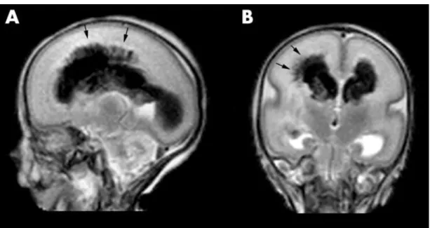
Global hypoxia-ischemia most often damages specific structural groups (4, 15, 16). Gestational age and the level of CNS maturity determine the characteristics of the HI lesions and the associated clinical patterns of disability. The lack of cerebral circulatory autoregulation and the presence of fragile, poorly anastomosed blood vessels in the germinal matrix (17, 18) put preterm neonates at extremely high risk for germinal matrix - intraventricular hemorrhage (GM-IVH), one of the most devastating forms of permanent perinatal injury (Figure 1).
Figure 1. MRI image showing a characteristic injury of the premature brain, germinal matrix - intraventricular hemorrhage on both sides with parenchymal involvement on the right (arrows). A) Sagittal and (B) coronal T2 weighted FSE image of an infant at 27 weeks GA. The images were published by Counsell et al. (19).
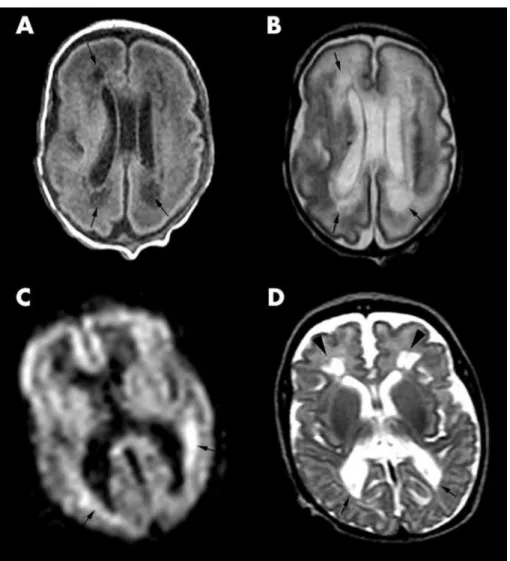
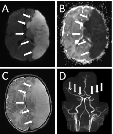
Figure 2. MRI image showing a characteristic form of injury of the premature brain, periventricular leukomalacia (PVL). Image was published by Counsell et al. (19), with the following figure legend: PVL in an infant at 28 weeks gestational age. (A) Transverse T1 weighted image at the mid-ventricular level showing cystic PVL as areas of low signal within the cerebral white matter posterior and anterior to the lateral ventricles (arrows). (B) Transverse T2 weighted FSE image at the mid- ventricular level showing the cystic lesions as high signal intensity (arrows). (C) DWI image showing areas of restricted diffusion around the lateral ventricles as high signal intensity (arrows). (D) T2 weighted FSE image of the same infant at 40 weeks GA showing squared off posterior horns of the lateral ventricles (arrows) and diminished white matter posteriorly. Cystic lesions are shown anterior to the anterior horns of the lateral ventricles (arrowheads)
Until the 32nd week of fetal development, the selective vulnerability of late oligodendrocyte progenitors (20) make the periventricular white matter especially susceptible to damage, making periventricular leukomalacia (PVL) (Figure 2), often associated with spastic palsy, the other common form of injury in preterm neonates (4). The consequent motor, sensory and cognitive deficits show specific patterns characteristic for preterm brain injury, for example cortical visual impairment (2).
Term neonates on the other hand, respond with different patterns of injury to severe or “near-total asphyxia”. Based on magnetic resonance imaging (MRI), the most common types of damage are parasagittal cortical injury and the symmetric injury of basal ganglia, the thalamus, hippocampus and the peri-Rolandic cerebral cortex (21-26), often along with brain stem injury (Figure 3). The fragility of vessels decreases by this time and if IVH is present, it usually is associated to thalamic bleeding (13). Periventricular white matter damage is also uncommon, although transient MRI-signal alterations often show the involvement of the posterior internal capsule (25, 27). The associated clinical symptoms in term neonates are severe, permanent motor impairment with rigidity, which affects the upper extremities more than the lower ones and motor speech impairment (22, 4, 24). One factor, which can influence the gestation-age dependent pattern of HI injury is the localization of the
“watershed” boundary zones, which changes as the vascularization of the fetal brain evolves (8). In term infants these lie between the major cerebral arteries and overlap with the structures most affected by HI. Although cerebrovascular factors might have an impact on the initial HI insult, it appears, that the endogenous vulnerability of certain structures in the developing brain might play a much more important role in determining the final pattern of injury and disability (4).
The selective vulnerability of the developing brain to hypoxic-ischemic injury has been the focus of intensive research for decades, however, many questions remain.
One working model proposes, that the key to understanding selective neuronal vulnerability in term infants lies in the position of the vulnerable structures within the maturing excitatory neuronal circuits (28). It suggests, that excitotoxicity, which is the selective death of neurons and glia due to the overstimulation of excitatory neurotransmitter - mainly glutamate – receptors, is one of the key processes in the pathological development of HIE. In fact, every vulnerable region in term neonates
(the thalamus, putamen and peri-Rolandic cortex) receives major excitatory glutamatergic input (29).
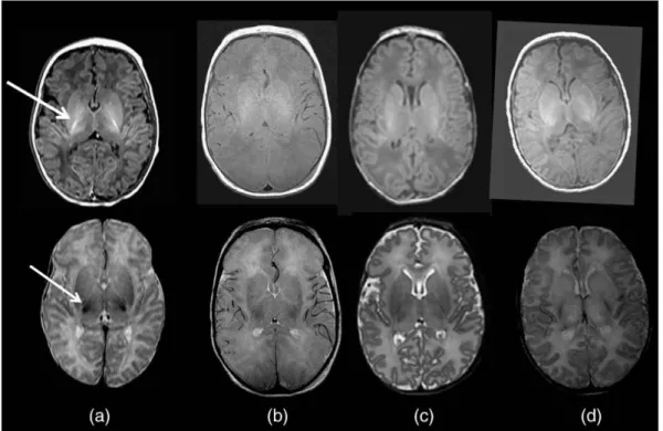
Figure 3. MRI images comparing the brain of a healthy term neonate with three term neonates with HIE. This image was published by Rutherford et al. (30) with the following legend: the posterior limb of the internal capsule. T1 weighted images (top row) with T2 weighted images (bottom row). (a) Normal appearances of the posterior limb of the internal capsule (arrows). (b, c, d) Abnormal appearances of paired T1 and T2 weighted sequences in three different infants with HIE.
Authors hypothesize, that these circuits become hyperactivated and the synapses are flooded with glutamate, causing excitotoxicity, which triggers selective neuronal death (4, 31, 32, 6). This model also explains why structures such as the internal and external segments of the globus pallidus are spared during HIE, but not in other disorders, such as kernicterus (28), since they are strongly inhibited during the hyperactivation of these circuits (22). Furthermore, the inhibition of the globus pallidus, alleviates the inhibition of the thalamus, which leads to enhanced cortical stimulation. This is consistent with the clinical signs of HIE, which indicate cortical hyperactivity, such as seizures and abnormal electroencephalographic
(EEG) background activity (3, 6, 33). Further evidence suggests, that vulnerable regions show selective glucose hypermetabolism (6), which appears to reflect enhanced glutamate release and reuptake at the hyperactive excitatory synapses (34) and correlate with outcome (35).
Oxidative stress appears to be the other major cause of neuronal damage following perinatal HI injury by triggering apoptosis via the mitochondria along other effects (36). Oxidative stress emerges due to the imbalance between the production and neutralization of free radicals, such as reactive oxygen species (ROS) and reactive nitrogen species (RNS) (8). The developing brain is highly vulnerable to oxidative stress, much more than the adult brain due to its higher lipid content (37), the uniquely high levels of polyunsaturated fatty acids, its high oxygen requirement and thus, increased ROS-generating capacity, the low concentrations and activity of antioxidants and the higher levels of redox-active iron (38-43).
Nitric oxide (NO), although an important physiological mediator (44), when produced in excess amounts, has neurotoxic effects via the generation of RNS, such as peroxynitrite (OONO) (45). RNS-mediated damage includes lipid peroxidation, impairment of mitochondrial respiration and the induction of ROS production (46-48).
The nitric oxide synthase (NOS) enzyme has three major isoforms: the neuronal NOS (nNOS), the endothelial NOS (eNOS), and the inducible NOS (iNOS), which is mainly found in activated inflammatory cells (8, 49). Increased NO production in specific cerebral regions appears to be an important component of selective ischemic vulnerability (50). Cells expressing the immature form of N-methyl-D-aspartate (NMDA) glutamate receptor are generally highly resistant to HI injury and NMDA mediated excitotoxicity (51, 52) and are abundant in regions, which are known to be selectively vulnerable to HI, such as the basal ganglia and thalamus (53). Increased stimulation after HI injury via this NMDA receptor leads to robust NO production due to the up-regulation of nNOS (54). Both the elimination of these neurons and the inhibition of NMDA coupling lead to the amelioration of ischemic injury (55). Furthermore, nNOS knockout in mice proved to be highly neuroprotective (56, 57). The inhibition of nNOS and iNOS also improved long-term functional outcome (58). Overall, it appears, that excitotoxicity and oxidative stress are the two most important mediators of perinatal HI brain damage and are key to understanding
selective ischemic vulnerability (8, 50).
Another important characteristic of the immature brain is, that neurons are much more prone to undergo apoptosis than in the adult brain, as programmed cell death is an important mechanism for refining newly formed neuronal pathways in the developing brain (59, 50, 60, 61). Compared with adult acute ischemic stroke (AIS), the ratio of apoptotic vs necrotic cells is much higher in the basal ganglia and cerebral cortex of term neonates one week after HI injury (62, 4, 63). This suggests, that apoptosis is a key step in the pathomechanism of HI brain injury, as is indicated by the elevated level of death-receptor proteins in the brain and cerebrospinal fluid (CSF) of neonates after HI brain injury (64, 65).
There is clear evidence from imaging studies and animal models, that the HI injury in the neonatal brain evolves in the first days to weeks after the initial insult (66) and the majority of the cell loss happens as a consequence of the ensuing neuroimmune response (67-69). The release of excitatory and inflammatory signals promotes robust microglial activation, initiating leukocyte migration to the CNS, which in return induce further inflammation. This cascade of events known as neuroinflammation, leads to the release of a wide range of inflammatory mediators, increased excitotoxicity and oxidative stress and the further destruction of the BBB (8). These processes all lead to accelerated cell death in the immature brain via apoptosis or necrosis (50). The growing understanding of the selective ischemic vulnerability of the developing brain has also opened the chance for new immunomodulatory therapeutic approaches in the form of NMDA antagonists combined with caspase inhibitors, which could yield an enhanced neuroprotective effect by inhibiting excitotoxicity and apoptosis at the same time (4, 70, 71).
Along the increased vulnerability, it is also important to consider, that the neonatal brain has a remarkable ability to recover and remodel after HI injury, due to its high level of neuroplasticity. Synaptic organization and the development of white matter pathways continues for years after birth (72, 73). One of the most relevant of these processes, is the activity-dependent plasticity of the descending motor pathways, which can influence outcome after unilateral hypoxic brain injury (74). Therefore, there is intensive research regarding neurodevelopmental techniques, which could alter this process in a favorable manner (74). Several animal models have confirmed,
that normal corticospinal tract connectivity and motor cortical map can be re- established, and motor function can be improved (75, 76) with electrical stimulation and/or targeted physiotherapeutic training.
It appears, that the neurogenesis capacity of the developing brain is not damaged by HI, in fact, in vivo data shows, that neural stem cell proliferation increases in the subventricular zone after HI in mice (77, 78). Evidence also indicates, that compared to the mature, adult brain, where remodeling is very limited, the developing brain has great capacity to modify its structure and function to overcome functional loss after HI injury (79, 80, 73). The developing brain retains its ability to “recruit new areas and form new circuits by reorganizing the neural network” (81, 82). Jung et al.
utilized novel MR imaging techniques to demonstrate neuroplasticity on many levels, from cellular to systemic changes, such as cortical remapping during the spontaneous functional recovery of mice who sustained neonatal HI injury. They found, that the contralesional hemisphere was able to take over the governing of the motor function of both sides, which authors interpret as “spontaneously generated active plasticity with brain laterality established autonomously via the modification of neural circuitry”. This process was associated with the recovery of motor functions (73).
However, it is important to distinguish “good plasticity” from “bad plasticity”. As an example, excessive stimulation of developing neuronal circuits due to HI injury and subsequent seizures, can lead to altered signaling and neurotransmission, wrong targeted innervation and the interruption of developmental apoptosis. This could result in abnormal neural circuitry, which contribute to epilepsy, motor and cognitive impairment (83, 84).
In conclusion, although, a growing body of evidence supports the higher neuroplasticity of the developing brain compared to the adult brain, it still appears, that the immature brain sustains far worse neurodevelopmental outcomes following severe ischemic injury (2). This indicates, that the crucial vulnerabilities of the developing brain outweigh its enhanced capacity for regeneration (83).
2.2 PERINATAL ASPHYXIA
2.2.1 DEFINITION AND INCIDENCE
Several terms are often used as synonyms to describe the clinical syndrome, which evolves in the days following HI injury in neonates such as hypoxic-ischemic encephalopathy (HIE), neonatal encephalopathy (NE), perinatal asphyxia and birth asphyxia (85). As per the definition of Nelson and Leviton, NE is a “clinically defined syndrome of disturbed neurologic function in the earliest days of life in the term infant, manifested by difficulty with initiating and maintaining respiration, depression of tone and reflexes, subnormal level of consciousness, and often by seizures”. This definition does not however specify etiological cause, and in fact, not all neonates who present with NE have an obvious history of perinatal hypoxic-ischemic episode (86).
According to the definition of Volpe, hypoxemia is the “diminished amount of oxygen in the blood supply”, whereas cerebral ischemia is the “diminished amount of blood perfusing the brain”. As detailed previously, evidence shows, that the latter has greater impact in neonates, as glucose is also deprived from the developing brain along with oxygen, contributing to the primary energy failure (85, 87). The term asphyxia in addition refers to the “impairment of the exchange of blood gases oxygen and carbon dioxide”, which occurs in the fetus if the umbilical or placental perfusion is compromised (85). To try to maintain a temporary homeostasis, the fetal circulation becomes rearranged, reducing nonobligatory energy consumption and increasing the blood flow of the brain, heart and adrenal glands. However, glucose reserves are rapidly consumed, leading to anaerobic metabolism and severe metabolic acidosis, due to lactate accumulation (2). In the clinical practice, indirect markers are used to estimate the degree of hypoxia-ischemia, such as low Apgar scores, decreased peripheral blood pH and acid-base parameters indicating metabolic acidosis. If these alterations are present along with the signs of encephalopathy (altered state of consciousness, hypotonia, abnormal reflexes, clinical seizures (88)), the diagnosis of hypoxic-ischemic encephalopathy can be established (85). Although there is a significant overlap in the above definitions, for the sake of clarity, the terms perinatal asphyxia and consequent hypoxic-ischemic encephalopathy (HIE) will be used in this thesis.
The incidence of HIE ranges between 1.0 and 8.0 per 1000 live births worldwide, with higher percentages in the developing world (85). In the developed world, the incidence of neonatal encephalopathy (NE) is between 2.5 and 3.5 per 1000 live births with a combined point estimate of 3.0 per 1000 live births, while the incidence of HIE is estimated to be between 1.3 and 1.7 per 1000 live births with a combined point estimate of 1.5 per 1000 live births. (85).
Perinatal asphyxia continues to be a major cause of perinatal mortality and long-term disability, despite the advances in neonatal healthcare and the introduction of therapeutic hypothermia as part of standard care (2). Perinatal asphyxia is accountable for approximately one million (23% of all) neonatal deaths worldwide (89). In the developed world, at least 0.5% of term neonates required resuscitation after birth due to a 5-min Apgar score of 6 or less (90), and in 2013 in the Netherlands at least 0.2% of all term neonates were severely depressed with a 5-min Apgar score of 3 or less. Among the latter group mortality was 24% (91).
Even though some surviving children show favorable neurological outcome, many sustain neurodevelopmental disabilities ranging from fine motor impairments and developmental delay to severe hindrances such as mental retardation, sensory impairment, cerebral palsy and epilepsy (92). Data indicates, that despite the significant reduction of perinatal morbidity in the developed world, the incidence of cerebral palsy has remained almost the same in the past 40 years (8, 93). The degree of individual variability in neurological outcome after perinatal asphyxia is especially intriguing, since it shows little correlation with the initial clinical status. It is thus crucial to constantly improve our understanding of the pathophysiology of HIE, since the key pathological factors regarding outcome are likely to be part of the neuroimmune response of the neonate to the initial HI insult.
2.2.2 RISK FACTORS
Several studies aimed to identify the potential risk factors of HIE, however, there was a high level of variability between the studies. In an Italian case-controlled study, out of the included 30,580 infants, 27 (0.09%) developed NE. In 26% of the cases only antepartum risk factors were present, in 22% only intrapartum risk factors could be identified, including acute intrapartum events, whereas in 44% of the cases both antenatal and peripartum risk factors were present. In 2 cases (7%) however, no risk factors could be identified (94). In the largest case-controlled study performed in Western Australia, in 69% of cases only antepartum risk factors were present, whereas only intrapartum risk factors could be identified in just 4% of the cases. In 22% of cases both were present, and in 2% no risk factors could be identified. There is also much discussion regarding the timing of the HI insult, however, prospective MRI based studies indicate, that the majority of HI injury is likely to occur at or close to the time of birth (95).
Several risk factors of NE have been identified, many of which are common in developed and developing countries. Identifying modifiable risk factors is a key point in current research. Risk factors, which have been shown to be associated with perinatal HIE are shown in Table 1. The presented information has been collected from the two largest case-controlled studies, one from Western Australia and the other from Nepal. Interestingly, there is a considerable overlap in the risk factors, despite the major differences in the socioeconomic background of the patients in the two studies.
Antepartum maternal risk factors include older maternal age, hypertension, thyroid disease, infertility treatments, severe pre-eclampsia and several sociodemographic factors (employment, insurance, etc.). Intrapartum complications such as cord abnormalities, maternal fever or prolonged membrane rupture to delivery interval resulted in a non-significant increase in the risk of NE in the Western Australian study. Non-cephalic presentation, forceps delivery, emergency section and catastrophic intrapartum events such as placental abruption or uterine rupture were associated with HIE (96, 97, 50, 85).
Of the fetal risk factors the most common were growth restriction in Western Australia and twin pregnancy in Nepal. Risk factors also included fetal distress and meconium stained amniotic fluid, however these are also the consequences of
intrauterine hypoxia and thus cannot be considered independent risk factors (96, 97, 85). Postnatal complications were present in less than 10% of cases and were severe respiratory distress, shock and sepsis (50).
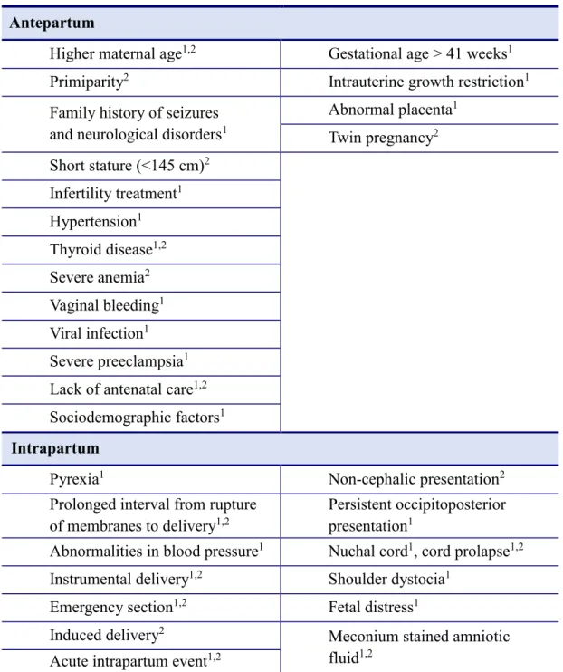
Table 1. Summary of the risk factors of neonatal encephalopathy (NE). The data were collected from the two largest case-control studies. 1 significant risk factor in Western Australian study, 2 significant risk factor in Nepal study (98, 96, 97).
Maternal risk factors Fetal risk factors Antepartum
Higher maternal age1,2 Gestational age > 41 weeks1 Primiparity2 Intrauterine growth restriction1 Family history of seizures
and neurological disorders1
Abnormal placenta1 Twin pregnancy2 Short stature (<145 cm)2
Infertility treatment1 Hypertension1 Thyroid disease1,2 Severe anemia2 Vaginal bleeding1 Viral infection1 Severe preeclampsia1 Lack of antenatal care1,2 Sociodemographic factors1 Intrapartum
Pyrexia1 Non-cephalic presentation2
Prolonged interval from rupture of membranes to delivery1,2
Persistent occipitoposterior presentation1
Abnormalities in blood pressure1 Nuchal cord1, cord prolapse1,2 Instrumental delivery1,2 Shoulder dystocia1
Emergency section1,2 Fetal distress1
Induced delivery2 Meconium stained amniotic
fluid1,2 Acute intrapartum event1,2
2.2.3 PATHOPHYSIOLOGY
Perinatal asphyxia evokes the injury of the central nervous system (CNS) due to the severe lack of oxygen and perfusion, leading to hypoxic-ischemic brain injury.
In the initial phase, due to circulatory redistribution, a temporary compensation can be maintained, where nonobligatory energy consumption is minimized, cerebral blood flow is increased regionally, and neuronal activity is suppressed (99, 100, 2, 101). After a while cerebral blood flow gradually decreases due to either regional vasoconstriction or the progressive collapse of cardiac output (8, 102, 100). This leads to reduced oxygen delivery, decreased oxidative phosphorylation causing the depletion of high energy phosphate compounds (i.e. ATP), the rapid consumption of glucose reserves, and severe lactate acidosis, as a result of anaerobic metabolism (103-105, 2). This primary energy failure damages the BBB, leads to cerebral edema and initiates excitotoxicity and oxidative stress, culminating in the first wave of necrotic and apoptotic cell death. (8, 106, 107, 14).
If neonatal resuscitation is successful, glucose and ATP reserves are temporarily restored. However, this recovery phase is followed by the second wave of energy failure within 6 hours, as the HI brain injury continues to develop over the next weeks (2, 108, 6). The main features of the second phase are the secondary energy failure, characterized by an even more pronounced exhaustion of cellular ATP stores, further increase in lactate levels and pH alterations (109, 110, 2), the robust neuroinflammatory response and the decrease of cerebral autoregulation (8, 111, 112, 40). ATP deprivation combined with lactate acidosis leads to the failure of active membrane transport, leading to intracellular accumulation of Na+, Ca2+, and Cl− ions.
Consequently, prolonged neuronal membrane depolarization occurs, leading to excessive presynaptic glutamate release (113). In the absence of ATP and glucose, the function of glutamate reuptake pumps on perisynaptic glial cells is compromised (4, 34), leading to the accumulation of extracellular glutamate and other excitatory neurotransmitters, which causes the overstimulation of ionotropic glutamate receptors such as NMDA and α-amino-3-hydroxy-5-methyl-4-isoxazole (AMPA) receptors (40).
This leads to excessive calcium influx, initiating the excito-oxidative cascade.
Intracellular calcium overload activates lipases, proteases and endonucleases, leading to the degradation of the cytoskeleton, mitochondria dysfunction, the swelling of the
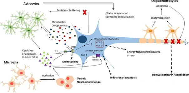
cell and eventually apoptosis or necrosis (8, 4, 114, 2). Membrane depolarization and excessive excitatory stimulation can also induce seizures (115). Since glutamate receptors are vital regulators of the fetal brain development, they are expressed abundantly, therefore severe fluctuations in excitatory input have massive detrimental consequences (116, 40). The neuroinflammatory response is interconnected with this vicious circle in many ways. The activation and invasion of inflammatory cells results in the robust release of cytokines and other mediators, which continue to exacerbate the excito-oxidative cascade (8, 40) (Figure 4). In light of the above, it is not very surprising, that the majority of accelerated cell death happens during this second phase, where most of the free radicals, inflammatory mediators and excitatory amino acids are released (67, 68, 2, 69).
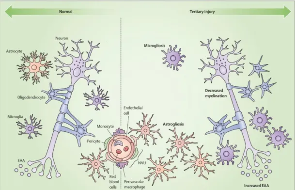
Figure 4. Key features of the neuroinflammatory response
This image was originally published by Sajja et al. in Frontiers (117) illustrating the contribution of glial cells to secondary brain injury.
Following the initial few days, the tertiary phase of the HI brain injury begins, which can continue for weeks after the initial insult. The primary mediators of this phase are the activated inflammatory cells and depending on the local homeostasis created by the neuroinflammatory response, the excito-oxidative injury can be sustained, and repair mechanisms can be stalled (118). The neuroinflammatory response is a complex process, which may have dual aspects being a hindrance, but
also a significant help in the recovery of the CNS. The substantial variability in outcome is likely related to the characteristics of the individual response to the initial HI insult, therefore understanding its characteristics is the primary aim of current research in the field. The neuroinflammatory response will be discussed in detail in Chapter 2.4.
2.2.4 CLINICAL PRESENTATION
Evidence indicates, that the majority of HI injuries happens during or in close proximity to birth (119). Term neonates generally present with symptoms of perinatal asphyxia directly after birth. The first mode of assessment is the Apgar score (120, 91), which is an evaluation of the cardiac, pulmonary and neurological status of the neonate at 1, 5 and 10 minutes after birth. The consequences of a severe HI insult are often already present at birth, indicated by a 5-min Apgar score under 5. Further signs of severe asphyxia include acidosis within 60 minutes of birth indicated by “umbilical- cord, arterial, or capillary pH of <7.00 or base deficit of ≥16 mmol per liter, signs of moderate-to-severe encephalopathy (lethargy, stupor, or coma), abnormal tone (focal or general hypotonia or flaccidity), abnormal reflexes (absent or weak suck or Moro response, sometimes oculomotor or pupillary abnormalities) and clinical seizures”
(121).
Symptoms usually evolve during the first days of life and their severity and persistence is in correlation with the severity and duration of the HI insult (50, 91, 122, 6). Based on the clinical presentation, HIE can be classified as mild, moderate or severe (Table 2). Mild HI insult might manifest in subtle neurological abnormalities (such as transient drowsiness) without multi-organ damage, whereas a severe insult usually leads to moderate-to-severe HIE and variable degree of multi-organ failure (91, 123).
During the first 12-24 hours neonates with mild HIE generally show some neurological alterations, such as restlessness, moderate hypertonia, often associated with periodic breathing and feeding difficulty, which are likely associated with
“abnormal stimulation or transient malfunction of the brain” (91). The most common systemic symptom is decreased urinary output. After 24 hours neonates with mild-to- moderate HIE can improve in their overall clinical and neurological status. They often
reach normal level of consciousness and begin to tolerate feeds during the first week of life. Laboratory parameters usually show a similar tendency (91).
Neonates with moderate-to-severe HIE on the other hand often show more profound neurological alterations during the first days, such as “depressed state of consciousness, gazing with abnormal papillary size, hypotonia, periodic breathing with apnea, bradycardia, and signs of seizure activity” (50, 91). In the most severe cases, neonates are often lethargic, and stupor and early onset, overt seizure activity is also common (91). In term infants, seizures are usually multifocal and clonic and can manifest in apneic spells (91). Systemic symptoms almost always include kidney damage and respiratory insufficiency with prolonged apnea, which often makes mechanic ventilation necessary around 48-72 h of life. HI injury can cause cardiomyopathy, with the need for inotropes, the severity of which is in close relations with the severity of the HI insult (124). As the HIE progresses, after 24 hours either a gradual stabilization or deterioration can be observed. In the first case, a gradual stabilization of physical parameters and improvement of alertness level occurs, however, neurological abnormalities, such as “hypo- or hypertonia (in the case of basal ganglia damage), feeding difficulties, decreased suck, abnormal swallow” likely reside (91). Hypotonia and proximal limb, facial and bulbar muscle weakness with amplified reflexes can persist for months after HI brain injury (50). In the second case, a gradual deterioration occurs, most likely as a consequence of the secondary phase of HI injury and neuroinflammation (109, 68). Seizures often become refractory, indicated by
“apneic episodes, shrill cry, and jitteriness” (50). Neonates’ consciousness level progressively deteriorates, as brain stem functions start to be affected, and mechanic ventilation is almost always inevitable. Responsiveness decreases, seizure activity increases, and pupils become dilated (91). In the most severe cases, neonates die within the first week of life. Autopsy-based studies revealed cytotoxic brain edema and extensive neuronal damage after lethal perinatal asphyxia (125, 91).
2.2.5 DIAGNOSIS
2.2.5.1 CLINICAL STAGING
Prenatal diagnostic methods (such as fetal heart rate patterns, scalp blood gas, meconium passing) may indicate fetal distress, however, they are poor predictors of the development of HIE and of long term neurological outcome (91, 126). The Apgar score is a somewhat better tool for direct postnatal assessment, however it is still far from reliable. A 10 min Apgar score of 5 or less predicts perinatal HI with 43%
sensitivity and 95% specificity (127). However, HI injury cannot be ruled out in neonates with a good Apgar score (91), as 1 min Apgar score of 3 or less was found in only 31% of neonates with HIE (128).
The clinical features of the first few days show better correlation with future neurodevelopmental outcome, therefore, the need for clinical staging became evident.
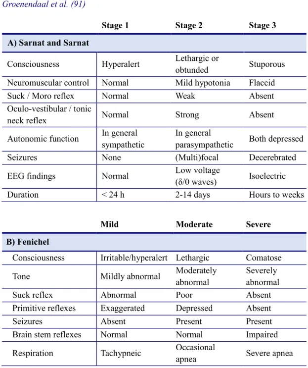
The first clinical staging system of HIE was established by Sarnat and Sarnat in 1976, it defines 3 stages of HIE based on neurological status and EEG characteristics of affected neonates (91, 6). This scoring system was then modified by Fenichel in 1983, to define mild, moderate and severe HIE, by including only clinical features (129) (Table 2). The latter system became the standard mode of clinical staging of HIE in neonates in the past decades worldwide, however, neurodevelopmental outcome cannot be reliably predicted based on solely the clinical severity of HIE.
In the “TOBY (TOtal Body hYpothermia) - Whole body hypothermia for the treatment of perinatal asphyxial encephalopathy trial”, term neonates with moderate- to-severe perinatal asphyxia were selected for total-body moderate hypothermia, initiated within 6 hours of birth and continued for the first 72 hours of life (121). They established the inclusion criteria to identify clinical features indicative of a developing encephalopathy early on (88). A modified version of this criteria system is used in the clinical practice to select neonates eligible for therapeutic hypothermia (see below).
Table 2. Clinical staging of hypoxic-ischemic encephalopathy according to Sarnat and Sarnat (6) and Fenichel (129) This table is based on the publication of Groenendaal et al. (91)
Stage 1 Stage 2 Stage 3
A) Sarnat and Sarnat
Consciousness Hyperalert Lethargic or
obtunded Stuporous
Neuromuscular control Normal Mild hypotonia Flaccid
Suck / Moro reflex Normal Weak Absent
Oculo-vestibular / tonic
neck reflex Normal Strong Absent
Autonomic function In general sympathetic
In general
parasympathetic Both depressed
Seizures None (Multi)focal Decerebrated
EEG findings Normal Low voltage
(δ/0 waves) Isoelectric
Duration < 24 h 2-14 days Hours to weeks
Mild Moderate Severe
B) Fenichel
Consciousness Irritable/hyperalert Lethargic Comatose
Tone Mildly abnormal Moderately
abnormal
Severely abnormal
Suck reflex Abnormal Poor Absent
Primitive reflexes Exaggerated Depressed Absent
Seizures Absent Present Present
Brain stem reflexes Normal Normal Impaired
Respiration Tachypneic Occasional
apnea Severe apnea
The following criteria are used to select neonates eligible for therapeutic hypothermia.
Neonates meeting both criteria A and B may be considered for treatment with cooling.
A) Infants ≥36 completed weeks gestation admitted to the neonatal unit with at least one of the following:
Apgar score of ≤ 5 at 10 minutes after birth
Continued need for resuscitation, including endotracheal or mask ventilation, at 10 minutes after birth
Acidosis within 60 minutes of birth (defined as any occurrence of umbilical cord, arterial or capillary pH < 7.00)
Base Deficit ≥ 16 mmol/L in umbilical cord or any blood sample (arterial, venous or capillary) within 60 minutes of birth
B) Seizures or moderate to severe encephalopathy, consisting of:
Altered state of consciousness (reduced response to stimulation or absent response to stimulation) AND
Abnormal tone (focal or general hypotonia, or flaccid) AND
Abnormal primitive reflexes (weak or absent suck or Moro response)
2.2.5.2 NEUROPHYSIOLOGY
The assessment of neonates’ brain activity by EEG has become an important tool in clinical staging, evaluating eligibility for therapies such as hypothermia, and predicting outcome (130, 131, 91, 132-134). Since the characteristics of the background activity in the first few days of life appear to be more important than those of the seizure activity, conventional multi-channel EEG recordings have limited value, as they give a detailed overview of a short time period (30-45 minutes) (135). In clinical practice, the usage of a simplified method, the amplitude-integrated EEG (aEEG) has become widespread, as it is easy to apply, correlates well with conventional EEG data and allows a continuous monitoring of brain activity in the first days of life (91, 136). Data from aEEG monitoring has predictive value, especially of severe injury, as suppressed background activity after 24 hours, or flat or continuous low voltage in the first 6 hours was almost always associated with poor outcome (137, 91, 138, 139). However, in neonates, where the background activity becomes normal within 24 hours, 39% still developed some neurological disability later on (139). On the other hand, continuous normal background activity was associated with normal
outcome (91, 138). Evoked potentials (EP) are electrical responses to repetitive visual, auditory or somatosensory stimulation, which can be assessed on the EEG. Visual and somatosensory evoked potentials in the first week of life (VEP and SEP respectively) also correlate with outcome, however, their clinical use is much less common than that of the aEEG (137, 91).
2.2.5.3 NEUROIMAGING
Although cranial ultrasound has been used for many years and is a good tool for bedside monitoring, its sensitivity after perinatal HI injury is far exceeded by magnetic resonance techniques (91). In the past decades, cranial magnetic resonance imaging (MRI) has become the standard diagnostic method following perinatal HI injury (95, 140) (Figure 5). MRI is the most informative, when performed between 4- 7 days of life, as HI brain lesions acquired in the perinatal period are the most likely to be visible at this time period (141, 91). Following perinatal HI, the classical pattern of injury on MRI images is abnormalities in the thalamus and basal ganglia, and the surrounding white matter in the most severe cases (91, 142). Specific modalities, such as diffusion-weighted (DW) MRI are highly sensitive to early changes elicited by HI injury, especially if cranial mapping is performed by calculating the apparent diffusion coefficient (ADC) of water, based on which early cytotoxic edema can be differentiated from vasogenic edema (91, 143). Although DWI is a sensitive mode of evaluating the ischemic injury in the early phase, it often underestimates the final extent of damage, especially in the basal ganglia and thalamus (30).
Cranial magnetic resonance spectroscopy (MRS) can also be a valuable tool, as it provides insight into the metabolic changes of the CNS following perinatal HI.
Poor neurological outcome was associated with elevated lactate level in the basal ganglia and thalamus of neonates on 1H-MRS (144). Lower levels of phosphocreatine and ATP also correlated with worse outcome (91, 109, 68).
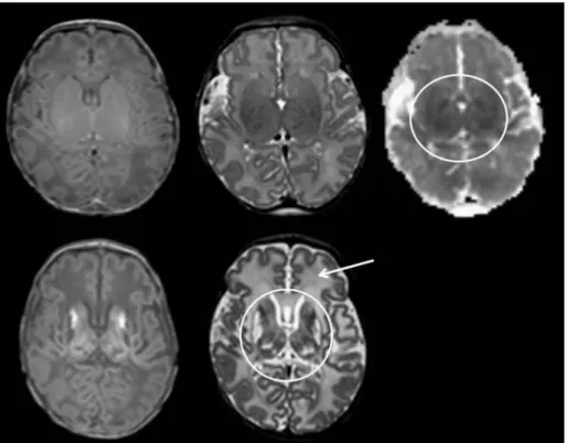
Figure 5. Different MRI modalities for assessing perinatal HIE. This image was published by Rutherford et al. (30) with the following legend: Diffusion-weighted imaging in the basal ganglia and thalami. Top row. T1 (left) and T2 (middle) weighted images and ADC map (right) acquired on day 3. The ADC map shows several regions of abnormal low signal intensity within the basal ganglia and thalami (BGT) that look relatively subtle (circular region of interest). Bottom row. Day 22. T1 (left) and T2 (right) weighted images. The BGT abnormalities are very severe with no normal looking tissue within the BGT (circular region of interest). Any abnormality within the BGT on an early ADC map is likely to be clinically significant and any one diffusion- weighted examination is likely to underestimate the final lesion load. By day 22 the white matter signal intensity (arrow) has become abnormally high and there has been no head growth in this neonate.
2.2.6 THERAPY
Neonates born with poor Apgar score due to perinatal asphyxia often require perinatal resuscitation and subsequent intensive supportive care in the first weeks of life. The most important aspects of supportive care are adequate ventilation and oxygenation, correction of acid-base status, cardiac support, anti-convulsive treatment, the maintenance of normal blood glucose and electrolyte levels, and the management of blood count abnormalities (145, 146). Hypoxia (91) and hyperoxia(147), hypocapnia (148), hyperglycemia (149), hypotension and hypoperfusion (124), metabolic acidosis, and seizures (150) have all been documented to aggravate perinatal HI brain injury (146, 91). Therefore, careful management of these parameters is essential in the first week after perinatal asphyxia. In the first 48 h of life enteral feedings should be withheld due to an increased risk of necrotizing enterocolitis, however, sufficient caloric intake should be established as soon as possible (151, 91).
Research data indicates, that the most critical time window for neuroprotective intervention is the first 6 hours after birth, as this is likely the time between the initial HI insult and resuscitation and the secondary brain damage (146, 152). In the current clinical practice, hypothermia is the only neuroprotective treatment, which specifically targets the consequences of HI brain injury and has been proven to be beneficial in large clinical trials (146). Following the TOBY trial, published in 2014, hypothermia has become the standard of care for term neonates with perinatal asphyxia and consequent HIE. Authors found, that total body moderate hypothermia, did not alter combined rates of death and severe disability, but improved neurological outcome in survivors at 18 months (130). However, according to a Cochrane review published in 2013 of 11 randomized control trials with therapeutic hypothermia, moderate hypothermia also reduced mortality in term and late preterm newborns with HIE (153).
There is evidence, which indicates, that treatment with moderate hypothermia after perinatal asphyxia led to improved neurocognitive outcome at school-age, however, the opposite has also been reported (130, 154). In the current clinical practice, therapeutic moderate hypothermia is induced within the first 6 h of life and is maintained for 72 hours (146). There is also evidence, that hypothermia initiated before 3 hours of life yields further benefits regarding outcome (155). The neuroprotective effects of hypothermia include the suppression of cerebral
metabolism, the reduction of excitatory neurotransmitter and free radical release, the inhibition of apoptosis, seizure prophylaxis and the amelioration of the neuroinflammatory response (146).
In the past decades, novel neuroprotective strategies have been the primary focus of research in the field of perinatal HI brain injury (156). Many neuroprotective agents are being investigated, which either target the excito-oxidative cascade or the neuroinflammatory response. Blocking the inflammation after perinatal HI injury appears to confer neuroprotective effect, which could yield important therapeutic benefits (157-160). So far, few pharmacological agents proved to be effective in human preclinical trials (161). One of the most promising agents is erythropoietin (EPO), which has pleiotropic effects, such as neuroregenerative, angiogenic, anti- inflammatory, and antiapoptotic effects (162-165, 161). Neurons, astrocytes and microglial cells all express EPO receptors, upregulate its expression upon HI insult and neuroinflammation (165) and EPO has been shown to exert anti-inflammatory and neuroprotective effects (166). A pilot trial of recombinant human EPO (rhEPO) in perinatal asphyxia showed that treatment resulted in improved EEG background activity and decreased serum NO levels at 2 weeks of age and fewer neurologic and developmental abnormalities at 6 months of age (167). However, in NAIS rhEPO treatment did not alter the extent of the brain infarction or neurodevelopmental outcome (168). Phase II and III clinical trials are currently in process (165). The administration of melatonin is also currently being assessed in clinical trials (NCT02621944). Melatonin also has pleiotropic effects, targeting several steps of the neuroinflammatory cascade and has been shown to exert antioxidative, free radical scavenging and anti-inflammatory effects (169, 170, 161). In piglets, administration of melatonin together with hypothermia decreased neuronal loss and preserved function (171). Xenon is another promising neuroprotective agent, showing synergic neuroprotective effects when combined with hypothermia (172). It is an antagonist of the NMDA glutamate receptor (173) exerting its protective effect primarily via inhibiting intrinsic apoptotic pathways. It has a very low blood-gas partition coefficient (174) enabling it to pass the through the BBB quickly. Xenon has been shown to reduce neuronal injury (175). There are ongoing clinical trials investigating the efficacy of hypothermia combined with xenon inhalation (NCT01545271). Doxycycline is an
important inhibitor of neuroinflammatory response, which reduces of IL-1β and TNF- α expression and is approved for use in neonates, has few side effects, thus making it a promising candidate (176). Histone deacetylases exert pleiotropic anti-inflammatory and neuroprotective effects, via inhibiting the components excito-oxidative cascade, inhibition neuroinflammation and proapoptotic factors (177-179, 161). Further promising anti-inflammatory and neuroprotective agents include selective cyclooxygenase-2 inhibitors, resveratrol, N‑acetylcysteine, TNF-α soluble receptor, IL‑1ra and magnesium, which acts as an (endogenous cationic NMDA channel blocker) (180, 161).
One intriguing recent discovery is, that mesenchymal stem cell (MSC) and neural progenitor cell therapy can protect the developing brain from HI injury. The hypothesis is, that MSCs are either able to “migrate to the site of injury, differentiate into specific cell lineages and exert anti-inflammatory effects” or stimulate the differentiation of endogenous precursors, inducing neuroregeneration and plasticity (77, 2). The strenuous efforts to find novel, individualized anti-inflammatory and neuroprotective strategies clearly outline the paramount importance of this aspect of managing neonates with perinatal HI brain injury. Overall, the past decade has brought much insight into the underlying pathomechanism of perinatal HI injury and potential ways to counteract the detrimental consequences, however, continuous efforts in this field are essential in the future as well.
2.2.7 OUTCOME
In developed countries, perinatal asphyxia continues to be a great burden on society as a leading cause of neonatal mortality and neurodevelopmental disability.
Mortality and morbidity rates vary among the literature, but according to a recent review on outcome by Antonucci et al., in term neonates, the mortality rate is around 20% and about 25% of survivors will develop neurological disabilities after perinatal asphyxia (146, 181, 182). Another review assessed the prevalence of individual neurodevelopmental disabilities after perinatal asphyxia and found, that of all children with adverse outcomes, 45% sustained “cognition and developmental delay or learning difficulties, 29% cerebral palsy, 26% blindness or vision defects, 17% gross motor and coordination problems, 12% epilepsy, 9% hearing loss or deafness, and 1% behavioral problems” (146, 183). A follow-up study by Van Handel et al. however, revealed, that when assessing at school-age, behavioral, social and attention problems were much more common in children, who sustained perinatal asphyxia (184). If neurological outcome was assessed in correlation with the initial clinical stage of HIE, mild insult lead to no serious neurodevelopmental alterations, whereas cognitive impairments and/or sensory-motor impairments or death occurred in 32% of children who initially had moderate HIE and 100% of children with severe initial HIE (185). However, further studies revealed, that subtler cognitive deficits and behavioral problems were also present following mild HIE (186). It appears, that the outcome is the most variable and the most challenging to predict in neonates with moderate HIE (146, 187, 188).
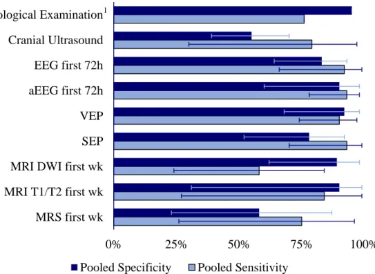
A recent meta-analysis aimed to assess the prognostic value of currently available clinical diagnostic methods in patients with perinatal asphyxia and HIE. Data was collected from 29 studies and they found that overall, clinical and neurological examination and cerebral ultrasound had poor predictive value. In the first week of life, aEEG (sensitivity 0.93, specificity 0.90), conventional EEG (sensitivity 0.92, specificity 0.83), and VEP (sensitivity 0.90, specificity 0.92) had the best prognostic value regarding neurological outcome. From the imaging techniques, if performed in the first week of life, diffusion weighted MRI had the highest specificity (0.89), whereas T1/T2-weighted MRI (performed within the first 2 weeks of life) had the highest sensitivity (0.98). Early MRS had a sensitivity of 0.75 and poor specificity (0.58) (189). The data of this meta-analysis is summarized on Figure 6.
Figure 6. Prognostic value of the diagnostic methods available in the clinical practice. The original data which this figure is based on was published by van Laerhoven et al. (189) Pooled specificity and sensitivity values with confidence intervals are shown for each diagnostic test. 1The sensitivity and specificity of neurological examination is based on data from a single study (190). EEG = Electroencephalogram, aEEG = Amplitude-integrated Electroencephalogram, VEP = Visual evoked potential, SEP = Somatosensory evoked potential, MRI DWI = Magnetic resonance imaging, Diffusion-weighted imaging, MRS = Magnetic resonance spectroscopy.
0% 25% 50% 75% 100%
MRS first wk MRI T1/T2 first wk MRI DWI first wk SEP VEP aEEG first 72h EEG first 72h Cranial Ultrasound Neurological Examination
Pooled Specificity Pooled Sensitivity
1
2.3 NEONATAL ARTERIAL ISCHEMIC STROKE (NAIS)
2.3.1 DEFINITION AND INCIDENCE
The perinatal period carries the highest risk for stroke in the entire childhood, with almost similar incidence to that in the elderly population. “Perinatal stroke” is a broad term defining a group of heterogeneous cerebrovascular events which occur between 28 weeks of gestation and 7 days of age. However, many studies discuss the perinatal and the neonatal period together, extending inclusion to the first 28 days of life and the variation in terminology can be somewhat confusing. Although this definition also includes in utero stroke, most perinatal stroke events are thought to occur within a few hours to a few days post-delivery (191).
Neonatal stroke can be categorized based on the distinct characteristics of the lesion. The most common subtypes in term neonates are neonatal arterial ischemic stroke (NAIS), primary Heamorrhagic stroke and cerebral sinus venous thrombosis (CSVT). Heamorrhagic events associated with prematurity, especially intraventricular hemorrhages account for a significant proportion of neonatal cerebrovascular events, however, we only included term neonates in our studies, therefore these events will not be discussed in detail. NAIS by definition is an arterial ischemic stroke, with clinical symptoms occurring in the neonatal period (within the first 28 days of life) which are supported by radiological evidence of a focal arterial infarction. (192-194).
Many cases of perinatal stroke are only diagnosed later in infancy, with no evidence of an acute neurological event during the neonatal period. “Presumed perinatal stroke”
refers to cases, where the diagnosis is made after 28 days of age, when NAIS most likely remained undetected in the acute phase (191).
The exact incidence of neonatal stroke is difficult to determine and estimates are based on studies of neonatal seizures and population-based studies of childhood stroke (195). The incidence of neonatal stroke is estimated to be around 1/4000 live births, with NAIS occurring in 1/8000 live births.
NAIS generally occurs in term neonates and presents a major risk for life-long motor, cognitive and behavioral disabilities ranging from fine motor impairments to unilateral cerebral palsy, which develops in around 20-30% of affected neonates. Thus NAIS is a leading cause of cerebral palsy (196).
2.3.2 RISK FACTORS
The predisposing factors of NAIS are still under strong investigation and there is little consensus among field experts. There are several challenges in identifying the truly relevant risk factors and their possible impact on the course of AIS in neonates.
Firstly, the number of studies is relatively low and those which are available are mostly case reports or small case series, only very few included enough cases to be representative. Secondly, the enrolling criteria and the recorded data are not homogenous, many studies include AIS and borderzone infarction as well and some do not distinguish between neonatal stroke cases and stroke occurring in older children (192). Another important challenge is the overlap of the risk factors and presentation NAIS with perinatal asphyxia. Furthermore, in the case of certain risk factors, similarly to HIE, it is very difficult to determine their place in the causal pathway. Authors Lee et al. propose the following possibilities for the role of factors such as fetal heart rate abnormalities in the development of NAIS (197):
1) Plays a primary causal role
2) Is on the causal pathway between preceding factor and NAIS 3) Adverse effect of causal factor with no impact on NAIS 4) Direct consequence of NAIS
5) Combination of the above
Further such factors include prolonged second stage of labor, vacuum assistance, and emergency cesarean section. These factors show a clear association with NAIS cases, however the impact of these factors is yet to established, and this must be considered when interpreting findings. Overall, there is little consistency among the different studies (193). Therefore, instead of true risk factors, it is more correct to discuss “associations”, which have been documented in numerous studies (Table 3).