R E S E A R C H A R T I C L E
Horse riding and the shape of the acetabulum: Insights from the bioarchaeological analysis of early Hungarian mounted archers (10th century)
William Berthon
1,2,3 |Balázs Tihanyi
2,4 |Luca Kis
2 |László Révész
4 |Hélène Coqueugniot
1,3,5 |Olivier Dutour
1,3,6 |György Pálfi
21Chaire d'Anthropologie Biologique Paul Broca, EPHE, PSL University, Paris, France
2Department of Biological Anthropology, University of Szeged, Szeged, Hungary
3UMR 5199 PACEA, CNRS/Université de Bordeaux, Pessac, France
4Department of Archaeology, University of Szeged, Szeged, Hungary
5Department of Human Evolution, Max Planck Institute for Evolutionary Anthropology, Leipzig, Germany
6Department of Anthropology, University of Western Ontario, London, Ontario, Canada Correspondence
William Berthon, Chaire d'Anthropologie Biologique Paul Broca, EPHE, PSL University, 4‐14 rue Ferrus, 75014 Paris, France.
Email: william.berthon@etu.ephe.psl.eu Funding information
National Research, Development and Innova- tion Office, Hungary, Grant/Award Number:
K125561; French‐Hungarian Hubert Curien Partnership“Balaton”; Scientific Council of Region Ile‐de‐France; Tempus Public Foundation
Abstract
Horse riding is a human activity that has particularly interested bioanthropologists and paleopathologists working on the reconstruction of activities from skeletal changes in ancient populations. However, various sample and methodological limitations, such as the absence of direct evidence connecting the individuals and the activity, result in a lack of confidence regarding what changes should be included in the so
‐called horse riding syndrome. Focusing on the ovalization of the acetabulum, regularly men- tioned in literature, we analyzed comparative samples of presumed riders and non
‐riders to evaluate its reliability for the identification of horse riding.
We relied on a Hungarian Conquest period collection (10th century CE), including several individuals associated with horse riding equipment or horse bones in the graves.
Direct and easily repeatable measurements were used to calculate an index of ovalization of the acetabulum (IOA). The index values were compared according to the presence or absence of archaeological deposit. An extra
‐group of presumed non
‐riders from the documented Luís Lopes Skeletal Collection (Lisbon) was used for comparison.
Early Hungarians buried with horse
‐related grave goods exhibited a higher overall IOA compared with the ones without and those known not to ride (p = 0.049 in the latter case, with left and right values combined).
Our results suggest that the ovalization of the acetabulum may indeed be a promising indicator to be included in a set of markers for horse riding. The analysis of further different types of pathological and nonpathological skeletal changes (e.g., joint and entheseal changes) will contribute to a more reliable identification of horse riders in anthropological collections.
K E Y W O R D S
activity‐related skeletal changes, anthropometrics, bioarchaeology, equestrian, hip, Hungarian Conquest period, morphology
1
|I N T R O D U C T I O N
Bone changes of different types have been interpreted for decades as activity, occupational, or stress markers, with an important step forward from the 1980s (e.g., Capasso, Kennedy, & Wilczak, 1999;
Dutour, 1986; Hawkey & Merbs, 1995; Henderson & Alves Cardoso, 2013; Jurmain, 1999; Kennedy, 1989; Knüsel, 2000; Mariotti, Facchini, & Belcastro, 2004; Milella, Alves, Assis, Perréard Lopreno,
& Speith, 2015; Niinimäki, 2012; Pálfi & Dutour, 1996; Perréard Lopreno, 2007; Villotte et al., 2010). Among all human activities, horse DOI: 10.1002/oa.2723
Int J Osteoarchaeol. 2019;29:117–126. wileyonlinelibrary.com/journal/oa © 2018 John Wiley & Sons, Ltd. 117
and unmistakable sign of riding (Anthony, 2007). Unfortunately, the existence of such a direct link between specific skeletal changes and the practice of horse riding has not been yet unarguably demonstrated regarding humans. Bioarchaeological research on horse riding has developed strongly, with a growing interest from the beginning of the 1990s especially and the early contributions of Miller on historic Native American Omaha and Ponca skeletal remains (Miller, 1992;
Miller & Reinhard, 1991; Reinhard, Tiezen, Sandness, Beiningen, &
Miller, 1994) and Pálfi on a Hungarian cemetery of the 10th century CE (Pálfi, 1992, 1997; Pálfi & Dutour, 1996).
“Horse(back) riding syndrome”commonly refers to a combination of skeletal changes repeatedly observed and interpreted as signs of riding. It covers various types of pathological and nonpathological changes, which were previously reviewed (Baillif‐Ducros, Truc, Paresys, & Villotte, 2012; McGrath, 2015). To summarise, they can be categorised in different groups of indicators: (a) spinal changes:
osteoarthritis, Schmorl's nodes, and thoracic and lumbar wedging (e.g., Baillif‐Ducros et al., 2012; Blondiaux, 1994; Karstens, Littleton, Frohlich, Amgaluntugs, & Pearlstein, 2017; Reinhard et al., 1994;
Üstündağ & Deveci, 2011; Wentz & de Grummond, 2009); (b) extraspinal joint changes: coxarthrosis and gonarthrosis (e.g., Baillif‐ Ducros & McGlynn, 2013; Fornaciari et al., 2014); (c) entheseal changes: adductor, gluteal, quadriceps, hamstring, external obturator, and soleus muscles (e.g., Belcastro, Facchini, Neri, & Mariotti, 2001;
Blondiaux, 1994; Djukic, Miladinovic‐Radmilovic, Draskovic, & Djuric, 2018; Miller, 1992; Pálfi, 1992; Tichnell, 2012); (d) morphological var- iations: femoral head–neck junction changes (Poirier's facet, plaque, and cervical fossa of Allen) and elongated acetabula (e.g., Baillif‐ Ducros et al., 2012; Courtaud & Rajev, 1998; Dutour & Buzhilova, 2014; Erickson, Lee, & Bertram, 2000; Molleson & Blondiaux, 1994;
Pálfi, 1992); and (e) traumatic lesions resulting from riding accidents, at the clavicula, radius/ulna, carpal bones, skull, tibia/fibula, and ribs (e.g., Anđelinović, Anterić, Škorić, & Bašić, 2015; Khudaverdyan, Khachatryan, & Eganyan, 2016; Ki et al., 2018).
The lack of specificity of the markers and problems of equivalence between contemporary references and historical cases are among the many limitations to the use of skeletal changes as activity markers in archaeological samples (e.g., Dutour, 1992; Jurmain, Alves, Henderson,
& Villotte, 2012). Although clinical and sport medicine data may pro- vide valuable information and guide the bioarchaeological analyses, the existence of a clear archaeological, historical, or ethnographic context is crucial to support the link between skeletal changes and a specific activity. Unfortunately, the contextual data provided in the studies are often insufficient. Although it must not be considered as absolute evidence, only the presence of archaeological goods related
quite consensual (Baillif‐Ducros & McGlynn, 2013; Baillif‐Ducros et al., 2012; Courtaud & Rajev, 1998; Erickson et al., 2000; Fornaciari et al., 2014; Larentis, 2017; Miller, 1992; Miller & Reinhard, 1991; Pálfi, 1992; Pálfi & Dutour, 1996; Reinhard et al., 1994; Üstündağ& Deveci, 2011). Changes consist in an ovalization of the acetabulum or an expansion/elongation of the rim, which can be anterior‐superior or superior. The results of a visual examination greatly depend on the experience of the observer, thereby limiting the reliability and repro- ducibility of the observations. Therefore, different methodologies have been experimented to identify precise, reliable, and repeatable criteria to describe the acetabular shape changes. Erickson et al. (2000) per- formed a Fourier analysis on adult males from two Native American Arikara populations, respectively, presumed riders (24 acetabula) and non‐riders (37 acetabula). They observed significant differences (set level ofα= 0.1) between both groups, with expanded anterior‐superior acetabular rims in the presumed riders group. Later on, a morphometric analysis of the acetabular rim shape performed with the use of 3D scans on samples of ancient Mongolian pastoralists (34 coxae) revealed significant differences between the Scythians and the other Mongolian samples, but not with a reference collection (Eng, Baker, Tang, Thompson, & Gomez, 2012). According to the authors, these results could be related to the riding style or sample and methodological limitations. Finally, through direct measurements and the calculation of an index, one probable Merovingian horse rider revealed very elon- gated acetabula compared with a reference collection (Baillif‐Ducros et al., 2012), whereas, with the same method, Sarry, Courtaud, and Cabezuelo (2016) did not observe particularly elongated acetabula in a La Tène period mass burial associating eight horses and eight human individuals. However, bone preservation was a limiting factor. This last methodological approach was the one adopted in this paper.
The position that is privileged by riders is characterised by a pelvis in retroversion (tilted backward) and a diminished lumbar lordosis that allows a better absorption of vertical stresses (Auvinet, 1999;
Humbert, 2000). Baillif‐Ducros et al. (2012) explain how changes in the acetabular shape can result from pressure of femoral head on the superior and anterior‐superior parts of the acetabula when the hip is in a rider position (Figure 1).
Historical and archaeological sources attest that Hungarian, or Magyar, tribes conquered the Carpathian Basin at the end of the 9th and the beginning of the 10th century with powerful armies of mounted archers, resulting in the foundation of the Kingdom of Hungary by Stephen I in the year 1000 or 1001 (Engel, 2001). In many cemeteries from that period, pieces of archery and horse riding equipment, as well as horse bones, were deposited in the graves (Révész, 2003). The close association between these items and the skeletons, together with the
well‐known historical context, allows postulating that the concerned individuals were riders. This population is, therefore, one of the most rel- evant for methodological investigations of activity‐related skeletal changes, especially those regarding horse riding, already identified in skeletal remains of medieval Hungarians (Józsa, Pap, & Fóthi, 1991;
Pálfi, 1992; Pálfi & Dutour, 1996; Pap, 1985).
We followed an approach previously applied in studies about skeletal changes related to archery and horse riding in particular (Belcastro et al., 2007; Belcastro & Facchini, 2001; Thomas, 2014;
Tihanyi et al., 2015), which provided promising results in an explor- atory investigation (Berthon et al., 2016). The objective is to analyse the acetabular shape according to the presence or absence of archaeological deposits related to the investigated activity in the graves. Specifically, through a comparison between two subgroups of Hungarians, those with and without a horse‐related deposit, as well as with an extra‐group of non‐riders, we test the hypothesis that the skeletons associated with a funeral deposit present a stronger ovalization of the acetabulum, indicating a horse riding practice.
2
|M A T E R I A L S A N D M E T H O D S
2.1
|A unique cemetery from the Conquest period
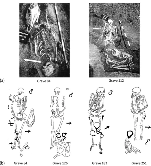
The cemetery of Sárrétudvari‐Hízóföld, located in the Northern Great Plain region of eastern Hungary, was excavated by Ibolya M. Nepper between 1983 and 1985. The graves delivered a large number of archaeological goods, such as jewels, elements of clothing, or weapons (Nepper, 2002). Those included, in particular, pieces of archery (antler bow plates, quivers, and arrowheads) and horse riding equipment (stir- rups, bits, girth buckles, and saddles). Several graves also contained horse bones (skull and extremities) that were, in some cases, folded inside the horsehide before being deposited in association with the individuals (Révész, 2003; Figure 2a,b). Sárrétudvari‐Hízóföld was exhaustively excavated, well documented and published, and yielded a large number of archaeological remains that can be related to mounted archery. The signs of riding were originally identified in sev- eral individuals in a comprehensive paleopathological study (Pálfi, 1992, 1997; Pálfi & Dutour, 1996).
2.2
|Composition of the sample of early Hungarians
Two hundred sixty‐two graves from the 10th century provided a total of 265 individuals. According to previous studies, the population is composed of 101 subadults (including three fetuses), 162 adults, and two undetermined subjects (Oláh, 1990; Pálfi, 1997). The recon- struction of activities in past populations involves numerous limita- tions (e.g., Dutour, 1992; Jurmain et al., 2012). We included in this study only adult males to limit the influence of nonmechanical factors such as hormonal and developmental changes. All adult age catego- ries were included as a large number of subjects could not be categorised due to the state of preservation. Furthermore, age does not seem to be a bias factor for the changes of the acetabular shape as it is not related to degenerative processes (Baillif‐Ducros et al., 2012). Erickson et al. (2000) found no significant correlation between the estimated age at death and the coefficients used in a Fourier analysis of the acetabular shape.
On the basis of a reassessed sex diagnosis (Bruzek, 2002; Bruzek, Santos, Dutailly, Murail, & Cunha, 2017; Murail, Bruzek, Houët, &
Cunha, 2005) and age estimation (Bruzek, Schmitt, & Murail, 2005;
Scheuer & Black, 2004), we identified a total of 67 adult male individ- uals in the population. In Sárrétudvari‐Hízóföld, 32 graves presented an archaeological deposit that can be related to horse riding (equip- ment, horse bones, or both). There are at least 16 adult males in this group (RD⊕ =“riding deposit”). The other subjects, not included in the study, are one adult female, seven adults with indeterminate sex, seven subadults, and one nonidentified subject. On the other hand, 51 adult males did not have any horse riding equipment or horse bones in their graves (RD⊝=“no riding deposit”).
2.3
|Out ‐ group comparison
The Hungarian samples were also compared with an extra‐group for which the intensive and regular practice of horse riding was highly improbable. As the use of horses was very widespread in medieval Hungary, we used, among the limited possibilities available, a European documented skeletal collection with a known sociocultural context. Bearing in mind the potential limits that this might involve, FIGURE 1 Reconstruction illustrating the
possible contact between the femoral head and the superior part of the acetabulum (white arrow) in the riding position, with the pelvis in retroversion. Drawing by Luca Kis
we believed that it represents a valid alternative to provide input to discuss the link between bone changes and horse riding.
We used the Luís Lopes Skeletal Collection, curated in the National Museum of Natural History and Science (MUHNAC), in Lisbon, Portugal.
It is composed of 1,692 skeletons with information regarding the sex, age at death, occupation, the cause of death, and so forth (Alves Cardoso
& Henderson, 2013; Cardoso, 2006). Our sample was composed of Por- tuguese people who mainly lived during the first half of the 20th cen- tury, in the urban area of Lisbon, with electricity and modern means of transport. It is therefore very unlikely that they were practicing horse riding intensively and on a daily basis. The sample selected in the collection is composed of 47 adult males, aged 20 and older.
2.4
|Metric acquisition
The analysis of the acetabular shape relied on direct measurements of the vertical and horizontal diameters of the acetabulum and the calcu- lation of an index, following the approach taken by Baillif‐Ducros et al.
(2012) and Sarry et al. (2016) that we also previously experimented (Berthon et al., 2016). Although the Fourier analysis used by Erickson
et al. (2000) seems particularly relevant to capture the changes of the shape of the entire acetabular rim, several portions of the acetabular rim are frequently not preserved in ancient materials. Second, the ver- tical and horizontal diameters allow assessing the anterior‐superior rim expansion previously described (Baillif‐Ducros et al., 2012; Erickson et al., 2000). Finally, we applied a methodology that does not raise technical and time issues in broad studies of large samples as our results aim to be combined with those of a more comprehensive analysis including the scoring of various other types of observations.
The metrics are well‐defined measurements that can be easily taken: VEAC (M22) corresponds to the maximum vertical diameter of the acetabulum, measured as a prolongation of the longitudinal axis of the ischium, and HOAC (M22) is the maximum horizontal diameter, measured perpendicularly to VEAC (Baillif‐Ducros et al., 2012; Bräuer, 1988; Bruzek et al., 2017; Murail et al., 2005). The measurements are neither external nor internal but taken precisely on the rim of the ace- tabulum (Figure 3a).1They were used to calculate a vertical/horizontal diameter index (VEAC/HOAC) that allows comparing the acetabular
1Bruzek et al. (2017) also provide an illustration of the technique to measure the vertical diameter of the acetabulum in the DSP2 software.
FIGURE 2 Examples of graves from the Hungarian Conquest period cemetery of Sárrétudvari‐Hízóföld (10th century) with deposits of horse riding equipment (stirrups, girth buckles, and bits), sometimes associated with horse bones: (a) field photographs and (b) archaeological drawings of graves. From Nepper (2002)
shape between individuals, groups, and studies. This index of ovalization of the acetabulum (IOA) indicates a vertical ovalization when it is higher than 1 (VEAC > HOAC), whereas an IOA close to 1 corresponds to circular acetabula, and an IOA inferior to 1 indicates acetabula wider than their height (HOAC > VEAC; Figure 3b,c). The metrics were recorded with a digital sliding caliper with a precision of 0.1 mm. Both left and right acetabula were measured to address asymmetry questions. We excluded suspected cases of hip dysplasia, subluxation or dislocation, and acetabula showing strong osteophytosis at the rim.
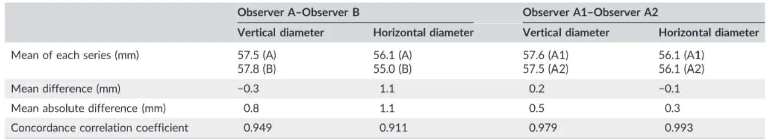
The measurements were taken by one observer (A), without knowledge of the presence or absence of horse riding deposit in the graves, concerning the Hungarian samples. Additionally, a set of measurements was taken by a second observer (B), independently, to evaluate the reproducibility of the measurements. Observer A had more practical experience than Observer B in metrics of the coxal bone, but both observers are experienced in osteometry. The defini- tions and techniques of measurements were clarified before the test.
Observer A also performed a second round of measurements (A2), with an interval of several months, to assess the intraobserver agree- ment. Observer B's measurements were compared with the A2 set.
Thirty coxae from 15 individuals were measured for comparing the vertical and the horizontal diameters, respectively. The individuals were selected randomly among the Hungarian adults, provided that the acetabular rim was well preserved. The concordance between the series of measurements was evaluated using the concordance cor- relation coefficient that allows quantifying the agreement between two measures of the same variable (Lin, 1989). Values are ranged between−1 and 1, with ±1 denoting a perfect concordance or discor- dance and 0 denoting an absence of correlation.
2.5
|Statistical analyses
Boxplots were constructed to visually represent the distribution of the indices in each group. The non‐parametric Mann–Whitney test was used to assess the differences between the groups. It was applied to the indices, both sides combined and taken independently.
We used the non‐parametric Wilcoxon test, applied on the paired indices in each group, to evaluate the asymmetry (Nikita, 2017).
The difference between both sides' indices was also calculated (right index–left index) and compared between the groups. The set signifi- cance level wasα= 0.05. The analyses were performed using SPSS Statistics 25.
3
|R E S U L T S
3.1
|Interobserver and intraobserver agreement evaluation
There was a very good concordance between the series of measure- ments taken by Observer A and Observer B for the diameters of the acetabulum (Table 1). The mean absolute difference was indeed close to 1 mm in each case, and the concordance correlation coefficient was superior to 0.900, which is considered as very satisfying. Additionally, we observed an excellent concordance for both diameters between the two series of measurements taken by Observer A, with a mean absolute difference not higher than 0.5 mm and concordance correla- tion coefficients superior to 0.970 (Table 1). The concordance was almost perfect in the case of the horizontal diameter, with a coefficient of 0.993.
FIGURE 3 (a) Measurements of the maximum vertical and horizontal diameters of the acetabulum, and examples of (b) an acetabulum with a shape intermediate between circular and a horizontal ovalisation (index = 0.971), and (c) an acetabulum exhibiting a strong vertical ovalization (index = 1.082) [Colour figure can be viewed at wileyonlinelibrary.com]
3.2
|Asymmetry in each group and differences between groups
The preservation of the coxae was a limiting factor in the Hungarian archaeological groups. The composition of the samples of acetabula for which the index could be calculated is presented in Table 2.
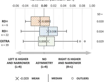
As a first step, we evaluated the bilateral asymmetry of the acetabular shape, excluding the individuals with only one observ- able acetabulum (Table S1 and Figure 4). In all cases, the acetabula were not circular on average, but they tended to be a bit higher
than wide (IOA lightly superior to 1), which corresponds to a normal morphology (Bräuer, 1988). In all groups, we observed a bilateral asymmetry, although it was not significant (Wilcoxon signed‐ranks test: RD⊕: Z=−0.943,p = 0.345; RD⊝:Z= −1.542, p = 0.123; EXTRA: Z = −1.451, p = 0.147), but this asymmetry was inverted in the case of the RD⊕ group compared with the RD⊝ and EXTRA groups. Although the median and the variation were greater on the left side concerning the RD⊕ group, the spread of the values appeared to be higher on the right side in the two other groups (median and interquartile range in particular).
TABLE 2 Composition of the samples of observable acetabula in the Hungarian archaeological groups with riding deposit (RD⊕) and without riding deposit (RD⊝), and in the extra‐group (EXTRA)
Sample groups Adult males
Adult males with at least one observable coxal bone
Observable acetabula
Pairs of observable acetabula
Left Right Total
RD⊕ 16 12 8 10 18 6
RD⊝ 51 33 26 29 55 22
EXTRA 47 46 43 42 85 39
Total 114 91 77 81 158 67
FIGURE 4 Asymmetry of the index of ovalization (vertical/horizontal diameter) of the acetabulum (IOA) on paired coxae of the Hungarians with riding deposit (RD⊕) and without riding deposit (RD⊝), and in the extra‐group (EXTRA).SD= standard deviation [Colour figure can be viewed at wileyonlinelibrary.com]
Thus, the acetabula tended to exhibit a vertical ovalization more important on the left side in the RD⊕group and on the right side in the RD⊝ and EXTRA groups.
We also calculated the difference between the right and left index values for each pair of acetabula to compare the degree and direction of bilateral asymmetry between the groups (Table S1 and Figure 5).
The tendencies concerning the bilateral asymmetry were confirmed, with a negative median in the RD⊕ group and a positive median in the RD⊝and EXTRA groups, respectively. The left acetabula in the RD⊕group appeared to be higher and narrower than the right ones, but they tended to be shorter and wider than on the right side in
the RD⊝ and EXTRA groups. Although the differences between groups were not significant (Mann–Whitney test: RD⊕/RD⊝: U= 38,p = 0.117; RD⊕/EXTRA:U= 76,p = 0.171; RD⊝/EXTRA:
U= 389, p= 0.548, two‐tailed), we observed that the direction of the asymmetry differed between the RD⊕and the other groups.
3.3
|Differences of acetabular shape between groups
The index values were then compared between each group, for the left and right acetabula taken separately and combined, without con- sidering if one or both sides were observable in an individual (Table S1 and Figure 6). The RD⊕ group was characterised by a higher median value and a greater variation (especially interquartile range) compared with the RD⊝ and EXTRA groups, which showed similar distributions. The differences were not statistically significant between the groups for the left side (Mann–Whitney test: RD⊕/ RD⊝L: U= 80,p = 0.330; RD⊕/EXTRA L: U= 125.5,p = 0.228;
RD⊝/EXTRA L:U= 520.5,p= 0.634, two‐tailed) and for the right side (RD⊕/RD⊝ R: U = 108, p = 0.234; RD⊕/EXTRA R: U = 146.5, p= 0.140; RD⊝/EXTRA R:U= 566.5,p= 0.619, two‐tailed). When left and right indices were combined, the differences were not signif- icant between RD⊕and RD⊝groups (U= 374.5,p= 0.123) and RD⊝ and EXTRA groups (U = 2158.5, p = 0.445), but the differences observed between RD⊕and EXTRA groups were statistically signifi- cant (U= 538.5,p= 0.049, two‐tailed).
4
|D I S C U S S I O N
Our results must be regarded in close connection with the archaeolog- ical data concerning the Hungarian samples. In a cemetery of a semi‐ FIGURE 5 Difference between the right and left indices of
ovalization of the acetabulum (IOA) in the Hungarians with riding deposit (RD⊕) and without riding deposit (RD⊝), and in the extra‐ group (EXTRA; a positive result indicates right values superior to the left values, and a negative result indicates the contrary).SD= standard deviation [Colour figure can be viewed at wileyonlinelibrary.com]
FIGURE 6 Index of ovalization (vertical/horizontal diameter) of the acetabulum (IOA), by side and combined, on unpaired coxae of the Hungarians with riding deposit (RD⊕) and without riding deposit (RD⊝), and in the extra‐group (EXTRA).SD= standard deviation [Colour figure can be viewed at wileyonlinelibrary.com]
their grave may have been finally included in the RD⊝group, contrib- uting to mitigate significant differences between both Hungarian groups.
Our study revealed that the subjects from burials with archaeo- logical items related with horse riding showed an increased overall acetabular vertical ovalisation compared with the ones from the graves with no funeral goods of this type. The IOA values of individ- uals of this last group are similar to the ones of the extra‐group sub- jects, which almost certainly did not practise horse riding, intensively or regularly.
Although we should not exclude the possibility that this skeletal modification could also result from other postures, it is clearly compat- ible with horse riding: The vertical ovalisation of the acetabulum results from the pressure applied by the femoral heads on the acetab- ula when the hip is placed in the rider position (Auvinet, 1999; Baillif‐ Ducros et al., 2012; Humbert, 2000).
Concerning the difference of direction of the bilateral asymmetry in the case of the RD⊕group, Molleson (2007) discussed, for instance, that the occurrence of degenerative joint changes on the medial con- dyle of the left knee in individuals from Spitalfields, London, might be explained by riders mounting a horse from the left side. In addition, experienced riders can develop a pelvic asymmetry in response to back pain (Hobbs et al., 2014). In the context of the Hungarian Con- quest period, we might wonder if an asymmetry could result from the movements involved in mounted archery in particular.
Despite strict sampling criteria and bone preservation limitations, this group of 32 ancient Hungarian individuals including at least 16 adult males represents, to the best of our knowledge, the largest homogeneous anthropological sample investigated for horse riding with direct archaeological evidence for each individual. Our promising results support the hypothesis that the vertical ovalisation of the ace- tabulum may represent a potential indicator for the practice of horse riding, among a set of other skeletal changes that will be tested. The question of the riding style will be another point of interest as we ignore so far if the steppe nomad style of the early Hungarians (Uray‐Kőhalmi, 1972) and other riding styles may result in different bone modifications. A future comparative analysis should allow us to identify the possible existence of specific or common features accord- ing to the riding style.
A C K N O W L E D G E M E N T S
The authors are grateful to the National Museum of Natural History and Science (MUHNAC), University of Lisbon, to Dr. Susana Garcia, Curator of the Anthropological Collection, and Dr. Judite Alves, Head
International Mobility of Doctoral Students 2015–2018), the French‐ Hungarian Hubert Curien Partnership“Balaton”(2015–2016, 2018– 2019), and the National Research, Development and Innovation Office, Hungary (K125561).
C O N F L I C T S O F I N T E R E S T
The authors have no conflicts of interest to declare.
O R C I D
William Berthon http://orcid.org/0000-0003-1776-4798
R E F E R E N C E S
Alves Cardoso, F., & Henderson, C. (2013). The categorisation of occupa- tion in identified skeletal collections: A source of bias?International Journal of Osteoarchaeology, 23, 186–196. https://doi.org/10.1002/
oa.2285
Anđelinović,Š., Anterić, I.,Škorić, E., & Bašić,Ž. (2015). Skeleton changes induced by horse riding on medieval skeletal remains from Croatia.
The International Journal of the History of Sport,32, 708–721. https://
doi.org/10.1080/09523367.2015.1038251
Anthony, D. W. (2007).The horse, the wheel, and language: How Bronze‐Age riders from the Eurasian steppes shaped the modern world. Princeton, NJ:
Princeton University Press.
Auvinet, B. (1999). Lombalgies et équitation.Synoviale,83, 25–31.
Baillif‐Ducros C., McGlynn G. 2013. Stirrups and archaeological popula- tions: Bio‐anthropological considerations for determining their use based on the skeletons of two Steppe riders. Poster presented at the Schweizerischen Gesellschaft für Anthropologie annual meeting, Neuchâtel, 16 November 2013.
Baillif‐Ducros, C., Truc, M. C., Paresys, C., & Villotte, S. (2012). Approche méthodologique pour distinguer un ensemble lésionnel fiable de la pra- tique cavalière. Exemple du squelette de la tombe 11 du site de « La Tuilerie » à Saint‐Dizier (Haute‐Marne), VIe siècle.Bulletins et Mémoires de la Société d'Anthropologie de Paris, 24, 25–36. https://doi.org/
10.1007/s13219‐011‐0049‐8
Belcastro, G., Rastelli, E., Mariotti, V., Consiglio, C., Facchini, F., &
Bonfiglioli, B. (2007). Continuity or discontinuity of the life‐style in cen- tral Italy during the Roman imperial age‐early middle ages transition:
Diet, health, and behavior.American Journal of Physical Anthropology, 132, 381–394. https://doi.org/10.1002/ajpa.20530
Belcastro, M. G., & Facchini, F. (2001). Anthropological and cultural fea- tures of a skeletal sample of horsemen from the medieval necropolis of Vicenne‐Campochiaro (Molise, Italy).Collegium Antropologicum,25, 387–401.
Belcastro, M. G., Facchini, F., Neri, R., & Mariotti, V. (2001). Skeletal markers of activity in the Early Middle Ages necropolis of Vicenne‐ Campochiaro (Molise, Italy).Journal of Paleopathology,13, 9–20.
Berthon W., Tihanyi B., Révész L., Coqueugniot H., Pálfi G., Dutour O.
2016. A contribution to the definition of “horse riding syndrome”: The mounted archers from the Hungarian Conquest (Xth century
AD). Poster presented at the 21st European Meeting of the Paleopa- thology Association, Moscow, 15–19 August 2016.
Blondiaux, J. (1994). À propos de la dame d'Hochfelden et de la pratique cavalière: discussion autour des sites fonctionnels fémoraux. In L.
Buchet (Ed.),Actes des 6e Journées Anthropologiques (Valbonne, 9–11 juin 1992).Dossier de Documentation Archéologique n° 17. (pp. 97–109).
Paris: CNRS Éditions.
Bräuer, G. (1988). Osteometrie. In R. Knussman (Ed.), Anthropologie, Handbuch der vergleichenden Biologie des Menschen. Zugleich 4. Auflage des Lehrbuchs der Anthropologie begründet von R. Martin. Band I/1.
Wesen und Methoden der Anthropologie (pp. 160–232). Stuttgart:
Gustav Fisher Verlag.
Bruzek, J. (2002). A method for visual determination of sex, using the human hip bone. American Journal of Physical Anthropology, 117, 157–169. https://doi.org/10.1002/ajpa.10012
Bruzek, J., Santos, F., Dutailly, B., Murail, P., & Cunha, E. (2017). Validation and reliability of the sex estimation of the human os coxae using freely available DSP2 software for bioarchaeology and forensic anthropology.
American Journal of Physical Anthropology,164, 440–449. https://doi.
org/10.1002/ajpa.23282
Bruzek, J., Schmitt, A., & Murail, P. (2005). Identification biologique individuelle en paléoanthropologie. Détermination du sexe et estima- tion de l'âge au décès à partir du squelette. In O. Dutour, J.‐J. Hublin,
& B. Vandermeersch (Eds.),Objets et méthodes en paléoanthropologie (pp. 217–246). Paris: Comité des Travaux Historiques et Scientifiques.
Bulatović, J., Bulatović, A., & Marković, N. (2014). Paleopathological changes in an early iron age horse skeleton from the Central Balkans (Serbia).International Journal of Paleopathology,7, 76–82. https://doi.
org/10.1016/J.IJPP.2014.07.001
Capasso, L., Kennedy, K. A. R., & Wilczak, C. A. (1999).Atlas of occupational markers on human remains. Teramo: Edigrafital.
Cardoso, H. F. V. (2006). Brief communication: The collection of identified human skeletons housed at the Bocage Museum (National Museum of Natural History), Lisbon, Portugal.American Journal of Physical Anthro- pology,129, 173–176. https://doi.org/10.1002/ajpa.20228
Courtaud, P., & Rajev, D. (1998). Osteomorphological features of nomadic riders: Some examples from Iron Age populations located in south- western Siberia. In M. Pearce, & M. Tosi (Eds.),Papers from the EAA Third Annual Meeting at Ravenna, 1997.BAR International Series 717, Vol. 1, Pre‐and protohistory. (pp. 110–113). Oxford: Archaeopress.
Djukic, K., Miladinovic‐Radmilovic, N., Draskovic, M., & Djuric, M. (2018).
Morphological appearance of muscle attachment sites on lower limbs:
Horse riders versus agricultural population. International Journal of Osteoarchaeology. Advance online publication. DOI: https://doi.org/
10.1002/oa.2680
Dutour, O. (1986). Enthesopathies (lesions of muscular insertions) as indi- cators of the activities of Neolithic Saharan populations. American Journal of Physical Anthropology, 71, 221–224. https://doi.org/
10.1002/ajpa.1330710209
Dutour, O. (1992). Activités physiques et squelette humain: le difficile passage de l'actuel au fossile. Bulletins et Mémoires de la Société d'Anthropologie de Paris, 4, 233–241. https://doi.org/10.3406/
bmsap.1992.2319
Dutour, O., & Buzhilova, A. (2014). Palaeopathological study of Napoleonic mass graves discovered in Russia. In C. J. Knüsel, & M. Smith (Eds.), The Routledge handbook of the bioarchaeology of human conflict (pp. 511–524). London: Routledge.
Eng J.T., Baker A., Tang P., Thompson S., Gomez J.M. 2012. Morphometric analysis of acetabular rim shape among ancient Mongolian pastoralists.
Poster presented at the 19th Annual Meeting of the Midwest Bioarchaeology and Forensic Anthropology Association, Carbondale, IL, October 2012.
Engel, P. (2001). The realm of St Stephen. A history of medieval Hungary, 895–1526. London: I.B. Tauris Publishers.
Erickson, J. D., Lee, D. V., & Bertram, J. E. A. (2000). Fourier analysis of ace- tabular shape in Native American Arikara populations before and after
acquisition of horses.American Journal of Physical Anthropology,113, 473–480. https://doi.org/10.1002/1096‐8644(200012)113:4<473::
AID‐AJPA3>3.0.CO;2‐5
Fornaciari, G., Bartolozzi, P., Bartolozzi, C., Rossi, B., Menchi, I., & Piccioli, A. (2014). A great enigma of the Italian Renaissance: Paleopathological study on the death of Giovanni dalle Bande Nere (1498–1526) and his- torical relevance of a leg amputation.BMC Musculoskeletal Disorders, 15, 301. https://doi.org/10.1186/1471‐2474‐15‐301
Härke, H. (1997). The nature of burial data. In C. K. Jensen, & K. H. Nielsen (Eds.),Burial and society: The chronological and social analysis of archae- ological burial data (pp. 19–27). Aarhus – Oakville, Conn: Aarhus University Press.
Hawkey, D. E., & Merbs, C. F. (1995). Activity‐induced musculoskeletal stress markers (MSM) and subsistence strategy changes among ancient Hudson Bay Eskimos. International Journal of Osteoarchaeology, 5, 324–338. https://doi.org/10.1002/oa.1390050403
Henderson, C., & Alves Cardoso, F. (2013). Special issue “Entheseal changes and occupation”: Technical and theoretical advances and their applications.International Journal of Osteoarchaeology, 23, 127–134.
https://doi.org/10.1002/oa.2298
Hobbs, S. J., Baxter, J., Broom, L., Rossell, L. A., Sinclair, J., & Clayton, H. M.
(2014). Posture, flexibility and grip strength in horse riders.Journal of Human Kinetics, 42, 113–125. https://doi.org/10.2478/hukin‐2014‐ 0066
Humbert C. 2000. L'équitation et ses conséquences sur le rachis lombaire du cavalier: à propos de 123 observations (MD thesis). Université Henri Poincaré Nancy I: Nancy.
Józsa, L., Pap, I., & Fóthi, E. (1991). Enthesopathies (insertion tendopathies) as indicators of overuse of tendons and muscles in ancient Hungarian populations. Annales Historico‐Naturales Musei Nationalis Hungarici, 83, 269–276.
Jurmain, R. (1999).Stories from the skeleton. Behavioral reconstruction in human osteology. Gordon and Breach: Amsterdam.
Jurmain, R., Alves Cardoso, F., Henderson, C. Y., & Villotte, S. (2012).
Bioarchaeology's Holy Grail: The reconstruction of activity. In A. L.
Grauer (Ed.),A companion to paleopathology(pp. 531–552). Chichester, UK: Wiley‐Blackwell. https://doi.org/10.1002/9781444345940.ch29 Karstens S., Littleton J., Frohlich B., Amgaluntugs T., Pearlstein K. 2017.
Palaeopathological indicators of mounted pastoralism during the Mongolian Bronze Age. Poster presented at the 86th Annual Meeting of the American Association of Physical Anthropologists, New Orleans, LA, 19‐22 April 2017.
Kennedy, K. A. R. (1989). Skeletal markers of occupational stress. In M. Y.
İşcan, & K. A. R. Kennedy (Eds.),Reconstruction of life from the skeleton (pp. 129–160). New York: Alan R. Liss, Inc.
Khudaverdyan, A., Khachatryan, H., & Eganyan, L. (2016). Multiple trauma in a horse rider from the Late Iron Age cemetery at Shirakavan, Armenia.Bioarchaeology of the Near East,10, 47–68.
Ki, H. C., Shin, E. K., Woo, E. J., Lee, E., Hong, J. H., & Shin, D. H. (2018).
Horse‐riding accidents and injuries in historical records of Joseon Dynasty, Korea. International Journal of Paleopathology, 20, 20–25.
https://doi.org/10.1016/j.ijpp.2017.12.001
Knüsel, C. (2000). Bone adaptation and its relationship to physical activity in the past. In M. Cox, & S. Mays (Eds.),Human osteology in archaeology and forensic science (pp. 381–402). London: Greenwich Medical Media Ltd.
Larentis, O. (2017). San Martino di Lundo (Trento) Grave 1. Case study of an individual introducing possibilities markers of horse riding.Medicina Historica,1, 103–110.
Lin, L. I. K. (1989). A concordance correlation coefficient to evaluate repro- ducibility.Biometrics,45, 255–268. https://doi.org/10.2307/2532051 Mariotti, V., Facchini, F., & Belcastro, M. G. (2004). Enthesopathies—
Proposal of a standardized scoring method and applications.Collegium Antropologicum,28, 145–159.
and Ponca: Paleopathological indications of European contact. Paper presented at the Plains Anthropology Conference, Lawrence, KS, 13– 16 November 1991.
Molleson, T. (2007). A method for the study of activity related skeletal morphologies.Bioarchaeology of the Near East,1, 5–33.
Molleson, T., & Blondiaux, J. (1994). Riders' bones from Kish, Iraq.
Cambridge Archaeological Journal, 4, 312–316. https://doi.org/
10.1017/S095977430000113X
Murail, P., Bruzek, J., Houët, F., & Cunha, E. (2005). DSP: A tool for proba- bilistic sex diagnosis using worldwide variability in hip‐bone measurements. Bulletins et Mémoires de la Société d'Anthropologie de Paris,17, 167–176. DOI:10670/1.3p2nbs
Nepper I.M. 2002. Hajdú‐Bihar megye 10–11. századi sírleletei I‐II.
(Magyarország honfoglalás és kora Árpád‐kori sírleletei 3): Budapest– Debrecen.
Niinimäki, S. (2012). The relationship between musculoskeletal stress markers and biomechanical properties of the humeral diaphysis.Amer- ican Journal of Physical Anthropology,147, 618–628. https://doi.org/
10.1002/ajpa.22023
Nikita, E. (2017). Osteoarchaeology. A guide to the macroscopic study of human skeletal remains. London, UK: Academic Press.
Oláh S. 1990. Sárrétudvari‐Hízóföld honfoglalás kori temetőjének történeti embertani értékelése (Doctoral dissertation). József Attila Tudományegyetem: Szeged.
Outram, A. K., Stear, N. A., Bendrey, R., Olsen, S., Kasparov, A., Zaibert, V., … Evershed, R. P. (2009). The earliest horse harnessing and milking. Science, 323, 1332–1335. https://doi.org/10.1126/
science.1168594
Pálfi, G. (1992). Traces des activités sur les squelettes des anciens Hongrois.Bulletins et Mémoires de la Société d'Anthropologie de Paris, 4, 209–231. https://doi.org/10.3406/bmsap.1992.2318
Pálfi, G. (1997). Maladies dans l'Antiquité et au Moyen‐Âge.
Paléopathologie comparée des anciens Gallo‐Romains et Hongrois.
Bulletins et Mémoires de la Société d'Anthropologie de Paris,9, 1–205.
https://doi.org/10.3406/bmsap.1997.2472
Pálfi, G., & Dutour, O. (1996). Activity‐induced skeletal markers in histori- cal anthropological material.International Journal of Anthropology,11, 41–55. https://doi.org/10.1007/BF02442202
Pap, I. (1985). A Dabas (Gyón)‐paphegyi XI. századi embertani széria (The anthropological series of Dabas (Gyón)‐paphegy from the 11th century). In N. Ikvai (Ed.), Régészeti tanulmányok Pest megyéből.
Studia Comitatensia 17(pp. 387–407). Szentendre: Museums of Pest County.
Perréard Lopreno G. 2007. Adaptation structurelle des os du membre supérieur et de la clavicule à l'activité: Analyse de l'asymétrie des propriétés géométriques de sections transverses et de mesures linéaires dans une population identifiée (collection SIMON)(Doctoral dissertation).
Université de Genève: Genève.
Sarry, F., Courtaud, P., & Cabezuelo, U. (2016). La sépulture multiple laténienne du site de Gondole (Le Cendre, Puy‐de‐Dôme). BMSAP, 28, 72–83. https://doi.org/10.1007/s13219‐016‐0151‐z
Scheuer, L., & Black, S. (2004). The juvenile skeleton. London: Elsevier Academic Press.
Taylor, W. T. T., Bayarsaikhan, J., & Tuvshinjargal, T. (2015). Equine cranial morphology and the identification of riding and chariotry in late Bronze Age Mongolia. Antiquity, 89, 854–871. https://doi.org/10.15184/
aqy.2015.76
Thomas, A. (2014). Bioarchaeology of the middle Neolithic: Evidence for archery among early European farmers.American Journal of Physical Anthropology,154, 279–290. https://doi.org/10.1002/ajpa.22504 Tichnell T.A. 2012.Invisible horsewomen: Horse riding and social dynamics on
the Steppe (Doctoral dissertation). Michigan State University: East Lansing, MI.
Tihanyi, B., Bereczki, Z., Molnár, E., Berthon, W., Révész, L., Dutour, O., &
Pálfi, G. (2015). Investigation of Hungarian Conquest period (10th c.
AD) archery on the basis of activity‐induced stress markers on the skeleton—Preliminary results.Acta Biologica Szegediensis,59, 65–77.
Uray‐Kőhalmi, K. (1972).A steppék nomádja lóháton, fegyverben. Budapest:
Akadémiai.
Üstündağ, H., & Deveci, A. (2011). A possible case of Scheuermann's dis- ease from Akarçay Höyük, Birecik (Şanlıurfa, Turkey). International Journal of Osteoarchaeology, 21, 187–196. https://doi.org/10.1002/
oa.1120
Villotte, S., Castex, D., Couallier, V., Dutour, O., Knüsel, C. J., & Henry‐ Gambier, D. (2010). Enthesopathies as occupational stress markers:
Evidence from the upper limb.American Journal of Physical Anthropol- ogy,142, 224–234. https://doi.org/10.1002/ajpa.21217
Wentz, R. K., & de Grummond, N. T. (2009). Life on horseback:
Palaeopathology of two Scythian skeletons from Alexandropol, Ukraine. International Journal of Osteoarchaeology, 19, 107–115.
https://doi.org/10.1002/oa.964
S U P P O R T I N G I N F O R M A T I O N
Additional supporting information may be found online in the Supporting Information section at the end of the article.
How to cite this article: Berthon W, Tihanyi B, Kis L, et al.
Horse riding and the shape of the acetabulum: Insights from the bioarchaeological analysis of early Hungarian mounted archers (10th century).Int J Osteoarchaeol. 2019;29:117–126.
https://doi.org/10.1002/oa.2723