Entomophtfarales Infections
1D O N A L D M . M A C L E O D
Insect Pathology Research Institute, Canada Department of Forestry, Sault Ste. Marie, Ontario, Canada
I. Introduction 189 II. T h e Genus Entomophthora Fresenius 191
A. Historical 191 B. Morphology and D e v e l o p m e n t 193
C. Host-Parasite Relationships 205 III. T h e Genus Massospora Peck 219
A. Historical 219 B. Morphology and D e v e l o p m e n t 220
C. Host-Parasite Relationships 221
References 226
I. INTRODUCTION
T h e Entomophthorales (Table I) constitute a small, b u t rather distinc
tive, order of Phycornycetes (algal fungi). T h e order, as here treated, con
sists of the single family Entomophthoraceae, which may be divided into six genera. W h i l e differing greatly with respect to habitat, the genera ap
pear to be closely allied structurally. T h e i r mycelium is often m u c h re
duced, forming short, thick-walled hyphal bodies. Asexual reproduction is by means of modified sporangia (conidia), uninucleate or multinucleate, which are shot off singly from the apex of club-shaped conidiophores. In Massospora they are produced within the body of the host insect. Sexual reproduction is by the u n i o n of mycelial fragments or hyphal bodies to form thick-walled zygospores. I n many species, morphologically similar spores (the azygospores) are formed parthenogenetically.
ι Contribution N o . 33, Insect Pathology Research Institute, Canada Department of Forestry.
189
T h r e e genera are not parasitic on insects and will not be considered in detail in this work. T h e genus Completoria with its single species in para
sitic on the prothalli of various ferns (Lohde, 1874). T h e three species of Ancylistes are parasitic on desmids of the genus Closterium (Bessey, 1950;
Sparrow, 1960). At least two species of Conidiobolus have been isolated from plant debris (Drechsler, 1954), and others are weakly parasitic or saprophytic on the pilei of Auricularia, Hypochnus, and other Basidio- mycetes (Wolf and Wolf, 1947).
T A B L E I
CHARACTERIZATION OF THE ORDER ENTOMOPHTHORALES, FAMILY ENTOMOPHTHORACEAE Forms chiefly parasitic and entomogenous, a few forms saprophytic, a few others parasitic o n plants; mycelium not very extensive, at first coenocytic but sooner or later becoming septate and often falling apart into hyphal bodies; asexual reproduction almost always by conidia borne at the ends of specialized conidiophores and shot away (with violence) at maturity; sexual reproduction by the u n i o n of mycelial fragments (or of hyphal bodies) to form zygospores; zygospores frequently replaced by the par- thenogenetic development of azygospores.
Key to genera of Entomophthoraceae Mycelium not entomogenous (i.e., not living o n insects)
Parasitic in the gametophytes of ferns Parasitic in the desmid genus Closterium Saprophytic in the excrement of frogs and lizards Saprophytic or weakly parasitic o n fungi Mycelium entomogenous
Conidia borne on conidiophores extruded through the body wall of the host, smooth-walled, discharged forcibly from
the conidiophore Entomophthora Conidia borne within the body of the host and freed by its
disintegration, not shot away Massospora
Basidiobolus, though wholly saprophytic, is noteworthy because of its unusual developmental cycle, which, at one stage, involves insects. T h e species B. ranarum Eidam (Eidam, 1886; G ä u m a n n and Dodge, 1928), grows on the excrement of frogs and lizards. T h e sporangia produced on this substrate are eaten by beetles, principally Carabidae, Scarabaeidae, and Silphidae, that prowl the excrement; these beetles, in turn, may be devoured by frogs. D u r i n g digestion within the frog's stomach, the spores are delimited within the sporangia and set free. T h e spores, if retained for a long time, may then multiply further by division or by budding. T h e y remain d o r m a n t in this state until excreted with the feces; but, once ex
posed to the outer air they germinate to form mycelium on which are pro
duced other sporangia.
Completoria Ancylistes Basidiobolus Conidiobolus
T h e remaining genera,2 Entomophthora (= Empusa) and Massospora, are true entomogenous3 fungi that develop in the bodies of immature a n d / o r adult stages of various insects. T h e former includes the vast majority of known species (see A p p e n d i x I at end of chapter) of the order. These two genera will be considered, separately, in Sections I I a n d I I I .
I I . T H E GENUS Entomophthora FRESENIUS A . Historical
Entomophthora Fresenius, Botan. Ztg., 14, 882; 1856.
syn. Empusa Cohn, Nova Acta Leop. Carol., 25, 301-360; 1855.
Tarichium Cohn, Beitr. Biol. Pflanz., 1, 58; 1875.
Lamia Nowakowski, Pamietnik Akad. Umiejejnosci zu Krakau, 8, 153-183; 1884.
Delacroixia Costantin, Bull. Soc. mycol. France, 13, 38-43; 1897.
T h e genus, as originally described by Cohn was named Empusa, based on Empusa muscae Cohn, the type species. Fresenius (1856), rec
ognized that the n a m e Empusa h a d already been pre-empted by a genus of orchids a n d proposed that the genus be called Entomophthora. Bre
feld (1877), concluded that there were actually two genera involved among the known species; to those h e applied the names Empusa a n d Ento
mophthora. Nowakowski (1884) subdivided the g r o u p still further and created a new genus Lamia. According to this arrangement Empusa is characterized by the possession of u n b r a n c h e d conidiophores a n d t h e formation of azygospores a n d Entomophthora has branched conidio
phores, forms rhizoids a n d cystidia, a n d produces zygospores. Lamia was intermediate, differing from Empusa chiefly in the possession of cystidia.
These proposals subsequently led to some confusion in the taxonomy of this g r o u p of fungi.
After a critical examination, T h a x t e r (1888) concluded that an abundance of borderline species m a d e Nowakowski's separation invalid.
He, therefore, reunited all the species u n d e r the generic n a m e Empusa, which he selected because of its priority a n d weight of authority.
2 Weiser (1951) described a n e w fungus, Zygaenobia intestinalis Weiser, growing in the midgut epithelium of a n u m b e r of larvae of Zygaena carniolica Scopoli. T h e mycelium is coenocytic and penetrates between the cells of the epithelium. Its conidia are mononuclear and form in the mesenteron, eventually leaving the insect along w i t h the feces. Resting spores were not seen. Since these unusual characteristics are n o t shared by any other entomogenous fungus, Weiser established a n e w genus with this fungus as the only species. T h e genus appears to be close to Massospora (Steinhaus, 1957).
3 T h e term entomogenous simply means the growing in or on the bodies of insects.
It usually denotes an intimate or parasitic relationship (Steinhaus, 1949).
Eleven years after the appearance of Thaxter's publication, Cavara (1899a, b) found from a cytological investigation of E. muscae and En
tomophthora delpiniana Cavara that the conidia of the former are typically multinucleate whereas those of the latter are typically uninu
cleate. I n a paper presented in 1906, Riddle reported similar results after studying six additional species of Entomophthora and one of Empusa, and he concluded that they should be considered as two dis
tinct genera (Riddle, 1907). Shortly thereafter the same cytological condition, though less definite, was again demonstrated by Olive (1906).
Although he recognized the importance of the nuclear condition as a distinctive feature, Olive did not think that this character alone would justify the separation of Entomophthora from Empusa. However, Gold
stein (1927), on the basis of the cytological evidence, together with mor
phological differences, concluded that the separation into two genera was valid.
I n general, contemporary workers have treated this g r o u p of fungi as a single genus. Some authors (Fitzpatrick, 1930; MacLeod, 1956) have preferred to follow T h a x t e r in the use of Empusa, others ( G ä u m a n n and Dodge, 1928; Kevorkian, 1937; Bessey, 1950; Hall a n d D u n n , 1957b; Hall and Bell, 1962) have subscribed to a strict interpretation of the homonym r u l e4 of section 12 (Rejection of names. Art. 61), International Rules of Botanical Nomenclature and have adopted the generic n a m e Ento
mophthora.
I n order to resolve this nomenclatorial problem authoritatively, the question at issue was thoroughly discussed with Dr. J. W . Groves, Head, Mycology Section, Plant Research Institute, Science Service, Ottawa.
His views expressed in a personal communication are as follows: (1) Thaxter's point, that, since Empusa Lindley h a d been relegated to synonymy, Empusa Cohn could be used, is inadmissible. T h e Interna
tional Code clearly states that a later homonym must be rejected even if the earlier homonym is illegitimate or generally treated as a synonym on taxonomic grounds. (2) It is impossible to use both names Empusa Cohn and Entomophthora Fresenius since they are obligate synonyms based on the same type. W h e n Fresenius recommended that Entomoph
thora replace Empusa in 1856, he had not described a new genus, b u t rather only proposed a new name. (3) Empusa Cohn could be officially
4 International Rules of Botanical Nomenclature. T h i r d edition. Fischer, Jena, 1935. "Sect. 12 (Art. 61). A name of a taxon is illegitimate and must be rejected if it is a later h o m o n y m , that is, if it duplicates a name previously and validly published for a taxon of the same rank based o n a different type. Even if the earlier h o m o n y m is illegitimate, or is generally treated as a synonym o n taxonomic grounds, the later h o m o n y m must be rejected" (Hall and D u n n , 1957bV
conserved against Empusa Lindley. Entomophthora Fresenius would then become an obligate synonym of Empusa C o h n and would not be available as a generic n a m e for any taxa subsequently segregated from Empusa. T h i s proposal, however, has already been m a d e and was re
jected by the Special Committee for Fungi at Stockholm in 1950. (Dr.
Groves, a member of this committee, voted to reject conservation at that time a n d would certainly do so again if the proposal were resubmitted.
H e is of the opinion that conservation should be invoked only when supported by very strong arguments and to avoid extremely undesirable consequences. T h e reasoning for conservation of Empusa Cohn is con
sidered to be very weak.) (4) Dr. Groves' recommendation is that the rules be applied. Entomophthora Fresenius is thus the legitimate n a m e for the genus, and Empusa Cohn is a synonym.
Finally, it must be recognized, while treating this g r o u p of fungi as a single genus, that there are cytological and morphological differences among the members. T h e i r value, however, as taxonomic criteria has not been investigated thoroughly. Nevertheless, the possibility remains that a definitive study may eventually show that more than one genus is involved (Steinhaus, 1949). If this situation should arise, it must be remembered that Empusa is invalid and that a new generic n a m e will have to be proposed.
T h e genus Delacroixia Costantin (1897), represented by the single species D. coronata, was studied by Kevorkian (1937), who concluded that the fungus warranted a new combination and applied the n a m e Entomophthora coronata (Costantin) Kevorkian.
T h e identity of another genus, n a m e d Tarichium by Cohn (1875), was based on the resting spore condition of an u n k n o w n species of Entomophthora. According to T h a x t e r (1888) and Fitzpatrick (1930) there is no reason for recognizing it as a distinct genus.
B. Morphology and Development 1. Vegetative Phase
a. Mycelium. T h e vegetative phase of all species of Entomophthora occurs within the body of living insects. W i t h some species the mycelium persists to maturity as a filamentous, coenocytic, branching thallus, about 13 μ in diameter (Thaxter, 1888; Olive, 1906; Ullyett and Schonken, 1940). T h e r e is a tendency in many forms for the hyphae to be limited in development and to separate early into their component cells (Fitz
patrick, 1930; Wolf and Wolf, 1947; Steinhaus, 1949; MacLeod, 1956).
These develop into hyphal bodies (Fig. 1) which are short, thick, multi
nucleate, of variable form and size, and contain p r o m i n e n t globules of
fat or oil. T h e y rapidly increase in n u m b e r by fission and budding, more or less filling the insect hemocoel.
b. Rhizoids. As the host sickens and dies, hyphae are sometimes pushed out from the ventral surface to anchor the body firmly to the substrate. These are termed rhizoids and, according to T h a x t e r , may be simple or variously branched, each end terminating in a type of expanded holdfast that apparently secretes a viscous substance. Rhizoids appear to be confined to certain species and usually accompany the digi
tate type of conidiophore.
c. Paraphyses or cystidia. Occasionally, sterile hyphae p r o t r u d e be
yond the layer of conidiophores. These have been termed paraphyses by some authors and cystidia by others (Ainsworth and Bisby, 1945).
2. Reproductive Phase
a. Asexual development. W i t h some Phycomycetes asexual reproduc
tion takes place with the production of sporangiospores. These spores are formed within sporangia at the apex of sporangiophores. I n En
tomophthora the sporangia have become true conidia supported on branched club-shaped structures called conidiophores ( G ä u m a n n and Dodge, 1928; W ö l f a n d Wolf, 1947).
(1) Conidiophores. Shortly after the death of the insect, conidio
phores grow out from the hyphal bodies and emerge through the less resistant portions of the exoskeleton. These conidiophores form tufts or definite palisade layers, evident to the unaided eye as feltlike masses.
Frequently, there is considerable variation in the general appearance of the conidiophores, depending u p o n the species of fungus involved and the conditions u n d e r which they develop. T h e i r color is usually white b u t may vary to some shade of gray, brown, or green. T h e conidio
phores may be simple or u n b r a n c h e d (E. muscae), barely projecting
FIG. 1. Prepared section from an infected aphid, Macrosiphum pisi, showing hyphal bodies characteristic of Entomophthora aphidis. ( χ 220.) (From MacLeod, 1955.)
FIG. 2. Smooth-walled resting spores of Entomophthora grylli, from Melanoplus hwittatus Say. ( χ 703.) (From MacLeod, 1956.)
FIG. 3. Resting spores of Entomophthora megasperma, from Malacosoma disstria;
note sinuous outline, ( χ 623.) (From MacLeod, 1956.)
FIG. 4. Resting spores of a species of Entomophthora from Sarcophaga aldrichi Parker, note outer wall with knoblike projections, ( χ 600.) (From MacLeod, 1956.)
FIG. 5. Resting spores of Entomophthora aphidis, from Schizolachnus pini-radiatae;
note that the resting spore is partially released from the epispore. ( χ 1000.) (From Grobler et al, 1962.)
FIG. 6. Resting spore of Entomophthora aphidis without epispore; note granular cytoplasm, ( χ 1400.) (From Grobler et al, 1962.)
beyond the body wall of the host; or the external growth may be so extended as to cover the entire body with a continuous layer (Entomoph
thora sciarae Olive) of digitately branched conidiophores.
I n either case the growth of the conidiophores takes place very rap
idly, particularly u n d e r o p t i m u m conditions of temperature and mois
ture, a n d may assume its characteristic external appearance within a few hours. Soon after the clusters of conidiophores appear, each abjoints terminally a single conidium.
(2) Conidia. T h e conidium as described by Fitzpatrick (1930), is formed as a b u d at the apex of the clavate termination of the conidio- phore. I n those species (E. muscae) with u n b r a n c h e d conidiophores all the nuclei in the vegetative cell from which the conidiophore arises flow out into the conidium. I n species like E. sciarae, on the other hand, the coenocytic conidiophore is divided by septa into uninucleate segments each of which forms a branch that abjoints terminally a single uninucle
ate conidium. As the protoplasm of the conidiophore passes into the co
nidium, it enlarges until its m a t u r e shape and size is attained. It is then cut off from the conidiophore by a transverse septum. W h e n the conid
ium is fully developed the contents of the spore as well as those of the conidiophore absorb water rapidly. T h e osmotic force exerted is greater in the conidiophore and the septum is pushed into the conidium as a definite columella. Later, as the contents of the conidium become more dense, they exert the greater pressure, and the columella is forced back into the conidiophore, its former position thus being reversed. Finally the pressure exerted is so strong that the outer of the two walls enclosing the conidium ruptures in a circle about its base, and the conidium is discharged violently into the air a n d carried a considerable distance.
T h e ejected conidia u n d e r n a t u r a l conditions form the aureole fre
quently observed a r o u n d insects infected by species of this genus (Fig. 7).
T h e conidia are unicellular, thin-walled structures densely filled with a rather granular protoplasm containing one or more nuclei and a large fat or oil globule. T h e y are usually hyaline, rarely slightly colored. T h e walls of the conidia are smooth a n d possess an adhesive quality which serves to attach them to any surface u p o n which they fall. T h e basal por-
FIG. 7. T h e cast-off conidia of Entomophthora aphidis forming an aureole around a diseased aphid, Macrosiphum pisi. ( χ 496.) (From MacLeod, 1955.)
FIG. 8. A n Entomophthora species of the "grylli type" growing o n Sabouraud maltose agar. T h e head (black spot) of a naturally infected larva of Choristoneura fumiferana that had been used as a source of i n o c u l u m can be seen in the center of the colony. N o t e that the fungus seems to grow in deep folds or convolutions, ( χ 2.) (From MacLeod, 1956.)
tion of the spore is always more or less papillate, the papilla being in reality that portion of the spore which projects from within the mother cell and from which it is distinguished by the ring of dehiscence (Thax
ter, 1888).
T h e conidia are of different size and shape, often varying consider
ably in the same species, for example, Entomophthora aphidis Hoffman (MacLeod, 1955). T h e extremes of shape are well represented by the nearly spherical, bell-shaped conidia of E. muscae and the slender taper
ing form of Entomophthora gracilis T h a x t e r . T h e y vary from 10 to 75 μ or more in length (Thaxter, 1888; Steinhaus, 1949).
If the conidium comes in contact with a suitable host after ejection, it sends out a germ tube that penetrates the outer integument. I n liquid media one or more germ tubes may be formed. As the germ tubes elon
gate the protoplasm remains at the growing tip and septa are formed at intervals, cutting off the tip from the empty rear portion. T h i s type of growth may continue until the protoplasm is spent.
As a rule germination must take place very soon after discharge (Speare, 1912; Rees, 1932), as the ability to germinate is rarely retained for more than 2 weeks, and usually m u c h less (Thaxter, 1888; Skaife, 1925; Ullyett and Schonken, 1940). Should a conidium fail to strike an insect host it may form a germ tube and, on this, produce a secondary conidium, which is in t u r n discharged. T h i s process may be repeated until the vitality of the protoplasm is exhausted or a susceptible host is encountered (Burger and Swain, 1918; Steinhaus, 1949). Other abnormal variations have been found to occur, u n d e r unfavorable environmental conditions, in which secondary conidia b u d directly from the primary, or in which thicker-walled resting conidia are formed (Speare, 1912; Fitz- patrick, 1930).
(3) Chlamydospores. U n d e r unfavorable environmental conditions small portions of hyphae, or hyphal bodies in some entomophthorous species, may contract, r o u n d u p , and secrete a special outer m e m b r a n e of variable thickness. These enter a period of rest as a form of secondary spore; they are frequently termed "chlamydospores" (Snell and Dick, 1957), or gemmae ( G ä u m a n n and Dodge, 1928). T h e y are essentially vegetative cells with thickened walls which germinate readily on the return of favorable conditions and proceed to form germ tubes (see section on azygospores).
b. Sexual development. Hyphal bodies that produce conidiophores and conidia may also reproduce by sexual fusion. Sexual reproduction occurs through the u n i o n of two specific hyphal bodies to form zygo
spores. T h e hyphal cells that fuse to form zygospores may be regarded as gametangia which, instead of forming gametes, have taken over the
function of gametes and by their fusion form a large multinucleate zygote (Brown, 1935).
(1) Zygospores. Entomophthora species may form zygospores in a n u m b e r of ways (Thaxter, 1888; G ä u m a n n and Dodge, 1928, see their Figs. 76 and 77). Entomophthora americana T h a x t e r shows two methods of producing zygospores (Riddle, 1907). I n the first, two hyphal bodies fuse at one point. In the second, the fusion of the two hyphal bodies is distinctly lateral, forming what may be compared to an Η with a very short crossbar. T h e young zygospore in this case usually buds out from one of the cells at a place far removed from the point of fusion (Riddle, 1907, see his Plate 1, Figs. 11 and 12).
T h e essential cytological conditions in the young zygote, regardless of the mode of development, are basically the same. A b u d grows out from one of the conjugating cells at the point of u n i o n or on a copula
tion branch. T h i s outgrowth, u n d e r the pressure of inflowing proto
plasm, continues to swell until a definite ampulla is formed. After the entire contents of the two hyphal bodies have passed into the ampulla the latter is cut off by a cross wall. At this time the young zygospore is a spherical body surrounded by a single thin wall which develops into a triple-layered wall (Hall and Halfhill, 1959) approximately 3 μ thick (MacLeod, 1956) at maturity. T h e epispore, or outer wall of the zygote, is derived directly from the walls of the hyphal bodies. T h e internal wall (endospore), according to R i d d l e (1907), is formed by the direct transformation of an outer zone of the cytoplasm of the zygote; though Fitzpatrick (1930) has suggested that it is formed directly from that of the gametangium. In any case, the m a t u r e zygote must lie free within its enclosure, since they can be liberated in most Entomophthora species through pressure (Figs. 5 and 6).
It might be expected that fusion of the nuclei in pairs takes place at the time of germination, b u t this has not been determined, owing mainly to the difficulty of germinating the zygospores u n d e r artificial conditions.
(2) Azygospores. In some species—for example, Entomophthora grylli Fresenius—the hyphal bodies fail to react sexually (Riddle, 1907).
I n such cases, spores morphologically equivalent to the sexual spores (zygospores) are formed parthenogenetically and are termed azygospores.
T h e simplest process by which azygospores are formed is that in which the hyphal body rounds u p and a thick wall is formed. Alternatively they may be formed by direct lateral b u d d i n g from chlamydospores, or hyphal bodies, or at the tips of hyphae arising from these structures.
Sometimes azygospores may be produced interstitially—between fungus cells—and this frequently results in spores having very irregular shapes (Steinhaus, 1949). T h e y usually develop internally like the zygospores,
b u t the azygospores associated with infected green apple bugs, Lygus communis var. novascotiensis Knight, occur on the outside of the body (Dustan, 1924b).
T h e azygospores again resemble the zygospores in that they are mul
tinucleate. Since, however, they show neither nuclear divisions nor nu
clear fusions, Riddle (1907) is inclined to consider them as being more in the nature of chlamydospores, possibly differing from the latter only in that they are thicker walled. Goldstein (1923) also considered this point b u t decided there was not enough evidence to determine whether they were more closely related to chlamydospores or to zygospores.
(3) Conditions that lead to the development of resting spores. Un
questionably both zygospores and azygospores are a type of resting spore especially adapted to withstand conditions that would prove fatal to conidia in a short time. It is a general belief that they furnish the fungus with a means of withstanding adverse conditions for periods of time or of hibernating for one or more seasons (MacLeod, 1956; Hall and Half- hill, 1959).
T h e circumstances that give rise to the development of resting spores are imperfectly understood. T h a x t e r (1888) found that their formation starts toward the end of the growing season, so that at first b o t h conidia and resting spores develop side by side b u t eventually the latter is the only form produced. Dustan (1927) concluded that the time of year is not critical, since resting spores were formed as readily in J u n e as in October and appeared at the same time as the conidial stage. Probably a lowering of the temperature or a short period of dry weather at a certain stage in the development of the organism may supply the stimulus necessary for the formation of these spores. G ä u m a n n and Dodge (1928) are also of the opinion that resting spores form only with the onset of unsuitable growth conditions. Schweizer (1947) found that in E. muscae they form after the substratum has been used u p , or even earlier if other unfavor
able conditions develop. I n a m u c h earlier investigation, Speare (1912) concluded that darkness is one of the factors essential for the develop
m e n t of the resting spores.
Dustan (1923, 1927) reported that the hyphal bodies giving rise to the conidial stage in the European apple sucker, Psylla mali (Schmidberger), infected with Entomophthora sphaerosperma Fresenius are smooth and regular, whereas those that develop resting spores are very rough and irregular. O n the other hand, in the green apple b u g infected with Entomophthora erupta Dustan, it is the less irregular hyphal bodies that form azygospores, whereas the more variable or amoeboid forms give rise to conidiophores and conidia (Dustan, 1924b). It may be concluded from these observations that, if morphological differences exist between
the hyphal bodies giving rise to the conidial and resting spore stages, there is every possibility that physiological differences may exist as well.
(4) Germination. T h e r e are a n u m b e r of papers dealing with ger
mination of resting spores of various species of Entomophthora, b u t only a very few authors give enough details of their techniques so that their results might be repeated by others. T h i s is especially true in regard to the isolation and preparation of the resting spores used in their germina
tion tests. T h a x t e r (1888), for example, reported that a n u m b e r of earlier workers including Nowakowski, Sorokin, and Krassilstschik, claimed to have germinated resting spores of certain Entomophthora species. T h e spores, according to Nowakowski, when placed in water in the a u t u m n germinate d u r i n g the following spring. T h a x t e r ques
tioned the accuracy of this statement on the basis that he h a d never ob
tained germination even though spores were kept in water for upwards of three months. H e did feel, however, that more satisfactory results could be obtained if, as suggested by Eidam, the spores were cultivated in nutritive solutions.
Gilliatt (1925) recorded germination of resting spores of E. sphaero- sperma in Van T i e g h e m cells 16 days after their suspension in water.
Sawyer (1931), who failed in every attempt to bring about germination of the azygospores of the same fungus, although he tried freezing, drying, heating, treatment with acid, and simple suspension in water, suggested that the illustrations used by Gilliatt indicate that he may have suc
ceeded in germinating hyphal bodies rather than true resting spores.
T h e spores of E. muscae, according to Schweizer (1947), require a spe
cial biocatalytic stimulus which may be simulated in the laboratory by means of p u r e cultures of chitin-splitting bacteria. As a result of their laboratory tests with Entomophthora virulenta, Hall and Halfhill (1959) concluded that 2 to 5 percent of the resting spores are ready to germinate when removed from the dry state and placed on Sabouraud maltose agar. T h e y found that presoaking the spores led to a fivefold increase from 3.1 to 14.5 percent in the n u m b e r of germinating spores.
I n addition, they reported that a considerable a m o u n t of germination occurred following 10-minute exposures to temperatures u p to 93 °C.
T h e general inability to induce germination readily u n d e r laboratory conditions suggests that a certain period of rest is necessary before the spores are in the proper condition to initiate growth. Dustan (1923) reported that, u n d e r field conditions, the resting spores of E. erupta germinate and produce conidia in the spring soon after the eggs of the green apple bug hatch. Resting spores collected in the a u t u m n , however, failed to germinate u n d e r laboratory conditions, even when submitted to a variety of treatments. It was not until the spring of the following
year that resting spores, which h a d overwintered normally in naturally infected green apple bugs u n d e r the bark of apple trees, were seen to germinate in hanging drops of sterile water. T o determine whether these resting spores retained their power of germination over a second year, lots of diseased adults from the previous year were covered with layers of cheesecloth, so as to keep them free from the current year's population of diseased adults. T h e protective covering was then removed and the spores were allowed to overwinter in the customary manner. Numerous germination tests were again made during the second spring, b u t in n o case was there any evidence of growth, a result suggesting that u n d e r natural conditions a single season is the normal period of rest. W i t h other Entomophthora species, or under other environmental conditions, the period of viability may be extended. Brefeld (cited by T h a x t e r , 1888) believed that it may extend over more than one season, and Schweizer (1947) found that the resting spores of several species of Entomophthora, when preserved dry in naturally infected insect material, maintain their viability for two to three years.
T h e pattern of morphological development d u r i n g and following germination has not been definitely established. Fitzpatrick (1930) has suggested that the spore probably puts out a germ tube that functions as a conidiophore or grows directly intp mycelium. Dustan (1924b) ob
served that a germinating resting spore of E. erupta swells slightly, rup
tures, and then a stout germ tube, b l u n t and r o u n d e d at the point, slowly grows out of the cleft in the spore wall. W h e n fully formed (meas
uring 34 to 42 by 16 to 24 μ) a small constriction appears at the tip and a tiny spore which resembles a conidium is pinched off. T h i s spore is probably responsible for the primary infection in the spring. O n the other hand, resting spores of E. sphaerosperma produced hyphae b u t not conidia (Dustan, 1927). H e concluded that since primary infection takes place with many other fungi in this way, it seems reasonable to infer that a similar method is utilized by E. sphaerosperma, particularly as germination of the resting spores had been actually observed.
T . C. Loughheed of this Institute found that resting spores (azygo
spores) of E. grylli, following a prolonged incubation in liquid n u t r i e n t media, form a germ tube of varying length, with the entire contents massed at the growing tip, leaving the basal portion as a series of empty cells separated by cross walls. T h i s development is similar to that re
ported for conidia.
(5) Resting spore characteristics of taxonomic significance. W h e n fully developed, both zygospores and azygospores have a granular ap
pearance and contain highly refractive fat globules. T h e y are all spheri
cal with the exception of Entomophthora fresenii Nowakowski and En-
tomophthora coleopterorum Petch; spores of the former are elliptical or ovoid and of the latter globose to broadly oval or sometimes pyriform.
In the majority of species the resting spores are smooth-walled (Fig. 2) and indistinguishable except for slight variations in size. T h e r e are, however, at least 10 Entomophthora species recorded in the literature as having rough-walled resting spores. A n u m b e r of these are incompletely described, b u t on the basis of current information they appear to fall into three groups as follows: (a) Resting spore outer wall reticulate, verrucose, or with sinuous furrows: Entomophthora reticulata Petch (Petch, 1939); E. coleopterorum Petch (Petch, 1932); and Entomophthora megasperma Cohn (Cohn, 1875) (Fig. 3). (b) Resting spore outer wall with knoblike projections: Entomophthora calliphorae Giard (Giard, 1879); Entomophthora muscivora Schroeter (Schroeter, 1889); and En
tomophthora bullata T h a x t e r (Povah, 1935) (Fig. 4). (c) Resting spore outer wall with spines or hairlike appendages: Entomophthora echino- spora T h a x t e r (Thaxter, 1888); Entomophthora atrosperma Petch (Petch,
1932); and E. coronata (Costantin) Kevorkian (Harris, 1948, see his Fig. 2).
3. Cultivation of Species of E n t o m o p h t h o r a
Because of the potential economic importance of species of Ento
mophthora as insect pathogens, many attempts have been m a d e to estab
lish them on artificial media, with little success among earlier endeavors.
Consequently obligate parasitism has been assumed to be predominant in this group. I n more recent years attempts to culture Entomophthora species have been more successful (Schweizer, 1947; Wolf, 1951; Mac
Leod, 1956), and it now seems probable that many of them will be found to develop saprophytically on natural media (Fig. 8). Probably the most extensive studies concerning the growth of this g r o u p are those of Sawyer (1929). H e successfully cultivated E. sphaerosperma from the yellow-headed fireworm, Acleris minuta (Robinson), on over 40 different natural media, including swordfish, pork, and others rich in protein.
It was concluded that neither carbohydrates nor fats were essential for growth.
Schweizer (1947) has shown that following cold sterilization5 the fly parasite E. muscae can be grown satisfactorily on meat extract-gelatin with added blood or serum and glucosamine (a constituent of chitin).
H e also found that fly fat exerts a stimulating effect on the growth of young Entomophthora mycelium and on conidial germination. Resting
5 Shell N o . I l l (boiling point 38°C) was reported to be the best sterilization agent. It is composed essentially of paraldehyde, methylal, ethyl chloride, and ethy]
mercaptan. A n interval of 5 hours was recommended for sterilization.
spores, both zygospores and azygospores, are believed to require a special biocatalytic stimulus for germination, which may be simulated in the laboratory by a p u r e culture of chitin-splitting bacteria.
T h e first report of a parasitic species of the Entomophthorales being cultivated on a m e d i u m of defined chemical composition was by Wolf (1951). H e found that Entomophthora apiculata T h a x t e r and E. coro- nata can be grown u p o n a dextrose-asparagine-salts synthetic medium.
Both species are autotrophic with respect to vitamins and other accessory growth factors. Smith (1953) showed that E. coronata also grows well in a synthetic m e d i u m containing mineral salts, arginine hydrochloride, dextrose, and distilled water.
For a more detailed account of the literature of the nutrition of species of Entomophthora, the reader is referred to the recent publica
tion by Müller-Kögler (1959). H e found that an egg-yolk m e d i u m sup
ported the growth of a n u m b e r of species. T h e yolk is separated from the egg white and egg shell (previously washed and sterilized in a 70 percent ethyl alcohol-1 percent acetone solution) u n d e r aseptic condi
tions. Five milliliters of egg yolk is placed into a sterilized tube, the tube is plugged, and then the yolk is coagulated by heating at 80°C for 40 to 50 minutes in a slanted position. H e suggested that penicillin (25 I.U. per milliliter) and streptomycin (50 μg per milliliter) may be added to media to lessen the danger of contamination by the saprophytic fungi and bacteria often found associated with naturally infected ma
terial.
Freshly thrown off conidia, for use as inoculum, were collected on a sterile glass slide supported 2 to 3 m m above a freshly dead infected in
sect placed in a dry sterile petri plate. T h e conidia were used within 12 to 24 hours, as a longer waiting period may lead to decreased viability and also increases the danger of contamination with saprophytic fungi.
T o inoculate the egg-yolk tubes a small portion of the m e d i u m was removed with an inoculating needle, r u b b e d over the collecting surface of the slide, and then replaced on the medium.
4. Effect of Temperature on Growth of Species of E n t o m o p h t h o r a on Artificial Media
T e m p e r a t u r e has always been considered one of the important fac
tors affecting the natural activity of parasitic fungi. T h e r e are very few records dealing with the temperature requirements of entomophthorous fungi. Sawyer (1929, 1931) reported that E. sphaerosperma grew best at temperatures of 18° to 21 °C. T h e fungus also grew, although slowly, and produced conidia at 8°C. Growth was very slow at 34°C, and the organ
ism was unable to survive at 35°C.
W i t h five other species of Entomophthora, Hall and Bell (1960, 1961), using constant temperatures and an artificial medium, found that there was considerable variation in the temperature range and optima for growth. T h e i r study, which attempts to measure the differences by a quantitative evaluation, showed that Entomophthora ignobilis Hall and D u n n , Entomophthora obscura Hall and D u n n , and Entomophthora exitialis Hall and D u n n , grew best at 24°C, E. coronata at 27° to 33°C, a n d E. virulenta at 30°C. T h e limits for survival also varied among these species, one organism making some growth at a temperature as low as 1°C and another species able to survive as high as 36°C. Hall and Bell concluded that temperature, by interacting with other restrictive en
vironmental factors such as humidity, may play a very important role in regulating the activities of this g r o u p of fungi in nature.
C. Host-Parasite Relationships 1. Host Species and Distribution
T h e following list is not exhaustive; it is intended merely to show the worldwide distribution of species of Entomophthora and to illustrate that these organisms play an effective role in destroying many insects of economic importance. T h a t members of the order Acarina are also attacked is noted.
Insects attacked by this fungus in Canada (MacLeod, 1956) have been found in various localities in each of the provinces where insect collec
tions have been made. T h e y represent a wide range of species involving 20 different families from the following orders: Orthoptera, Homoptera, Hemiptera, Coleoptera, Lepidoptera, Diptera, and Hymenoptera.
I n apple orchards in the Annapolis Valley, Nova Scotia, where E.
sphaerosperma is present, nymphs of the European apple sucker are frequently so reduced in numbers that they cause very little injury to the trees. T h e green apple b u g has been practically held in check by Ε., erupta.
A strain of E. muscae, the house-fly pathogen, is frequently isolated from infected onion maggots, Hylemya antiqua (Meigen) (Miller and Mc- Clanahan, 1959) (Fig. 9). Entomophthora megasperma is important in the natural control of the forest tent caterpillar, Malacosoma disstria H ü b n e r (Fig. 10). T h i s fungus caused high larval mortality at widely separated points in Ontario from 1949 to 1952. An unidentified En
tomophthora species is the most i m p o r t a n t fungal pathogen among feed
ing larvae of the larch sawfly, Pristiphora erichsonii (Hartig), and some localized populations are known to have suffered heavy mortality.
Approximately 50 species of this fungus are known to occur in the
United States (Thaxter, 1888; Steinhaus, 1949). O n e of the better known, E. aphidis, is capable of destroying heavy infestations of pea aphids, Macrosiphum pisi (Harris) (Fig. 12). Actually, Entomophthora spe
cies are regarded as the most i m p o r t a n t pathogens of aphids; at least 10 are known a n d recently an additional four, isolated from the spotted alfalfa aphid, Therioaphis maculata (Buckton), have been named and described by Hall and D u n n (1957b). Rockwood (1950) concluded that larvae of the clover leaf weevil, Hyper a punctata Fabricius, are pre
vented from causing appreciable injury to clovers and lucerne by E.
sphaerosperma. Caterpillars, particularly noctuids, are frequently killed by Entomophthora virescens T h a x t e r ; another species, probably En
tomophthora forficulae Giard, is frequently found on Forficula auricu- iaria Linnaeus and is believed to be an i m p o r t a n t factor in the natural control of this earwig.
In Argentina, E. americana has been recorded on the tachinid Pa- rexorista caridei Brethour and E. aphidis on various aphids (Marchi- onatto, 1945), and Entomophthora dysderci Viegas was observed infect
ing several species of Dysdercus in Säo Paulo, Brazil (Viegas, 1939).
A m o n g the many species of Entomophthora recorded by Picard (1914a), Entomophthora aulicae Reichardt is described as the great enemy of the woolly bear, Arctia caja Linnaeus, and saves the vineyards of France from destruction; Plusia gamma Linnaeus, another agricul
tural pest, is successfully checked by Entomophthora plusiae Giard.
Other species attacking a variety of lepidopterous larvae cited by Picard include: E. apiculata, Entomophthora geometralis T h a x t e r , Entomoph
thora saccharina Giard, and E. virescens.
Petch (1940), in the course of a p o p u l a t i o n study of Halotydeus de
structor T u c k e r in western Australia, discovered that the mite was at
tacked by a fungus later n a m e d Entomophthora acaricida Petch.
Entomophthora delphacis H o r i has been included among the natural enemies of H o m o p t e r a infesting rice near Oita in Kyushu, J a p a n (Sakai, 1932).
Eighty percent of the adults of a heavy infestation of Agriotes sputator Linnaeus and A. obscurus Linnaeus were attacked by E.
sphaerosperma in the vicinity of Moscow (Durnovo, 1935).
It is apparent (Thaxter, 1888; Speare, 1912; Steinhaus, 1949) that
FIG. 9. A n onion maggot, Hylemya antiqua, destroyed by a strain of Entomo
phthora muscae; note that fungus is evident between the abdominal terga and sterna, ( χ 15.) (From Perron and Crete, 1959.)
FIG. 10. Naturally infected larvae of Malacosoma disstria killed by Entomophthora megasperma. ( χ 0.8.) (From MacLeod, 1956.)
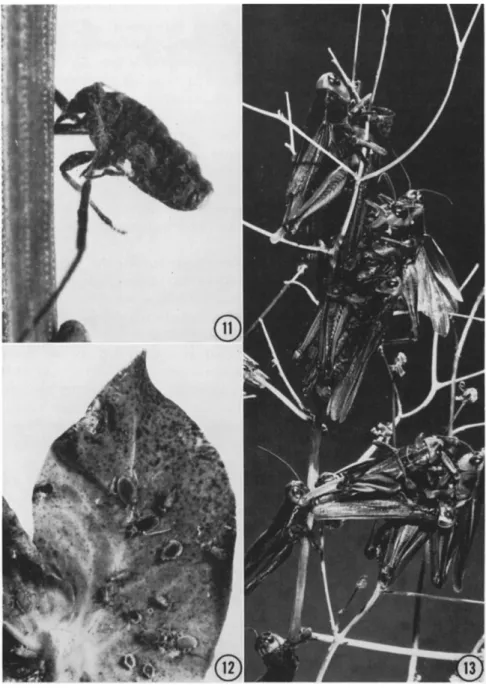
FIG. 11. A woolly pine needle aphid, Schizolachnus pini-radiatae, killed by Ento
mophthora aphidis, attached to the needle by its proboscis. Insects fastened in this manner are characteristic of many species of Entomophthora. ( χ 22.) (From Grobler et al., 1962.)
FIG. 12. A cluster of aphids, Macrosiphum pisi, destroyed by Entomophthora
there is some specificity of hosts for each species of Entomophthora. T h i s specificity, however, is by no means certain or uniform: some species of Entomophthora infect a wide range of hosts, including insects in differ
ent orders; others have been found only on a single insect species or a closely related g r o u p of insects. Entomophthora sphaerosperma, for ex
ample, has a wide host range (Ullyett a n d Schonken, 1940) whereas E. erupta has only three known host species, all from the hemipteran family Miridae (Dustan, 1924b; Hall, 1959).
It has been further shown that among the various species attacked, susceptibility to infection is shared by larvae, pupae, and adults, al
though the last are most commonly affected in many hosts.
2. The Introduction of Disease Agents by Inoculation
Every investigator who has attempted to infect healthy insects with species of Entomophthora has attested to the difficulties he h a d en
countered (Thaxter, 1888; Olive, 1906; Speare and Colley, 1912; Baird, 1957). I n most cases it seems the difficulty was to reproduce the exact conditions u n d e r which the infection naturally occurred.
It has been repeatedly stated that fungi may infect insects through the ingestion of spores (Picard, 1914b, cited by Skaife, 1925; Gabriel, 1959). Others differ in opinion and believe that the chances of infection in this way are very slight indeed (Glaser, 1926; Ullyett and Schonken, 1940). Skaife (1925) and M c M a r t i n (1935) reported that all carefully controlled experiments have proved that infection does not take place when grasshoppers are fed on E. grylli. I n the case of such species as E. aphidis which attacks aphids, and Entomophthora culicis Braun found on mosquitoes, the mode of infection must be through contact with the conidia, for the food of the insects cannot be contaminated with spores. Dustan (1924b) pointed out that the diameter of the conidia of E. erupta is greater t h a n that of the feeding tube of the green apple bug, making infection by this route impossible. It would seem, therefore, that the typical mode of infection among the entomophthorous forms is through contact, not through ingestion.
It must be assumed then that when conidia alight on an appropriate
aphidis o n the under surface of a leaf like stipule from a pea plant. T h e cadavers are firmly attached by rhizoids or "holdfasts." ( χ 3.) (From MacLeod, 1955.)
FIG. 13. Melanoplus bivittatus killed by Entomophthora grylli. Several typical characteristics of the disease are shown, e.g., congregation in a vertical position near the top of the plant stem, clasping, and partial disintegration of some of the speci
mens. T h e s e insects contain resting spores rather than conidia. ( χ 1.5.) (Courtesy of Canada Department of Agriculture Research Station, Lethbridge, Alberta.)
host they quickly give rise to a germ tube that enters the body of the insect either through one of the spiracles or else directly through the thinner areas of the integument. I n the latter case, the growing tip must penetrate the cuticle mechanically a n d / o r by the excretion of an enzyme that can degrade and weaken the cuticle. Penetration may take u p to 12 hours for completion (Ullyett and Schonken, 1940), after which mycelia can grow within the body cavity.
T h e conditions that induce a highly contagious state are not gener
ally known. T h e r e are, however, a n u m b e r of factors that affect the infectivity of various species of Entomophthora. T h e degree of infec
tivity, for example, may be influenced by the n a t u r e of the outer integu
ment (Thaxter, 1888). It is suspected that the thin body wall of some insects, particularly flies, mosquitoes, and aphids, are readily penetrated at almost any site on the body. T h e more highly chitinized outer coating of other insects, including beetles and grasshoppers, must diminish the chances of infection considerably, thus limiting the primary points of entrance to the intersegmental membranes between the body segments and at the joints of the appendages.
It has been suggested that an inoculation suspension may decrease the effectiveness of conidia by washing off or dissolving their protoplasmic coating (Baird, 1957). W h e n this substance is removed, the conidia are deprived of the adhesive qualities that enable the spores to stick readily to an object; of their source of protection from desiccation; and of the stimulatory effect of the coating u p o n germination.
Baird (1957) reported that u n d e r laboratory conditions E. muscae was transmitted to colonies of Kellymyia kellyi (Aldrich) reared in cages covered with plastic screening whereas flies in cages covered with cheese
cloth did not become cross-infected. According to Baird, the loose fibers of the cheesecloth prevented entry of airborne conidia.
H u m i d i t y is another important factor affecting the spread of fungal diseases, b u t information about its effect on species of Entomophthora is sparse and in some cases contradictory. Sawyer (1929) obtained maxi
m u m germination of E. sphaerosperma on glass slides at any relative humidity above 74.6 percent. However, Ullyett and Schonken (1940) found that, unless free water was present, n o infection was obtained when conidia of this fungus were applied to larvae of Plutella maculipennis (Curtis). Another situation was reported by Baird (1957), who found that disease caused by E. muscae could be transmitted among adults of K. kellyi kept in cages at 50 percent relative humidity.
I n inoculation tests with Entomophthora pseudococci Speare on the sugarcane mealybug, Pseudococcus calceolariae (Maskell), Speare (1912) demonstrated that best results were obtained when the insects
were kept overnight in poured potato agar petri plates, in which the fungus was growing, then transferred the following m o r n i n g to sterile rearing jars. T h e fungus normally discharged its spores at night, but only occasionally and sparingly in the daytime. D u r i n g the vegetative stage of growth, from conidial germination to m a t u r e hyphal bodies, the fungus was not affected by light b u t the young conidiophores remained absolutely d o r m a n t until 3 P.M., after which they grew vigorously and sporulated before morning. W i t h respect to germination, however, Speare's observations are not in agreement with Sawyer's statement that more conidia of E. sphaerosperma germinate in darkness than in light.
T h e fact that certain stages of the fungus do not develop in daylight may account for some of the p h e n o m e n a noted below in the section on disease symptoms.
It is apparent from the literature that some success has been obtained in the transmission of diseases by species of Entomophthora u n d e r experi
mental conditions. Still, in the main, this passage has been d u e to conidial dissemination directly from freely sporulating infected insects (Brefeld,
1873, 1881; Olive, 1906; Pole Evans, 1911; Dustan, 1924a; Schweizer, 1947) and in a few instances from sporulating culture media (Speare, 1912; Har
ris, 1948; Hall and D u n n , 1958). T h u s infection through artificial inocu
lation is still very uncertain. Indeed, to get an indication of how meager basic information on the infection process really is, it is only necessary to consider a few of the tests that have been reported on E. grylli, the important fungal pathogen of the grasshopper (Fig. 13). T h i s fungus, described and named in 1858 has, u n d e r favorable conditions, reduced large destructive outbreaks to negligible proportions in various countries throughout the world; yet the m a n n e r in which healthy grasshoppers become infected is still unknown. Skaife (1925), though unable to infect grasshoppers through the ingestion of spores, reported high mortality among 400 to 500 caught and placed in a cage for conveyance by train.
T h e first individuals to die in the cage were infected before they were captured, and the conidia thrown off from their bodies served to infect the others in the cage. Well over 75 percent of the total was lost in this manner. T e n years later M c M a r t i n (1935) stated that every attempt to infect healthy grasshoppers by placing conidia on their bodies had failed.
T h i s was the case whether the conidia were fresh or old, whether they were from dead specimens or from vegetation on which the insects died, or whether the grasshoppers on which they were placed were kept in a d a m p or dry atmosphere. I n the following year Schaefer (1936), d u r i n g field observations with E. grylli on the red locust, Nomadacris septem- fasciata (Serville), attempted to transfer the fungus obtained from re
cently dead specimens to 200 kept in a circular enclosure. T h i s was done
by squashing 25 grasshoppers that h a d died of the disease the previous evening, mixing them thoroughly in a p i n t of water, and then applying the suspension with an atomizer. W i t h i n the next 3 weeks only three of the sprayed insects died from the fungus. These were thought to have been naturally infected, since grasshoppers immediately next to and at some distance from the enclosure were also dying in the same proportion from the same fungal infection.
Findings that may develop following a far more critical approach and a search for additional factors involved in the infection process would unquestionably resolve some of the difficulties now encountered in the artificial utilization of fungal pathogens in insect control.
3. Mode of Development
I n nature, infection of the host results when a germ tube from an airborne spore or conidium penetrates the body wall of the insect and enters the body cavity.
Once inside, the fungus continues to grow by a peculiar b u d d i n g process in which hyphal bodies are produced until the body cavity is almost filled (Speare and Colley, 1912; Dustan, 1923; Fitzpatrick, 1930;
Steinhaus, 1946, 1949). H y p h a l bodies, however, do not always develop in the initial stages of the disease. I n larvae of P. maculipennis infected with E. sphaerosperma, for example, Ullyett and Schonken (1940) found that the fungus develops as mycelial threads that r u n longitudinally in the body cavity of the insect. W h e n these primary hyphae have ranged through the whole length of the host branching occurs freely and the body cavity gradually becomes filled with the mycelium of the fungus. As the final stages of growth are attained, the mycelium becomes stouter and then breaks u p into short lengths or hyphal bodies just before the death of the host.
In both cases the fungus invades all parts of the host; the head, thorax, and abdomen are readily attacked and mycelium may even be found in the legs as far down as the tarsi. T h e organism continues to grow until all the internal structures of the host have been destroyed and, when the disease has fully developed, what was once an insect con
sists merely of a chitinous shell filled with fungal threads. T h e r e appear to be a few exceptions to this general condition: Dustan (1924b) found that E. erupta, instead of working its way into all parts of the body of the green apple bug, confined itself chiefly to the abdomen of the insect.
Here it developed until all that was left of the internal tissues were the more heavily chitinized parts, such as portions of the h i n d intestine, the large trachea, etc. Examination of the head, thorax, and legs of the same insect showed that, for the most part, none of the vital organs had
been destroyed. Usually the fungus invaded the posterior half of the metathorax and there destroyed the larger wing and leg muscles, while the muscles of the pro- and mesothorax were left untouched. T h i s means that the insect was still able to use the first two pairs of legs and the first pair of wings, and it explains why diseased forms are able to move about freely (see section on dissemination).
As a rule, u p o n the death of the host, and occasionally even before death, (Dustan, 1927), the vegetative growth of the fungus terminates, whereupon the reproductive phase begins. T h e hyphal bodies send out very stout tubes that give rise to large club-shaped structures called co- nidiophores which break through the outer integument of the host a n d form conidia. At the same time entozoic rhizoids may grow out through the ventral surface of the dead insect, anchoring it to the substrate.
U n d e r conditions that are still undetermined, conidia formation may at times be interrupted by a sudden change in the morphology (Dustan, 1924b, 1927) and physiology (Speare and Colley, 1912; Ullyett and Schonken, 1940) of some of the hyphal bodies. Instead of producing conidiophores, they produce either zygospores or azygospores. Such spores eventually fill the body of the host a n d can be discerned in the head, thorax, abdomen, and legs (Dustan, 1927). A modification of this process sometimes occurs in that chlamydospores are formed. These germinate readily u n d e r favorable conditions, whereas the thicker- walled resting spores remain within the mummified host until the latter breaks u p or until they germinate the following spring.
Apart from the p r e m a t u r e death of the host, infection by species of Entomophthora is also believed to produce sterility in adults (Ullyett and Schonken, 1940). Dustan (1924b) concluded that infected females lose the power of depositing eggs before they are wholly overcome by E. erupta. Vinokurov (1949) has studied an epidemic reputedly caused by E. grylli among Acrididae which spread over the greater part of Siberia in 1937. H e found that the female insects were rendered sterile and concluded that this physiological sterilization was the p r e d o m i n a n t factor in reducing the heavy infestations. Despite the emphasis placed on the fungal pathogen, however, it is i m p o r t a n t to note that a micro
bial complex was involved in this investigation.
T h e length of the period between infection and death of the host insect varies to some extent. T h e length of time which elapses between the initial infection of P. maculipennis by E. sphaerosperma and the production of fruiting bodies may be as short as 3 days (Ullyett and Schonken, 1940). Speare and Colley (1912) reported that E. aulicae con
tinues to grow within the body of Nygmia phaeorrhoea (Donovan) for about 5 days after infection. T h e usual incubation period is from 5 to 8
days in larger hosts, b u t death may not take place for 12 days (Thaxter, 1888; Steinhaus, 1949). I n m i n u t e hosts, such as many gnats that are commonly attacked, this period is often m u c h shorter and may not exceed 2 or 3 days (Thaxter, 1888). It is thus possible to appreciate how a fungal disease may decimate a p o p u l a t i o n when conditions are favora
ble.
O n the basis of his observations on an outbreak of E. erupta in Lygus communis var. novascotiensis, Dustan (1924b) reported that there was an interval of 14 days between the first appearance of conidia and the first appearance of resting spores; he assumed that this period was the time required for resting spores, and possibly conidia, to develop.
T h i s assumption may not be quite correct since resting spores and conidia may develop concurrently in a population (Thaxter, 1888;
Speare, 1912). However, it is apparent from his observations that the growing period of the fungus is not more than about 6 weeks. T h e balance of the year must then be spent in the resting spore stage.
4. Disease Signs and Symptoms
D u r i n g the early stages of infection it is not possible to distinguish a diseased insect from a healthy one by external examination. It is only when the disease is well advanced that the first outward symptom, a general restlessness on the p a r t of the host, appears. T h e brown-tail m o t h caterpillar is not visibly affected by the presence of E. aulicae until the afternoon of the fifth or sixth day, when, after a brief period of nervous activity, its movement becomes sluggish or ceases altogether (Speare, 1912). A nervous restlessness lasting for a comparatively short time was also noted by Ullyett a n d Schonken (1940) among Plutella larvae infected with E. sphaerosperma. T h e sluggish movements of the walnut aphid, Chromaphis juglandicola (Kaltenbach), are among the early indications of parasitism of the insect by Entomophthora chro- maphidis Burger and Swain (Burger and Swain, 1918).
It has also been noted (Burger and Swain, 1918; Petch, 1940; Ullyett a n d Schonken, 1940; MacLeod, 1955) that, as the insects become more and more sluggish in habit, the living specimens may assume a yellowish tinge, which is intensified as the disease progresses.
Shortly before the last stage in the development of the fungus is reached, some insects, including infected adults of P. mali (Dustan,
1923), lose their power of locomotion and either d r o p to the ground or settle down on the underside of branches or leaves, a favorite position of many ailing insects. O t h e r insects, including the grasshopper, climb to the top of grass stems or twigs of bushes and die there with the head pointing u p w a r d (Skaife, 1925; Schaefer, 1936; Steinhaus, 1949). After
death the legs stiffen and the cadavers remain clasping the stalk until they are blown away by winds or washed off by rain. T h e forelegs of caterpillars of N. phaeorrhoea on the other h a n d , lose their power u p o n reaching the highest point of the twig or b r a n c h u p o n which they have been resting, with the result that the forepart of the body droops back
ward or to one side in a position so characteristic as to be easily recog
nized (Speare and Colley, 1912; see their Plate VI). Sometimes, when about to die, they may even leave their n o r m a l habitat and cluster on the tops of fences and stone walls. A n o t h e r remarkable characteristic of the disease is that the great majority of infected insects die in the late after
noon between 3 and 7 P.M. (Speare, 1912; Dustan, 1924b; Skaife, 1925;
Schaefer, 1936). T h e conidiophores form overnight and discharge the conidia early the next m o r n i n g while dew is still on the leaves.
Immediately after death and before the appearance of the aerial hyphae, the insect body is extended a n d turgid and contains a great a m o u n t of moisture. Once conidiophores are formed and conidia re
leased, the abdomen shrivels u p , becomes quite firm and brittle, and crumbles readily u n d e r pressure.
Insects in which resting spores are developing show the same general symptoms at first, b u t after death there is n o penetration of the body wall; instead they remain more or less mummified.
Infected sucking insects sometimes die with the proboscis firmly in
serted into the substratum and remain hanging long after death (Speare, 1922; Petch, 1940; Grobler et al, 1962) (Fig. 11); others are stuck fast by rhizoids (Burger and Swain, 1918; Petch, 1932; MacLeod, 1955; Hall and D u n n , 1957a,b; Grobler et al., 1962) and may remain firmly attached until the following spring (Fig. 12).
5. Dissemination
a. Natural spread. A remarkable characteristic of species of Ento
mophthora is their particularly effective means of survival and dis
semination. T h e y can endure the severe n o r t h e r n winters; they persist d u r i n g periods of extremely dry conditions in arid regions; and they maintain their viability under the extremely warm and h u m i d condi
tions of the tropics. They, therefore, have a capacity to survive in a wide variety of adverse conditions. Information on the underlying mechanism is meager and m u c h even of this is questionable. I n fact, our reasoning is largely deductive, being based on comparison with other microorganisms. T h u s conclusions regarding the p h e n o m e n o n of survival, together with those on n a t u r a l spread as considered here, will obviously require a m e n d m e n t as further knowledge is accumulated.
It seems to be a general characteristic that species of Entomophthora
form resting spores toward the end of the infection phase, their func
tion at this time being to ensure survival over prolonged adverse periods until growth can be resumed. T h u s the resting spores reestablish the disease in new insect populations. T h e r e is some question how this takes place since most of the spores are by this time on the ground and could inoculate only those insects that hibernate in the soil. Despite this, some insect hosts such as aphids (Grobler et al., 1962) spend their whole life cycle on plant foliage. Presumably a sufficient n u m b e r of spores remain on the foliage to serve as a source of inoculum.
It is believed that by spring the resting spores reach a condition or state where germination can occur. It is possible though that only a fraction of the resting spores ever germinate. W h e n germination occurs one of two processes may take place. If the resting spore is on an insect, the germ tube may function as an infection thread leading to direct infection. Alternatively, if no host is present the germ tube may func
tion as a conidiophore that abjoints terminally a single conidium that is in turn discharged. If contact is m a d e with an insect host, infection follows; otherwise secondary or even tertiary, conidia may be formed.
A n u m b e r of modifications of this general scheme may occur in the genus as a whole, the actual system, in any one case, depending on the fungal species involved and the host being attacked.
Once the initial infection is established, the disease develops rapidly in the insect with subsequent conidia formation. These, the "summer conidia," are produced in great numbers, are forcibly ejected, usually from an elevated position, and germinate rapidly. T h i s combination of factors tends to effect a rapid spread of the disease throughout a population.
T h e r e are additional factors which may be peculiar to a few, or even one, fungus-insect relationship that further aid in the natural dis
semination of the fungal pathogen. T h e migration of infected winged adults with subsequent spore discharge among healthy colonies has proved most effective in spore dispersal. Young insects which are ex
tremely active can in the early stages of the disease travel to previously uninfected groups of insects (Speare, 1912). I n the case of the green apple bug, some specimens retain the ability to move over the foliage quite rapidly even while conidia are being discharged, so distributing the spores in a far more efficient m a n n e r than would otherwise be pos
sible (Dustan, 1924b). T h e tendency of healthy insects to feed on diseased ones is another factor that contributes very largely to the spread of E.
erupta among the green apple bugs; Dustan has reported that often four or five nymphs or adults may be found clustered around a dorsally rup
tured insect with their beaks inserted in the moist fruiting layer, and this