UNCORRECTED
PROOF
Contents lists available at ScienceDirect
Journal of Organometallic Chemistry
journal homepage: http://ees.elsevier.com
Naphthoquinones of natural origin: Aqueous chemistry and coordination to half-sandwich organometallic cations
János P. Mészáros
a,b,1, Heiko Geisler
c,1, Jelena M. Poljarević
a,d, Alexander Roller
c, Maria S. Legina
c, Michaela Hejl
c, Michael A. Jakupec
c,e, Bernhard K. Keppler
c,e, Wolfgang Kandioller
c,e, Éva A. Enyedy
a,b,∗aDepartment of Inorganic and Analytical Chemistry, Interdisciplinary Excellence Centre, University of Szeged, Dóm tér 7, H-6720, Szeged, Hungary bMTA-SZTE Momentum Functional Metal Complexes Research Group, University of Szeged, Dóm tér 7, H-6720, Szeged, Hungary
cInstitute of Inorganic Chemistry, Faculty of Chemistry, University of Vienna, Währinger Str. 42, A-1090, Vienna, Austria dFaculty of Chemistry, University of Belgrade, Studentski trg. 12-16, 11000, Belgrade, Serbia
eResearch Cluster“Translational Cancer Therapy Research”, University of Vienna, Währinger Str. 42, A-1090, Vienna, Austria
A R T I C L E I N F O
Article history:
Received 28 October 2019
Received in revised form 5 December 2019 Accepted 6 December 2019
Available online xxx
Keywords Solution stability X-ray crystal structures Cytotoxicity Natural ligands Half-sandwich complexes
A B S T R A C T
Half-sandwich organometallic complexes featuring Ru(II), Os(II) and Rh(III) metal centers and naturally occur- ring bidentate 2-hydroxy-[1,4]-naphthoquinone ligands (lawsone and phthiocol) have been synthesized and char- acterized in both solid state and solution phase by analytical, spectroscopic, electrochemical and single crystal X-ray diffraction techniques. Comparative studies revealed the influence of the respective metal center (Ru, Os, Rh), leaving group (Cl, Br, I) and arene (p-cymene, toluene, pentamethylcyclopentadienyl), as well as the naph- thoquinone ligand on the structural properties and solution speciation. Additionally, cytotoxicity was tested in SW480, CH1/PA-1 and A549 human cancer cell lines showing a broad range of IC50values.
© 2019
Abbreviations
BOLD-100 sodiumtrans-[tetrachloridobis(1H-indazole)ruthen- ate(III)], KP-1339
CV cyclic voltammetry
deferiprone 1,2-dimethyl-3-hydroxy-pyridin-4(1H)-one DMF dimethylformamide
Hlaw lawsone, 2-hydroxy-[1,4]-naphthoquinone
Hphth phthiocol, 3-methyl-2-hydroxy-3-methyl-[1,4]-naphtho- quinone
maltol 3-hydroxy-2-methyl-pyran-4(1H)-one menadione 3-methyl-[1,4]-naphthoquinone
MTT 3-(4,5-dimethylthiazol-2-yl)-2,5-diphenyltetrazolium bromide
NAMI-A trans-[tetrachlorido(DMSO)(imidazole)ruthenate(III) ROS reactive oxygen species
∗Corresponding author. Department of Inorganic and Analytical Chemistry, Interdisciplinary Excellence Centre, University of Szeged, Dóm tér 7, H-6720, Szeged, Hungary.
E-mail address:enyedy@chem.u-szeged.hu (É.A. Enyedy) 1 Both authors contributed equally to this work.
TGA thermogravimetric analysis
1. Introduction
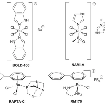
Due to the reoccurring limitations with well-established Pt(II) drugs (e.g.cisplatin) in cancer treatment, much research effort has been fo- cused on the search for other metallodrugs with different activity pro- files and lower toxicity [1,2]. In this context, metal center variation brought to light the possible suitability of Ru based drugs. At this time Ru(III) compounds, as well as Ru(II)“piano-stool” complexes are ex- tensively investigated. Two well-known examples of Ru(III) drug can- didates are BOLD-100 (sodium trans-[tetrachloridobis(1H-inda- zole)ruthenate(III)], formerly KP-1339 or IT-139), which is about to enter clinical phase II studies, and NAMI-A (trans-[tetrachlo- rido(DMSO)(imidazole)ruthenate(III)]). Both of these complexes high- light the importance of the metal's coordination sphere (Fig. 1). While BOLD-100 features two indazole ligands and is active against primary tumors as well as metastases, its structurally related congener NAMI-A only shows activity against metastases [3,4].
On the other hand, organometallic Ru(II) complexes often feature a stabilizing arene moiety, and mono-, or bidentate ligands, as well as a leaving group. The name of these so-called“piano-stool”complexes is https://doi.org/10.1016/j.jorganchem.2019.121070
0022-328/© 2019.
UNCORRECTED
PROOF
2 J.P. Mészáros et al. / Journal of Organometallic Chemistry xxx (xxxx) 121070
Fig. 1.Structural formulae of Ru(III) and Ru(II) drug candidates, which are currently un- dergoing or have undergone (pre)-clinical trials.
derived from the resulting geometrical form, which resembles a three legged stool, where the arene is the seat and the other ligands are the legs. The most advanced and extensively studied drug candidates, RM175 and RAPTA-C (Fig. 1) are believed to have completely differ- ent modes of action. While Sadler et al. postulated that RM175 interacts with DNAviaintercalation of the biphenyl moiety and covalent binding to nucleobases; activity of RAPTA-C is ascribed to protein interaction [5]. However, no“piano-stool”complex has been evaluated in clinical studies so far.
Another approach is the synthesis of Ru(II)“piano-stool”complexes featuring bioactive natural products with anticancer properties. Many natural compounds such as quinones and naphthoquinones are found in plants, algae, and bacteria and used as versatile building blocks for or- ganic synthesis of medicinal drugs [6,7]. Other members of this family are phthiocol and lawsone, which are considered as derivatives of vita- min K (Fig. 2) [6,8,9].
Naphthoquinones readily act as reducing agents leading to the for- mation of radicals and reactive oxygen species (ROS) [10]. In the past, several quinones and naphthoquinones were biologically tested and re- vealed cytostatic properties [6,7,11–13]. Half-sandwich organometallic complexes containing 2-hydroxy-[1,4]-naphthoquinones as ligand scaf- fold have already been reported in literature with promising results [14–16]. Furthermore, heterobimetallic compounds with a pendant fer- rocene moiety have been investigated for their anticancer properties [17,18]. Physicochemical properties (e.g.solubility, lipophilicity), bio- logic parameters (e.g.IC50values, mode of action) as well as solution equilibrium constants are crucial factors in drug development. One of the main advantages of using“piano-stool”complexes is the possibility to fine-tune these physicochemical properties, namely stability, redox properties, solubility via ligand modification and variation of the metal center or the leaving group [19,20].
Fig. 2.Structural formulae of vitamin K3 (menadione) and its naphthoquinone deriva- tives: phthiocol, lawsone, and lapachol (from left to right).
Herein we report the synthesis, characterization, detailed solution stability and complex formation properties of six new “piano-stool”
complexes. These organometallics feature either 3-methyl-2-hy- droxy-3-methyl- [1,4]-naphthoquinone (phthiocol, Hphth) or its non-methylated analogue 2-hydroxy- [1,4]-naphthoquinone (lawsone, Hlaw) asO,O-chelates for Ru(II)-, Os(II)- and Rh(III)-arene organometal- lic ions. Additionally, cytotoxicity tests in human cancer cell lines were conducted in order to assess the anticancer potency of these compounds.
2. Results and discussion
2.1. Synthesis
Lawsone is commercially available, phthiocol was synthesized in a two-step synthesis, starting from commercially available 3-methyl- [1,4]-naphthoquinone (menadione), according to literature with minor modifications [21,22]. Firstly, an epoxide intermediate was formed, us- ing aqueous hydrogen peroxide solution (30%). Ring opening reaction was performed with concentrated sulfuric acid and silica gel in THF and provided good yields (72%) over two steps (Scheme S1).
The phthiocol-based Ru(II)-p-cymene complex (KP2048) showed promising anticancer activity in vitro and in vivo experiments [14].
Within this work, we wanted to investigate the effect of different metal centers (Ru(II), Os(II), Rh(III)), halide leaving groups (Cl−, Br−, I−), arenes (p-cymene, toluene, C5Me5-) and ligands on the solution behavior and cytotoxic potency.
The desired complexes (1–7) were obtained by the deprotonation of the 2-hydroxy-naphthoquinone (phthiocol or lawsone) with sodium methoxide in dry methanol, followed by the addition of the respec- tive dimeric metal precursor ([MX2(arene)]2) (see Scheme 1). Fol- lowing the standard purification reported in literature [16], the com- plexes were isolated in moderate-to-good yields. Two different meth- ods were applied to obtain the desired complexes. In method A: the starting materials were reacted under standard conditions (20–40 °C) for 2–24 h. The other method (method B) included microwave irradiation for 6–14 min at 40–50 °C. Both methods were used for all complexes.
However, the microwave assisted method reduced the reaction time of complexes2,4and5considerably. After purification, yields between 15 and 90% were obtained.
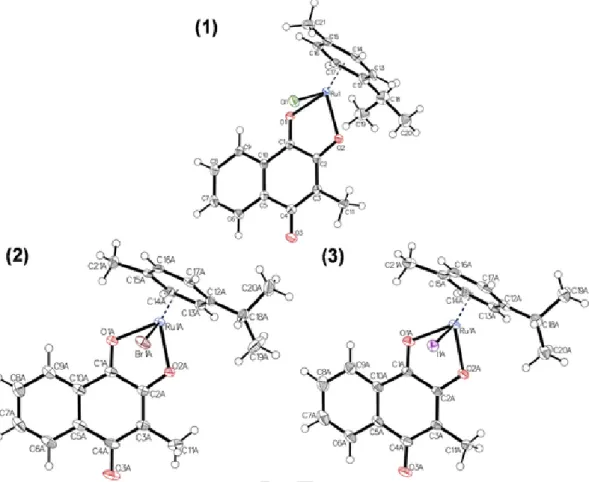
2.2. Crystal structures of phthiocol complexes
Single crystals of five complexes (1–3,5–6) were obtained by liq- uid-liquid diffusion from dichloromethane/n-hexane (see Figs. 3 and 4). These complexes exhibit a pseudo-octahedral geometry with a lig- and-to-metal ratio of 1:1. The arene, the bidentate naphthoquinone lig- and (Hphth) and the monodentate halide ion are coordinated to the metal center and adopt the characteristic “piano-stool” configuration.
Scheme 1.Synthetic pathway for complex synthesis.
UNCORRECTED
PROOF
Fig. 3.Molecular structures of1,2and3drawn at 50% probability level. Solvent molecules were omitted for clarity. Where both enantiomers crystallized one of them was omitted as well.
Fig. 4.Molecular structures of5and6drawn at 50% probability level. Solvent molecules were omitted for clarity. Where both enantiomers crystallized one of them was omitted as well.
Complexes2,3, and6crystallized in the triclinic space group P1̅. Com- pounds1and5crystallized in the monoclinic space group P21/c.
The bond lengths between the donor atoms of phthiocol and the metal center and bond angles are shown in Table 1. The variation of the metal center Ru(II) (1) to Os(II) (5) has no relevant impact on the bond lengths and angles. However, the Rh(III) complex6 exhibits a
slight elongation of the coordination bonds, compared to the Ru(II) ana- log1.
Based on these data the variation of halide ions has no relevant effect on geometric parameters in the coordination sphere. These organometal- lic complexes possess a chiral metal center and both enantiomers were found in the unit cell of complexes2,3and6.
UNCORRECTED
PROOF
4 J.P. Mészáros et al. / Journal of Organometallic Chemistry xxx (xxxx) 121070
Table 1
Selected bond lengths, bond angles and torsion angles for the phthiocol complexes: (1–3, 5–6).
1 2a 3a 5 6a
Bond distances [Å]
M-Cg 1.651 1.646 1.651 1.651 1.743
M-O1 2.119(2) 2.123(2) 2.122(3) 2.119(3) 2.160(4)
M-O2 2.092(2) 2.082(2) 2.085(3) 2.092(4) 2.095(5)
M-X 2.403(1) 2.532(4) 2.711(4) 2.394(1) 2.390(2)
Bond angles [°]
O1-M-O2 76.31(8) 76.39(8) 76.18(9) 75.60(1) 76.25(2) O-M-Xb 84.89(7) 85.61(5) 86.17(8) 83.66(8) 87.63(4)
aCalculated average of the two isomers in one unit cell.
b Calculated average value of O1-M-X and O2-M-X.
2.3. In vitro anticancer activity of compounds
The cytotoxicity of 2-hydroxy- [1,4]-naphthoquinones depends on the nature of alkyl substituents at position 3 [13]. Cytotoxic activities of Hphth,Hlawand the respective complexes1,4–7were determined in human colon carcinoma (SW480), ovarian teratocarcinoma (CH1/PA-1) and non-small cell lung carcinoma (A549) cells by means of the col- orimetric 3-(4,5-dimethylthiazol-2-yl)-2,5-diphenyltetrazolium bromide (MTT) assay (Table 2). No IC50values could be determined for com- plexes2and3,due to the poor solubility in aqueous systems.
All tested complexes 1, 4–7 showed at least a tendency for in- creased cytotoxicity in the intrinsically chemo-resistant (P-glycopro- tein-expressing) human cancer cell line SW480, compared to the free lig- ands (Hphth,Hlaw). The combination of Ru(II) andp-cymene yields a clearer increase of cytotoxicity in all cancer cell lines employed, whereas the other variants with a different metal center (6) or arene ligand (4) showed only marginally increased cytotoxicity in the cell line SW480 and no enhanced activity in A549 and CH1/PA-1 cells.
Based on these data we could conclude that the tested compounds exhibited a broad range of anticancer potency with IC50in the range of 15μM to >200μM. Generally the dimeric organometallic precursors do not exhibit strong cytotoxicity (>100μM [RuCl2(η6-p-cymene)]2re- ported for several cancer cell lines [25,26], and both naphthoquinone ligands showed IC50values > 100μM. The arene change resulted in a loss of activity compared to thep-cymene derivative1, as did the metal center variation from Ru(II) to Os(II) or Rh(III). Introduction of the law- sone ligand was not beneficial with regard to cytotoxicity either, but complex7was still more active than the free ligandHlaw.
Table 2
Cytotoxicity of ligands, their corresponding half-sandwich organometallic complexes (1, 4–7) and cisplatin and RAPTA-C for comparison in three different human cancer cell lines using 96 h incubation time.
IC50[μM] A549 SW480 CH1/PA-1
1a 47 ± 4 15 ± 3 31 ± 10
4 IC50> 100 87 ± 6 IC50> 100
5 111 ± 23 81 ± 18 59 ± 12
6 IC50> 200 96 ± 17 IC50~ 200
Hphth 210 ± 32 116 ± 37 129 ± 29
7 98 ± 24 86 ± 20 84 ± 15
Hlaw 157 ± 13 247 ± 17 246 ± 24
cisplatinb 6.2 ± 1.2 3.3 ± 0.2 0.077 ± 0.006
RAPTA-C c >500 170 ± 60 65 ± 15
aData are taken from Ref. [14].
b Data are taken from Ref. [23].
c Data are taken from Ref. [24].
2.4. Solution chemistry
Investigation of solution behavior and determination of stability con- stants of bioactive metal complexes are key steps in drug development processes since from speciation data the biologically active chemical forms can be predicted. Thus this kind of information can help in deep- ening the understanding of pharmacokinetic properties and the mech- anism of action. Currently no speciation data have been reported for half-sandwich organometallic complexes of 2-hydroxy-[1,4]-naphtho- quinones so far. Only a few studies on the time-dependent stability in buffered solutions can be found in literature [15].
In this work we investigated systematically the aqueous solution chemistry of metal-ligand systems containing phthiocol and organometallic triaqua complex cation ([Ru(η6-p-cymene)(H2O)3]2+, [Ru(η6-toluene)(H2O)3]2+ and [Rh(η5-C5Me5)(H2O)3]2+) in the pres- ence of 0.2 M chloride ions. pH-potentiometry is not suitable for deter- mining stability constants for these naphthoquinone complexes due to their low water solubility and high concentration demand of the tech- nique. Therefore UV–visible (UV–Vis)titrations were performed where lower concentrations can be employed. Though the osmium complex5 was synthesized and itsin vitrocytotoxicity was determined, the solu- tion chemical properties were not characterized here since the more pro- nounced and slower hydrolytic behavior of [Os(η6-p-cymene)(H2O)3]2+
compared with [Ru(η6-p-cymene)(H2O)3]2+ or [Rh(η5-C5Me5)(H2O)3]2+ would make the studies fairly difficult [27,28]. Lawsone was also involved the studies to reveal the impact of the methyl group on the ligand scaffold on the solution chemical prop- erties.
2.4.1. Proton dissociation processes of naphthoquinones and hydrolysis of the organometallic cations
ThepKavalue is a key parameter of bioactive compounds, as it has a large effect on the pharmacokinetic properties considering the actual protonation state (and charge), and it also affects the pH-dependence of the lipophilicity. with the help of these constants it is possible to calculate the distribution of different protonated species and the aver- age charge at a given pH. phthiocol and lawsone have one dissociable proton, as only the hydroxyl group can lose a proton with increasing pH. although the structures are similar, the methyl group in the third position has a great impact on the neighboring hydroxyl group. pro- ton dissociation constants of lawsone and phthiocol were determined by UV–Vis titrations (I = 0.2 M KCl, Fig. s1) and deprotonation occurs at much lower pH values in the case of lawsone(pKa(Hl) = 3.90 and 5.08 for lawsone and phthiocol, respectively, see Table 3). These data
Table 3
Proton dissociation constants (pKa(HL)) ofHlawandHphthand stability constants (logΚ [M(arene)L]) of their complexes. {T = 25.0 °C,I= 0.2 M (KCl)}.
Ru(η6-p-
cymene) Ru(η6-
toluene) Rh(η5- C5Me5)
Hlaw pΚa(HL)a 3.90 ± 0.01
logΚ
[M(arene)L]a 3.45 ± 0.01 3.31 ± 0.02 <3.1
% of[M(arene)L]b 0.01% <0.01% 6.6%
Hphth pΚa(HL)a 5.08 ± 0.01
logΚ
[M(arene)L]a 4.04 ± 0.03 3.78 ± 0.04 3.52 ± 0.03
% of[M(arene)L]b 0.04% <0.01% 14.5%
aFrom UV–Vis spectrophotometric titrations.
b Calculated atc(M(arene)) =c(L) = 100μM, pH = 7.40.
UNCORRECTED
PROOF
are in good agreement with literature data, although different conditions were used (I = 0.5 M KCl) [29,30].
Hydrolysis of the [Rh(η5-C5Me5)(H2O)3]2+, [Ru(η6-p-cymene)(H2O)3]2+ and [Ru(η6-toluene)(H2O)3]2+
organometallic complex cations has been characterized in detail previ- ously [27,28]. In these organometallic triaqua cations and (their stud- ied complexes) the bond between the metal ion and the arene is sta- ble under the used conditions (pH = 2.0–11.5). Increasing the pHμ-hy- droxido bridged dinuclear species are formed with the formula [(M(arene))2(OH)2]2+ and [(M(arene))2(OH)3]+ [27,28]. Although Bíró et al. examined extensively the species formed from [Ru(η6-p-cymene)(H2O)3]2+in chloride ion containing medium and de- termined the overall stability constants for five different species, all the titrations can be described sufficiently by only the overall stability con- stants determined for [(M(arene))2(OH)2]2+and [(M(arene))2(OH)3]+ (I= 0.2 M KCl) [31]. These constants are log β [(M(arene))2(OH)2] =−6.50, −7.12, −11.12 and log β [(M(arene))2(OH)3] =−10.56, −11.88, −19.01 for M(arene) = Ru(η6-toluene), Ru(η6-p-cymene) and Rh(η5-C5Me5), re- spectively, and these data were used in this work as well [27,28].
2.4.2. Complex formation equilibria of naphthoquinones with organometallic ions
Based on the crystallographic structures and literature data of other half-sandwich Ru(II) and Rh(III) complexes with (O,O) donors, mono complex formation is the most probable process under the applied con- ditions. The complexes of half-sandwich Rh and Ru complexes with (O,O) donors usually reaches the equilibrium state rather fast [28,32–34].Because of the extended delocalized electron system, the complexes of phthiocol and lawsone have absorbance in the UV–Vis re- gion, so spectrophotometric titration is a suitable method for follow- ing the complex formation and spectra were recorded at 1:1 and 1:2 metal-to-ligand ratios.
In Fig. 5a the pH-dependent UV–Vis spectra of the [Ru(η6-toluene)(H2O)3]2+–lawsone (1:1) system are shown. The spec- tra above 590 nm (see Fig. 5a inserted diagram) give the most valu-
able informa
tion: neither the metal ion nor the ligand has absorption in that re- gion. Spectral changes here directly show the formation of the formed metal complex. In the system with 1:2 metal-to-ligand ratio the ab- sorbance change is relatively high making the determination of the stability constants more precise (Table 3). In Fig. 5b the concentra- tion distribution curves calculated with the stability constants and the absorbance at 590 nm are shown. The maximum of the formed com- plex is 11.4% at pH 4.72, which demonstrates low solution stability.
With [Ru(η6-p-cymene)(H2O)3]2+the spectral changes are similar (Fig.
S2) and the formation of the [M(arene)L] complex is detectable also around 590–600 nm. However, with Rh(η5-C5Me5) no complex forma- tion occurred under the used conditions. Titration of lawsone with the organometallic ion at pH 7.4 shows no interaction between them, and the linearity of the curve in Fig. S3 proves the lack of complex forma- tion in the system. Only an upper limit could be provided for the stabil- ity constant of this complex in Table 3.
The pH-dependence of UV–Vis spectra of
[Ru(η6-p-cymene)(H2O)3]2+, phthiocol and their 1:1 metal-to-ligand ra- tio system is shown in Fig. 6a. The detection of complex formation in the case of phthiocol is more difficult: there is only a minor spec- tral change at ~600 nm with a maximum of 0.02 absorbance (see Fig.
6a inserted figure); in this region only the metal complex absorbs pho- tons. Based on this absorbance change, the complex formation shows a maximum at pH 5.1. Calculations approved the low stability of the formed complex as the concentration distribution curves represent in Fig. 6b; [M(arene)L] has a maximum with 26.7% at 100μM concen- tration and at pH 5.06. With [Ru(η6-toluene)(H2O)3]2+ the spectral changes and the extent of complex formation are even smaller than with [Ru(η6-p-cymene)(H2O)3]2+(shown in Fig. S4). Additionally, the ab- sorbance at 600 nm reaches only 0.015, which makes the detection of the complex formation even more problematic. The formation of the half-sandwich Rh(η5-C5Me5) complex of phthiocol is also observable at 600 nm and the absorbance changes are negligible for this system (ΔA
~0.015, see Fig. 7) (see Fig. 8).
Fig. 5.a) UV–Vis absorption spectra of the [Ru(η6-tol)(H2O)3]2+–lawsone 1:1 system at pH = 2.0–11.5. b) Concentration distribution curves calculated with the determined constants from Table 3. Absorbance at 590 nm (○) shows the complex formation {c(lawsone) =c([Ru(η6-tol)(H2O)3]2+) = 200μM;I= 0.2 M (KCl);T= 25.0 °C;l= 2 cm}.
Fig. 6.a) UV–Vis absorption spectra of phthiocol, [Ru(η6-p-cymene)(H2O)3]2+and the [Ru(η6-p-cymene)(H2O)3]2+–phthiocol 1:1 system at pH = 2.0–11.5. b) Concentration distribu- tion curves calculated with the determined constants from Table 3. Absorbance at 600 nm (○) shows the complex formation {c(phthiocol) =c([Ru(η6-p-cymene)(H2O)3]2+) = 100μM;
I= 0.2 M (KCl);T= 25.0 °C;l= 2 cm}.
UNCORRECTED
PROOF
6 J.P. Mészáros et al. / Journal of Organometallic Chemistry xxx (xxxx) 121070
Fig. 7.a) Molar absorbance spectra calculated from the spectrophotometric titration of the [Rh(η5-C5Me5)(H2O)3]2+–phthiocol system at pH = 2.0–11.5. b) Concentration dis- tribution curves calculated with the determined constants from Table 3. Absorbance at 600 nm (□) shows the formation of [M(arene)L] complex {c(phthio- col) =c([Rh(η5-C5Me5)(H2O)3]2+) = 100μM;I= 0.2 M (KCl);T= 25.0 °C;l= 1 cm}.
Fig. 8.Calculated pM*-curves obtained for the [Rh(η5-C5Me5)(H2O)3]2+‒(O,O) biden- tate ligand systems plotted against the pH. L = lawsone (- - -); phthiocol (‒‒); acety- lacetone (-∙-∙-); maltol (-∙∙-∙∙-); deferiprone (∙∙∙∙∙∙∙) [28,32–34]
{c(L) =c([Rh(η5-C5Me5)(H2O)3]2+) = 100μM;T= 25.0 °C;I= 0.2 M (KCl)}.
Comparison of the stability constants for both ligands shows a clear trend. The stability constant is the highest for Ru(η6-p-cymene) com- plexes, which is followed by Ru(η6-toluene) and the Rh(η5-C5Me5) com- plexes have the lowest stability constants (Table 3). The same tendency has been found previously in the case of other (O,O) donor bidentate ligands [33]. Based on the determined stability constants (Table 3) the calculated concentration distribution curves show different characteris- tics for the [Rh(η5-C5Me5)(H2O)3]2+–phthiocol (1:1) system (Fig. 7b);
namely the complex is present in a wider range (>5% at pH 4.4–8.0) than the ruthenium analogues (>5% at pH 3.8–6.0). For the interpre- tation of the biological activity data, Table 3 shows the amount of the [M(arene)L] complex formed at pH = 7.4. It can be concluded that only the [Rh(η5-C5Me5)(phth)Cl] complex is present in the solution in more than 10% and it is the least cytotoxic of them.
Quinones and naphthoquinones are redox active molecules and their redox chemistry is well characterized [35]. As complex formation often has a great impact on the ligand's redox chemistry cyclic voltammetry (CV) was used to follow the changes of the voltammograms upon com- plexation in a 9:1 solvent mixture of dimethylformamide (DMF) and wa- ter due to the low solubility of the compounds in water (Fig. S5). How- ever, variation of the metal center had only a small effect on the shape of the voltammograms, most probably as a result of the low fraction of complex formation under the applied conditions.
2.4.3. Comparison the stability of naphthoquinone complexes with other (O,O) donors
To compare the stability of complexes of the same organometallic ion formed with ligands of different pKavalues, herein a modification
of the method introduced by Raymond et al. is used, namely the di- rect comparison of pM* values [36]. The original pM is the negative logarithm of the free metal ion concentration, while pM* is calculated for the nonbound metal ion concentration: not only the free metal ion but the hydroxido species are taken into account as well. The higher pM* value shows higher stability. The calculated pM* curves are shown in Fig. 8 and Fig. S6, illustrating the stability difference between the complexes of 2-hydroxy-[1,4]-naphthoquinones (Table 3), 2,4-dike- tonates (acetylacetone), 3-hydroxy-2-methyl-pyran-4(1H)-one (maltol) and 1,2-dimethyl-3-hydroxy-pyridin-4(1H)-one (deferiprone) [28,32–34]. The stability constants for the Ru(η6-toluene)–deferiprone system were missing for this comparison, thus pH-potentiometric titra- tions were performed in this work to determine these data, which are: log K [M(arene)L] = 11.74 ± 0.08 and pKa
[M(arene)L] = 9.34 ± 0.09. The trend of stability is the same indepen- dently from the type of the organometallic cations: deferiprone > mal- tol > acetylacetone > 2-hydroxy- [1,4]-naphthoquinones is the order in a wide pH range.
3. Experimental 3.1. Chemicals
All solvents were of analytical grade and used without further pu- rification. Dry solvents were used for synthesis and stored under argon.
Lawsone, menadione (Acros Organics), RuCl3× xH2O, RhCl3× xH2O, IrCl3× xH2O, OsO4 (Johnson Matthey), α-terpinene (Alfa Aesar), 1,2,3,4,5-pentamethylcyclopentadiene (TCI Europe), deferiprone, H2O2 (30%), KCl, HCl, KOH, KH-phthalate, NaOMe, Na2CO3, KH2PO4, NaH2PO4, Na2HPO4, MeOH, CHCl3, CH2Cl2, DMF and DMSO (Sigma Aldrich) were purchased from commercially available suppliers. For aqueous solutions Milli-Q water was used. The exact concentrations of the metal precursor stock solutions were determined by pH-potentio- metric titrations with the aim of Hyperquad 2013 program [37], em- ploying the stability constants for hydroxido complexes from (I= 0.2 M KCl) [27,28]. Stock solutions of the ligands were prepared on a weight-in-volume basis. In the case of buffered samples, 20 mM phos- phate buffer was used instead of water with pH = 7.40.
For characterization of the compounds NMR spectra (Figs. S7–13) were recorded with a Bruker FT-NMR spectrometer Avance III™
500 MHz with an UltraShieldPlus magnet at 25 °C. The measurement frequency for proton NMR (1H) was 500.10 MHz and for carbon NMR (13C{1H}) 125.75 MHz CDCl3was used as solvent. Elemental analysis for carbon, hydrogen, nitrogen, sulfur and oxygen was performed at the Microanalytical Laboratory of the University of Vienna with Eurovec- tor EA 3000 CHNS–O elemental analyzer (2009) equipped with a high temperature pyrolysis furnace (HT, Hekatech, Germany, 2009). Ther- mogravimetric analysis (TGA) was carried out on a TGA/SDTA851ein
UNCORRECTED
PROOF
strument with a Sample Robot TS0801R0 from Mettler Toledo (Figs.
S14–16). The recorded TGA curves revealed thermal decomposition at T < 100 °C, which did not allow the determination of the water content of the samples by this method.
3.2. pH-potentiometric measurements
pH-potentiometric measurements determining concentrations of [Rh(η5-C5Me5)(H2O)3]2+, [Ru(η6-p-cymene)(H2O)3]2+ and [Ru(η6-toluene)(H2O)3]2+were carried out at 25.0 °C ± 0.1 °C in water and at a constant ionic strength of 0.2 M KCl. The titrations were per- formed with a carbonate-free KOH solution (0.20 M). The exact concen- trations of HCl and KOH solutions were determined by pH-potentiomet- ric titrations. An Orion710A pH-meter equipped with a Metrohm elec- trode (type 6.0234.100) filled with 3 M KCl and a Metrohm 665 Dosi- mat burette was used. The volume resolution of the burette is 0.001 mL and its precision is 0.002 mL. The electrode system was calibrated to the pH = -log [H+] scale by means of blank titrations (strong acid vs.
strong base: HCl vs. KOH), as suggested by the Irving method [38].
The water ionization constant (pKw) was determined as 13.76 ± 0.01 at 25.0 °C ± 0.1 °C,I= 0.2 M (KCl) [39]. The reproducibility of the titra- tion points included in the calculations was within 0.005 pH units. The pH-potentiometric titrations were performed in the pH range between 2.0 and 11.5. The initial volume of the samples was 5.0 mL. The metal ion concentration was 2.0 mM. The goodness-of-fit measured in Hyper- quad2013 [37] by sigma (σ) represents the overall goodness-of-fit de- rived from the sum of squared residuals (calculated-experimental titra- tion data). The model was accepted whenσwas close to one (<1.5).
The standard deviation of the log β values of species included into the model was always lower than 0.1. Samples were degassed by bub- bling purified argon through them for about 10 min prior to the mea- surements and the inert gas was also passed over the solutions dur- ing the titrations. Log βvalues for the various hydroxido complexes [(Rh(η5-C5Me5))2(μ-OH)i](4−i)+, [(Ru(η6-p-cymene))2(μ-OH)i](4−i)+and [(Ru(η6-toluene))2(μ-OH)i](4−i)+(i = 2 or i = 3) were calculated based on the pH-potentiometric titration data in the presence of chloride ions and were found to be in good agreement with the previously published data [27,28]. Stability constants for (M(arene))pLqHrcomplexes cannot be determined by pH-potentiometry because of solubility problems of the ligands except the case of the deferiprone complexes.
3.3. UV–Vis spectrophotometric measurements
An Agilent Cary 8454 diode array spectrophotometer was used to record the UV–Vis spectra in the interval 200–800 nm. The path length was 1 or 2 cm. Equilibrium constants (proton dissociation and stability constants) and the individual spectra of the species were calculated with the computer program PSEQUAD [40]. The spectrophotometric titra- tions were performed in aqueous solution on samples containing the lig- ands with or without the organometallic cations and the concentration of the ligands was 50–200μM. The metal-to-ligand ratio was 1:1 in the pH range from 2 to 11.5 at 25.0 °C ± 0.1 °C, I = 0.2 M (KCl).
3.4. Cyclic voltammetric measurements
Electrochemical experiments of phthiocol containing samples were performed on an Autolab-PGSTAT 204 potentiostat/galvanostat and monitored with Metrohm's Nova software. Measurements of the lig- and (1 mM) in the presence and the absence of the metal ions (1 mM) were obtained in a 9:1 DMF/water mixture because of low solubil- ity of phthiocol. The aqueous part contained 20 mM phosphate buffer (pH = 7.40). The supporting electrolyte was 0.2 M [n-Bu4N][BF4]. A three-electrode configuration cell was used with two glassy carbon
electrodes: one was the working and another was the counter electrode, the reference electrode was an Ag/AgCl/(1 M) KCl electrode. The sys- tem was calibrated for 0.01 M ferrocene before every experiment and gave a potential at +0.688 Vvs. NHE.
3.5. Synthesis phthiocol (Hphth)
Menadione (1.00 g, 5.81 mmol, 1 eq.) was dissolved in methanol (10 mL) and stirred under ice cooling. Na2CO3(0.207 g, 1.95 mmol, 0.3 eq.) and H2O2solution (36%, 1 mL, 32.6 mmol, 1.7 eq.) were dis- solved in H2O (10 mL). The aqueous solution was added dropwise to the yellow suspension. After complete addition, the mixture was stirred at room temperature for 1 h. H2O (100 mL) was added to the mix- ture and the formed precipitate was separated by filtration and driedin vacuo. The colorless solid was suspended in water (20 mL) and concen- trated sulfuric acid was added until complete dissolution. The dark red solution was diluted with water and extracted with dichloromethane.
The organic layer was separated and extracted with saturated NaHCO3
solution. The aqueous layer was acidified with concentrated HCl and extracted with dichloromethane. Afterwards, the yellow solution was dried over anhydrous Na2SO4, evaporated and driedin vacuo. Yield:
0.788 g (4.19 mmol, 72%), yellow powder. Characterization:1H NMR (500.10 MHz, CDCl3) δ= 2.11 (s, 3H, H1’), 7.29 (s, 1H, HOH), 7.68 (ddd,J= 8 Hz, 1 Hz, 1H, H6/7.), 7.75 (ddd,J =8 Hz, 1 Hz, 1H, H6/7.), 8.08 (dd,J= 8 Hz, 1H, H5/8), 8.13 (dd,J= 8 Hz, 1H, H5/8).
3.6. Synthesis of dimeric metal precursors
The dimeric metal precursors [RuCl2(η6-p-cymene)]2 [41], [RuCl2(η6-tolouene)]2 [42], [RuBr2(η6-p-cymene)]2 [43], [RuI2(η6-p-cymene)]2 [43], [RhCl2(η5-C5Me5)]2 [44] and [OsCl2(η6-p-cymene)]2[45] were synthesized according to literature.
3.7. General procedure
Phthiocol (3 eq.) and sodium methoxide (3.2 eq.) were dissolved in dry methanol (12 mL) and stirred for 10 min. Afterwards, the re- spective metal precursor dimer (1 eq.) was added to the dark red mix- ture. Depending on the desired complex, two different methods were applied. Method A: The mixture was stirred at room temperature or at 40 °C for 2–24 h. Method B: The mixture was stirred at 40–50 °C under microwave irradiation for 6–14 min. In both methods, the pu- rification procedure was conducted in the same way. The precipitate was separated from the supernatant, which was further evaporated. The solid were dissolved in dichloromethane, filtrated, and the solvent was evaporated. The remaining black residues were combined, dissolved in dichloromethane andn-hexane was added until precipitation started.
The mixture was cooled to ensure complete crystallization of the desired complexes. The black crystals were separated and driedin vacuo. Minor changes to the general procedure are described in the respective synthe- sis.1H NMR spectra recorded for complexes are shown in Figs. S7–13 with the general numbering scheme for clarity (Chart S1).
3.7.1. Synthesis of Ru(η6-p-cymene)(phth)Cl](1)
The synthesis of complex 1 was introduced earlier [14] and the reaction was performed according to this procedure (method A), us- ing [Ru(η6-p-cym)Cl2]2 (150 mg, 0.24 mmol, 1 eq.), Hphth(135 mg, 0.72 mmol, 3 eq.) and sodium methoxide (42 mg, 0.77 mmol, 3.2 eq.).
The mixture was stirred at room temperature for 2 h. Yield: 122 mg (0.27 mmol, 56%), black crystals. 1H NMR: (500.10 MHz, CDCl3) δ= 1.41 (d,J= 7 Hz, 3H, Hg), 1.43 (d,J= 7 Hz, 3H, Hg), 2.10 (s, 3H, H1’), 2.41 (s, 3H, He), 2.96–3.03 (m, 1H, Hf), 5.48 (d,J= 6 Hz, 2H, Hb), 5.75 (d,J= 5 Hz, 2H, Hc), 7.52 (dd,J= 8 Hz,J= 8 Hz, 1H, H8), 7.68 (dd,J= 8 Hz,J= 8 Hz, 1H, H5), 7.94–7.99 (m, 2H, H6/7).
UNCORRECTED
PROOF
8 J.P. Mészáros et al. / Journal of Organometallic Chemistry xxx (xxxx) 121070
13C NMR (125.76 MHz, CDCl3)δ= 196.1 (C1), 183.4 (C4), 169.3 (C3), 136.1 (C8), 133.0 (C4a), 131.4 (C5), 128.1 (C8a), 126.6 (C7), 126.5 (C6), 123.5 (C2), 100.7 (Cd), 96.8 (Ca), 82.1 (Cc), 81.1 (Cc), 79.5 (Cb), 78.5 (Cb), 31.6 (Cf), 22.6 (Cg), 22.6 (Cg), 18.8 (Ce), 9.0 (C1‘). Anal. Calc.
for C21H21ClO3Ru·0.2H2O: C 54.65%, H 4.67%, O 11.09%. Found: C 54.52%, H 4.56%, N < 0.05% S < 0.02%, O 11.07%.
3.7.2. Synthesis of [Ru(η6-p-cymene)(phth)Br](2)
The reaction was performed according to method B, using compound [RuBr2(η6-p-cymene)]2 (150 mg, 0.19 mmol, 1 eq.), Hphth (107 mg, 0.57 mmol, 3 eq.) and sodium methoxide (33 mg, 0.61 mmol, 3.2 eq.).
The mixture was stirred under microwave irradiation at 50 °C for 15 min (10–20 W). Yield: 126 mg (0.25 mmol, 66%), black crystals. 1H NMR (500.10 MHz, CDCl3)δ= 1.41 (d,J= 7 Hz, 3H, Hf), 1.44 (d,J= 7 Hz, 3H, Hf), 2.08 (s, 3H, H1’), 2.41 (s, 3H, He), 3.01 (hept,J= 7 Hz, 1H, Hg), 5.48 (d,J= 6 Hz, 1H, Hb), 5.50 (d,J= 6 Hz, 1H, Hb), 5.74 (d, J= 6 Hz, 1H, Hc), 5.77 (d, J= 6 Hz, 1H, Hc), 7.52 (dd, J= 8 Hz, J= 8 Hz, 1H, H8), 7.68 (dd,J= 8 Hz,J= 8 Hz, 1H, H5), 7.94–8.00 (m, 2H, H6/7).13C NMR (125.76 MHz, CDCl3) δ= 196.2 (C1), 183.3 (C4), 169.4 (C3), 136.1 (C8), 132.9 (C4a), 131.4 (C5), 128.0 (C8a), 126.5 (C7), 126.5 (C6) 123.5 (C2), 100.9 (Cd), 96.5 (Ca), 82.3 (Cc), 80.8 (Cc), 80.3 (Cb), 78.6 (Cb), 31.7 (Cf), 22.7 (Cg), 22.6 (Cg), 19.0 (Ce), 8.9 (C1’).
Anal. Calc. for C21H21BrO3Ru: C 50.21%, H 4.21%, O 9.55%. Found: C 49.90%, H 4.07%, N < 0.05%, S < 0.02%, O 9.80%.
3.7.3. Synthesis of [Ru(η6-p-cymene)(phth)I](3)
The reaction was performed according to method A, using [RuI2(η6-p-cymene)]2 (120 mg, 0.12 mmol, 1 eq.), Hphth (68 mg, 0.36 mmol, 3 eq.) and sodium methoxide (21 mg, 0.61 mmol, 3.2 eq.).
The mixture was stirred at 40 °C for 23 h. Yield: 96 mg (0.17 mmol, 71%), black crystals. 1H NMR (500.10 MHz, CDCl3) δ= 1.42 (d, J= 7 Hz, 3H, Hg), 1.45 (d,J= 7 Hz, 3H, Hg), 2.05 (s, 3H, H1’), 2.41 (s, 3H, He), 2.97–3.07 (m, 1H, Hf), 5.50 (d,J= 6 Hz, 1H, Hb), 5.58 (d, J= 6 Hz, 1H, Hb), 5.76 (d, J =6 Hz, 1H, Hc), 5.84 (d, J= 5.8, 1H, Hc), 7.52 (dd,J= 8 Hz,J= 8 Hz, 1H, H8), 7.69 (dd,J =8 Hz, J= 8 Hz, 1H, H5), 7.92–8.01 (m, 2H, H6/7).13C NMR (125.76 MHz, CDCl3)δ= 196.2 (C1), 183.3 (C4), 169.6 (C3), 136.0 (C5), 132.8 (C2), 131.4 (C8), 127.9 (C8a), 126.5 (C6/7), 126.4 (C6/7), 123.4 (C4a), 101.0 (Cd), 96.0 (Ca), 82.7 (Cc), 81.6 (Cc), 80.7 (Cb), 79.1 (Cb), 31.8 (Cf), 22.8 (Cg), 22.7 (Cg), 19.3 (Ce), 8.9 (C1’). Anal. Calc. for C21H21IO3Ru·0.1H2O:
C 45.76%, H 3.88%, O 9.00%. Found: C 45.43%, H 3.86%, N < 0.05%
S < 0.02%, O 8.66%.
3.7.4. Synthesis of [Ru(η6-toluene)(phth)Cl](4)
The reaction was performed according to method B, using Hphth (85 mg, 0.454 mmol, 2.4 eq.) and sodium methoxide (27 mg, 0.491 mmol, 2.6 eq.). They were dissolved in 6 mL dry methanol and stirred for 10 min at room temperature. [Ru(η6-toluene)Cl2]2(100 mg, 0.189 mmol, 1 eq.) was added and the mixture was stirred under mi- crowave irradiation at 40 °C for 7 min. Afterwards, the dark mixture was stored in the fridge overnight for complete precipitation. The black solid was separated, washed twice with 2 mL ice cold methanol, twice with 4 mLn-hexane and driedin vacuo. 141 mg (0.339 mmol, 90%).1H NMR (500.10 MHz, CDCl3) δ= 7.99 (ddd, J= 7.6, 4.0, 1.2 Hz, 2H), 7.69 (ddd,J= 7.6, 1.3 Hz, 1H), 7.53 (ddd,J= 7.6, 1.3 Hz, 1H), 6.01–5.96 (m, 2H), 5.71 (t,J= 5.6 Hz, 1H), 5.50–5.47 (m, 2H), 2.41 (s, 3H), 2.11 (s, 3H).13C NMR (151 MHz, CDCl3)δ196.5 (C1), 183.5 (C4), 169.2 (C2), 136.3(C6/7), 132.9 (C8a/4a), 131.5 (C6/7), 128.1(C8a/4a), 126.6 (2xC5/
8), 123.9 (C3), 99.9 (Ca), 86.4 (Cc), 85.7 (Cc), 78.3 (Cb), 75.9 (Cb), 19.17 (Cg), 9.0 (C1’). Anal. Calc. for C16H15ClO3Ru·0.1H2O: C 51.77%, H 3.67%, N < 0.05% S < 0.02%, O 11.88%. Found: C 51.47%, H 3.54%, N < 0.05% S < 0.02%, O 12.22%.
3.7.5. Synthesis of [Os(η6-p-cymene)(phth)Cl](5)
The reaction was performed according to method A, using [Os(η6-p-cymene)Cl2]2 (126 mg, 0.159 mmol, 1 eq.), Hphth (63 mg, 0.335 mmol, 2.2 eq.) and sodium methoxide (21 mg, 0.382 mmol, 2.4 eq.). The mixture was stirred at 40 °C for 1.5 h. Yield: 26 mg (0.048 mmol, 15%), black crystals. 1H NMR (500.10 MHz, CDCl3) δ= 1.37 (d,J= 7 Hz, 3H, Hg), 1.39 (d,J= 7 Hz, 3H, Hg), 2.15 (s, 3H, H1’), 2.44 (s, 3H, He), 2.79–2.89 (m, 1H, Hf), 5.93 (d,J= 5 Hz, 1H, Hb), 5.96 (d,J= 5 Hz, 1H, Hb), 6.22 (d,J= 5 Hz, 1H, Hc), 6.25 (d, J= 5 Hz, 1H, Hc), 7.55 (dd, J= 8 Hz, J= 8 Hz, 1H, H8), 7.73 (dd,J= 8 Hz,J= 8 Hz, 1H, H5), 8.00–8.04 (m, 2H, H6/7).13C NMR (125.76 MHz, CDCl3) δ= 198.0 (C1), 183.5 (C4), 170.2 (C3), 136.4 (C5), 133.0 (C2), 131.6 (C8), 128.0 (C8a), 126.8 (C6/7), 126.7 (C6/7), 124.2 (C4a), 91.3 (Cd), 88.2 (Ca), 73.7 (Cc), 72.7 (Cc), 70.3 (Cb), 69.3 (Cb), 32.3 (Cf), 23.0 (Cg), 22.9 (Cg), 19.2 (Ce), 9.0 (C1’). Anal. Calc.
for C21H21ClO3Os·0.5H2O: C 45.36%, H 3.99%, O 10.07%. Found: C 45.07%, H 3.98%, N 0.07%, O 9.67%.
3.7.6. Synthesis of [Rh(η5-C5Me5)(phth)Cl](6)
The reaction was performed according to method B, using com- pound [RhCl2(η5-C5Me5)]2(100 mg, 0.16 mmol, 1 eq.),Hphth(90 mg, 0.48 mmol, 3 eq.) and sodium methoxide (28 mg, 0.51 mmol, 3.2 eq.).
The mixture was stirred under microwave irradiation at 50 °C for 6 min (10–20 W). Yield: 111 mg (0.24 mmol, 75%), black crystals. 1H NMR (500.10 MHz, CDCl3)δ= 1.79 (s, 15H, Hb), 2.10 (s, 3H, H1’), 7.49 (ddd, J= 8 Hz,J= 7 Hz,J= 1 Hz, 1H, H7), 7.67 (ddd,J= 8 Hz,J= 8 Hz, J= 1 Hz, 1H, H6), 7.96–8.02 (m, 2H, H5/8).13C NMR (125.76 MHz, CDCl3)δ= 194.2 (C1), 183.7 (C4), 169.2 (C3), 135.7 (C6), 133.6 (C4a), 131.2 (C7), 129.2 (C8a), 126.4 (C8), 126.4 (C5), 122.6 (C2), 93.1(Ca), 9.2 (Cb), 9.2 (C1’). Anal. Calc. for C21H22ClO3Rh·0.1H2O: C 54.53%, H 4.84%, N < 0.05% S < 0.02%, O 10.72%. Found: C 54.15%, H 4.75%, N < 0.05% S < 0.02%, O 10.34%.
3.7.7. Synthesis of [Ru(η6-p-cymene)(law)Cl](7)
The complex of lawsone was synthesized according to method A.
A solution of [Ru(η6-p-cymene)Cl2]2 (200 mg, 0.33 mmol, 1 eq.) in CH2Cl2(10 mL) was added to a solution ofHlaw(124 mg, 0.73 mmol, 2 eq.) and sodium methoxide (43 mg, 0.80 mmol, 2.2 eq.) in methanol (15 mL). The reaction mixture was stirred at room temperature and un- der argon atmosphere for 3 h. The reaction mixture was filtered and sol- vent was evaporated under reduced pressure. The residue (deep violet solid) was extracted with dichloromethane and concentrated. The solid was dissolved in 2 mL MeOH and precipitated by addition of ether and n-hexane. Yield: 202 mg (0.455 mmol, 69%). 1H NMR (500.10 MHz, CDCl3):δ 1.41 (dd,J= 6.9, 2.4 Hz, 6H, Hg), 2.40 (s, 3H, He), 3.00 (hept,J= 6.7 Hz, 1H, Hf), 5.53–5.49 (m, 2H), 5.80–5.74 (m, 2H), 6.13 (s, 1H, H1’), 7.58 (ddd,J= 7.6, 1.2 Hz, 1H, H6/7), 7.75 (ddd,J= 7.6, 1.3 Hz, 1H, H6/7), 8.04–7.99 (m, 2H, H5/8).13C NMR (126 MHz, CDCl3) δ197.8 (C1), 183.7 (C4), 172.0 (C3), 136.8 (Carom.), 133.1 (Carom.), 131.7 (Carom.), 128.3 (Carom.), 127.0 (Carom.), 126.7 (Carom.), 113.0 (C2), 101.3 (Cd), 96.8 (Ca), 81.4 (2xCc), 79.2 (2xCb), 31.6 (Cf), 22.6 (2xCg), 19.0 (Ce). Anal. Calc. for C20H19ClO3Ru·0.25H2O: C 53.57%, H 4.38%. Found:
C 53.71%, H 4.04%, N < 0.05% S < 0.02%.
3.8. Single-crystal X-ray structure analysis
The X-ray intensity data were measured on a Bruker D8 Venture diffractometer equipped with multilayer monochromators, Mo K/αIN- COATEC micro focus sealed tubes and Oxford system. The structures were solved by direct methods and refined by full-matrix least-squares techniques. Non-hydrogen atoms were refined with anisotropic displace- ment parameters. Hydrogen atoms were inserted at calculated positions
UNCORRECTED
PROOF
and refined with riding model. The following software was used: Bruker SAINT software package [46] using a narrow-frame algorithm for frame integration, SADABS [47] for absorption correction, OLEX2 [48] for structure solution, refinement, molecular diagrams and graphical user-interface, SHELXLE [49] for refinement and graphical user-inter- face SHELXS-2015 [50] for structure solution, SHELXL-2015 [51] for refinement, Platon [52] for symmetry check andπ-πinteractions. Ex- perimental data for complexes [Os(η6-p-cymene)(phth)Cl], [Ru(η6-p-cymene)(phth)Cl], [Ru(η6-p-cymene)(phth)Br], [Ru(η6-p-cymene)(phth)I] and [Rh(η5-C5Me5)(phth)Cl] (available on- line: http://www.ccdc.cam.ac.uk/conts/retrieving.html, codes:
1961117–1961121) can be found in Table S1. Crystal data and struc- ture refinement details for complexes are given in Tables S1–S6.
3.9. Cell lines, culture conditions and cytotoxicity tests in cancer cell lines 3.9.1. Cell lines and culture conditions
CH1/PA-1 cells (identified via STR profiling as PA-1 ovarian tera- tocarcinoma cells by Multiplexion, Heidelberg, Germany) were a gift from Lloyd R. Kelland, CRC Center for Cancer Therapeutics, Institute of Cancer Research, Sutton, UK. SW480 (human adenocarcinoma of the colon), and A549 (human non-small cell lung cancer) cells were provided by Brigitte Marian (Institute of Cancer Research, Department of Medicine I, Medical University of Vienna, Austria). All cell culture reagents were obtained from Sigma-Aldrich and plasticware from Star- lab (Germany). Cells were grown in 75 cm2 culture flasks as adher- ent monolayer cultures in minimum essential medium (MEM) supple- mented with 10% heat-inactivated fetal calf serum (Gibco™, Thermo Fisher),1 mM sodium pyruvate, 4 mM l-glutamine, and 1% non-essen- tial amino acids (from 100 × ready-to-use stock). Cultures were main- tained at 37 °C in humidified atmosphere composed of 95% air and 5%
CO2.
3.9.2. MTT assay
Cytotoxic effects were determined by means of a colorimetric micro- culture assay (MTT assay). For this purpose, cells were harvested from culture flasks by trypsinization and seeded in 100μL/well aliquots into 96-well microculture plates. Cell densities of 1.0 × 10³cells/well (CH1/
PA-1), 2.0 × 10³cells/well (SW480), and 3.0 × 10³cells/well (A549) were chosen in order to ensure exponential growth of untreated controls throughout the experiment. Cells were allowed to settle and resume ex- ponential growth in drug-free complete culture medium for 24 h. The compounds were then dissolved in DMF (compounds4,6) or DMSO (all other compounds) first, diluted in complete culture medium and added to the plates where the final DMF/DMSO content did not exceed 0.5%.
After 96 h of exposure, all media were replaced with 100μL/well of a 1:7 MTT/RPMI 1640 solution (six parts of RPMI 1640 medium supple- mented with 10% heat-inactivated fetal bovine serum and 4 mM l-glu- tamine; one part of 5 mg/mL MTT reagent in phosphate-buffered saline (PBS)). After incubation for 4 h, the supernatants were removed and the formazan crystals formed by viable cells were dissolved in 150μL DMSO per well. Optical densities at 550 nm were measured with a microplate reader (BioTek ELx808) using a reference wavelength of 690 nm to cor- rect for unspecific absorption. The quantity of viable cells was expressed as percentage of untreated controls, and 50% inhibitory concentrations (IC50) were calculated from concentration-effect curves by interpolation.
Evaluation is based on means from at least three independent experi- ments, each comprising three replicates per concentration level.
4. Conclusions
Phthiocol and lawsone are 2-hydroxy-[1,4]-naphthoquinones with pronounced bioactivity and redox chemistry; therefore the complexa
tion with metal ions, such as Ru(II)-, Os(II)- and Rh(III)-arene is a promising approach for the development of novel organometallic com- pounds with anticancer properties. Within this work, six naphtho- quinone-based complexes with different metal centers, arenes, chelat- ing ligands and leaving groups were synthesized and characterized.
The analysis of the single-crystal X-ray data of the phthiocol-based organometallic complexes showed that the exchange of the metal center with a higher charged metal ion affects the bond lengths of the coordi- nation bonds considerably. The cytotoxic properties of the compounds in three human cancer cell lines and their solution chemistry were inves- tigated. The performed structural variations did not lead to an increase in cytotoxicity in the used cancer cells compared to the most cytotoxic compound1.
Solution equilibrium chemistry was investigated systematically for Ru(η6-p-cymene), Ru(η6-toluene) and Rh(η5-C5Me5) complexes of both ligands with natural origin, and stability constants were determined.
The observed trend of the stability constants is the following:
Ru(η6-p-cymene) > Ru(η6-toluene) > Rh(η5-C5Me5), which is the same as it was found earlier for other (O,O) bidentate ligand complexes. De- spite this trend the [Rh(η5-C5Me5)(phth)Cl] complex has the highest so- lution stability at physiological pH among the studied complexes. No- tably, the tested complexes have generally lower stability compared with those of other ligands bearing (O,O) donor set.
Declaration of competing interest
The authors declare that they have no known competing financial in- terests or personal relationships that could have appeared to influence the work reported in this paper.
Acknowledgement
This work was supported by National Research, Development and Innovation Office-NKFIA through projects GINOP-2.3.2-15-2016-00038, FK 124240 and FIKP program TUDFO/47138–1/2019-ITM.
Appendix A. Supplementary data
Supplementary data to this article can be found online athttps://doi.
org/10.1016/j.jorganchem.2019.121070.
References
[1] I. Ott, R. Gust, Arch. Pharm. 340 (2007) 117–126, doi:10.1002/
ardp.200600151.
[2] N. Muhammad, Z. Guo, Curr. Opin. Chem. Biol. 19 (2014) 144–153, doi:10.1016/j.cbpa.2014.02.003.
[3] R. Trondl, P. Heffeter, C.R. Kowol, M.A. Jakupec, W. Berger, B.K. Keppler, Chem. Sci. 5 (2014) 2925–2932, doi:10.1039/C3SC53243G.
[4] E. Alessio, Eur. J. Inorg. Chem. 2017 (2017) 1549–1560, doi:10.1002/
ejic.201600986.
[5] J. Coverdale, T. Laroiya-McCarron, I. Romero-Canelón, Inorganics 7 (2019) 31–45, doi:10.3390/inorganics7030031.
[6] Y. Kumagai, Y. Shinkai, T. Miura, A.K. Cho, Annu. Rev. Pharmacol. Toxicol. 52 (2012) 221–247, doi:10.1146/annurev-pharmtox-010611-134517.
[7] C.O. Salas, M. Faúndez, A. Morello, J.D. Maya, R.A. Tapia, Curr. Med. Chem. 18 (2011) 144–161, doi:10.2174/092986711793979779.
[8] H. Babich, A. Stern, J. Appl. Toxicol. 13 (1993) 353–358, doi:10.1002/
jat.2550130510.
[9] Q. Zhang, J. Dong, J. Cui, G. Huang, Q. Meng, S. Li, Chem. Pharm. Bull. 66 (2018) 612–619, doi:10.1248/cpb.c18-00013.
[10] K.W. Wellington, RSC Adv. 5 (2015) 20309–20338, doi:10.1039/C4RA13547D.
[11] I.T. Crosby, D.G. Bourke, E.D. Jones, P.J. de Bruyn, D. Rhodes, N. Vandegraaff, S. Cox, J.A.V. Coates, A.D. Robertson, Bioorg. Med. Chem. 18 (2010) 6442–6450, doi:10.1016/j.bmc.2010.06.105.
[12] O. Pawar, A. Patekar, A. Khan, L. Kathawate, S. Haram, G. Markad, V. Puranik, S. Salunke‒Gawali, J. Mol. Struct. 1059 (2014) 68–74, doi:10.1016/
j.molstruc.2013.11.029.
[13] S.H. Wang, C.Y. Lo, Z.H. Gwo, H.J. Lin, L.G. Chen, C.D. Kuo, J.Y. Wu, Mole- cules 20 (2015) 11994–12015, doi:10.3390/molecules200711994.
![Fig. 6. a) UV–Vis absorption spectra of phthiocol, [Ru(η 6 -p-cymene)(H 2 O) 3 ] 2+ and the [Ru(η 6 -p-cymene)(H 2 O) 3 ] 2+ – phthiocol 1:1 system at pH = 2.0–11.5](https://thumb-eu.123doks.com/thumbv2/9dokorg/964775.57128/5.892.155.717.930.1098/fig-vis-absorption-spectra-phthiocol-cymene-cymene-phthiocol.webp)
![Fig. 7. a) Molar absorbance spectra calculated from the spectrophotometric titration of the [Rh(η 5 -C 5 Me 5 )(H 2 O) 3 ] 2+ – phthiocol system at pH = 2.0–11.5](https://thumb-eu.123doks.com/thumbv2/9dokorg/964775.57128/6.892.180.732.90.271/fig-molar-absorbance-spectra-calculated-spectrophotometric-titration-phthiocol.webp)
