O R I G I N A L A R T I C L E
Worse lung cancer outcome in patients with lower
respiratory tract infection con fi rmed at time of diagnosis
Attila Nagy†, Veronika Müller†, Abigel M. Kolonics-Farkas , Noemi Eszes, Krisztina Vincze &
Gabor Horvath
Department of Pulmonology, Semmelweis University, Budapest, Hungary
Keywords
Lower respiratory tract infection; lung cancer;
survival; treatment.
Correspondence
Abigel M. Kolonics-Farkas, Department of Pulmonology, Semmelweis University, Budapest 1125, Diósárok u. 1/c, Hungary.
Tel: +36 1 3559733 Fax: +36 1 2142498 Email: kolonics-farkas.abigel@
med.semmelweis-univ.hu
†These authors contributed equally to this work.
Received: 3 June 2019;
Accepted: 5 July 2019.
doi: 10.1111/1759-7714.13153 Thoracic Cancer10(2019) 1819–1826
Abstract
Background: Pulmonary malignancy is one of the most frequent and fatal can- cers in older patients. As data on lower respiratory tract infection (LRTI) and the outcome of lung cancer are scarce, our objective was to determine the impact of LRTI on therapeutic possibilities and one-year mortality.
Methods: Patients undergoing bronchoscopy in 2017 who had bronchial micro- bial sampling at the time of the lung cancer diagnosis (n= 143) were included.
Group 1 (LRTI+) included patients with confirmed infection (n = 74) while Group 2 (LRTI-) included patients without infection (n= 69). Clinical character- istics, pathogen profile and one-year survival were analyzed.
Results: Age, gender, TNM stage, histology type, comorbidities or underlying lung disease did not differ among groups. The most common LRTI pathogens included aerobic (n = 49), anaerobic (n = 14) and fungal (n = 26) infections. Chemo/- immune/target therapy alone, or in combination with radiotherapy were significantly less frequently used, whilst palliative care was more common in Group 1 (LRTI+).
Multiple pathogen LRTI patients were significantly older, less frequently diagnosed with adenocarcinoma and had worse performance status compared to solitary patho- gen LRTI patients. One-year median survival was 274 days (235 vs. 305 days Group 1 vs. Group 2). Risk factors for increased one-year mortality included performance status≥2 (OR 30.00, CI 95% 5.23–313.00), performance status 1 (OR 11.87, CI 95%
4.12–33.78), male gender (OR 4.04, CI 2.03–8.04), LRTI with multiple pathogens (OR 2.72, CI 1.01–6.81) and nonadenocarcinoma histology (OR 2.26, CI 1.15–4.56).
Conclusion: LRTIs in lung cancer patients, especially multiple pathogen infec- tions, are associated with less oncotherapeutic possibilities and significant risk for lower one-year median survival.
Key Points
Significantfindings of the study
Patients with LRTI less frequently had adencocarcinoma, sig- nificantly worse ECOG performance status withholding several treatment possibilities and lower one-year survival. Patients with multiple pathogen LRTI were less eligible for oncotherapy and had significantly increased risk of one-year mortality.
What this study adds
More attention should be given to LRTI lung cancer patients and the pathogen profile described in our series
could assist with empiric treatment selection. Treatable threats are important elements to improve survival of this special patient population.
Introduction
Lung cancer is one of the leading causes of malignancy- associated mortality worldwide.1 The prevalence in older age has risen considerably in the past decade,2 with more than 2 000 000 patients recognized yearly with pulmonary malignancy.3
Lung cancer is often asymptomatic in the early stages;
on the other hand, most cases are diagnosed only when the disease is at an advanced stage.4 No established screening strategies are available; however, the results of the NEL- SON trial, using a low dose CT, are promising for the future.5
Advanced disease, older age and comorbidities often make histological verification difficult and may also be associated with less favorable treatment options.6 Lung cancer often develops in damaged lungs (e.g., chronic obstructive lung disease, emphysema, idiopathic pulmo- nary fibrosis [IPF]) and underlying lung diseases might make diagnosis and treatment even more difficult.7,8 In addition to lung disease, numerous factors can additionally predispose lung cancer patients to develop lower respira- tory tract infection (LRTI), including damage to anatomi- cal barriers during invasive procedures.9 With subtle or absent respiratory symptoms, the diagnosis of infection is often delayed, which can readily lead to increased morbid- ity and mortality especially for elderly individuals.10–12
Despite the high mortality rate of lung cancer, additional treatable threats should be considered when treating patients, particularly with extensive disease. In this study, we aimed to determine LRTI in Hungarian lung cancer patients and assess its impact on treatment possibilities and one-year survival.
Methods
Study population
The medical records of 966 patients undergoing bronchos- copy at Semmelweis University, Department of Pulmonology in the year 2017 were reviewed. All patients who underwent bronchoscopy for microbiological sam- pling (n = 648) were selected, out of whom all with con- firmed pulmonary malignancy (n= 143) were included in this retrospective analysis. The selection of the study popu- lation is summarized in Fig 1.
Two groups were compared: Group 1, (n = 74) con- sisting of patients with concomitant lower respiratory tract infection (LRTI+) at the time of the diagnosis, and Group 2 (LRTI-;n= 69). Demographic information (age, gender), smoking habits, stage (TNM classification of malignant tumors), body mass index (BMI), underlying lung disease (presence of chronic obstructive pulmonary (COPD) and/or interstitial lung disease (ILD), anatomical localiza- tion of the cancer, ECOG (Eastern Cooperative Oncology Group) performance status, comorbidities, peripheral blood neutrophil/lymphocyte ratio (NLR), tumor histology were summarized. Forced vital capacity (FVC), forced expiratory volume in one second (FEV1), FEV1/ FVC|) was measured by means of electronic spirometer and
plethysmography (PDD-301/s, Piston, Budapest, Hungary) according to the American Thoracic Society guidelines.13 Three technically acceptable maneuvers were performed and the highest used.
Additionally, cancer therapy, including curative intent surgery/radiotherapy, chemotherapy and/or radiotherapy as well as the best supportive care were analyzed. All treat- ment decisions were made at the multidisciplinary tumor board according to national regulations and ESMO (European Society for Medical Oncology) guidelines.14–16 All patients were followed for at least one year. One-year survival was assessed in all patients and the results of the groups were analyzed.
Lower respiratory tract infection
Bronchoscopies with microbiological test results from bronchial lavage were selected. Microbial analysis was more frequently performed in bronchoscopic procedures in patients with purulent endobronchial mucus, which may have contributed to some extent to selection bias. The sam- ples were analyzed for aerobic and anaerobic bacteria, fun- gal and Mycobacterium infections. Infections appearing in
966 patients with bronchoscopic examination
318 without microbiological bronchoscopic samples 318 patients excluded
648 patients with microbiological bronchoscopic samlpes
502 patients excluded
502 without malignancy 146 patients with
malignancy at care in pulmonology
3 patients excluded
3 without pulmonary malignancy
143 patients with pulmonary malignancy
69 patients without lower respiratory
tract infection (LRTI-) 74 patients with lower
respiratory tract infection (LRTI+)
29 patients with multiple pathogen LRTI
45 patients with solitary pathogen LRTI
Figure 1 Selection of the study population.
further areas, such as global septicemia or urinary tract infection, were not considered for the current research.
LRTI was confirmed when samples contained pathogens of colony forming units (CFU) ≥102, while lower CFU was considered as potential upper-airway contamination. Infec- tions with samples positive for only one pathogen were acknowledged as solitary pathogen infections. LRTI with at least two microorganisms were determined as multiple pathogen LRTI. All clinical data, oncotherapy and one-year survival were additionally assessed in solitary pathogen and multiple pathogen subgroups of Group 1 (LRTI+).
Statistical analysis
Data are reported as meansSEM or median (range). Sta- tistical analysis was performed with the GraphPad software (Graph Pad Prism 5.0 by Graph Pad Software Inc., San Diego, USA). Normally distributed data were analyzed by an unpairedt-test, for categorical data, the Chi-square test was used. Survival was analyzed with the Kaplan Meier test. Risk factors for one-year mortality were calculated with determining odds ratios (Graph Pad Prism 5.0 by Graph Pad Software Inc., San Diego, USA and IBM SPSS, Armonk, New York, USA).P< 0.05 was considered as sta- tistically significant.
Results
Baseline characteristics
Our analysis included slightly more men than women, and these were mostly ever-smokers (Table 1.). Histology dis- tribution, anatomical location, and underlying lung dis- eases were not different between the groups and TNM stages were similarly distributed. Most of the patients were discovered in more advanced stages, not eligible for cura- tive intent therapies, which was similar in both groups.
Most patients had an ECOG performance status of 0–1;
however, we could see significantly more patients with the best performance state in Group 2 (LRTI-). No differences in the number of comorbidities were noted. Significantly higher NLR was observed in Group 1 (LRTI+).
In Group 1 (LRTI+) the pathogen profile is summarized according to the histology type in Table 2. Aerobic, anaero- bic and fungal infections did not differ, but significantly more solitary pathogens were noted in adenocarcinoma patients. Most common solitary pathogens in Group 1 (LRTI+) included Candida albicans (n = 19), H. influenzae (n= 15), S. aureus(n= 13),S. pneumoniae (n= 10),Enterobacterspp. (n= 10),P. aeruginosa(n= 7) and other pathogens (n= 42). Isolates of multiple patho- gen LRTI were similarly distributed, and includedCandida albicans (n = 14), S. pneumoniae (n = 7), S. aureus (n = 7),
P. aeruginosa (n = 6), H. influenzae (n = 6) and other pathogens (n = 31); although there were proportionally more Candida albicans, S. pneumonia and P. aeruginosa infections detected. The percentage of H. influenzae was notably lower than in the solitary pathogen LRTI individ- uals (8.45% vs. 20%).
Oncotherapy possibilities are summarized in Table 3.
Curative intent interventions were not different between groups; less than 15% were eligible. Chemo/immune/target therapy alone or in combination with radiotherapy were significantly less frequent in Group 1 (LRTI+) as compared to Group 2 (LRTI-). Conversely, significantly more patients in Group 1 (LRTI+) could only receive the best supportive care (BSC) as compared to Group 2 (LRTI-). In general, nearly one-quarter of the patients were not eligible for pal- liative oncotherapeutic interventions.
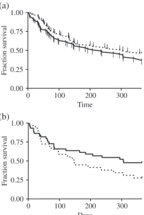
The one-year median survival for all patients was 274 days, lower for Group 1 (LRTI+) with 235 days as compared to Group 2 (LRTI-) where it was 305 days.
Kaplan Meier analysis did not show any statistically signifi- cant differences between the groups (P= 0.244, HR: 1292 [95% CI: 0.84–1.99], Fig 2).
Between the subgroups, made according to the presence of solitary or multiple pathogens, patients in the latter group were significantly older and had worse ECOG per- formance status (Table 1). Significantly less adenocarci- noma was noted in this subgroup and the curative intent interventions were less common in these cases. Signifi- cantly more patients could only receive BSC compared to solitary pathogen LRTI (Table 1). Lung cancer patients with solitary pathogen LRTI had longer median one-year survival as compared to patients with multiple pathogens (306 vs. 146 days, HR: 1.57 [95% CI: 0.87–2.99],P= 0,318;
Fig 2). Patients with multiple pathogen infections had shorter median one-year survival than lung cancer patients in Group 2 (LRTI-) (146 vs. 305 days, HR: 1.67 [95% CI:
0.98–3.23],P= 0.057).
Odds ratio analysis data are presented in Table 4. Signif- icantly higher risk could be observed for one-year mortality in case of performance status≥2, male gender, LRTI with multiple pathogens and nonadenocarcinoma histology.
Discussion
In our study, lung cancer treatment possibilities and one- year survival were analyzed according to the presence of LRTI at the time of bronchoscopy intervention. Interaction of LRTI and lung cancer outcome data is scarce, and our data demonstrated worse outcome in patients with lung malignancies, having LRTI at diagnosis. In the case of LRTI patient’s ECOG performance status was significantly worse; consequently, patients had less therapeutic possibili- ties. This is in line with the Italian survey, confirming the
Table1Baselinepatientcharacteristics ParameterAll (n=143)Group1:LRTI+ (n=74)Group2:LRTI- (n=69)P-valueGroup1vs. Group2Solitarypathogen (n=45)Multiplepathogen (n=29)P-valueSolitaryvs. Multiplepathogen Age:years66.269.366.778.3865.7210.230.5064.938.6969.627.10.01 Gender:(n[%]) Male:female79:64(55.24:44.76)43:31(58.11:41.89)36:33(52.17:47.83)0.4725:20 (55.55:44.44)18:11 (62.07:37.93)0.57 Smoking:(n[%]) Ever-smoker117(81.82)65(87.84)52(75.36)39(86.66)26(89.65) Never-smoker18(12.59)6(8.11)12(17.39)0.154(8.88)2(6.90)0.92 Smokingstatusnotavailable8(5.59)3(4.05)5(7.25)2(4.44)1(3.45) BMI:kg/m225.8410.9325.025.6226.7214.630.3725.845.5623.765.580.12 Underlyinglungdisease: COPD/ILD Yes(n[%])63(44.06)36(48.65)27(39.13)20(44.44)16(55.17) 0.250.36 No(n[%])80(55.94)38(51.35)42(60.87)25(55.55)13(44.83) Anatomicaltype Peripheralcarcinoma(n[%])83(58.04)44(59.46)39(56.52)28(62.22)16(55.17) 0.720.54 Centraltypecarcinoma(n[%])60(41.96)30(40.54)30(43.48)17(37.77)13(44.83) Histology(n[%])0.71 Adenocarcinoma56(39.17)31(41.89)26(37.68)25(55.55)6(20.69)0.01 Squamouscellcarcinoma42(29.37)24(32.43)18(26.09)12(26.66)12(41.38)0.18 SCLC23(16.08)10(13.51)12(17.39)5(11.11)5(17.25)0.45 Mixedtumor9(6.29)4(5.41)5(7.25)1(2.22)3(10.34)0.13 Otherlungcancer13(9.09)5(6.76)8(11.59)2(4.44)3(10.34)0.32 TNM(n[%]) T1-245(31.46)23(31.08)22(31.88)0.9115(33.33)8(27.59)0.60 T3-498(68.54)51(68.92)47(68.12)30(66.66)21(72.41) N025(17.48)16(21.62)9(13.04)9(20.00)7(24.14) N1-294(65.73)44(59.46)50(72.47)0.2429(64.44)15(51.72)0.52 N324(16.79)14(18.92)10(14.49)7(15.55)7(24.14) M057(39.86)31(41.89)26(37.68)0.6020(44.44)11(37.93)0.57 M186(60.14)43(58.11)43(62.32)25(55.55)18(62.07) I7(4.90)3(4.05)4(5.80)2(4.44)1(3.45) II14(9.79)10(13.51)4(5.80)9(20.00)1(3.45) IIIA13(9.09)5(6.76)8(11.60)0.493(6.66)2(6.90)0.29 IIIB-C24(16.78)13(17.57)11(15.94)6(13.33)7(24.14) IV85(59.44)43(58.11)42(60.86)25(55.55)18(62.06)
most common cause of patient exclusion from first-line chemotherapy, which is the poor ECOG performance sta- tus (2–4).17The ECOG performance status is a crucial pre- dictive factor when allowing for treatment with chemotherapy, and the higher the number is connected with shorter survival as also noted in previous studies18,19 and confirmed in our analysis.
Similarly to other researches, the NLR was higher in patients with lung malignancy, associated with LRTI and lower in those with noninfectious cases.20 High NLR at diagnosis is an accepted prognostic marker of worse prog- nosis and therapy response for patients with lung cancer.21–23 As infections are often associated with high neutrophil count it could have contributed to the worse outcome in Group 1 (LRTI+) patients, most pronounced in patients with multiple pathogen infections.
The incidence of adenocarcinoma was the highest in our study population in agreement with international litera- ture.4 Adenocarcinoma histology was associated with a more favorable outcome in our study population. Lung adenocarcinoma is mainly observed as a peripheral lesion, but in advanced stages, it also appears centrally,24similarly to the other histological subtypes.25 Central carcinoma is readily associated with bronchial stricture and underlying pneumonia, which can advance in lung atelectasis. Conse- quently, microorganism colonization may evolve to infec- tion. These known differences in the location could have contributed to the higher number of solitary pathogens (80.65% vs. 46.51%) in adenocarcinoma patients; however, in our analysis, the locations of the tumors did not notably differ in the case of LRTI.
Patients with LRTI had worse one-year survival as com- pared to patients without an infection and significant increase of risk of one-year mortality. This is in line with previous observations, where patients with multiple patho- gen LRTI got shorter median survival than patients with the solitary pathogen LRTI (8.0 vs. 15.0 months, P= 0.003).26
The lungs although previously considered sterile in health are regularly colonized with varied communities of microbes from the oropharynx and other locations. The most common Bacteroidetes and Firmicutes are the Prevotella, Veillonellaand Streptococcusspp. Microbiota of the lung which mostly correspond to those of the mouth than of other body sites. In cases of respiratory tract inflammation, intra-alveolar catecholamines and inflamma- tory cytokines advance the growth of selected bacterial spe- cies (e.g., P. aeruginosa, S. pneumoniae, Staphylococcus aureus, Burkholderia cepaciacomplex).27
The most predominant pathogen in our lung cancer patients wereCandida albicans, pursued byH. influenzae, S. aureus,S. pneumoniaeandEnterobacter spp. These path- ogens have been regularly recognized in lung infections,
Table1Continued ParameterAll (n=143)Group1:LRTI+ (n=74)Group2:LRTI- (n=69)P-valueGroup1vs. Group2Solitarypathogen (n=45)Multiplepathogen (n=29)P-valueSolitaryvs. Multiplepathogen ECOGperformancestatus(n[%]) 072(50.35)30(40.54)42(60.87)0.0123(51.11)7(24.14)0.02 140(27.97)23(31.08)17(24.64)0.3914(31.11)9(31.04)0.99 214(9.79)9(12.16)5(7.24)0.323(6.66)6(20.69)0.07 39(6.29)7(9.46)2(2.90)0.103(6.66)4(13.79)0.30 48(5.60)5(6.76)3(4.35)0.532(4.44)3(10.34)0.32 Numberofcomorbidities(n[%]) 026(18.18)11(14.86)15(21.75)8(17.77)3(10.34) 142(29.37)22(29.73)20(28.98)0.6613(28.88)9(31.04)0.70 247(32.87)27(36.49)20(28.98)17(37.77)10(34.48) ≥328(19.58)14(18.92)14(20.29)7(15.55)7(24.14) NLRvalue5.134.305.955.554.241.990.015.385.626.835.410.27 P-valuewascalculatedforGroup1vs.Group2andforsolitaryvs.multiplepathogen.BMI,bodymassindex;COPD,chronicobstructivepulmonarydisease;ECOG,EasternCooperativeOncology Group;IPF,idiopathicpulmonaryfibrosis;LRTI,lowerrespiratorytractinfections,NLR,neutrophil:lymphocyteratio;n,number;SCLC,smallcelllungcancer;TNM,classificationofmalignanttumors.
but the incidence of the microorganisms is different in recent studies, where the isolates of Enterobacter spp.
(40.86%), followed by S. aureus (21.51%), H. influenzae (16.13%) andS. pneumoniae (7.53%) were recorded.28The nonfermenting Gram-negative bacteria were Pseudomonas spp. (6.45%) andAcinetobacter spp. (3.23%). Among fun- gal species, the most common was Candida albicans (63.77%).28,29
Infections increase the incidence of several malignancies (e.g., Human papillomavirus types 6 and 11 DNA sequences in cervical cancers, Helicobacter pylori infection in colorectal carcinoma).30,31Lung cancer often evolves in damaged lungs (e.g., COPD, emphysema, IPF).7,8 In these lung diseases, mucociliary abnormality can grant mutagens from the smoke or further air pollution longer contact period at these locations, promoting the progress of pul- monary malignancy formation.32 Constant irritation, cau- sed by airway obstruction and the imbalance among oxidants and antioxidants may lead to DNA changes.33 The incidence of pulmonary malignancy in patients with IPF (4.8% to 48%) is significantly higher than in patients without IPF (2.0% to 6.4%).34,35 The mechanism of
increased cancer development in IPF might be associated with increased inflammatory reaction, cell damage, abnor- malfibroblast production and the activation of specific sig- naling pathways (e.g., Wnt/β-catein).36–39 In our data set, about half of the patients had a significant underlying lung disease (COPD/IPF); however, this was not associated with differences in patients’ characteristics of histology in the presence of LRTI.
In conclusion, LRTI, detected in bronchial samples at the time of diagnosis of lung malignancies influences the treatment options and outcome of these patients. Our data confirmed that LRTI+ patients had a worse ECOG perfor- mance status, withholding several treatment possibilities and so resulting in lower one-year survival. Patients with multiple pathogen LRTI were particularly less eligible for oncotherapy; however, no differences in stage, cancer his- tology subtype, gender or age were noted.
Our data emphasize that more attention should be given to LRTI and its treatment in lung cancer patients. The pathogen profile described in our series may assist with the selection of empiric treatment and hopefully decrease the observed risk of one-year mortality. Treatable threats are
Table 2 LRTI pathogens in bronchoscopic samples at the time of cancer diagnosis Pathogen
Group 1 LRTI + (n= 74)
Adenocarcinoma (n= 31)
Squamous cell carcinoma (n= 24)
SCLC (`n= 10)
Other lung cancer (n= 9)
P- value
Aerobic (n[%]) 49 (55.06) 21 (60.00) 16 (59.26) 7 (58.33) 5 (33.33) 0.32
Anaerobic (n[%]) 14 (15.73) 5 (14.29) 4 (14.81) 2 (16.66) 3 (20.00) 0.96
Fungal (n[%]) 26 (29.21) 9 (25.71) 7 (25.93) 3 (25.00) 7 (46.66) 0.44
Solitary pathogen (n[%])
45 (60.81) 25 (80.65) 12 (50.00) 5 (50.00) 3 (33.33) 0.02
Multiple pathogen (n[%])
29 (39.19) 6 (19.35) 12 (50.00) 5 (50.00) 6 (66.66)
P-value was calculated for different cancer types in Group 1. LRTI, Lower respiratory tract infections;n, number; SCLC, small cell lung cancer.
Table 3 Oncotherapy in patients with pulmonary malignancy
Oncotherapy
All (n= 143)
Group 1:
LRTI+
(n= 74)
Group 2:
LRTI- (n= 69)
P-value Group 1 vs.
Group 2
Solitary pathogen
(n= 45)
Multiple pathogen
(n= 29)
P-value Solitary vs.
multiple pathogen Curative intent surgery+/−
chemo/radiotherapy (n[%])
19 (13.29) 11 (14.86) 8 (11.60) 0.56 10 (22.22) 1 (3.45) 0.02
Chemo−/immune−/target therapy (n[%])
62 (43.36) 25 (33.78) 37 (53.62) 0.02 15 (33.33) 10 (34.48) 0.91
Chemo + radiotherapy (n[%]) 16 (11.19) 4 (5.41) 12 (17.39) 0.02 2 (4.44) 2 (6.90) 0.64
Radiotherapy (n[%]) 5 (3.49) 5 (6.76) 0 Not valid 2 (4.44) 3 (10.34) 0.32
BSC (n[%]) 34 (23.78) 24 (32.43) 10 (14.49) 0.01 11 (24.44) 13 (44.83) 0.06
Lost from medical attendance (n[%])
7 (4.89) 5 (6.76) 2 (2.90) 0.28 5 (11.11) 0 Not valid
P-value was calculated for Group 1 vs. Group 2 and for Solitary vs. multiple pathogen. BSC, best supportive care; LRTI, lower respiratory tract infec- tions;n, number.
important elements to improve therapy and survival of this special patient population.
Acknowledgments
The manuscript has been professionally edited and proof- read by the proof reading service of Semmelweis University.
Disclosure
There are no conflicts of interest.
References
1 Albano JD, Ward E, Jemal Aet al. Cancer mortality in the United States by education level and race.JNCI J Natl Cancer Inst2007;99: 1384–94. https://doi.org/10.1093/jnci/
djm127.
2 Pang HH, Wang X, Stinchcombe TEet al. Enrollment tTrends and dDisparity aAmong pPatients wWith lLung cCancer in National Clinical Trials, 1990 to 2012.J Clin Oncol2016;34: 3992–9. https://doi.org/10.1200/JCO.2016.
67.7088.
3 Bray F, Ferlay J, Soerjomataram I, Siegel RL, Torre LA, Jemal A. Global cancer statistics 2018: GLOBOCAN estimates of incidence and mortality worldwide for 36 cancers in 185 countries.CA Cancer J Clin2018;68:
394–424. https://doi.org/10.3322/caac.21492.
4 Blandin Knight S, Crosbie PA, Balata H, Chudziak J, Hussell T, Dive C. Progress and prospects of early detection in lung cancer.Open Biol2017;7: 170070. https://doi.org/10.
1098/rsob.170070.
5 Walter JE, Heuvelmans MA, de Bock GHet al. Relationship between the number of new nodules and lung cancer probability in incidence screening rounds of CT lung cancer screening: The NELSON study.Lung Cancer2018;125:
103–8. https://doi.org/10.1016/j.lungcan.2018.05.007.
6 Janssen-Heijnen MLG, Maas HAAM, Houterman S, Lemmens VEPP, Rutten HJT, Coebergh JWW. Comorbidity in older surgical cancer patients: Influence on patient care and outcome.Eur J Cancer2007;43: 2179–93. https://doi.
org/10.1016/j.ejca.2007.06.008.
7 Zhang X, Jiang N, Wang L, Liu H, He R. Chronic obstructive pulmonary disease and risk of lung cancer: A meta-analysis of prospective cohort studies.Oncotarget2017;
8: 78044–56. https://doi.org/10.18632/oncotarget.20351.
8 Li J, Yang M, Li P, Su Z, Gao P, Zhang J. Idiopathic pulmonaryfibrosis will increase the risk of lung cancer.
Chin Med J (Engl)2014;127: 3142–9.
9 Mao Q, Jiang F, Yin Ret al. Interplay between the lung microbiome and lung cancer.Cancer Lett2018;415: 40–8.
https://doi.org/10.1016/j.canlet.2017.11.036.
10 Sarihan S, Ercan I, Saran A, Çetintas SK, Akalin H, Engin K.
Evaluation of infections in non-small cell lung cancer patients treated with radiotherapy.Cancer Detect Prev2005;
29: 181–8. https://doi.org/10.1016/j.cdp.2004.11.001.
11 Remiszewski P, Słodkowska J, Wiatr Eet al. Infection as a main or additional cause of death in patients treated for small cell lung cancer.Pneumonol Alergol Pol1999;67:
347–53.
12 Torres VB, Azevedo LC, Silva UVet al. Sepsis-associated outcomes in critically ill patients with malignancies.Ann Am Thorac Soc2015;12: 150618124156002. https://doi.org/
10.1513/AnnalsATS.201501-046OC.
13 Miller MR, Hankinson J, Brusasco Vet al. Standardisation of spirometry.Eur Respir J2005;26: 319–38. https://doi.org/
10.1183/09031936.05.00034805.
(a)
(b)
0 100 200 300
0.00 0.25 0.50 0.75 1.00
Time
Fraction survival
0 100 200 300
0.00 0.25 0.50 0.75 1.00
Days
Fraction survival
Figure 2One-year survival of patients with pulmonary malignancy according to the presence of LRTI (a) ( ) Group 1 (LRTI+), and ( ) Group 2 (LRTI−) and solitary and multiple pathogen LRTI (b) ( ) Mul- tiple pathogen (LRTI+), and ( ) Solitary pathogen (LRTI+).
Table 4 Risk factors for one-year mortality
One-year mortality OR CI 95% P-value
LRTI 1.19 0.62–2.28 >0.05
LRTI with multiple pathogen 2.72 1.01–6.81 0.04 LRTI with solitary pathogen 1.19 0.45–1.640 >0.05
Male gender 4.04 2.03–8.04 <0.01
Nonadenocarcinoma 2.26 1.15–4,56 0.02
Ever-smoker 2.89 0.87–8.88 0.09
ECOG performance status 1 11.87 4.12–33.78 <0.01 ECOG performance status≥2 30.00 5.23–313.00 <0.01 ECOG, Eastern Cooperative Oncology Group; LRTI, lower respiratory tract infection. [Correction added on 25 July 2019, after first online publication: in Table 4, ‘One-year survival' in first column has been corrected to 'One-year mortality’].
14 Fruh M, De Ruysscher D, Popat S, Crino L, Peters S, Felip E. Small-cell lung cancer (SCLC): ESMO clinical practice guidelines for diagnosis, treatment and follow-up.
Ann Oncol2013;24: vi99–vi105. https://doi.org/10.1093/
annonc/mdt178.
15 Planchard D, Popat S, Kerr Ket al. Metastatic non-small cell lung cancer: ESMO clinical practice guidelines for diagnosis, treatment and follow-up.Ann Oncol Off J Eur Soc Med Oncol2018;29: iv192–237. https://doi.org/10.1093/annonc/
mdy275.
16 Postmus PE, Kerr KM, Oudkerk Met al. Early and locally advanced non-small-cell lung cancer (NSCLC): ESMO clinical practice guidelines.Ann Oncol2017;28: iv1–iv21.
https://doi.org/10.1093/annonc/mdx222.
17 Gridelli C, Ardizzoni A, Barni Set al. Medical treatment choices for patients affected by advanced NSCLC in routine clinical practice: Results from the Italian observational
“SUN”(survey on the lUng cancer maNagement) study.
Lung Cancer2011;74: 462–8. https://doi.org/10.1016/j.
lungcan.2011.04.011.
18 Minami S, Ogata Y, Ihara S, Yamamoto S, Komuta K.
Outcomes and prognostic factors of chemotherapy for patients with locally advanced or metastatic pulmonary squamous cell carcinoma.Lung Cancer (Auckl)2016;7:
99–110. https://doi.org/10.2147/LCTT.S107560.
19 Minami S, Ogata Y, Ihara S, Yamamoto S, Komuta K.
Retrospective analysis of outcomes and prognostic factors of chemotherapy for small-cell lung cancer.Lung Cancer (Auckl)2016;7: 35. https://doi.org/10.2147/LCTT.S100184.
20 Naess A, Nilssen SS, Mo R, Eide GE, Sjursen H. Role of neutrophil to lymphocyte and monocyte to lymphocyte ratios in the diagnosis of bacterial infection in patients with fever.Infection2017;45: 299–307. https://doi.org/10.1007/
s15010-016-0972-1.
21 Yao Y, Yuan D, Liu H, Gu X, Song Y. Pretreatment neutrophil to lymphocyte ratio is associated with response to therapy and prognosis of advanced non-small cell lung cancer patients treated withfirst-line platinum-based chemotherapy.Cancer Immunol Immunother2013;62:
471–9. https://doi.org/10.1007/s00262-012-1347-9.
22 Unal D, Eroglu C, Kurtul N, Oguz A, Tasdemir A. Are neutrophil/lymphocyte and platelet/lymphocyte rates in patients with non-small cell lung cancer associated with treatment response and prognosis?Asian Pac J Cancer Prev 2013;14: 5237–42.
23 Yin Y, Wang J, Wang Xet al. Prognostic value of the neutrophil to lymphocyte ratio in lung cancer: A meta- analysis.Clinics2015;70: 524–30. https://doi.org/10.6061/
clinics/2015(07)10.
24 Moon Y, Lee KY, Sung SW, Park JK. Differing
histopathology and prognosis in pulmonary adenocarcinoma at central and peripheral locations.J Thorac Dis2016;8:
169–77. https://doi.org/10.3978/j.issn.2072-1439.2016.01.15.
25 Lemjabbar-Alaoui H, Hassan OU, Yang Y-W, Buchanan P.
Lung cancer: Biology and treatment options.Biochim
Biophys Acta2015;1856: 189–210. https://doi.org/10.1016/j.
bbcan.2015.08.002.
26 Qiao D, Wang Z, Lu Y, Wen X, Li H, Zhao H. A
retrospective study of risk and prognostic factors in relation to lower respiratory tract infection in elderly lung cancer patients.Am J Cancer Res2015;5: 423–32.
27 Dickson RP, Erb-Downward JR, Martinez FJ, Huffnagle GB.
The microbiome and the respiratory tract.Annu Rev Physiol 2016;78: 481–504.
28 Szymankiewicz M, Kowalewski J, Dancewicz M.
Bacteriological and mycological analysis of material taken from lower respiratory tract in patients with malignancy.Pol Merkur Lekarski2006;21: 218–22.
29 Watanabe A, Nakai Y, Saito Jet al. Clinical significance of respiratory infections associated with lung cancer patients.
Nihon Kyobu Shikkan Gakkai Zasshi1992;30: 1250–6.
30 Gissmann L, Wolnik L, Ikenberg H, Koldovsky U, Schnürch HG, zur Hausen H. Human papillomavirus types 6 and 11 DNA sequences in genital and laryngeal papillomas and in some cervical cancers.Proc Natl Acad Sci U S A 1983;80: 560–3.
31 Abbass K, Gul W, Beck G, Markert R, Akram S. Association of Helicobacter pylori infection with the development of colorectal polyps and colorectal carcinoma.South Med J 2011;104: 473–6. https://doi.org/10.1097/SMJ.
0b013e31821e9009.
32 Åstrand ABM, Hemmerling M, Root Jet al. Linking increased airway hydration, ciliary beating, and mucociliary clearance through ENaC inhibition.Am J Physiol Lung Cell Mol Physiol2015;308: L22–32. https://doi.org/10.1152/
ajplung.00163.2014.
33 Caramori G, Casolari P, Cavallesco GN, Giuffrè S, Adcock I, Papi A. Mechanisms involved in lung cancer development in COPD.Int J Biochem Cell Biol2011;43: 1030–44. https://
doi.org/10.1016/j.biocel.2010.08.022.
34 Wells C, Mannino DM. Pulmonaryfibrosis and lung cancer in the United States: Analysis of the multiple cause of death mortality data, 1979 through 1991.South Med J1996;89: 505–10.
35 Ma Y, Seneviratne CK, Koss M. Idiopathic pulmonary fibrosis and malignancy.Curr Opin Pulm Med2001;7:
278–82.
36 Hoyne GF, Elliott H, Mutsaers SE, Prêle CM. Idiopathic pulmonaryfibrosis and a role for autoimmunity.Immunol Cell Biol2017;95: 577–83. https://doi.org/10.1038/icb.
2017.22.
37 Ballester B, Milara J, Cortijo J. Idiopathic pulmonaryfibrosis and lung cancer: Mechanisms and molecular targets.Int J Mol Sci2019;20: 593. https://doi.org/10.3390/ijms20030593.
38 Takahashi T, Munakata M, Ohtsuka Yet al. Expression and alteration of ras and p53 proteins in patients with lung carcinoma accompanied by idiopathic pulmonaryfibrosis.
Cancer2002;95: 624–33. https://doi.org/10.1002/cncr.10708.
39 Bowley E, O’Gorman DB, Gan BS.β-Catenin signaling in fibroproliferative disease.J Surg Res2007;138: 141–50.
https://doi.org/10.1016/j.jss.2006.07.026.
![Table 2 LRTI pathogens in bronchoscopic samples at the time of cancer diagnosis Pathogen Group 1 LRTI+ ( n = 74) Adenocarcinoma(n= 31) Squamous cellcarcinoma (n = 24) SCLC(`n = 10) Other lungcancer (n = 9) P -value Aerobic (n [%]) 49 (55.06) 21 (60.00) 16](https://thumb-eu.123doks.com/thumbv2/9dokorg/1385381.114576/6.892.101.811.123.280/pathogens-bronchoscopic-diagnosis-pathogen-adenocarcinoma-squamous-cellcarcinoma-lungcancer.webp)