Characteristics of allergic colitis in breast-fed infants in the absence of cow’s milk allergy
Kriszta Molnár, Petra Pintér, Hajnalka Győrffy, Áron Cseh, Katalin Eszter Müller, András Arató, Gábor Veres
Kriszta Molnár, Petra Pintér, Áron Cseh, Katalin Eszter Mül- ler, András Arató, Gábor Veres, 1st Department of Pediatrics, Semmelweis University, 1083 Budapest, Hungary
Hajnalka Győrffy, 2nd Department of Pathology, Semmelweis University, 1091 Budapest, Hungary
Author contributions: Molnár K and Pintér P contributed equally to the writing of this paper; Veres G designed the re- search; Arató A and Veres G enrolled the patients; Molnár K, Pintér P, Cseh Á and Müller KE performed the analyses; Győrffy H analyzed the histological data; Molnár K, Pintér P and Veres G wrote the paper; Arató A critically reviewed the paper.
Supported by OTKA-K105530, -K81117 and ETT-028-02; the János Bolyai Research Grant, to Veres G; and the János Bolyai Research Scholarship of the Hungarian Academy of Sciences Correspondence to: Gábor Veres, MD, PhD, 1st Department of Pediatrics, Semmelweis University, Bókay u 53, 1083 Buda- pest, Hungary. veres.gabor@med.semmelweis-univ.hu
Telephone: +36-20-8258163 Fax: +36-1-3036077 Received: October 4, 2012 Revised: February 5, 2013 Accepted: March 23, 2013
Published online: June 28, 2013
Abstract
AIM: To investigate the characteristics of mucosal le- sions and their relation to laboratory data and long- term follow up in breast-fed infants with allergic colitis.
METHODS: In this study 31 breast-fed infants were prospectively evaluated (mean age, 17.4 wk) whose rectal bleeding had not ceased after a maternal elimi- nation diet for cow’s milk. Thirty-four age-matched and breast-fed infants (mean age, 16.9 wk) with no rectal bleeding were enrolled for laboratory testing as con- trols. Laboratory findings, colonoscopic and histological characteristics were prospectively evaluated in infants with rectal bleeding. Long-term follow-up with differ- ent nutritional regimes (L-amino-acid based formula or breastfeeding) was also included.
RESULTS: Iron deficiency, peripheral eosinophilia and
thrombocytosis were significantly higher in patients with allergic colitis in comparison to controls (8.4 ± 3.2 µmol/L vs 13.7 ± 4.7 µmol/L, P < 0.001; 0.67 ± 0.49 G/L vs 0.33 ± 0.17 G/L, P < 0.001; 474 ± 123 G/L vs 376 ± 89 G/L, P < 0.001, respectively). At colonosco- py, lymphonodular hyperplasia or aphthous ulceration were present in 83% of patients. Twenty-two patients were given L-amino acid-based formula and 8 contin- ued the previous feeding. Time to cessation of rectal bleeding was shorter in the special formula feeding group (mean, 1.4 wk; range, 0.5-3 wk) when com- pared with the breast-feeding group (mean, 5.3 wk;
range, 2-9 wk). Nevertheless, none of the patients ex- hibited rectal bleeding at the 3-mo visit irrespective of the type of feeding. Peripheral eosinophilia and cessa- tion of rectal bleeding after administration of elemental formula correlated with a higher density of mucosal eosinophils.
CONCLUSION: Infant hematochezia, after cow’s milk allergy exclusion, is generally a benign and probably self-limiting disorder despite marked mucosal abnor- mality. Formula feeding results in shorter time to cessa- tion of rectal bleeding; however, breast-feeding should not be discouraged in long-lasting hematochezia.
© 2013 Baishideng. All rights reserved.
Key words: Rectal bleeding; Breast-feeding; Allergic colitis; Colonoscopy; Amino-acid formula
Core tip: Rectal bleeding is a common problem in oth- erwise healthy breast-fed infants; our primary aim was to find characteristic lesions at colonoscopy and deter- mine the cessation of rectal bleeding when administer- ing different nutritional regimes (L-amino-acid based formula or breast-feeding). Our secondary aim was to find correlations between laboratory data, severity of mucosal lesions and cessation of rectal bleeding in al- lergic colitis infants with no cow’s milk allergy.
BRIEF ARTICLE
doi:10.3748/wjg.v19.i24.3824 © 2013 Baishideng. All rights reserved.
Molnár K, Pintér P, Győrffy H, Cseh Á, Müller KE, Arató A, Veres G. Characteristics of allergic colitis in breast-fed infants in the absence of cow's milk allergy. World J Gastroenterol 2013;
19(24): 3824-3830 Available from: URL: http://www.wjgnet.
com/1007-9327/full/v19/i24/3824.htm DOI: http://dx.doi.
org/10.3748/wjg.v19.i24.3824
INTRODUCTION
Rectal bleeding is a common problem in otherwise healthy breast-fed infants[1]. The differential diagnosis of this condition includes anal fissures, infectious colitis, congenital bleeding disorders, inflammatory bowel dis- ease (IBD) and, most frequently, allergic colitis (AC)[2]. Currently there is no reliable diagnostic test available for AC, and the diagnosis is often made presumptively in healthy infants with rectal bleeding who show no anal fissures or infectious colitis[3]. AC is believed to be a hy- persensitive gastrointestinal disorder to allergens present in breast milk, and formula is regarded as a form of food allergy in infancy[4]. As a resulting first-line therapeutic in- tervention, cow’s milk (CM) is generally eliminated from the maternal diet[5].
Recent guidelines state that the elimination diet for the mother should be continued for a minimum of at least 2 wk, and up to 4 wk in cases of AC[6]. According to the guidelines, if an infant has no cessation of rectal bleeding after 4 wk of maternal CM-free diet then the patient has no CM allergy. It is of interest that the ma- jority of infants with rectal bleeding, despite the general belief, have no CM allergy as an underlying cause of he- matochezia. This phenomenon was supported by recent studies showing that even in breast-fed infants with rectal bleeding, CM allergy is more uncommon than previously believed[7]. Only 18% of 40 infants with rectal bleeding had proven CM allergy, none had positive specific im- munoglobulin E (IgE) to cow’s milk, egg or wheat on admission, and only 5% had a positive skin-prick test to cow’s milk[8]. Nevertheless, an atopy patch test was useful to identify sensitization for cow’s milk (50%), soy (28%), egg (21%), rice (14%) and wheat (7%) in 14 AC infants with multiple food allergy[9].
Lymphonodular hyperplasia (LNH), patchy granu- larity and aphthous lesions were found at colonoscopy in patients with AC; however, most patients showed no abnormal mucosal lesions[5,7,8]. These studies included pa- tients with short-term rectal bleeding which may not have shown the development of characteristic lesions at colo- noscopy. Therefore, in our study, there was a minimum 4-wk timeline between onset of rectal bleeding and the procedure of colonoscopy. We hypothesized that 4 wk would be enough to find a typical mucosal abnormality in otherwise healthy breast-fed infants with rectal bleeding not caused by CM allergy.
Taken together, our primary aim was to find character- istic lesions at colonoscopy and determine the cessation of rectal bleeding when administering different nutritional
regimes (L-amino-acid based formula or breast-feeding).
Our secondary aim was to find correlations between labo- ratory data, severity of mucosal lesions and cessation of rectal bleeding in AC infants with no CM allergy.
It should be noted that there is no previous, prospec- tive study in AC patients with no CM allergy.
MATERIALS AND METHODS
Patients
During a 4-year period (January 2006-February 2010) at the First Department of Pediatrics, Semmelweis Univer- sity, breast-fed infants were invited to join the present study. Inclusion criteria were age less than 6 mo, exclusive breast-feeding, normal stool cultures (Salmonella, Shigella, E. coli, Yersinia, Campylobacter), and no cessation of rectal bleeding after introduction of a maternal elimination diet for CM for a minimum of 4 wk. All subjects were evalu- ated by history and physical examination for signs of fissure, infection and allergic diseases. Rectal bleeding as bloody spots or streaks with or without mucus was con- firmed macroscopically before the colonoscopy. A com- plete blood count with differential, C-reactive protein, prothrombin time, activated partial thromboplastin time, serum albumin, serum IgE, and specific IgE to common food antigens including CM, egg, wheat, rye, soy, fish, nuts, peanuts, sesame, almond, tomato, banana, celery, carrots, apple, peach, lemon, and orange were obtained.
Thirty-four age-matched and breast-fed infants (mean age, 16.9 wk; range, 4-25 wk; girls, 15) with no rectal bleeding (apnea, 10; gastro-esophageal reflux, 12; minor trauma, 7; minor surgical intervention, 5) were enrolled as controls for the laboratory testing. Characteristics of patients and controls are summarized in Table 1.
The Institutional Ethical Committee approved the study; written parental informed consent was obtained.
Evaluation of colonoscopy
To characterize allergic colitis and exclude other rare forms of rectal bleeding in infancy such as angiodys- plasia, hemangioma, polyp, IBD or blue rubber bleb syndrome, colonoscopy with multiple biopsies was per- formed using a flexible pediatric gastroscope with narrow band imaging technique included (Olympus GIF-Q180, Olympus Hungary KFT). Ambulatory colonoscopy was done by the same gastroenterologist (Veres G) with the assistance of a trained endoscopy nurse. The procedure was done under general anesthesia. The colon was gently cleansed by a single water enema (5 mL/kg) before the procedure.
Evaluation of histology
Using standard biopsy forceps, 4 biopsies were taken from the sigmoid colon (about 10 cm from the anal verge) targeted to areas with gross endoscopic findings. Biopsy samples were fixed in 4% buffered formalin for 24 h then embedded into paraffin. Hematoxylin eosin (HE) stain was performed on 3-4 µm thin slides. The biopsy was ex-
amined for routine histology by a pathologist (Győrffy H).
Eosinophils were counted in 10 high-power fields (HPF) and the average number of cells was recorded. Although there are no standard accepted criteria for the diagnosis of AC, several studies have demonstrated eosinophilic infiltration (≥ 6/HPF) in the lamina propria of the left colon or rectum[10-12]. Based on previous studies[11,13], patients with AC were subdivided according to the num- ber of eosinophils on biopsy specimens. AC1 consisted of patients with eosinophils between 6 and 19/HPF, and AC2 with eosinophils ≥ 20/HPF. Using these crite- ria, AC2 patients fulfill the diagnostic criteria for eosino- philic colitis.
Treatment and follow-up
All subjects were followed up for 6 mo to assess out- come, including visits after one week following colonos- copy, after 3 mo, and after 6 mo. Elemental L-amino acid formula(Neocate; SHS Int., Liverpool, United Kingdom) was offered to the parents to treat their children. Assess- ment of rectal bleeding during the follow-up period was qualitative and assessed by parental self-report. In addi- tion, if macroscopic bleeding eased, parents were asked to provide feces to exclude occult bleeding (HSV10;
Diagnosticum Zrt, Budapest, Hungary). Because macro- scopic bleeding was the main inclusion criteria, the ces- sation of macroscopic bleeding was considered to be the endpoint.
Up to 1 year of age, patients received AA formula or continued breast-feeding. At the age of 1 year, CM was introduced to both groups with no macroscopic bleed- ing in the follow-up. After 4 wk of the challenge, parents were asked to provide feces to exclude occult bleeding (HSV10; Diagnosticum Zrt, Budapest, Hungary). In ad- dition to CM, parents were asked to avoid eggs, wheat, soya, nuts and fish (“six food elimination diet”) up to one year of age. After CM introduction, other foods were in- troduced gradually (wheat, soya, eggs, fish and nuts).
At the age of 6 mo, introduction of supplemental feeding (except the 6 foods listed above) was recom- mended irrespective of whether the patient was on the formula or the breast-feeding arm. Subjects with worsen- ing of symptoms or having a severe form of colitis un- derwent repeat colonoscopy with biopsy. Six month time- point was chosen for this procedure to allow enough time for healing in the latter group.
Statistical analysis
Our data followed normal distribution, therefore para- metric statistical tests were implicated and data were ex- pressed as means with standard deviation. For compari- son of datasets, unpaired t test and analysis of variance (ANOVA) with Bonferroni’s post hoc tests were used.
For analyses of contributing effects, factorial ANOVA, logistic regression and Pearson’s correlation were used.
The level of significance was 5% (P < 0.05) and Bonfer- roni’s adjustment for multiple comparisons was intro- duced where needed. Statistical analysis was performed with Statistica 8 (Statsoft, Tulsa, OK, United States).
RESULTS
Clinical characteristics of infants with rectal bleeding Thirty-one healthy, breast-fed infants (mean age, 17.4 wk;
range, 5-26 wk; 12 girls) were enrolled. The mean (range) duration of bloody stools before colonoscopy was 8.7 (4-16) wk (Table 1). All mothers strictly followed a CM- free diet with the help of a trained dietician. In addition to CM-free diet, 7 mothers had an elimination diet for egg and 4 had an exclusion diet for egg, fish, wheat, nuts and soy. In all patients, rectal bleeding was the main symptom that prompted the request for gastroentero- logical evaluation. In addition to bloody stools, watery (42%) or mucous stools (68%) were common. None of the patients had fissures at rectal examination. Stool fre- quency averaged 4.2 (range, 1-7) bowel movements per day. Three patients had a classic history of infantile colic.
One patient had hemangioma on the thorax with no involvement of the liver. One patient had transient hypo- thyroidism. The others had no previous hospitalization or chronic disease. Growth was reported as normal in all patients whose weight and height charts were reviewed for an objective assessment of their growth pattern.
During follow-up, one subject had failure to thrive and was subsequently diagnosed with infantile Crohn’s disease.
She was 24 wk old at study entry with a history of rectal bleeding for 12 wk. Her data were excluded from the study (final study group consisted of 30 infants).
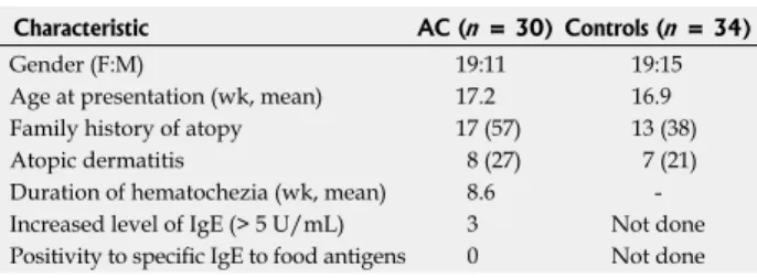
History of atopy, analysis of IgE-mediated hypersensitivity
Eight patients (27%) had atopic dermatitis which is com- parable with the percentage of control subjects (21%).
Seventeen patients (57%) had a positive family history of atopy among first-degree relatives which was higher than in controls (38%). Only 3 patients had an elevated level of serum IgE (> 5 kU/L). Specific IgE to the most com- mon food antigens was determined in 25 of 31 patients (81%) with negative results (Table 1).
Laboratory investigation
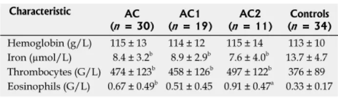
The laboratory abnormalities are presented in Table 2.
The mean iron level was significantly decreased in pa- tients (8.43 ± 3.2 µmol/L) in comparison to controls (13.7 ± 4.7 µmol/L, P < 0.001). We found a markedly increased thrombocyte count in patients (474 ± 123 G/L)
Characteristic AC (n = 30) Controls (n = 34)
Gender (F:M) 19:11 19:15
Age at presentation (wk, mean) 17.2 16.9 Family history of atopy 17 (57) 13 (38) Atopic dermatitis 8 (27) 7 (21) Duration of hematochezia (wk, mean) 8.6 - Increased level of IgE (> 5 U/mL) 3 Not done Positivity to specific IgE to food antigens 0 Not done
Table 1 Clinical characteristics and parameters related to atopy in infants with allergic colitis and control subjects n (%)
AC: Allergic colitis; M: Male; F: Female; IgE: Immunoglobulin E.
(Figure 1). LNH was found in 73% of patients (22/30) and aphthous ulceration in 47% of the patients (14/30).
Subdividing the patients with regard to the number of eosinophils on biopsies, LNH was seen in 15 patients with AC1 (79%) and in 7 patients with AC2 (64%). Aph- thous ulceration was visualized in 8 patients with AC1 (42%) and in 6 patients with AC2 (55%). More patients with AC2 (45%) depicted both phenomenon, which were present in 32% of subjects with AC1. However, the dis- crepancies failed to reach significance (Table 4).
LNH and aphthous ulceration without any specific signs for IBD were present in the patient with Crohn’s disease at her first colonoscopy. She was the only patient with no cessation of rectal bleeding at the 3-mo visit.
Deep ulceration in the transverse colon and non-caseat- ing granulomas on histology established the diagnosis of Crohn’s disease at her second colonoscopy (after 6-mo visit). Abnormal findings at colonoscopy are depicted in Table 4. There were no adverse events recorded under general anesthesia and colonoscopy.
Characterization of histology
Light microscopy revealed normal mucosal architecture in all patients studied, including the first biopsy of the patient with Crohn’s disease which showed normal mucosal archi- tecture with an elevated number of eosinophils (29/HPF).
All patients had more than 6 eosinophils/HPF on histol- when compared with controls (376 ± 89 G/L, P < 0.001).
Similarly, patients exhibited increased peripheral eosino- philia (6.7 ± 4.9 G/L) when compared with controls (3.3 ± 1.7 G/L, P < 0.001). On the other hand, 60% of patients had thrombocytosis, 50% showed low iron level, and 40% demonstrated eosinophilia (Table 3). There was no difference concerning hemoglobin level between pa- tients and controls.
Patients were subdivided into two groups (AC1 and AC2: eosinophilic colitis) according to the number of eosinophils determined by histology on biopsies (see above). Iron level, hemoglobin and thrombocytes were comparable in AC1 and AC2. However, peripheral eosin- ophils were significantly elevated in AC2 (9.1 ± 4.7 G/L) when compared with AC1 (5.1 ± 4.5 G/L, P < 0.01).
Only one patient had marked anemia which required blood transfusion. This patient was the only one who had low albumin level (< 35 g/L). Her other characteristics (including macroscopic and microscopic mucosal abnor- malities) were not different from those of the other sub- jects. Laboratory data in patients who were subsequently diagnosed with Crohn’s disease showed iron deficiency (4 mol/L) without anemia (113 g/L), and thrombocytosis (524 × 109/L).
Characterization of colonoscopy
At colonoscopy, the cecum was reached in 23 (74%) patients. In all other subjects the colonic mucosa was visualized from the anus to the splenic flexure, two of them to the hepatic flexure. Three patients had normal visual colonoscopy findings. Twenty-seven (90%) showed abnormal mucosal lesions such as LNH, aphthous ul- ceration, and marked erythema (2 patients). LNH or aphthous ulceration was present in 25/30 patients (83%)
Table 2 Laboratory data on admission in infants with allergic colitis, allergic colitis 1, allergic colitis 2 and control subjects (mean ± SD)
Characteristic AC
(n = 30) AC1
(n = 19) AC2
(n = 11) Controls (n = 34) Hemoglobin (g/L) 115 ± 13 114 ± 12 115 ± 14 113 ± 10 Iron (µmol/L) 8.4 ± 3.2b 8.9 ± 2.9b 7.6 ± 4.0b 13.7 ± 4.7 Thrombocytes (G/L) 474 ± 123b 458 ± 126b 497 ± 122b 376 ± 89 Eosinophils (G/L) 0.67 ± 0.49b 0.51 ± 0.45 0.91 ± 0.47a 0.33 ± 0.17
aP < 0.05 vs allergic colitis (AC), bP < 0.01 vs controls. Allergic colitis 1 (AC1) consisted of patients with eosinophils between 6 and 19/ high power field (HPF), and allergic colitis 2 (AC2) with eosinophils ≥ 20 eosinophils/HPF.
Characteristic On admission 6-mo visit ND
Thrombocytosis (> 450 × 109/L) 16 (60) 5 2
Iron deficiency (< 9 µmol/L) 15 (50) 4 2
Eosinophilia (> 5% of leukocytes) 12 (40) 5 1
Table 3 Frequency of laboratory abnormalities in infants with allergic colitis on admission and at 6-mo visit n (%)
The number of patients who had previous abnormal parameters and no control analysis depicted in the last column. ND: Not done.
Characteristic AC1 (n = 19) AC2 (n = 11)
LNH 15 (79) 7 (64)
Aphthous ulceration 8 (42) 6 (55) LNH and aphthous ulceration 6 (32) 5 (45) Marked erythema 1 (5) 1 (9) No. of eosinophils/HPF (mean) 12.2 29.5
Table 4 Frequency of colonoscopic abnormalities and number of eosinophils on histology/high power field of infants with allergic colitis 1 and allergic colitis 2 n (%)
LNH: Lymphonodular hyperplasia; AC1: Allergic colitis 1; AC2: Allergic colitis 2; HPF: High power field.
Figure 1 Endoscopic findings of allergic colitis in a breastfed infant. Lym- phonodular hyperplasia with circumscribed erythema (arrow) and tiny bleeding spot (double arrow) in the descending colon in a breast-fed infant with allergic colitis.
ogy (Table 4). All three patients with normal macroscopic findings at colonoscopy showed an elevated eosinophil count on biopsies (6, 20 and 39 cells/HPF, respectively).
Nineteen patients had an eosinophil count between 6 and 19/HPF (AC1) and 11 patients had more than 20/HPF (AC2). Mean number of eosinophils was 12.2/HPF in AC1 and 29.5/HPF in AC2 (eosinophilic colitis).
Follow-up and clinical outcome, laboratory data
Based on marked mucosal abnormalities and the long- lasting rectal bleeding, we decided to treat patients with special formula. At the first visit one week after colo- noscopy, elemental L-amino acid formula was offered to the parents to treat their children. Parents refused the formula feeding in 8 cases (5 in AC1 and 3 in AC2) and continued the previous feeding type. Twenty-two patients received the special formula (14 in AC1 and 8 in AC2).
There were no significant differences concerning baseline characteristics between the 2 groups.
The duration of bleeding was significantly shorter in the special formula feeding group (mean, 1.4 wk; range, 0.5-3 wk) when compared with the breast-feeding group (mean, 5.3 wk; range, 2-9 wk). Nevertheless, none of the patients exhibited gastrointestinal complaints or visible rectal bleeding at the 3-mo visit irrespective of the type of feeding (Table 5). Three patients with serum iron level below 5 µmol/L were treated with iron-containing syrup (Maltofer; dosage, 2 drops/kg) for 6 wk. At 6-mo visit, control blood count and iron level were analyzed in pa- tients who had abnormal levels on admission.
Follow-up and colonoscopy, histology
Due to the cessation of bleeding, only four parents agreed for a control colonoscopy to be performed. At baseline all of them had LNH and aphthous ulceration. One patient was in AC1 (13 eosinophils/HPF) and 3 patients were in AC2 (eosinophils > 30/HPF). At control colonoscopy none of the patients had aphthous ulceration; only scat- tered LNH without bleeding spots were visible. The num- ber of eosinophils normalized in AC1 patients (5/HPF) and markedly decreased in AC2 patients (8-17/HPF).
Correlation analysis and risk factors
Correlation analysis revealed that the time to stop the bleeding was negatively correlated with the administra- tion of elemental formula (P < 0.001, r = -0.762). In ad-
dition, the number of eosinophils on biopsy specimens at baseline correlated negatively with elemental formula concerning cessation of rectal bleeding (P = 0.025, r = -0.667). There was no correlation between abnormal serum laboratory data (thrombocytes, eosinophils, iron) and the ending of rectal bleeding. In patients with AC1 and AC2, we found similar correlations between elemen- tal diet and the time to stop the bleeding.
DISCUSSION
The elimination of CM from the diet of the lactating mother is a commonly used and recommended prac-
tice[10-12] for infants with rectal bleeding. However, recent
studies have suggested that there are significant propor- tions of infants with rectal bleeding with no CM aller-
gy[8,9,13]. Based on these results we focused on AC patients
with no cessation of rectal bleeding after a minimum of 4 wk of maternal CM-free diet. Four weeks is a long enough time-span to exclude rare forms of surgical dis- eases, to cure an undetected viral gastrointestinal disorder or for elimination of CM protein from patients[6,8,14].
In our study, frequency of atopic dermatitis, measure- ment of serum IgE, and specific IgE to different foods were not significant factors in the diagnostic procedure, as has been suggested by previous studies[14,15]. On the other hand, a recent Italian study suggested a delayed-type im- munogenic mechanism in this process, hence the atopy patch test was useful in the diagnostic procedure in 14 AC infants with multiple gastrointestinal food allergy[9].
In our study, iron deficiency, peripheral eosino- philia and thrombocytosis were significantly higher in AC patients in comparison to controls. Surprisingly, no previous study has analyzed iron levels in patients with AC. Peripheral eosinophilia and thrombocytosis were re- ported previously[7,8,15]. To determine the risk factors for the clinical outcome, none of the 3 abnormal laboratory findings correlated with the cessation of rectal bleeding.
Theoretically, colonoscopy would have detected other rare causes of rectal bleeding such as polyps, heman- gioma, blue rubber bleb syndrome and angiodysplasia.
However, all but 3 patients showed mucosal abnormali- ties such as LNH, aphthous ulceration and marked focal erythema. As a bleeding source, LNH with circumscribed erythema or/and central pit-like bleeding spots (Figure 1) or aphthous ulceration were present in 25 patients with AC (83%). This is in contrast to previous studies, which reported a much lower percentage of characteristic ab- normalities at colonoscopy. In a prospective evaluation of 34 infants with rectal bleeding, only 10 (29%) showed LNH including three of them with normal histology[5]. Xanthakos et al[7] followed 22 infants younger than 6 mo of age with hematochezia. Only 12 patients showed gross endoscopic abnormalities at colonoscopy including dif- fuse erythema, friability, LNH and aphthous ulceration.
In addition, a recent study followed 40 infants with vis- ible rectal bleeding where 41% of patients had normal mucosa, 33% had aphthous ulceration, and 51% depicted focal erythema with no report of LNH[8]. One of the ex-
Characteristic Amino-acid based
formula feeding Breast-feeding
No. of patients 22 8
Age at presentation (wk) 16.0 (5-26) 20.6 (16-25)a Rectal bleeding at baseline (wk) 7.9 (4-14) 10.5 (6-16) Cessation of rectal bleeding (wk) 1.4 (0.5-3)
Rectal bleeding at 3-mo visit (n) 0 0 Table 5 Baseline characteristics and cessation of rectal bleeding in patients with amino acid-based formula feeding and breast-feeding [mean (range)]
aP < 0.05 vs formula feeding.
planations for the high rate of LNH and aphthous ulcer- ation found in our study is the long-lasting rectal bleeding (minimum 4 wk). LNH has been described as a sign of food allergy in children with CM allergy[16-18]. In our pa- tients with no CM allergy, LNH was also a characteristic abnormality at colonoscopy. We do not think that LNH is a specific sign of food allergy or AC, just an indicator of a focal inflammation beneath the epithelium. How- ever, LNH with circumscribed erythema or/and central pit-like bleeding spots and aphthous ulceration may have a role in the diagnostic procedure of AC.
The number of eosinophils on biopsies in patients with AC is a question of debate[19]. Previous studies[20,21]
have suggested, as a diagnostic criteria for AC, eosinophil- ic infiltration in the lamina propria of ≥ 6/HPF, whereas Machida et al[5] suggested for this entity of ≥ 20/HPF. All patients in the present study had ≥ 6 eosinophils/HPF in the lamina propria. To find a possible difference of labo- ratory, colonoscopy and clinical outcome, patients were subdivided to moderate (AC1) or high (AC2) mucosal density of eosinophils. It should be noted that AC2 pa- tients fulfill the present criteria for eosinophilic colitis (≥ 20 eosinophils/HPF)[22]. Peripheral eosinophilia and ces- sation of rectal bleeding after administration of elemental formula correlated with the higher number of eosinophils on biopsies. In conclusion, these data suggest that both groups represent similar entities, and a cut-off > 20 eo- sinophils/HPF for eosinophilic colitis may be artificial.
Due to the marked mucosal abnormality and the long-lasting rectal bleeding, elemental formula was given to the patients. It was refused by 8 cases who continued their previous feeding. Elemental diet shortened the time to cessation of hematochezia; nevertheless, it should be emphasized that none of the patients exhibited gastro- intestinal complaints or rectal bleeding at the 3-mo visit irrespective of the type of feeding. This result suggests that the underlying cause could be food-related other than CM, hence AA formula shortened the resolutions of symptoms. Probably, our study population may have multiple food allergy; at least, we cannot exclude this be- cause the nursing mothers conducted only limited types of food elimination diet (beside CM).
The beneficial effect of amino acid-based formula was reported previously[23], suggesting an allergy-related mechanism even in AC patients with no CM allergy.
On the other hand, we cannot exclude the possibility that our patients had no food allergy at all. This phe- nomenon is similar to infantile eczema[24] or eosinophilic esophagitis in infancy, where considerable proportions of patients have no food allergy but an allergic-immunologic component does exist[25]. Exclusive amino acid formula- based dietary trials resulted in more than 90% remission in children with eosinophilic esophagitis. Similarly to cur- rent guidelines in AC, empiric elimination diets of avoid- ance of foods commonly known to cause hypersensitivity reactions resulted in 50%-74% disease remission in eo- sinophilic esophagitis[26]. Taken together, previous studies and the beneficial effect of AA formula in the present study suggest that our study population probably has
multiple food allergy. One of the most interesting points of data in our study is that patients were able to tolerate a non-restricted diet after a period of elimination diet (AA- based formula) or after a more prolonged persistence of symptoms on breast-feeding. Acquired tolerance may explain this finding, which is supported by our previous study where depleted FOXP3 regulatory T cells normal- ized after feeding of AA formula[27].
Nevertheless, the only patient who had not ceased the rectal bleeding was subsequently diagnosed with infantile Crohn’s disease and she was excluded from the study. As reported previously[7,28,29], at baseline, none of her labora- tory, colonoscopy or histology parameters were different from the patients with AC.
In conclusion, rectal bleeding in infancy, even after ex- clusion of CM, is generally benign and is probably a self- limiting disorder, despite the marked mucosal abnormality.
Iron deficiency, peripheral eosinophilia and thrombocyto- sis are characteristic findings in these patients. At colonos- copy, LNH and aphthous ulceration may be characteristic signs. Administration of amino acid formula shortened the timeline of the rectal bleeding in patients with AC;
however, breast-feeding should not be discouraged in this population with long-lasting hematochezia.
COMMENTS
Background
Allergic inflammation of the large intestine is believed to be a hypersensitive gastrointestinal disorder to allergens present in breast milk, and formula is regarded as a form of food allergy in infancy. As a resulting first line therapeutic intervention, cow’s milk is generally eliminated from the maternal diet; however, significant numbers of infants with rectal bleeding, despite the general belief, have no cow’s milk allergy as an underlying cause of hematochezia. Currently there is no reliable diagnostic test available for allergic colitis, and the diagnosis is often made presumptively in healthy breast-fed infants with rectal bleeding who show no anal abnormalities or infectious colitis.
Research frontiers
A research group in Hungary investigated the characteristics of endoscopic find- ings in colonic mucosal lesions and their relation to laboratory data in breast-fed infants with allergic colitis. Long-term follow-up with different nutritional regimes (L-amino acid-based formula or breast-feeding) was also included. According to the literature there is no previous, prospective study in allergic colitis patients with no cow’s milk allergy.
Innovations and breakthroughs
In this study, 31 breast-fed infants were prospectively evaluated whose rectal bleeding had not ceased after a maternal elimination diet for cow’s milk allergy.
As controls, 34 age-matched and breast-fed infants without rectal bleeding were enrolled for the laboratory testing. In the laboratory findings, iron deficiency and elevation of peripheral eosinophilic cell and platelet counts were significantly higher in patients with allergic colitis in comparison to controls. At colonoscopy, increments of lymphoid tissue appearing as a lump (called lymphonodular hy- perplasia) or aphthous ulceration were present in 83% of patients. Twenty-two patients were given L-amino acid-based formula and 8 continued the previous feeding. Time to cessation of rectal bleeding was shorter in the special formula feeding group when compared with the breast-feeding group. Nevertheless, none of the patients exhibited rectal bleeding at the 3-mo visit irrespective of the type of feeding. The elevated peripheral eosinophilic cell count and cessa- tion of rectal bleeding after administration of elemental formula correlated with the higher density of mucosal eosinophilic cells in the colon.
Applications
Administration of amino acid formula shortened the timeline of rectal bleeding in patients with allergic colitis; however, breast-feeding should not be discour- aged in this population with long-lasting rectal bleeding. These data suggest that the significance of real food allergy in breast-fed infants with allergic colitis
COMMENTS
should be revised in the future. This phenomenon is similar to infantile eczema or eosinophilic esophagitis in infancy where considerable proportions of pa- tients had no food allergy but an allergic-immunologic component does exist.
It may indicate that the “food allergy” theory is currently much less applicable, and probably a significant number of nursing mothers continue an unnecessary restriction diet worldwide.
Terminology
Allergic colitis is explained as an allergic condition of the gastrointestinal tract in breast-fed infants that results from an immune response to the allergens pres- ent in breast milk.
Peer review
This is a very interesting article from a teaching institution in Hungary. It is well written and conclusions are valid.
REFERENCES
1 Eigenmann PA. Mechanisms of food allergy. Pediatr Allergy Immunol 2009; 20: 5-11 [PMID: 19154253 DOI: 10.1111/j.1399- 3038.2008.00847.x]
2 Fox VL. Gastrointestinal bleeding in infancy and child- hood. Gastroenterol Clin North Am 2000; 29: 37-66, v [PMID:
10752017 DOI: 10.1016/S0889-8553(05)70107-2]
3 Troncone R, Discepolo V. Colon in food allergy. J Pediatr Gastroenterol Nutr 2009; 48 Suppl 2: S89-S91 [PMID: 19300136 DOI: 10.1097/MPG.0b013e3181a15d1a]
4 Moon A, Kleinman RE. Allergic gastroenteropathy in chil- dren. Ann Allergy Asthma Immunol 1995; 74: 5-12; quiz 12-16 [PMID: 7719884]
5 Machida HM, Catto Smith AG, Gall DG, Trevenen C, Scott RB. Allergic colitis in infancy: clinical and pathologic aspects.
J Pediatr Gastroenterol Nutr 1994; 19: 22-26 [PMID: 7965472 DOI: 10.1097/00005176-199407000-00004]
6 Vandenplas Y, Koletzko S, Isolauri E, Hill D, Oranje AP, Brueton M, Staiano A, Dupont C. Guidelines for the diag- nosis and management of cow’s milk protein allergy in in- fants. Arch Dis Child 2007; 92: 902-908 [PMID: 17895338 DOI:
10.1136/adc.2006.110999]
7 Xanthakos SA, Schwimmer JB, Melin-Aldana H, Rothenberg ME, Witte DP, Cohen MB. Prevalence and outcome of allergic colitis in healthy infants with rectal bleeding: a prospective cohort study. J Pediatr Gastroenterol Nutr 2005; 41: 16-22 [PMID:
15990624 DOI: 10.1097/01.MPG.0000161039.96200.F1]
8 Arvola T, Ruuska T, Keränen J, Hyöty H, Salminen S, Iso- lauri E. Rectal bleeding in infancy: clinical, allergological, and microbiological examination. Pediatrics 2006; 117: e760-e768 [PMID: 16585287 DOI: 10.1542/peds.2005-1069]
9 Lucarelli S, Di Nardo G, Lastrucci G, D’Alfonso Y, Mar- cheggiano A, Federici T, Frediani S, Frediani T, Cucchiara S.
Allergic proctocolitis refractory to maternal hypoallergenic diet in exclusively breast-fed infants: a clinical observa- tion. BMC Gastroenterol 2011; 11: 82 [PMID: 21762530 DOI:
10.1186/1471-230X-11-82]
10 Sampson HA, Anderson JA. Summary and recommenda- tions: Classification of gastrointestinal manifestations due to immunologic reactions to foods in infants and young children. J Pediatr Gastroenterol Nutr 2000; 30 Suppl: S87-S94 [PMID: 10634304 DOI: 10.1097/00005176-200001001-00013]
11 Winter HS, Antonioli DA, Fukagawa N, Marcial M, Gold- man H. Allergy-related proctocolitis in infants: diagnostic usefulness of rectal biopsy. Mod Pathol 1990; 3: 5-10 [PMID:
2308921]
12 Hill SM, Milla PJ. Colitis caused by food allergy in infants.
Arch Dis Child 1990; 65: 132-133 [PMID: 2301977 DOI: 10.1136/
adc.65.1.132]
13 Hwang JB, Park MH, Kang YN, Kim SP, Suh SI, Kam S.
Advanced criteria for clinicopathological diagnosis of food protein-induced proctocolitis. J Korean Med Sci 2007; 22:
213-217 [PMID: 17449926 DOI: 10.3346/jkms.2007.22.2.213]
14 Lake AM. Food-induced eosinophilic proctocolitis. J Pediatr Gastroenterol Nutr 2000; 30 Suppl: S58-S60 [PMID: 10634300 DOI: 10.1097/00005176-200001001-00009]
15 Lake AM, Whitington PF, Hamilton SR. Dietary protein-in- duced colitis in breast-fed infants. J Pediatr 1982; 101: 906-910 [PMID: 7143166 DOI: 10.1016/S0022-3476(82)80008-5]
16 Anveden-Hertzberg L, Finkel Y, Sandstedt B, Karpe B. Proc- tocolitis in exclusively breast-fed infants. Eur J Pediatr 1996;
155: 464-467 [PMID: 8789762 DOI: 10.1007/BF01955182]
17 Ravelli A, Villanacci V, Chiappa S, Bolognini S, Manenti S, Fuoti M. Dietary protein-induced proctocolitis in childhood.
Am J Gastroenterol 2008; 103: 2605-2612 [PMID: 18684195 DOI:
10.1111/j.1572-0241.2008.02035.x]
18 Kokkonen J, Karttunen TJ, Niinimäki A. Lymphonodular hyperplasia as a sign of food allergy in children. J Pediatr Gastroenterol Nutr 1999; 29: 57-62 [PMID: 10400105 DOI:
10.1097/00005176-199907000-00015]
19 Yan BM, Shaffer EA. Primary eosinophilic disorders of the gastrointestinal tract. Gut 2009; 58: 721-732 [PMID: 19052023 DOI: 10.1136/gut.2008.165894]
20 Odze RD, Bines J, Leichtner AM, Goldman H, Antonioli DA. Allergic proctocolitis in infants: a prospective clini- copathologic biopsy study. Hum Pathol 1993; 24: 668-674 [PMID: 8505043 DOI: 10.1016/0046-8177(93)90248-F]
21 Odze RD, Wershil BK, Leichtner AM, Antonioli DA. Al- lergic colitis in infants. J Pediatr 1995; 126: 163-170 [PMID:
7844660 DOI: 10.1016/S0022-3476(95)70540-6]
22 Bischoff SC. Food allergy and eosinophilic gastroenteritis and colitis. Curr Opin Allergy Clin Immunol 2010; 10: 238-245 [PMID: 20431371 DOI: 10.1097/ACI.0b013e32833982c3]
23 Alfadda AA, Storr MA, Shaffer EA. Eosinophilic colitis:
epidemiology, clinical features, and current management.
Therap Adv Gastroenterol 2011; 4: 301-309 [PMID: 21922029 DOI: 10.1177/1756283X10392443]
24 Kvenshagen B, Jacobsen M, Halvorsen R. Atopic dermatitis in premature and term children. Arch Dis Child 2009; 94:
202-205 [PMID: 18829619 DOI: 10.1136/adc.2008.142869]
25 Hogan SP, Rothenberg ME. Eosinophil Function in Eosin- ophil-associated Gastrointestinal Disorders. Curr Allergy Asthma Rep 2006; 6: 65-71 [PMID: 16476198 DOI: 10.1007/
s11882-006-0013-8]
26 Chehade M, Aceves SS. Food allergy and eosinophilic esophagitis. Curr Opin Allergy Clin Immunol 2010; 10: 231-237 [PMID: 20410819 DOI: 10.1097/ACI.0b013e328338cbab]
27 Cseh A, Molnár K, Pintér P, Szalay B, Szebeni B, Treszl A, Arató A, Vásárhelyi B, Veres G. Regulatory T cells and T helper subsets in breast-fed infants with hematochezia caused by allergic colitis. J Pediatr Gastroenterol Nutr 2010; 51: 675-677 [PMID: 20818268 DOI: 10.1097/MPG.0b013e3181e85b22]
28 Ojuawo A, St Louis D, Lindley KJ, Milla PJ. Non-infective colitis in infancy: evidence in favour of minor immunode- ficiency in its pathogenesis. Arch Dis Child 1997; 76: 345-348 [PMID: 9166029 DOI: 10.1136/adc.76.4.345]
29 Cannioto Z, Berti I, Martelossi S, Bruno I, Giurici N, Crov- ella S, Ventura A. IBD and IBD mimicking enterocolitis in children younger than 2 years of age. Eur J Pediatr 2009; 168:
149-155 [PMID: 18546019 DOI: 10.1007/s00431-008-0721-2]
P- Reviewer Camilleri-Brennan J S- Editor Wen LL L- Editor Logan S E- Editor Li JY