A Localized Aldara (5% Imiquimod)–
Induced Psoriasiform Dermatitis Model in Mice Using Finn Chambers
Szabina Horváth,1,2Ágnes Kemény,2,3,4 Erika Pintér,2,3and Rolland Gyulai1,5
1Department of Dermatology, Venereology and Oncodermatology, University of Pécs Clinical Center, Pécs, Hungary
2János Szentágothai Research Center, University of Pécs, Pécs, Hungary
3Department of Pharmacology and Pharmacotherapy, University of Pécs Medical School, Pécs, Hungary
4Department of Medical Biology, University of Pécs Medical School, Pécs, Hungary
5Corresponding author:gyulai.rolland@pte.hu
The expanding number of research studies utilizing the imiquimod-induced psoriasiform dermatitis model attests to the usefulness of this procedure. Ad- vantages of this model include rapid development of the skin response and cost- effectiveness. A major limitation is that application of imiquimod cream over large areas of skin, as well as licking and ingestion of the cream, may lead to severe systemic inflammation, which can cause a general decline in health, splenomegaly, and death. In this protocol, Finn chambers are used to localize the imiquimod cream to a small area of the skin. This results in production of severe and reproducible psoriatic skin reactions with significantly less im- iquimod, greatly reducing the possibility of untoward systemic effects. More- over, having psoriasiform and control skin areas on the same mice decreases inter-animal differences. The protocol can be readily adapted for other skin dis- ease models involving topical application of test agents. This article also details functional measurements performed during assays, including skin thickness, blood perfusion, semiquantitative histopathological evaluation, determination of scaling score to monitor psoriatic symptoms, and collection of spleen and body weight data to identify systemic effects. © 2020 The Authors.
Basic Protocol: Use of Finn chambers to induce psoriasiform skin reactions with imiquimod
Support Protocol 1: Measurement of double-fold dorsal skin thickness Support Protocol 2: Measurement of blood perfusion
Support Protocol 3: Determination of scaling score
Support Protocol 4: Semiquantitative histopathological scoring
Support Protocol 5: Assessment of systemic side effects in response to imiquimod application
Keywords: Aldara rFinn chamber rimiquimod rmouse model rpsoriasis
How to cite this article:
Horváth, S., Kemény, Á., Pintér, E., & Gyulai, R. (2020). A localized aldara (5% imiquimod)–induced psoriasiform dermatitis
model in mice using Finn chambers.Current Protocols in Pharmacology,90,e78. doi: 10.1002/cpph.78
Current Protocols in Pharmacologye78, Volume 90 Published in Wiley Online Library (wileyonlinelibrary.com).
doi: 10.1002/cpph.78
© 2020 The Authors. This is an open access article under the terms of the Creative Commons Attribution-NonCommercial-NoDerivs License, which permits use and distribution in any medium, provided
Horváth et al.
1 of 12
INTRODUCTION
Imiquimod (IMQ) is a small nucleotide-like molecule commonly used to induce psoriasis-like dermatitis in animal models (Swindell et al., 2017; van der Fits et al., 2009). Imiquimod activates innate and adaptive immune system signaling pathways via TLR7/9, expressed on plasmacytoid dendritic cells (PDCs) (Hanna, Abadi, & Abbas, 2016). It is likely that other actions of imiquimod—such as NLRP1 inflammasome activation (pyroptosis) (Walter et al., 2013) and direct activation of TRPA1 nonselec- tive cation channels located on immune cells and peripheral nerve endings (Kemény et al., 2018)—also contribute to its proinflammatory effects. The popularity of the imiquimod model is due in large measure to the fact that this substance initiates IL-17, IL-23 cytokine-mediated immunological processes of the skin, thereby mimicking the Th17-induced autoimmune component of the human psoriatic condition. In addition, the imiquimod model is cost-effective and reproducible (Hawkes, Gudjonsson, & Ward, 2016). A major problem encountered with this approach in early studies was the robust systemic inflammation that often resulted from smearing 62.5 mg of 5% imiquimod on the shaved backs of mice. This systemic side effect results in a generalized deterioration of the health of the test animal, including fever, weight loss, splenomegaly (enlarged spleen), and death (Alvarez & Jensen, 2016). Such toxicity severely limits the utility of this approach. Moreover, the need to treat large surface areas required that different groups of animals be used to examine and compare results from imiquimod-treated and control subjects. The Basic Protocol below details a modified and improved method for the imiquimod-induced psoriasiform dermatitis model using Finn chambers to achieve localized application of imiquimod (Horváth et al., 2019). This new procedure decreases the likelihood of systemic side effects and makes possible comparisons between treated and untreated skin in the same subjects, thereby reducing inter-assay variability and cost.
In the mouse model of psoriasis, infiltration is measured by measuring double-fold skin thickness using a micrometer (see Support Protocol 1), erythema (increased blood perfusion) is measured using the LASCA laser Doppler method (see Support Proto- col 2), and desquamation is determined using the scaling score (see Support Proto- col 3). A semiquantitative histopathological evaluation is performed to determine the epidermal thickness as well as the presence of Munro’s microabscesses and dilated capillaries (see Support Protocol 4). Finally, measurement of spleen weight and body weight are recommended for monitoring the presence of systemic effects (see Support Protocol 5).
NOTE: All procedures were conducted according to the 1998/XXVIII Act of the Hungarian Parliament on Animal Protection and Consideration Decree of Scientific Procedures of Animal Experiments (243/1988). All protocols were approved by the Ethics Committee on Animal Research of the University of Pécs, in full accordance to the Ethical Codex of Animal Experiments. All protocols involving live animals must be reviewed and approved by an Institutional Animal Care and Use Committee (IACUC) and must conform to government regulations for the care and use of laboratory animals.
BASIC PROTOCOL
USE OF FINN CHAMBERS TO INDUCE PSORIASIFORM SKIN REACTIONS WITH IMIQUIMOD
In this procedure, imiquimod cream is applied to the dorsal skin of mice using small (8-mm-diameter) Finn chambers to induce typical psoriatic symptoms and histopatho- logical alterations while minimizing systemic inflammation. Such localized application of small amounts of imiquimod cream reduces side effects by decreasing the likelihood of
Horváth et al.
ingestion of the cream. In addition, it makes it possible to examine treated and control skin on the same animal, reducing inter-animal variability as well as the number of sub- jects needed for each experiment. Furthermore, like an occlusive dressing, the chamber may enhance penetration of active compounds. Treatment with active compound for as few as four consecutive days is sufficient to provide evidence of efficacy for the treatment of psoriasiform skin inflammation.
Materials
C57BL/6 female mice, 8-10 weeks old, 20-25 g Ketamine/xylazine solution (see recipe)
Depilatory cream (Veet)
Aldara cream (5% imiquimod; Meda Pharma) Vaseline
Small animal laboratory scale 1-ml syringes with 27-G needles Electric shaver
Rubber-edged spatula
Adjustable heating plate (Temperature Controller TMP-5a, Supertech Instruments) 8-mm (50-mm2) Finn chambers on Scanpor tape (SmartPractice)
Forceps
Day 0: Prepare animals
1. Select age- and sex-matched mice with at least six animals per group.
Female mice are recommended because development of imiquimod-induced inflamma- tory skin phenotype is exacerbated in female mice.
2. Measure body weight and calculate the amount of ketamine/xylazine solution needed for anesthesia.
Using the recipe provided, an i.p. injection of 100μl solution per 10 g body weight will give the desired doses of 100 mg/kg ketamine and 5 mg/kg xylazine.
3. Anesthetize by i.p. injection using a 1-ml syringe with a 27-G needle. Make sure that animals are completely anesthetized (e.g., by tail pinch).
4. Shave an∼3×4–cm area on the dorsal skin using an electric shaver.
5. Remove any remaining hairs with depilatory cream. Remove cream with a rubber- edged spatula, being careful to avoid even microscopic skin lacerations and abra- sions. Rinse the denuded area with wet cotton pads.
A rubber-edged spatula is best, as it will not damage the skin.
It is important to perform this step the day before the initial imiquimod treatment to avoid unwanted chemical interactions and to allow sufficient time for healing of any skin damage caused by depilation.
6. Place anesthetized animals on a 37°C heating plate to maintain constant body tem- perature during recovery, then return to their home cages.
Days 1-4: Induce psoriatic reaction
7. Measure body weight and anesthetize animal as before.
It is important to measure body weight prior to each treatment, as it is usually slightly decreased after the first and second treatments.
Horváth et al.
3 of 12
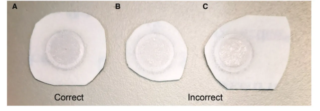
Figure 1 Desired shape and adhesive surface size of Finn chambers.
8. Place animals on a 37°C heating plate to maintain body temperature, then evaluate baseline dorsal skin thickness, blood perfusion, and scaling (see Support Protocols 1-3).
9. Cut out two individual circular Finn chambers per animal from the Scanpore tape, ensuring that there is sufficient adhesive surface to apply firmly to the skin.
Finn chambers adhere to the skin most firmly if the adhesive surface of the tape is larger than the chamber and circular in shape (Fig. 1).
10. Fill one chamber with 25 mg Aldara cream and the other with 25 mg Vaseline.
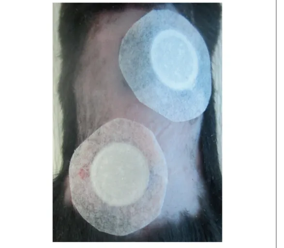
11. Place Finn chambers on two discrete spots on the dorsal skin at least 2 cm apart (Fig. 2).
It is best to place the chambers on opposite sides of the animal, with one on the upper region and the other on the lower region.
12. Return mice to their cages and house normally until the next treatment.
Finn chambers usually stay in place without taping or bandaging, although occasionally mice may pick at the chambers.
13. After 24 hr, use forceps to gently remove the Finn chambers from the dorsal skin.
14. Over the next three days (Days 2, 3, 4), repeat measurements and treatment (steps 7-13), placing fresh Finn chambers on the same locations as on Day 1.
Day 5: Perform functional measurements and collect tissue samples
15. On Day 5, repeat measurements in steps 7-8, then sacrifice animals, collect skin sam- ples for histology (see Support Protocol 4), and measure spleen weight (see Support Protocol 5).
SUPPORT PROTOCOL 1
MEASUREMENT OF DOUBLE-FOLD DORSAL SKIN THICKNESS
A typical symptom of imiquimod-induced psoriasiform dermatitis in mice is an increase in epidermal thickness. Dorsal skin thickness should be monitored daily after anesthe- sia but before treatment by measuring the treated skin areas in vivo with an engineer’s micrometer.
Materials
Mice enrolled in Finn chamber experiment (Days 1-5; see Basic Protocol) 70% (v/v) ethanol
Engineer’s micrometer (Moore and Wright, Sheffield, England)
Horváth et al.
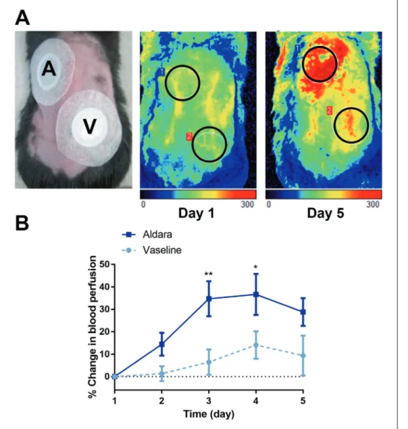
Figure 2 Placement of Finn chambers on the dorsal skin of a mouse.
Figure 3 Double-fold dorsal skin thickness measurement using an engineer’s micrometer. (A) Representative image of measurement on an 8-mm treated spot. (B) Percent change in skin thickness after Aldara or Vaseline treatment using Finn chambers on C57BL/6 mice. Data are mean±SEM forn=6/group. ***p<.001 for Aldara versus Vaseline based on repeated measures or two-way ANOVA followed by Bonferroni’s post hoc test.
1. With the anesthetized animal placed on a heating pad, pinch the dorsal skin in a rostral-caudal direction above the spine (Fig. 3A).
2. Place the treated area of the double-folded mouse skin between the anvil and spindle
face of the micrometer. Perform thickness measurement and record. Horváth et al.
5 of 12
The double-fold skin thickness is determined most precisely by having the anvil and spindle face touch the skin gently with little or no compression.
3. Thoroughly wipe the micrometer with 70% ethanol after each measurement to avoid cross-contamination of imiquimod- and Vaseline-treated areas.
4. Repeat each day of the 5-day experiment.
It is critical that the same areas of skin be measured each day.
5. Express data as percent increase in skin thickness compared with Day 1 baseline values (Fig. 3B).
SUPPORT PROTOCOL 2
MEASUREMENT OF BLOOD PERFUSION
Increased blood perfusion (erythema) is a hallmark of inflammatory skin conditions.
Here, mean perfusion of the treated skin area is quantified non-invasively using Laser speckle contrast analysis (LASCA) imaging. Blood perfusion should be measured after anesthesia but before treatment on each day of the 5-day experiment.
When measuring blood perfusion, anesthesia must be sufficiently stable to avoid involun- tary movements, which could affect the accuracy of the measurement. Additionally, body temperature must be maintained, as fluctuations in this parameter can have a significant influence on peripheral microcirculation.
Materials
Mice enrolled in Finn chamber experiment (Days 1-5; see Basic Protocol) Laser speckle contrast analysis laser Doppler (PeriCam PSI, Perimed) PimSoft software (Perimed, Sweden)
1. With the anesthetized animal placed on a heating pad, define a 3×4–cm area in the data acquisition software adjusted to the dorsal skin of mouse.
This area will be scanned by the instrument during the measurement. For further details, see manufacturer’s instructions.
2. Select regions of interest (ROI) according to the treated areas on the dorsal skin (Fig. 4A).
3. Measure the mean perfusion of the two ROIs for 2 min each.
4. Repeat each day of the 5-day experiment.
It is critical that the same areas of skin be measured each day.
5. Express data as percent change in blood perfusion relative to Day 1 baseline values (Fig. 4B).
SUPPORT PROTOCOL 3
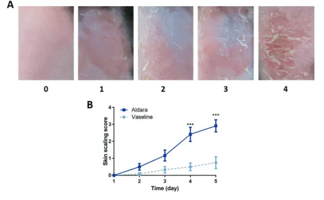
DETERMINATION OF SCALING SCORE
Scaling should be monitored daily after anesthesia but before treatment. The calculation of the scaling score is based on Luo, Wu, Zhao, Liu, & Wang (2016).
Materials
Mice enrolled in Finn chamber experiment (Days 1-5; see Basic Protocol) Digital camera (e.g., Sony Cybershot DSCW170)
Camera stand (e.g., Manfrotto 290)
1. With the anesthetized animal on a heating pad, use a digital camera on a camera stand to take individual photographs of each animal from a standard distance (determined by the focal distance of the camera).
Horváth et al.
Figure 4 Blood perfusion measurements by LASCA imaging. (A) Representative images of mea- surements on Finn chamber–covered sites on Days 1 and 5. (B) Percent change in skin blood flow after Aldara or Vaseline treatment using Finn chambers in C57BL/6 mice. Data are mean±SEM forn=6/group. *p<.05; **p<.01 for Aldara versus Vaseline based on repeated measures or two-way ANOVA followed by Bonferroni’s post hoc test.
2. Have three trained dermatologists who are blinded for the study score each of the pho- tographic images. Assign scores from 0-4, where 0=no scaling, 1=slight scaling, 2=moderate scaling, 3=marked scaling, and 4=maximal scaling (Fig. 5A).
3. Plot average score versus day of treatment (Fig. 5B).
SUPPORT PROTOCOL 4 SEMIQUANTITATIVE HISTOPATHOLOGICAL SCORING
An important characteristic of imiquimod-induced psoriasiform dermatitis is its ability to recapitulate the structural changes associated with human psoriatic skin. This includes an increase in epidermal thickness, the appearance of Munro’s microabscesses, and tortured dilated capillaries. Semiquantitative scoring of these parameters on hematoxylin-eosin stained tissue is used to determine the severity of psoriasiform skin inflammation.
Horváth et al.
7 of 12
Figure 5 Determination of skin scaling score. (A) Scaling is scored from 0-4, where 0=no scaling, 1=slight scaling, 2=moderate scaling, 3=marked scaling, and 4=maximal scaling. (B) Skin scaling scores after Aldara or Vaseline treatment using Finn chambers on the dorsal skin of C57BL/6 mice. Data are mean±SEM forn=6/group. ***p<.001 Aldara versus Vaseline based on repeated measures or two-way ANOVA followed by Bonferroni’s post hoc test.
Materials
Mice enrolled in Finn chamber (Day 5; see Basic Protocol) 6% formalin
Hematoxylin-eosin solution Forceps and scissors Microscope slides
Light microscope with 20×objective AnalySIS software (Olympus) or similar
Additional reagents and equipment for standard methods of sectioning fixed tissue and staining with hematoxylin-eosin
1. Sacrifice mice by cervical dislocation.
2. Excise the 8-mm treated full-thickness skin tissue using sterile forceps and scissors.
3. Fix in 6% formalin for 48 hr.
4. Prepare three 5-μm sections from the center of the treated area on microscope slides and stain with hematoxylin-eosin according to standard procedures.
5. Count the total number of Munro’s microabscesses throughout the entire section.
6. Determine the number of dilated capillaries present in the dermal-epidermal border of the entire section.
7. Measure the diameter of the epidermal layer of three separate sites in the field of view using the arbitrary distance function of AnalySIS software. Repeat for up to three fields of view on each tissue section. Average the collected data.
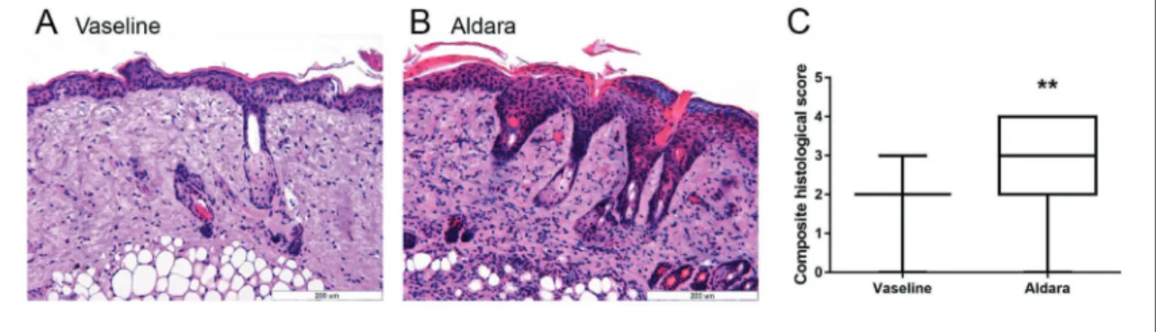
Horváth et al.
Figure 6 Histological analysis. (A,B) Representative images of Vaseline- and Aldara-treated dorsal mouse skin on Day 5. (C) Composite histological scores of mouse dorsal skin after different treatments. Data are mean
±SEM forn=6/group. **p<.01 for Aldara versus Vaseline based on Mann-Whitneyt-test.
8. Set the lowest and highest data values for each measured parameter (number of Munro’s microabscesses, number of dilated capillaries and thickness of epidermal layer) as the low and high reference points for that parameter, then divide the differ- ence between them into five equal ranges. Assign each range a point value from 0 to 4, with the lowest range being 0 and highest being 4. Plot each data set separatelyor calculate a composite score by averaging the three different scores (Munro’s, capillar- ies, thickness) derived from the same experimental setup (i.e., same treatment, same exposure time, etc.).
For an example of plotted composite scores, see Figure 6. For additional details on scoring, see Kemény et al. (2018).
SUPPORT PROTOCOL 5 ASSESSMENT OF SYSTEMIC SIDE EFFECTS IN RESPONSE
TO IMIQUIMOD APPLICATION
One of the major problems with the original imiquimod model was the appearance of systemic side effects including splenomegaly, loss of body weight, and increased serum concentrations of inflammatory cytokines, complicating the interpretation of the find- ings. This problem is largely overcome by using Finn chambers for localized application, which significantly minimizes the amount of irritant applied to the skin. Described below are ways to measure body weight and splenomegaly to assess the presence of imiquimod side effects.
Materials
Mice enrolled in Finn chamber experiment (Day 5; see Basic Protocol) Sterile forceps and scissors
Analytical balance
1. Using measurements from the Basic Protocol, plot the percent change in body weight on all days relative to Day 1 (Fig. 7A).
2. Sacrifice mice by cervical dislocation.
3. Open the peritoneum with a ventral midline incision using sterile forceps and scissors.
4. Remove and immediately weigh the spleen on an analytical balance (Fig. 7B).
The spleen must be weighed immediately to avoid changes in weight due to drying. Skin samples should be taken for histology first, then the animal can be opened and the spleen immediately excised and weighed.
Horváth et al.
9 of 12
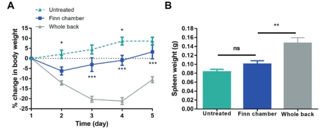
Figure 7 Changes in body and spleen weight. (A) Percent change in body weight after Aldara or Vaseline treatment using the Finn-chamber or conventional (whole-back) model in C57BL/6 mice. Data are mean± SEM forn=6/group, ***p<.001 Finn chamber versus whole back, *p<.05 untreated versus Finn chamber based on repeated measures or two-way ANOVA followed by Bonferroni’s post hoc test. (B) Effect of localized or whole-back Aldara treatment on development of splenomegaly. Data are mean± SEM forn = 6/group,
**p<.01 Finn chamber versus whole back based on one-way ANOVA followed by Bonferroni’s post hoc test.
ns: not significant.
REAGENTS AND SOLUTIONS Ketamine/xylazine solution
1200 μl 50 mg/ml ketamine (Richter Gedeon) 150 μl 20 mg/ml xylazine (Lavet Pharmaceuticals) 4650 μl physiological saline
Store up to 1 week at 4°C
Final concentrations are 10 mg/ml ketamine and 0.5 mg/ml xylazine. An intraperitoneal in- jection of 100μl solution per 10 g body weight will give the desired doses of 100 mg/kg ketamine and 5 mg/kg xylazine.
COMMENTARY
Background Information
In the original imiquimod-induced psori- asiform model, 62.5-80 mg of imiquimod or Vaseline control were smeared on the shaved whole back skin of the mouse (Kim et al., 2014; van der Fits et al., 2009). Often this quantity of imiquimod elicited severe systemic effects, as evidenced by a general decline in health, weight loss of 20-25%, fever, and death (Alvarez & Jensen, 2016). To minimize this problem, we modified the procedure to iden- tify the optimal dose of imiquimod and appli- cation method for inducing a consistent skin reaction. Given their design, Finn chambers were found to be ideal for administering the minimal amount of cream, greatly reducing the occurrence of systemic side effects. More- over, Finn chambers improved the penetration of active test substances into the skin through an occlusive effect, while minimizing scratch- ing and licking of the creams by experimental animals or their littermates.
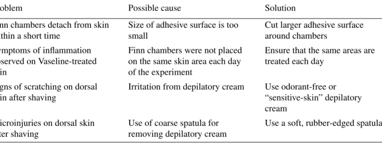
Critical Parameters and Troubleshooting
Imiquimod-induced psoriasiform dermati- tis results is induced by topical application of imiquimod cream. For proper penetration of the active ingredient and measurement of relevant symptoms, it is imperative that the fur be completely removed with an electric shaver and depilatory cream. As these proce- dures may cause minor skin injuries, care must be taken in to avoid such damage, and time should be allowed for the tissue to heal be- fore undertaking experiments. Typically, this is addressed by waiting at least one day af- ter fur removal before initiating a study. For optimal results, it is important to place Finn chambers in the same locations each day of the experiment.
To obtain consistent and reliable results us- ing Doppler measurement of blood perfusion, deep anesthesia of mice is necessary, as any Horváth et al.
Table 1. Troubleshooting Guide for Induction of Psoriasiform Skin Reactions Using Finn Chambers
Problem Possible cause Solution
Finn chambers detach from skin within a short time
Size of adhesive surface is too small
Cut larger adhesive surface around chambers
Symptoms of inflammation observed on Vaseline-treated skin
Finn chambers were not placed on the same skin area each day of the experiment
Ensure that the same areas are treated each day
Signs of scratching on dorsal skin after shaving
Irritation from depilatory cream Use odorant-free or
“sensitive-skin” depilatory cream
Microinjuries on dorsal skin after shaving
Use of coarse spatula for removing depilatory cream
Use a soft, rubber-edged spatula
involuntary movement can influence the as- sessment. It is also crucial to maintain animal body temperature using a 37°C heating pad, as fluctuations in body temperature can influence blood perfusion.
For troubleshooting guidance, see Table 1.
Understanding Results
The extent of the skin reaction to im- iquimod differs among mouse strains, with females being most sensitive in all cases (Alvarez & Jensen, 2016; Swindell et al., 2017). It is also possible that the extent of imiquimod-induced psoriasiform skin inflammation is dependent on the general health of the animal (e.g., body weight), which might explain variations in response to the irritant (Yu et al., 2019, 2020). Given these factors, all experiments should be repeated at least three times to obtain the most robust and reliable findings.
Time Considerations
A single experiment requires 6 days to complete. This includes 1 day for preliminary preparation (measurement of body weight, de- pilation), 4 days for imiquimod and Vaseline treatments and assessment of in vivo parame- ters, and 1 final day to complete in vivo mea- surements and obtain tissue samples for sub- sequent analysis.
Imiquimod and Vaseline treatments on the dorsal skin of mice using Finn chambers re- quire 15 min per six mice, as does the double- fold skin thickness measurement. Blood per- fusion testing requires∼25 min per six mice, with body weight measurements taking about 6 min per six mice. Collection of skin and spleen samples and measurement of spleen weights require 30 min per six mice. Process- ing excised skin samples for histological anal- ysis takes 7 days, and histopathological scor-
ing of hematoxylin-eosin slides takes∼15 min per sample.
Acknowledgments
This work was supported by NKFIH K_128210; PEPSYS-GINOP-2.3.2-15-2016- 00050, OTKA-NN-114458; EFOP 3.6.2-16- 2017-00009 Establishing and Internation- alizing the Thematic Network for Clinical Research. KÁ was supported by János Bolyai Research Scholarships of the Hungarian Academy of Sciences and ÚNKP-19-4 New National Excellence Program of the Ministry for Innovation and Technology.
Literature Cited
Alvarez, P., & Jensen, L. E. (2016). Imiquimod treatment causes systemic disease in mice re- sembling generalized pustular psoriasis in an IL-1 and IL-36 dependent manner. Mediators of Inflammation,2016, 5–8. doi: 10.1155/2016/
6756138.
Hanna, E., Abadi, R., & Abbas, O. (2016). Im- iquimod in dermatology: An overview. Inter- national Journal of Dermatology,55, 831–844.
doi: 10.1111/ijd.13235.
Hawkes, J. E., Gudjonsson, J. E., & Ward, N. L. (2016). The snowballing literature on imiquimod-induced skin inflammation in mice : A critical appraisal. Journal of Investigative Dermatology,137(3), 546–549. doi: 10.1016/j.
jid.2016.10.024.
Horváth, S., Komlódi, R., Perkecz, A., Pintér, E., Gyulai, R., & Kemény, Á (2019). Methodolog- ical refinement of Aldara-induced psoriasiform dermatitis model in mice.Scientific Reports,9, 3685. doi: 10.1038/s41598-019-39903-x.
Kemény, Á., Kodji, X., Horváth, S., Komlódi, R., Sz˝oke, É., Sándor, Z., … Gyulai, R.
(2018). TRPA1 acts in a protective manner in imiquimod-induced psoriasiform dermatitis in mice.Journal of Investigative Dermatology, 138(8), 1774–1784. doi: 10.1016/j.jid.2018.02.
040.
Kim, H. R., Lee, A., Choi, E. J., Hong, M. P.,
Kie, J. H., Lim, W., … Seoh, J. Y. (2014). Horváth et al.
11 of 12
Reactive oxygen species prevent imiquimod- induced psoriatic dermatitis through enhanc- ing regulatory T cell function.PLoS One,9(3), e91146. doi: 10.1371/journal.pone.0091146.
Luo, D.-Q., Wu, H.-H., Zhao, Y.-K., Liu, J.-H., &
Wang, F. (2016). Different imiquimod creams resulting in differential effects for imiquimod- induced psoriatic mouse models.Experimental Biology and Medicine, 241, 1733–1738. doi:
10.1177/1535370216647183.
Swindell, W. R., Michaels, K. A., Sutter, A. J., Diaconu, D., Fritz, Y., Xing, X., … Ward, N.
L. (2017). Imiquimod has strain-dependent ef- fects in mice and does not uniquely model hu- man psoriasis. Genome Medicine, 9, 24. doi:
10.1186/s13073-017-0415-3.
van der Fits, L., Mourits, S., Voerman, J. S. A., Kant, M., Boon, L., Laman, J. D., … Lub- berts, E. (2009). Imiquimod-induced psoriasis- like skin inflammation in mice is mediated via the IL-23/IL-17 axis.Journal of Immunol- ogy,182, 5836–5845. doi: 10.4049/jimmunol.
0802999.
Walter, A., Schäfer, M., Cecconi, V., Matter, C., Urosevic-Maiwald, M., Belloni, B., … van den Broek, M. (2013). Aldara activates TLR7- independent immune defence. Nature Com- munications, 4, 1560. doi: 10.1038/ncomms 2566.
Yu, S., Wu, X., Shi, Z., Huynh, M., Jena, P. K., Sheng, L., … Hwang, S. T. (2020).
Diet-induced obesity exacerbates imiquimod- mediated psoriasiform dermatitis in anti-PD- 1 antibody-treated mice: Implications for pa- tients being treated with checkpoint inhibitors for cancer.Journal of Dermatological Science, 97, 194–200. doi: 10.1016/j.jdermsci.2020.01.
011.
Yu, S., Wu, X., Zhou, Y., Sheng, L., Jena, P. K., Han, D., … Hwang, S. T. (2019). A West- ern diet, but not a high-fat and low-sugar diet, predisposes mice to enhanced suscepti- bility to imiquimod-induced psoriasiform der- matitis.Journal of Investigative Dermatology, 139, 1404–1407. doi: 10.1016/j.jid.2018.12.0 02.
Horváth et al.