One of the first studies of contraction-induced skeletal muscle damage was performed by Hugh et al. Accordingly, morphological evidence and direct quantification of skeletal muscle damage are necessary to clarify the appropriateness of continuing exercise with damaged muscles.
SIGNIFICANCE OF THE PROBLEM
Thus, the aim of this study is to study the impact of repeated bouts of eccentric training on several physiological aspects (mechanical, morphological, cellular-molecular) in order to determine the appropriateness of continuing training with "damaged" and/or affected muscles. Consequently, there is an emerging need to determine the effects and subsequently the appropriateness of continuing training with damaged muscles.
STATEMENT OF THE PROBLEM
To provide a more complete picture, regarding the effects and suitability of continuing pre-recovery exercises, we designed an integrated study that investigates several physiological aspects including mechanical, morphological and cellular-molecular.
DELIMITATIONS OF THE STUDY
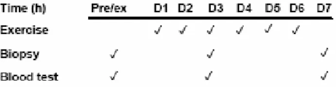
All blood and muscle biopsies were taken within one hour of the exercise protocol and blood always preceded the muscle biopsies. All blood and muscle biopsies were taken after a morning fast and always between 8:00 and 9:00 am.
LIMITATIONS OF THE STUDY
Skeletal muscle biopsies were taken under local anesthesia (1% lidocaine) by percutaneous needle biopsy using a 5 mm Bergström biopsy needle. Subjects were instructed to avoid any vigorous and/or intense activity during the experimental period; however, it was beyond the scope of this study to monitor the subject's activities.
DEFINITION OF TERMS
On days when exercise was accompanied by a muscle biopsy, subjects were instructed to attend the testing laboratory after a morning fast, but it was beyond the current study's ability to control the subject's diet.
HYPOTHESES
This chapter provides a review of related literature and is divided into four sections, some of which have multiple subsections: direct indices of skeletal muscle injury; Indirect indices of skeletal muscle damage; Myostatin and MRF for muscle regeneration; and Effect of resistance exercise on MRF and myostatin.
DIRECT INDICES OF SKELETAL MUSCLE DAMAGE
One of the first signs of muscle damage is the disruption of the cytoskeleton and especially the loss of the intermediate filaments of desmin. 1998 also found that the earliest manifestation of muscle damage is the loss of desmin intermediary in New Zealand white rabbits trained eccentrically (900 eccentric contractions in a 30 minute period).
INDIRECT INDICES OF SKELETAL MUSCLE DAMAGE
- Indirect Evidence for Eccentric Contraction-Induced Muscle Damage
- Indirect Evidence against Eccentric Contraction-Induced Muscle Damage 25
- Myogenic Regulatory Factors
- Myostatin
Chapman 2006 showed that contraction speed is another determining factor influencing the degree of muscle damage. simultaneously under tension; high-speed eccentric exercises caused greater muscle damage than low-speed exercises in untrained subjects. As can be deduced from the studies mentioned above, a single eccentric exercise results in the appearance of indirect markers of muscle damage.
RESISTANCE EXERCISE, MRFS AND MYOSTATIN
Myogenic Regulatory Factors
In a recent study, Yang et al. 2005), examined the time-course activation of all MRFs (MRF4, Myf5, MyoD, myogenin) genes after an acute bout of resistance exercise. Six relatively trained subjects performed a resistance exercise consisting of 3 sets of 10 knee extensions, on an isokinetic dynamometer, at 70% of the concentric maximum for one repetition. In addition, the author suggested that the higher resting myogenic mRNA levels in old women may reflect an attempt to preserve muscle mass and function. 2005) also investigated whether resistance exercise would induce expression of MRFs.
A main effect of exercise showed that MyoD mRNA expression levels increased by 20% 24 h after acute resistance exercise. The authors concluded that attenuated MyoD and myogenin mRNA expression in old subjects after resistance exercise indicates reduced growth and/or regenerative capacity.
Myostatin
Muscle biopsies and blood samples were taken before and after 6 and 12 weeks of resistance training of the vastus lateralis. Resistance training was performed three days per week and each session consisted of three sets of six to eight repetitions (85-90% of one repetition maximum) using leg press and knee extension exercises. Therefore, the authors concluded that the increased myostatin in response to cortisol and/or resistance training has no effect on the exercise-induced increase in muscle strength and mass.
At the end of the 9-week resistance training protocol, the study observed a 37% decrease in myostatin expression in all subjects. The authors suggested that myostatin mRNA levels are reduced in response to heavy resistance exercise in humans.
S ELECTION OF S UBJECTS
I NSTRUMENTS AND COLLECTION OF DATA
- Instruments and procedures for collecting biomechanical data and DOMS 36
- Instruments and procedures for collecting mRNA expression data
- Instruments and procedures for collecting Creatine kinase (CK), lactate
- Instruments and procedures for morphological data collection
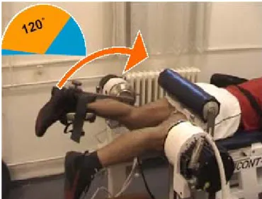
To initiate the lever arm of the dynamometer for each contraction, subjects had to exert 20 Nm of torque. Instruments and procedures for collection of muscle biopsies and experimental design. i) Skeletal muscle biopsies: were taken from the middle part of the vastus lateralis muscle (non-dominant exercised leg) under local anesthesia (1% Lidocaine) by percutaneous needle biopsy using a 5 mm Bergström biopsy needle (Fig. 2 ). The same time protocol was followed by the control subjects, except for the resistance exercise.
Note that the same timeline protocol was followed by the control subjects, with the exception of the resistance exercise. Analyzes of the real-time quantitative PCR data were performed using the comparative threshold cycle [Ct] method as proposed by Applied Biosystems (User Bulletin #2).
T REATMENT OF D ATA
Sections for all biopsies were viewed using a Nikon Eclipse, E600, microscope and the selected sections were photographed with a spot camera (Pixera Penguin 600CL model). All sections were blinded to the experimental treatment (exercise or control) by three different parties, one of which was independent of the study.
I NDIRECT M ARKERS OF M USCLE D AMAGE
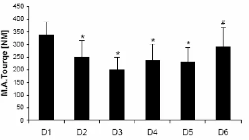
Delayed onset muscle soreness (DOMS). DOMS peaked on day 3 and then gradually decreased until day 5 was significantly lower than day 3. D1*: The measurement was taken 8 hours after the first exercise, and then the measurements were taken 24 hours after it. point. On day 6, MAT recovered significantly from day 3 and was not significantly different from the first training session.

D IRECT M ARKERS OF M USCLE D AMAGE
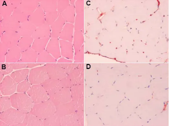
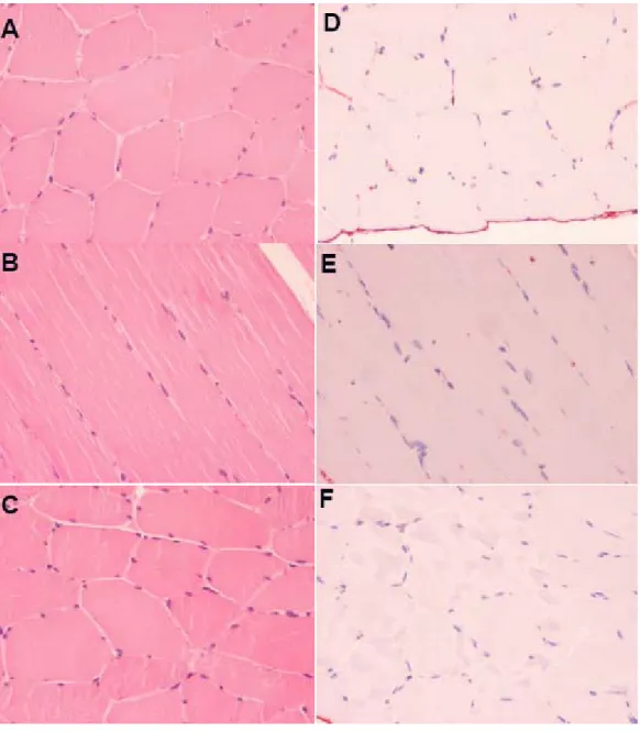
The left panel (A, B, and C) shows sections taken from a biopsy of the Vastus Lateralis muscle and stained with hematoxylin-eosin. Right panel (D, E and F) shows sections taken from a biopsy of the Vastus Lateralis muscle and stained with anti-human plasma fibnectin. Left panel (A and B) shows cross-sections taken from a biopsy of the Vastus Lateralis muscle and stained with hematoxylin-eosin.
Right panel (C and D) shows cross sections taken from a Vastus Lateralis muscle biopsy and stained with anti-human plasma fibronectin. Muscle biopsy longitudinal section from the only subject showing direct index of muscle damage on day 7.
M YOGENIC REGULATORY FACTORS (MRF S )
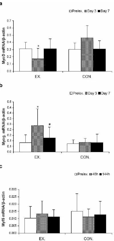
Relative RT-PCR results for MyoD (a), myogenin (b) and myf5 (c) mRNA expression using β-actin as internal standard.
M ARKERS OF PROLIFERATION AND DIFFERENTIATION
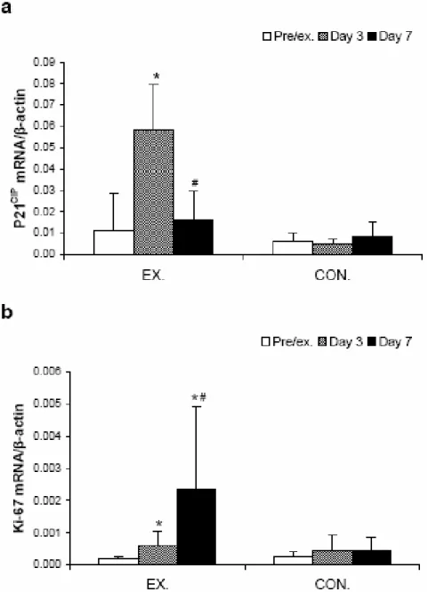
Relative RT-PCR results for P21 (a) and ki-67 (b) mRNA expression using β-actin as an internal standard. On day 7, the mRNA for p21 was not significantly different compared to the pre-exercise level, while ki-67 mRNA increased further (12.1 fold, P=0.007).
M YOSTATIN
The purpose of this study was to investigate the effects of repeated bouts of eccentric exercise over six consecutive days. To achieve this, we examined direct and indirect markers of muscle damage, as well as the expression of key myogenic genes. The results presented here indicate that repeated bouts of eccentric exercise for six consecutive days do not induce gross myofiber but may impair the adaptation process, as the expression of MRFs was attenuated.
M USCLE DAMAGE , D IRECT AND I NDIRECT MARKERS
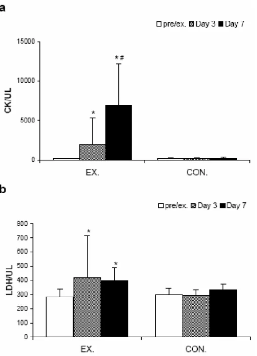
In contrast, the present study showed that torque recovery was not affected by the repeated bouts of EE, so that by day 6 there was no significant difference compared to pre-exercise levels. In the present study, we suspected that the single bout of EE used by the aforementioned studies was not sufficient to cause sarcolemma damage. Contrary to the latter logic, the increased activities of CK and LDH suggest that muscle damage in the present study was not attenuated, but rather exacerbated.
Given the latter paradox, it is important to investigate the reasons that may explain the increased CK and LDH activities despite the lack of sarcolemma damage found in this study. Therefore, there is a possibility that ultrastructural damage also occurred in this study.
M YOGENIC REGULATORY FACTORS
The reason for the delay between the peak of enzyme leakage and the overt muscle damage at the structural level observed by Jones et al. 1986 remains unknown, but it may be another determinant of not observing no sarcolemma and/or muscle fiber damage in the current research. The reason for the attenuated mRNA expression of MRFs observed by the current study is unknown, but may be a result of the relatively short resting intervals (24 hours) between exercise periods.
Indeed, Haddad and Adams (2002) reported that long rest intervals (48 hours) between two repeated bouts of isometric exercise resulted in much greater myogenic responses compared to short rest intervals (8 or 24 hours). Thus, it is not unreasonable to believe that this could have been the reason for the attenuated MRF expression observed in the current study.
M YOSTATIN
Myostatin is a negative regulator of skeletal muscle growth, and given its role and function, the contradictory findings mentioned above are not surprising. Myostatin could be such a chalon for muscle tissue, helping skeletal muscles to regulate its growth and regeneration (McCroskery S et al., 2005). A stimulus such as resistance exercise could complement the downregulation of myostatin so that skeletal muscle can achieve optimal recovery and/or growth.
Once optimal growth and/or recovery is achieved, skeletal muscle will most likely upregulate myostatin expression to avoid excessive/unwanted muscle growth. Accordingly, we propose that, depending on the skeletal muscle status, resistance exercise causes “up- or down-regulation” of myostatin and the net result of these actions will be optimal growth and/or recovery.
C ELL P ROLIFERATION AND D IFFERENTIATION
Muscle biopsies were obtained from the vastus lateralis muscle to determine muscle damage and gene expression of the selected genes. Effects of the number of eccentric muscle actions on the first and second bouts of eccentric exercise of the elbow flexors. Temporal pattern of the repeated bout effect of eccentric exercise on delayed onset muscle soreness.
Comparison between leg and arm eccentric exercises of the same relative intensity on indices of muscle damage. Recent advances in understanding the repeated bout effect: the positive effect against muscle damage of a single bout of eccentric exercise. I was informed that the purpose of the study is to determine the mechanical and morphological characteristics of human striae as a result of repeated bouts of eccentric exercise.
This rapid depolarization and subsequent repolarization of the muscle fiber is the action potential.
C ONCLUSIONS
R ECOMMENDATIONS FOR FUTURE STUDIES