C A S E R E P O R T Open Access
An unusual cause of infant ’ s stridor – congenital laryngocele
Andrea Ambrus1*, Balázs Sztanó1, Miklós Szabó2, Béla Vasas3, István Sziller4and László Rovó1
Abstract
Congenital laryngocele is an uncommon cause of neonatal stridor. There are only a few cases reported in the literature. The authors present a successfully treated case of an infant, whose life could only be saved by urgent tracheostomy. On the 5th postoperative day endoscopic excision and marsupialization provided patent airway. The patient could be decannulated. During follow-up no recurrence was observed.
Keywords:Congenital stridor, Laryngocele, Airway obstruction
Introduction
The neonatal laryngocele is an extremely rare phenomenon, which is defined as an air-filled cystic dilatation of the laryn- geal saccule. The incidence is estimated to be 1 per 2.5 million people per year [1]. Although it is a benign lesion, it may cause stridor, respiratory distress and serious airway obstruction in the narrow airway of a newborn which ne- cessitates urgent intervention [2]. The authors present a newborn baby successfully endoscopically treated with congenital laryngocele.
Case report
A 5-day-old male newborn was referred to our tertiary de- partment. He was a normal-appearing, 3135 g, full-term baby with no significant prenatal medical history. In an- other hospital, after birth (Apgar score: 7/7/9), immedi- ately severe stridor and respiratory distress occurred. He required DuoPAP ventilation. Direct laryngoscopy re- vealed a large cystic mass bulging from the right aryepi- glottic fold, which obstructed the glottis. He could not be intubated, so an urgent tracheotomy was performed. MRI described a well-circumscribed, thin-walled fluid attenu- ation mass, measuring 20 × 18 mm, localized at the level
of the glottis (Fig.1a, b). No other congenital malforma- tion occurred.
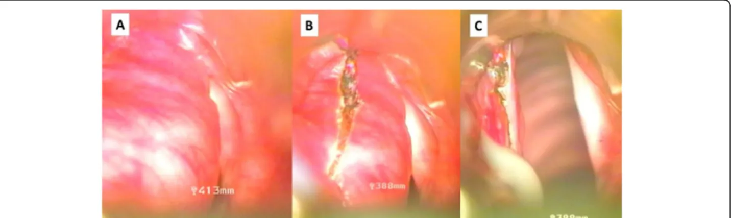
After his admission a direct laryngoscopy under gen- eral anaesthesia was performed: a swollen cystic mass bulging from the right pharyngoepiglottic and aryepi- glottic folds was found (Fig.2a). The cyst was incised by ultra-pulse mode CO2 laser (Dhaesin U-40, Dhaesin En- terprise, South Korea; 315 W, 90μs, 20 ms) (Fig. 2b).
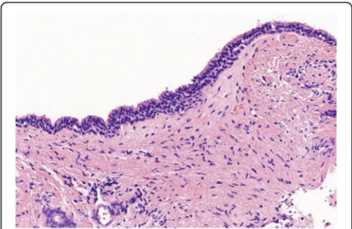
Thick mucoid fluid came out, then marsupialization was done. The supraglottic mass which caused laryngeal asymmetry immediately disappeared (Fig. 2c). At the end of the surgery, the baby was decannulated, but he was intubated for four days with a 3.5 cuffed tracheal tube. Parenteral antibiotic (Clindamycin 5 mg/ kg /3 times a day, Cefotaxime 25 mg/ kg/ 2 times a day) was administered for 4 days. After his extubation no dyspnea re-occurred and his stoma closed spontaneously. Postop- erative period was uneventful. Control endoscopy per- formed on the 7th postoperative day revealed no recurrence. During the 6-month follow up period the growing of the baby was appropriate and his parents no- ticed no voice disturbance. Histologic report confirmed the diagnosis of a congenital laryngocele. (Fig.3).
Discussion
The first air-containing ‘tumors’ of the neck were re- ported in 1829. The first laryngeal cyst in a newborn
© The Author(s). 2020, corrected publication 2020.Open AccessThis article is licensed under a Creative Commons Attribution 4.0 International License, which permits use, sharing, adaptation, distribution and reproduction in any medium or format, as long as you give appropriate credit to the original author(s) and the source, provide a link to the Creative Commons licence, and indicate if changes were made. The images or other third party material in this article are included in the article's Creative Commons licence, unless indicated otherwise in a credit line to the material. If material is not included in the article's Creative Commons licence and your intended use is not permitted by statutory regulation or exceeds the permitted use, you will need to obtain permission directly from the copyright holder. To view a copy of this licence, visithttp://creativecommons.org/
licenses/by/4.0/. The Creative Commons Public Domain Dedication waiver (http://creativecommons.org/publicdomain/zero/1.
0/) applies to the data made available in this article, unless otherwise stated in a credit line to the data.
* Correspondence:ambrus.andrea@med.u-szeged.hu
1Department of Otorhinolaryngology, Head and Neck Surgery, University of Szeged, Szeged, Hungary
Full list of author information is available at the end of the article
Ambruset al. Journal of Otolaryngology - Head and Neck Surgery (2020) 49:34 https://doi.org/10.1186/s40463-020-00430-9
was published in 1876 [3, 4]. The first reports were based on postmortem diagnosis - all newborns died with any type of airway obstruction. Since 1970s with modern neonatology, the introduction of intubation and ad- vanced endoscopic diagnostic and microsurgical tech- niques the patients’life could have been saved.
Laryngocele is an abnormal dilatation of the appendix of the laryngeal ventricle and is classified as internal and mixed or combined type according to its relationship with the thyrohyoid membrane. In reviewing the English-language literature and focusing on the pediatric population, there are very few cases of successfully treated neonatal laryngoceles. Embryologically, the ven- tricle of the larynx and the saccule develops at the end of the second intrauterine month as a secondary out- pouching from the laryngeal lumen. Congenitally large saccule, weakness of the periventricular connective tis- sues, and the thyroepiglottic and aryepiglottic muscles are accepted as predisposing factors [2].
Airway obstruction cannot always be diagnosed or pre- dicted prenatally [2]. The clinical features of this rare pathology depend on the size and type of laryngocele and are highly variable. In mild cases it is usually asymp- tomatic. But it may cause severe respiratory distress and inspiratory stridor and become a life-threatening event due to mechanical obstruction of the narrow and small airway of a neonate [5,6].
The diagnosis of laryngocele mainly depends on phys- ical examination, symptoms and laryngeal examination by direct laryngoscopy [2]. High resolution imaging mo- dalities such as computed tomography or magnetic res- onance imaging may help to delineate the structures involved and the exact location of the mass [7].
If the patient has respiratory distress and dyspnea, the airways should immediately be secured by intubation or tracheostomy. In our case the infant could not be intu- bated; only the urgent tracheostomy could save his life.
Various surgical techniques have been utilized such as
Fig. 1Preoperative MRI. A: Sagittal contrast MRI image shows well-circumscribed, thin-walled fluid attenuation mass. B: Axial contrast MRI image reveals the cystic mass at the level of the glottis
Fig. 2Endoscopic surgery for congenital laryngocele - intraoperative photos. A: Cystic mass of the left false vocal cord cause severe obstruction.
B: CO2 laser excision. C: After thick mucoid fluid came out, and marsupialization was done, the asymmetry of the supraglottis disappeared Ambruset al. Journal of Otolaryngology - Head and Neck Surgery (2020) 49:34 Page 2 of 3
external approach, endolaryngeal microlaryngoscopy, carbon-dioxide laser excision and transoral robotic sur- gery [1, 6–8]. Transoral laser endoscopic excision and marsupialization of the cyst’s wall is a minimally invasive solution. Temporary intubation and administration of an- tibiotics prevented postoperative edema. Although Mys- siorek et al. reported that marsupialization has a higher risk of recurrence, during the 6 month follow-up there was no evidence of laryngocele persistence [3,9,10].
Conclusion
Congenital laryngocele is a rare cause of laryngeal dyspnea in neonates. In severe cases urgent surgical intervention must be performed because of the life-threatening airway obstruction. Laryngoscopy may establish the proper diag- nosis. Laser excision with complete removal of the cyst’s wall under jet ventilation anesthesia might be a successful treatment without performing a tracheostomy.
Acknowledgements Not applicable.
Authors’contributions
AA collected the patient’s data and reviewed the literature, wrote the manuscript. BS and LR performed the operation and were the major contributers in writing the manuscript. IS and MS recognized the dyspnea immediately after birth, performed tracheostomy. BV performed the histological examination. All authors read and approved the final manuscript.
Funding
Authors received no funding.
Availability of data and materials
Video documentation of the pre- and postoperative status was achieved.
Ethics approval and consent to participate
The study was approved by our institutional ethics committee (registration number 49/2018).
Consent for publication Obtained from the patient’s family.
Competing interests
The authors declare that they have no competing interests.
Author details
1Department of Otorhinolaryngology, Head and Neck Surgery, University of Szeged, Szeged, Hungary.21st Department of Paediatrics, Semmelweis University, Budapest, Hungary.3Department of Pathology, University of Szeged, Szeged, Hungary.4Szent Imre University Teaching Hospital, Budapest, Hungary.
Received: 12 December 2018 Accepted: 24 May 2020
References
1. Yılmaz S, Yıldızbas S, Yaman H, et al. Stridor in a newborn caused by a congenital laryngocele and bifid epiglottis: a case report abd review of literature international. J Pediatric Otorhinolaryngol Extra. 2010;5:28–31.
2. Tasinklar H, Vayisoglu Y, Avlan D, et al. Congenital Laryngomucocele: a rare cause of airway obstruction is a newborn. J Craniofacial Surg. 2015;26:238– 40.
3. Borko R, Szucs S [Congenital laryngeal cysts] HNO 2000; 48:843–845.
4. DeSanto LW. Laryngocele, laryngeal mucocele, large saccules, and laryngeal saccular cysts: a developmental spectrum. 1974; 84:1291–1296.
5. Lim EH, Mohamad H, Hamid SSA. Congenital laryngeal cyst: a report of 2 cases. Egyptian Journal of Ear, Nose, Throat and Allied Sciences. 2017;18:
187–9.
6. Ahmed H, Ndiaye C, Barry MW, Thiongane A, et al. A rare cause of upper airway obstruction in a child. Case Rep Otolaryngol. 2017;2017:2017265.
7. Chu L, Gussack GS, et al. A cause for airway obstruction. Arch Otolaryngol Head Neck Surg. 1994;120:454–8.
8. Saha D, Sinha R, Pai RR, Kumar A, Chakraborti S. Laryngeal cysts in infants and children—a pathologist’s perspective (with review of literature). Int J Pediatr Otorhinolaryngol. 2013;77:1112–7.
9. Holinger LD, Barnes DR, Smid LJ. Laryngocele and saccular cysts. Ann Otol 87:1978.
10. Chu L, Gussack GS, Orr JB, Hood D. Neonatal Laryngoceles Arch Otolaryngol Head and Neck Surg. 1994;120:454–8.
Publisher’s Note
Springer Nature remains neutral with regard to jurisdictional claims in published maps and institutional affiliations.
Fig. 3Histologic finding. A hematoxylin-eosin (H&E) stained slide demonstrates the cyst lining, which is comprised of a ciliated pseudostratified columnar epithelium without atypia. There was no mucin accumulation observed in the lumen
Ambruset al. Journal of Otolaryngology - Head and Neck Surgery (2020) 49:34 Page 3 of 3