NIST Interlaboratory Study on Glycosylation
Analysis of Monoclonal Antibodies: Comparison of Results from Diverse Analytical Methods
Authors
Maria Lorna A. De Leoz, David L. Duewer, Adam Fung, Lily Liu, Hoi Kei Yau, Oscar Potter, Gregory O. Staples, Kenichiro Furuki, Ruth Frenkel, Yunli Hu, Zoran Sosic, Peiqing Zhang, Friedrich Altmann, Clemens Gr unwald-Grube, Chun Shao, Joseph Zaia, et al. Ⳋ
Correspondence lornadeleoz@gmail.com
In Brief
A broad-based interlaboratory study of glycosylation profiles of a reference and modified IgG antibody involving 103 reports from 76 laboratories.
Graphical Abstract
Highlights
• A broad-based interlaboratory study of the glycosylation of a reference antibody: NISTmAb.
• 103 reports were received from 76 diverse laboratories worldwide.
• Analysis involved two samples, the NISTmAb and an enzymatically modified sample, enabling within-lab separation of random and systematic errors using the “Youden two-sample” method.
• Consensus values were derived and similar performance across all experimental methods was noted.
De Leoz et al., 2020, Molecular & Cellular Proteomics
19, 11–30January 2020 © 2020 De Leoz et al. Published by The American Society for Biochemistry and Molecular
NIST Interlaboratory Study on Glycosylation
Analysis of Monoclonal Antibodies: Comparison of Results from Diverse Analytical Methods*
□SMaria Lorna A. De Leoz1,84,95, David L. Duewer2, Adam Fung3, Lily Liu3, Hoi Kei Yau3, Oscar Potter4, Gregory O. Staples4, Kenichiro Furuki5, Ruth Frenkel6, Yunli Hu6, Zoran Sosic6, Peiqing Zhang7, Friedrich Altmann8, Clemens Grunwald-GrubeⳊ 8, Chun Shao9, Joseph Zaia9, Waltraud Evers10, Stuart Pengelley10, Detlev Suckau10, Anja Wiechmann10, Anja Resemann10, Wolfgang Jabs10,11, Alain Beck12, John W. Froehlich13, Chuncui Huang14, Yan Li14, Yaming Liu14, Shiwei Sun15, Yaojun Wang15, Youngsuk Seo16, Hyun Joo An16, Niels-Christian Reichardt17, Juan Echevarria Ruiz17,85, Stephanie Archer-Hartmann18,
Parastoo Azadi18, Len Bell19, Zsuzsanna Lakos20, Yanming An21, John F. Cipollo21, Maja Pucic-Bakovic22, Jerko Sˇ tambuk22, Gordan Lauc22,23, Xu Li24, Peng George Wang24, Andreas Bock25, Rene´ Hennig25, Erdmann Rapp25,40, Marybeth Creskey26, Terry D. Cyr26, Miyako Nakano27, Taiki Sugiyama27, Pui-King Amy Leung28, Paweł Link-Lenczowski29, Jolanta Jaworek29, Shuang Yang30, Hui Zhang30, Tim Kelly31, Song Klapoetke31, Rui Cao31,86, Jin Young Kim32, Hyun Kyoung Lee32, Ju Yeon Lee32, Jong Shin Yoo32, Sa-Rang Kim33, Soo-Kyung Suh33, Noortje de Haan34, David Falck34, Guinevere S. M. Lageveen-Kammeijer34,
Manfred Wuhrer34, Robert J. Emery35, Radoslaw P. Kozak35, Li Phing Liew35, Louise Royle35, Paulina A. Urbanowicz35, Nicolle H. Packer36, Xiaomin Song36, Arun Everest-Dass36,87, Erika Lattova´37, Samanta Cajic38, Kathirvel Alagesan39,88, Daniel Kolarich39,88, Toyin Kasali40, Viv Lindo40, Yuetian Chen41, Kudrat Goswami41, Brian Gau42,89, Ravi Amunugama43, Richard Jones43, Corne´ J. M. Stroop44, Koichi Kato45,46, Hirokazu Yagi46, Sachiko Kondo46,47, C. T. Yuen48, Akira Harazono49, Xiaofeng Shi50, Paula E. Magnelli50, Brian T. Kasper51, Lara Mahal51,90, David J. Harvey52, Roisin O’Flaherty53, Pauline M. Rudd53,
Radka Saldova53, Elizabeth S. Hecht54, David C. Muddiman54, Jichao Kang55, Prachi Bhoskar56, Daniele Menard56, Andrew Saati56, Christine Merle57, Steven Mast58, Sam Tep58, Jennie Truong58, Takashi Nishikaze59, Sadanori Sekiya59, Aaron Shafer60,
Sohei Funaoka61, Masaaki Toyoda61, Peter de Vreugd62, Cassie Caron63, Pralima Pradhan63, Niclas Chiang Tan63, Yehia Mechref64, Sachin Patil65, Jeffrey S. Rohrer65, Ranjan Chakrabarti66, Disha Dadke66,91, Mohammedazam Lahori66,92, Chunxia Zou67,68,
Christopher Cairo67,68, Be´la Reiz68, Randy M. Whittal68, Carlito B. Lebrilla69, Lauren Wu69, Andras Guttman70, Marton Szigeti70,71, Benjamin G. Kremkow72, Kelvin H. Lee72, Carina Sihlbom73, Barbara Adamczyk74, Chunsheng Jin74, Niclas G. Karlsson74, Jessica O¨ rnros74, Go¨ran Larson75, Jonas Nilsson75, Bernd Meyer76, Alena Wiegandt76, Emy Komatsu77, Helene Perreault77, Edward D. Bodnar77,4, Nassur Said78, Yannis-Nicolas Francois78, Emmanuelle Leize-Wagner78, Sandra Maier79, Anne Zeck79,
Albert J. R. Heck80, Yang Yang80,93, Rob Haselberg81, Ying Qing Yu82, William Alley82,94, Joseph W. Leone83, Hua Yuan83, and Stephen E. Stein1
From the1Mass Spectrometry Data Center, Biomolecular Measurement Division, Material Measurement Laboratory, National Institute of Standards and Technology, 100 Bureau Drive Gaithersburg, Maryland 20899;2Chemical Sciences Division, Material Measurement Labora- tory, National Institute of Standards and Technology, 100 Bureau Drive Gaithersburg, Maryland 20899;3Analytical Development, Agensys, Inc., 1800 Steward Street Santa Monica, California 90404; 4Agilent Technologies, Inc., 5301 Stevens Creek Blvd Santa Clara, California 95051;5Astellas Pharma, 5-2-3 Tokodai, Tsukiba, Ibaraki, 300-2698, Japan;6Analytical Development, Biogen, 14 Cambridge Center Cambridge, Massachusetts 02142;7Bioprocessing Technology Institute, 20 Biopolis Way, Level 3 Singapore 138668; 8Department of Chemistry, University of Natural Resources and Life Science, Vienna (BOKU), Muthgasse 18 1190 Wien, Austria;9Center for Biomedical Mass Spectrometry, Boston University School of Medicine, 670 Albany Street Boston, Massachusetts 02118;10Bruker Daltonik GmbH, Fahrenheit- str. 4, 28359 Bremen, Germany;11Department of Life Sciences & Technology, Beuth Hochschule fu¨r Technik Berlin, Seestrae 64, 13347 Berlin, Germany;12Centre d’Immunologie Pierre Fabre, 5 Avenue Napole´on III, BP 60497, 74164 St Julien-en-Genevois, France;13Department of Urology, Boston Children’s Hospital, 300 Longwood Avenue Boston Massachusetts 02115;14Institute of Biophysics, Chinese Academy of Sciences, 15 Da Tun Road, Chaoyang District, Beijing 100101 China;15Key Lab of Intelligent Information Processing, Institute of Computing Technology, Chinese Academy of Sciences, 15 Da Tun Road, Chaoyang District, Beijing 100101 China;16Graduate School of Analytical Science and Technology, Chungnam National University, Gung-dong 220, Yuseong-Gu, Daejeon 305–764, Korea (South);17CICbiomaGUNE, Paseo Miramon 182, 20009 San Sebastian, Spain;18Analytical Services, Complex Carbohydrate Research Center, University of Georgia, 315 Riverbend Road Athens, Georgia 30602;19BioCMC Solutions (Large Molecules), Covance Laboratories Limited, Otley Road, Harrogate, North Yorks HG3 1PY, United Kingdom;20Biochemistry Method Development & Validation, Eurofins Lancaster Laboratories, Inc., 2425 New Holland Pike Lancaster, Pennsylvania 17601;21Center for Biologics Evaluation and Research, Food and Drug Administration, 10903 New Hampshire
Report
Author’s Choice
This is an open access article under the CC BY license.
Glycosylation is a topic of intense current interest in the development of biopharmaceuticals because it is related to drug safety and efficacy. This work describes results of an interlaboratory study on the glycosylation of the Pri-
mary Sample (PS) of NISTmAb, a monoclonal antibody reference material. Seventy-six laboratories from indus- try, university, research, government, and hospital sec- tors in Europe, North America, Asia, and Australia submit- Avenue, Silver Spring, Maryland 20993; 22Glycoscience Research Laboratory, Genos, Borongajska cesta 83h, 10 000 Zagreb, Croatia;
23Faculty of Pharmacy and Biochemistry, University of Zagreb, A. Kovacˇic´a 1, 10 000 Zagreb, Croatia;24Department of Chemistry, Georgia State University, 100 Piedmont Avenue, Atlanta, Georgia 30303;25glyXera GmbH, Brenneckestrasse 20 * ZENIT / 39120 Magdeburg, Germany;
26Health Products and Foods Branch, Health Canada, AL 2201E, 251 Sir Frederick Banting Driveway, Ottawa, Ontario, K1A 0K9 Canada;
27Graduate School of Advanced Sciences of Matter, Hiroshima University, 1-3-1 Kagamiyama Higashi-Hiroshima 739 – 8530 Japan;28Im- munoGen, 830 Winter Street, Waltham, Massachusetts 02451;29Department of Medical Physiology, Jagiellonian University Medical College, ul. Michalowskiego 12, 31–126 Krakow, Poland;30Department of Pathology, Johns Hopkins University, 400 N. Broadway Street Baltimore, Maryland 21287; 31Mass Spec Core Facility, KBI Biopharma, 1101 Hamlin Road Durham, North Carolina 27704; 32Division of Mass Spectrometry, Korea Basic Science Institute, 162 YeonGuDanji-Ro, Ochang-eup, Cheongwon-gu, Cheongju Chungbuk, 363– 883 Korea (South);33Advanced Therapy Products Research Division, Korea National Institute of Food and Drug Safety, 187 Osongsaengmyeong 2-ro Osong-eup, Heungdeok-gu, Cheongju-si, Chungcheongbuk-do, 363–700, Korea (South);34Center for Proteomics and Metabolomics, Leiden University Medical Center, P.O. Box 9600, 2300 RC Leiden, The Netherlands; 35Ludger Limited, Culham Science Centre, Abingdon, Oxfordshire, OX14 3EB, United Kingdom;36Biomolecular Discovery and Design Research Centre and ARC Centre of Excellence for Nanoscale BioPhotonics (CNBP), Macquarie University, North Ryde, Australia; 37Proteomics, Central European Institute for Technology, Masaryk University, Kamenice 5, A26, 625 00 BRNO, Czech Republic;38Max Planck Institute for Dynamics of Complex Technical Systems, Sand- torstrasse 1, 39106 Magdeburg, Germany;39Department of Biomolecular Sciences, Max Planck Institute of Colloids and Interfaces, 14424 Potsdam, Germany;40AstraZeneca, Granta Park, Cambridgeshire, CB21 6GH United Kingdom;41Merck, 2015 Galloping Hill Rd, Kenilworth, New Jersey 07033;42Analytical R&D, MilliporeSigma, 2909 Laclede Ave. St. Louis, Missouri 63103;43MS Bioworks, LLC, 3950 Varsity Drive Ann Arbor, Michigan 48108;44MSD, Molenstraat 110, 5342 CC Oss, The Netherlands;45Exploratory Research Center on Life and Living Systems (ExCELLS), National Institutes of Natural Sciences, 5–1 Higashiyama, Myodaiji, Okazaki 444 – 8787 Japan;46Graduate School of Pharmaceutical Sciences, Nagoya City University, 3–1 Tanabe-dori, Mizuhoku, Nagoya 467– 8603 Japan;47Medical & Biological Laboratories Co., Ltd, 2-22-8 Chikusa, Chikusa-ku, Nagoya 464 – 0858 Japan;48National Institute for Biological Standards and Control, Blanche Lane, South Mimms, Potters Bar, Hertfordshire EN6 3QG United Kingdom;49Division of Biological Chemistry & Biologicals, National Institute of Health Sciences, 1-18-1 Kamiyoga, Setagaya-ku, Tokyo 158 – 8501 Japan;50New England Biolabs, Inc., 240 County Road, Ipswich, Massachusetts 01938;51New York University, 100 Washington Square East New York City, New York 10003;52Target Discovery Institute, Nuffield Department of Medicine, University of Oxford, Roosevelt Drive, Oxford, OX3 7FZ, United Kingdom; 53GlycoScience Group, The National Institute for Bioprocessing Research and Training, Fosters Avenue, Mount Merrion, Blackrock, Co. Dublin, Ireland;54Department of Chemistry, North Carolina State University, 2620 Yarborough Drive Raleigh, North Carolina 27695;55Pantheon, 201 College Road East Princeton, New Jersey 08540;56Pfizer Inc., 1 Burtt Road Andover, Massachusetts 01810;57Proteodynamics, ZI La Varenne 20 –22 rue Henri et Gilberte Goudier 63200 RIOM, France;58ProZyme, Inc., 3832 Bay Center Place Hayward, California 94545;59Koichi Tanaka Mass Spectrometry Research Laboratory, Shimadzu Corporation, 1 Nishinokyo Kuwabara-cho Nakagyo-ku, Kyoto, 604 8511 Japan; 60Children’s GMP LLC, St. Jude Children’s Research Hospital, 262 Danny Thomas Place Memphis, Tennessee 38105;61Sumitomo Bakelite Co., Ltd., 1–5 Muromati 1-Chome, Nishiku, Kobe, 651–2241 Japan; 62Synthon Biopharmaceuticals, Microweg 22 P.O. Box 7071, 6503 GN Nijmegen, The Netherlands; 63Takeda Pharmaceuticals International Co., 40 Landsdowne Street Cambridge, Massachusetts 02139;64Department of Chemistry and Biochemistry, Texas Tech University, 2500 Broadway, Lubbock, Texas 79409;65Thermo Fisher Scientific, 1214 Oakmead Parkway Sunnyvale, California 94085;66United States Pharmacopeia India Pvt. Ltd. IKP Knowledge Park, Genome Valley, Shamirpet, Turkapally Village, Medchal District, Hyderabad 500 101 Telangana, India;67Alberta Glycomics Centre, University of Alberta, Edmonton, Alberta T6G 2G2 Canada;68Department of Chemistry, University of Alberta, Edmonton, Alberta T6G 2G2 Canada;69Department of Chemistry, University of California, One Shields Ave, Davis, California 95616;70Horva´th Csaba Memorial Laboratory for Bioseparation Sciences, Research Center for Molecular Medicine, Doctoral School of Molecular Medicine, Faculty of Medicine, University of Debrecen, Debrecen, Egyetem ter 1, Hungary;71Translational Glycomics Research Group, Research Institute of Biomolecular and Chemical Engineering, University of Pannonia, Veszprem, Egyetem ut 10, Hungary;
72Delaware Biotechnology Institute, University of Delaware, 15 Innovation Way Newark, Delaware 19711;73Proteomics Core Facility, University of Gothenburg, Medicinaregatan 1G SE 41390 Gothenburg, Sweden;74Department of Medical Biochemistry and Cell Biology, University of Gothenburg, Institute of Biomedicine, Sahlgrenska Academy, Medicinaregatan 9A, Box 440, 405 30, Gothenburg, Sweden;75Department of Clinical Chemistry and Transfusion Medicine, Sahlgrenska Academy at the University of Gothenburg, Bruna Straket 16, 41345 Gothenburg, Sweden;76Department of Chemistry, University of Hamburg, Martin Luther King Pl. 6 20146 Hamburg, Germany;77Department of Chemistry, University of Manitoba, 144 Dysart Road, Winnipeg, Manitoba, Canada R3T 2N2;78Laboratory of Mass Spectrometry of Interactions and Systems, University of Strasbourg, UMR Unistra-CNRS 7140, France;79Natural and Medical Sciences Institute, University of Tu¨bingen, Markwiesenstrae 55, 72770 Reutlingen, Germany; 80Bijvoet Center for Biomolecular Research and Utrecht Institute for Pharmaceutical Sciences, Utrecht University, Padualaan 8, 3584 CH Utrecht, The Netherlands;81Division of Bioanalytical Chemistry, Amsterdam Institute for Molecules, Medicines and Systems, Vrije Universiteit Amsterdam, de Boelelaan 1085, 1081 HV Amsterdam, The Netherlands;82Department of Chemistry, Waters Corporation, 34 Maple Street Milford, Massachusetts 01757;83Zoetis, 333 Portage St. Kalamazoo, Michigan 49007
Author’s Choice—Final version open access under the terms of the Creative CommonsCC-BYlicense.
Received July 24, 2019, and in revised form, August 26, 2019
Published, MCP Papers in Press, October 7, 2019, DOI 10.1074/mcp.RA119.001677
ted a total of 103 reports on glycan distributions. The principal objective of this study was to report and com- pare results for the full range of analytical methods pres- ently used in the glycosylation analysis of mAbs. There- fore, participation was unrestricted, with laboratories choosing their own measurement techniques. Protein gly- cosylation was determined in various ways, including at the level of intact mAb, protein fragments, glycopeptides, or released glycans, using a wide variety of methods for derivatization, separation, identification, and quantifica- tion. Consequently, the diversity of results was enormous, with the number of glycan compositions identified by each laboratory ranging from 4 to 48. In total, one hundred sixteen glycan compositions were reported, of which 57 compositions could be assigned consensus abundance values. These consensus medians provide community- derived values for NISTmAb PS. Agreement with the con- sensus medians did not depend on the specific method or laboratory type. The study provides a view of the current state-of-the-art for biologic glycosylation measurement and suggests a clear need for harmonization of glycosy- lation analysis methods. Molecular & Cellular Proteom- ics 19: 11–30, 2020. DOI: 10.1074/mcp.RA119.001677.
Biologics have recently emerged as critically important drugs from health and economic perspectives. Two-thirds of ap- proved biologics are glycoproteins,i.e.proteins containing gly- cans as post-translational modification. Alteration in glycosyla- tion may impact the safety and efficacy of the drug, including its clearance rates, effector functions, folding, immunogenicity, solubility, and biological activity. In addition to glycomic profiling of new drug candidates, analysis of glycoforms is essential for monitoring production batches of established drugs and com- paring biosimilars and biobetters to originator drugs.
This report describes results of a broad interlaboratory study designed to determine both the level of variability in current measurement methods as well as to support consen- sus measurement values for a reference material. Participa- tion was open to all laboratories, regardless of experience or preferred analytical method. Because specific methods se- lected by participating laboratories varied greatly, as did their degree of expertise, this study was not designed to determine
“best” methods, but to provide a “snapshot” of the currently used methods for biologic glycosylation measurement. Unfor- tunately, this diversity in experience and objective prevented a deeper analysis of the variability of results, with some highly experienced labs using well-developed standard operating procedures, and with others using novel approaches or ex- ploiting their unique capabilities. The study rationale and de- sign are presented in detail insupplementary Discussion S1.
Glycosylation analysis is inherently challenging because, unlike amino acids in proteins which are encoded by the genome, sequential addition of monosaccharide residues is not template-driven. It is rather dictated by competing enzy- matic activities, leading to heterogeneity. Even at the same
site of glycosylation, diverse glycans with different linkages, number of antenna, and monosaccharide compositions are possible, giving rise to challenges in separation (chromatog- raphy) and isomerization (mass spectrometry).
A common glycosylation in mAbs isN-glycosylation where the glycans are linked to the nitrogen of the Asn residue of the protein with a consensus sequence Asn-X-Ser/Thr or, more rarely, Asn-X-Cys where X is any amino acid except proline. Moreover,N-glycans have a common five-membered trimannosyl chitobiose core, Man␣1– 6(Man␣1–3)Man1- 4GlcNAc1– 4GlcNAc1-Asn-X-Ser/Thr. The highly complex nature of N-glycosylation analysis has given rise to a pro- liferation of different methods (1–14). Currently,N-glycosy- lation is examined at the level of intact proteins, protein fragments, peptides, glycans, or monosaccharides. Ana- lytes are then analyzed by mass spectrometry (MS)1 (1);
liquid chromatography (LC) with fluorescence detection (FD)(2) and/or MS detection; capillary electrophoresis (CE) with MS detection (3); CE-laser-induced fluorescence de- tection (CE-LIF); high performance anion exchange chroma- tography with pulsed amperometric detection (HPAEC- PAD); nuclear magnetic resonance (NMR) spectroscopy; or a combination of these techniques (4).
One popular approach is the release of glycans where N-glycans are cleaved from proteins using Peptide-N-Glyco- sidase F (PNGase F), which hydrolyzes the side-chain amide group of the glycosylated asparagine. Before analysis, gly- cans may be subjected to permethylation, reduction, or fluo- rophore labeling to increase sensitivity and specificity. Struc- ture elucidation and isomer separation is possible using the glycan-release approach, but it lacks information on the site of glycosylation because analysis is performed after the gly- cans are cleaved from the protein.
1The abbreviations used are: MS, generic mass spectrometry or first stage mass spectrometry; AA, aminobenzoic acid; AB, amino- benzamide; APTS, 9-aminopyrene-1,4 – 6-trisulfonate; C4, C4 (butyl) desalting column; C8, C8 (octyl) desalting column; CE, capillary elec- trophoresis; CFG, Consortium for Functional Glycomics; DI, direct infusion; Exo, exoglycosidase; Fab, antigen-binding fragment of a monoclonal antibody; Fc, crystallizable fragment of a monoclonal antibody; FD, fluorescence detection; GU, glucose units; HILIC, hy- drophilic interaction liquid chromatography; HPAEC-PAD, high per- formance anion exchange chromatography with pulsed amperomet- ric detection; IC, ion chromatography; IdeS, immunoglobulin G-degrading enzyme; LC, liquid chromatography; LIF, laser-induced fluorescence detection; LoR, limit of reporting; mAb, monoclonal antibody; MALDI, matrix-assisted laser desorption/ionization; MRV, minimum reported value; MS/MS, tandem mass spectrometry; MSn, nth stage MS; MT, migration time; ND, not detected; NISTIR, NIST internal report; NMR, nuclear magnetic resonance; NQ, not quanti- fied; PA, peak area/integration; PGC, porous graphitized carbon; PH, peak height; PNGase F, Peptide-N-Glycosidase F; PS, primary sam- ple; RP, reversed-phase; RT, retention time; SEC, size-exclusion chromatography; UOXF, Oxford Glycobiology Institute; xCGE, multi- plexed capillary gel electrophoresis.
Analysis of glycopeptides can provide glycosylation site information along with glycan compositions. In this approach, mAbs are digested with proteases such as trypsin (and less commonly used enzymes such as chymotrypsin, LysC, LysN, AspN, GluC, or ArgC) to produce peptides and glycopeptides that are then typically analyzed using MALDI-MS and LC- MS(/MS) methods (and less commonly CE-MS(/MS) methods (15)). The peptide attached to the glycoform gives information on the site of glycosylation. Potential disadvantages include challenges in differentiating isomers and suppression of gly- copeptide ions because of peptide ions at the precursor (MS1) level. The latter could be alleviated by (two-dimen- sional) LC or enrichment methods (16).
Middle-down and top-down approaches characterize the glycosylation by analyzing protein fragments and intact pro- teins, respectively. In the middle-down approach, mAbs are treated with immunoglobulin G-degrading enzyme (IdeS), an endopeptidase that cleaves heavy chains below the hinge region, resulting in antigen-binding (Fab) and crystallizable (Fc) fragments. These large fragments are then usually ana- lyzed by MS. Protein fragments have a lower molecular mass than the intact protein and could be better resolved in MS compared with the analysis of intact mAbs in the top-down approach. Compared with other techniques, the top-down approach provides the advantage that little-to-no sample preparation steps are needed before the analysis. Typically, only desalting of the intact mAb is necessary, which is nor- mally performed with a desalting column (e.g.C4, C8) fol- lowed by the analysis with MS. However, because top-down and middle-down analyses often result in higher masses, fewer glycan compositions can be distinguished because of lack of resolution compared with other MS-based methods.
The diversity of these methods presents a major challenge in the interpretation ofN-glycosylation measurements. Unfor- tunately, only a few multi-laboratory studies have been re- ported assessing the performance of the different approaches (17–21). In two studies by the Human Proteome Organization (HUPO), relative abundances ofN-glycans (in transferrin and IgG) (17) and O-glycans (in IgA1) (18) were analyzed by 20 and 15 laboratories, respectively. They observed that MS-based methods are efficient in identifying and quantifying glycans.
However, there were no participants from biopharmaceutical companies.
Here we present the design and results of our interlabora- tory study of two materials: primary sample (PS) 8670, com- monly referred to as NISTmAb (22), and mod-NISTmAb, a material derived from PS 8670 by modification with galacto- sidase. PS 8670 is the in-house standard for NIST Reference Material 8671 (23). The rationale for the use of these samples is presented insupplementary Discussion S2. This report is based on 103 reports submitted by 76 laboratories worldwide.
It builds on the NIST internal report (NISTIR) 8186 (24).
This interlaboratory study had two goals. The first goal was to determine measurement variability in identifying and quan-
tifyingN-glycosylation in monoclonal antibodies across labo- ratories in the glycomics and glycoproteomics community, including laboratories form biopharmaceutical companies and universities. The second goal was to aid in determining com- munity-based consensus medians for the glycosylation of the PS. The community’s consensus values for NISTmAb PS 8670 glycosylation, robustly estimated as medians, represent an unparalleled diversity of approaches applied to the same material and serve as a seminal baseline for comparing gly- coanalytical strategies.
Finally, we note two quite different levels of identification - by composition and by structure. Compositions are deter- mined by high mass accuracy mass spectrometry, whereas confident isomer identification often requires reference mate- rials or chromatographic retention matching.
EXPERIMENTAL PROCEDURES
Monoclonal Antibody Sample Preparation—Two materials were used in the study, (1 the Primary sample (PS) for NIST Reference Material 8671, NISTmAb, Humanized IgG1 Monoclonal Antibody produced in NS0 cells, and (2 a material derived from the PS by treatment with galactosidase, termed “mod-NISTmAb.”
NISTmAb was obtained as a bulk substance prepared using mam- malian cell culture and downstream processing. It has oneN-glyco- sylation site at the Fc region of the antibody. mod-NISTmAb was prepared by subjecting a portion of NISTmAb to-1,4-galactosidase (New England Biolabs, Ipswich, MA) and then adding the resulting solution back to the original NISTmAb (30:70 by mass).
Study Execution—The study was conducted in two stages: Stage 1 involved nine selected laboratories who volunteered to assist in final study design; Stage 2 was widely advertised and open to all labora- tories. Two samples were shipped to laboratories on June 2015 and August-September 2015 for Stage 1 and Stage 2, respectively. Lab- oratories received three vials consisting of two blinded monoclonal antibody samples and one buffer solution in 1.0 ml screw-top tubes (Matrix™ Thermo Fisher Scientific, #3740) as follows:
• Sample A: white label, frozen liquid, 0.4 mg, 100 mg/ml mAb
• Sample B: blue label, frozen liquid, 0.4 mg, 100 mg/ml mAb
• Buffer: yellow label, frozen liquid, 1 ml, 25 mmol/LL-Histidine, pH 6.0
Laboratories were informed that both samples are humanized IgG1k expressed in murine suspension culture and that the samples are “drug-like substances” not for human use. The buffer solution was provided as a diluent.
Participants used their method of choice to determine the glycan content in the two samples. Participants were requested to provide measurement results using NIST-provided data and method reporting templates (24) by July 30, 2015 (Stage 1) and November 6, 2015 (Stage 2). Some laboratories submitted more than one report; each report was assigned a confidential laboratory number (and was treated as a separate laboratory). Participants could enter other gly- cans or methods in the template; no other post-translational modifi- cations,e.g.lysine glycation, could be reported.
Data were analyzed as reported, i.e. no normalization, using a variety of robust statistical analysis techniques to assess measure- ment reproducibility and to characterize glycan distributions. Results were compiled and evaluated for determination of community’s con- sensus medians, within-laboratory precision, and concordance within the laboratories. A technical summary (24) of reported and derived values from all laboratories, a table of all identified glycans, and an
individualized graphical analysis of their performance for the exercise were sent to the participating laboratories on June 2, 2017.
Shipping—Package shipped to each laboratory consisted of three vials (Sample A, Sample B, and L-Histidine buffer solution) and a welcome packet (24). The three vials were stowed in a rolled, self- sealing bubble wrap bag and placed in an insulated box filled with dry ice. The welcome packet consisted of a cover letter; instructions;
packing list/shipment receipt confirmation form; and data, method, and comment reporting sheets. These documents were enclosed in a waterproof sleeve and placed at the top of the shipping box, between the cardboard covering and the foam insulation. A soft copy of the welcome packet was emailed to participants as one spreadsheet workbook with multiple worksheets. Participants were requested to return the filled shipment receipt confirmation form as soon as they received the shipped package.
Analysis Methods—Each laboratory was asked to perform glyco- sylation analysis of the two samples in triplicate using their own method(s), as summarized in Table I. Briefly, glycans were cleaved by incubating mAbs with PNGase F (74 reports), trypsin/PNGase F (1 report), and Pepsin/PNGase A (1 report). Cleaved glycans were de- rivatized using fluorescent (54 reports) or non-fluorescent (22 reports) methods. Next, glycans were separated with chromatography (CE (5 reports), HILIC (46 reports), IC (1 report), PGC (6 reports), RP (6 reports)) or without chromatography (12 reports), and then identified by various analytical methods.
Glycopeptides were cleaved from mAbs using trypsin (21 reports).
Cleaved glycopeptides were left underivatized (18 reports) or sub- jected to dimethylamidation (1 report), Ludger V-tag (1 report) or reduction (1 report). Glycopeptide separation was performed using RP (17 reports), HILIC (1 report) or CE (1 report) chromatography. MS (20 reports) or FD (1 report) was used for analysis.
To obtain protein fragments, Ides (2 reports) or Endo-S (1 report) enzymes were added to mAbs. No derivatization was performed before chromatography using CE (1 report), RP (1 report), or SEC (1 report). Analysis was performed using LC-MS. Intact mAbs were analyzed with (1 report) or without PNGase F (1 report) and with (1 report) or without RP (1 report) chromatography. Intact mAbs analysis was performed using MS.
Laboratories recorded in a provided template a) methods used and b) percent abundances of glycans. Laboratories were asked to create separate reports for each method of analysis. If a value obtained was below their limit of detection or quantification, participants were asked to indicate this result as “ND” (not detected) or “NQ” (not quantified), respectively.
Describing Glycans in the Data Reporting Template Naming Conventions—Currently, there is no standard way of nam- ingN-glycans. Common names using the G0F and Oxford naming conventions were used. In cases where, to the authors’ knowledge, there is no existing name for a glycan, every attempt was made to derive it from naming conventions, summarized below.
Common Name—
1. AllN-glycans have two core GlcNAcs;
2. High mannose glycans are named ManX where X is the number of mannoses after the two core GlcNAcs;
3. F is fucose, number after F indicates number of fucoses. No number indicates the presence of core fucose only;
4. Gx is galactose and x is the number of terminal Gal connected to two GlcNAcs, G0 is a biantennary complex glycan with two terminal GlcNAcs, G1 is a biantennary complex glycan with two GlcNAcs and one terminal Gal, G2 is a biantennary complex glycan with two GlcNAcs and two terminal Gal;
5. Gx-yN means y GlcNAc is missing, e.g.G1-N is biantennary complex glycan with one terminal GlcNAc and one terminal Gal;
6. S (NeuAc) or (NeuGc) is sialic acid. (NeuAc) or (NeuGc) indicates type of sialic acid;
7. Number in parenthesis indicates linkage: F(6) or S(6) means a
␣1– 6-linked core fucose or␣2,6-linked sialic acid, respectively;
8. Number in square brackets is the location of residue,e.g.(3) or (6) indicates that the residue is in the␣1,3 or␣1,6 mannose arm, respectively; and
9. xaGal is␣-1,3-linked galactose, x is the number of residues;
Oxford Name—
1. AllN-glycans have two core GlcNAcs;
2. F at the start of the abbreviation indicates a core fucose, (6) after the F indicates that the fucose is ␣1– 6 linked to the inner GlcNAc;
3. Mx, number (x) of mannose on core GlcNAcs;
4. Ax, number of antenna (GlcNAc) on trimannosyl core; A2, bian- tennary with GlcNAcs as1–2 linked; A3, triantennary with a GlcNAc linked 1–2 to both mannose and the third GlcNAc linked1– 4 to the␣1–3 linked mannose; A3⬘, triantennary with a GlcNAc linked1–2 to both mannose and the third GlcNAc linked1– 6 to the␣1– 6 linked mannose; A4, GlcNAcs linked as A3 with additional GlcNAc1– 6 linked to␣1– 6 mannose;
5. B, bisecting GlcNAc linked1– 4 to␣1–3 mannose;
6. Gx, number (x) of linked galactose on antenna, (4) or (3) after the G indicates that the Gal is1– 4 or1–3 linked; [3]G1 and [6]G1 indicates that the galactose is on the antenna of the␣1–3 or
␣1– 6 mannose;
7. Gax, number (x) of linked alpha galactose on antenna; and 8. Sx, number (x) of sialic acids linked to galactose; the numbers 3
or 6 in parentheses after S indicate whether the sialic acid is in an␣2–3 or␣2– 6 linkage.
Monosaccharide Composition—The monosaccharide composition is inside a square bracket. Small letters were used to avoid confusion with elements (hydrogen, nitrogen, fluorine, etc.): h⫽hexose,n⫽ N-acetylhexosamine, f⫽deoxyhexose (e.g.fucose), a⫽NeuAc, g⫽ NeuGc. Number after the letter denotes the number of residues. For example, [h6n4f1a1] has 6 hexoses, 4N-acetylhexosamine, 1 fucose, 1 NeuAc. For sulfonated glycans, S⫽sulfur.
Structure Conventions—Two structure notations were used: the Consortium for Functional Glycomics (CFG) (25) and the Oxford Gly- cobiology Institute (UOXF) notations. Glycoworkbench 2.1 (Eurocarb) (26) was used to draw the structures. The differences are in the monosaccharide residue representations (for example, NeuAc is pur- ple diamond in CFG but purple star in UOXF notations) and linkage notations (angle represents linkage in UOXF). A revised CFG format has been introduced but it was not used in this reporting template.
Calculations for Derived Attributes of NISTmAb—Calculations for the glycan attributes were estimated from the median results and based on an earlier reported method (27):
•Galactosylation⫽Sum of (% abundance)⫻(galactosylation fac- tor) for all glycans with terminal galactose where the factor is the fraction of antennae that are galactosylated. For example, the galactosylation factor of G0F is 0, G1F is 0.5, G2F is 1.
X ␣Galactosylation⫽As above but for␣galactosylation only
• Sialylation⫽Sum of (% abundance)⫻(sialylation factor) for all glycans with NeuAc or NeuGc sialic acid where the factor is the fraction of antanaee that are sialylated.
X NeuAc sialylation⫽As above, but for NeuAc only X NeuGc sialylation⫽As above, but for NeuGc only
• Fucosylation⫽Sum of (% abundance) for all glycans with fu- cose residues.
X Core fucosylation only⫽Sum of (% abundance) for all gly- cans with 1 fucose. This calculation assumes that the first fucose is always a core fucose.
X Difucosylation⫽Sum of (% abundance) for all glycans with 2 fucose residues.
•Bisecting GlcNAc⫽Sum of (% abundance) for all glycans with bisecting GlcNAc.
•High mannose level ⫽ Sum of (% abundance) for all high- mannose glycans.
•Sialic Acid/Galactose ratio⫽Sialylation/Galactosylation.
RESULTS
Overview—Because the participating labs selected their own methods of analysis and these methods can differ in many ways, merits and drawbacks of the methods can only be discussed in general terms. There are two principal factors distinguishing output: the number of different glycans re- ported and how the structures of those glycans were deter- mined. High mass accuracy mass spectrometry is currently capable of determining compositions of glycans over a wide range of abundance and therefore can yield the largest num- ber of different glycans, it is limited in isomer identification because spectra of different isomers are often indistinguish- able and do not generally contain sufficient information for full structure determination. For example, glycopeptide fragmen- tation appears incapable of yielding complete glycan struc- tural information. On the other hand, defined structures may be determined using chromatographic methods, coupled with standard materials and labeling or use of enzymes (exoglyco- sidases) capable to removing selected outer glycans. How- ever, only a limited number of standard glycans are available and enzymatic methods are of limited use in complex glycan mixtures. In the absence of direct structural information, structures are generally represented based on biological in- ference, which are, in effect, informed guesses.
Sample preparation methods, ranging from enzymatic gly- can release and labeling to protein digestion constitute a major source of variation. The effectiveness of these methods depends as much on laboratory skill than specific method and are, in effect, a hidden source of variation in these studies.
Differences in the objectives of the labs is a major contrib- utor to the diversity of methods and results. Probably the most critical measurements are made by biopharmaceutical companies who rely on glycan determinations for both prod- uct quality and government approval. Consequently, they generally use well-established, conservative methods that of- ten involve several targeted, derivatized glycans with pre- cisely defined established chromatographic methods. Fur- ther, they may limit their number of analytes to only the major glycans considered. Other groups, such as instrument com- panies and some academic institutions seek to maximize the number of glycans identified, which generally sacrifices struc- tural information. Other labs made these measurements for various educational and internal quality control purposes
whereas others wish to develop or demonstrate new methods of analysis.
Demographics of Laboratories—One hundred eighteen lab- oratories responded to the call for participation. However, because of challenges with timing, personnel, shipping (bad weather, customs delay), legal, technical (instrument, freezer malfunction), and other issues, several laboratories had to drop out of the study. Samples were sent to 90 laboratories;
76 laboratories submitted 103 reports.Supplemental Fig. S1A shows a map of the participating laboratories from Europe (42%) and North America (38%), Asia (18%), and Australia (2%). Laboratories were primarily from the industry sector, with almost half of these laboratories from biopharmaceutical com- panies, as shown insupplemental Fig. S1B.
Glycosylation Analysis Methods Used by Participating Lab- oratories—Table I summarizes the glycosylation analysis methods used by laboratories in this study. Out of 103 re- ports, 74% analyzed released glycans, 20% used glycopep- tides, and 6% used intact protein and protein fragments.
Fluorescently-labeled glycans were commonly analyzed by LC-FD or LC-FD-MS methods except for APTS-labeled gly- cans, which were analyzed by CE-LIF exclusively. Reduced and permethylated glycans were analyzed solely by MS (using MALDI, direct infusion- (DI) and LC-electrospray ionization techniques). Hydrophilic interaction liquid chromatography (HILIC) is the commonly used chromatographic method for glycans labeled with 2-AB, glycosylamine, and procainamide fluorophores whereas porous graphitized carbon (PGC) was used for reduced glycans.
In Table I, glycopeptides were typically analyzed without derivatization (18 out of 21 reports) by reversed-phase LC-MS (16 out of 21 reports). MS techniques (MS mass, MS/MS fragmentation data, or a combination) were frequently used for identification whereas MS peak area, MS intensity, and summation of isotope peaks were used for quantification.
Supplemental Fig. S2 shows an example of fragmentation data of glycopeptides using LC-MS/MS analysis. HCD spec- tra at 40% normalized collision energy (supplemental Fig.
S2A) shows the peptide backbone (supplemental Fig. S2B), oxonium ions (supplemental Figs. S2Cand S2J), and glycan fragmentation (supplemental Fig. S2D–S2I). Oxonium ions have been used to screen for the presence of glycopeptides and glycan motifs (28,29). The laboratory used oxonium ions from a HexNAc (m/z168.07,m/z186.08, andm/z204.09) to screen for the presence of glycopeptides (supplemental Fig.
S2C). Presence of m/z 512.20 oxonium ion is specific for antenna fucosylation (30) (supplemental Fig. S2E);m/z290.09 and m/z308.10 oxonium ions are specific for NeuGc resi- dues (supplemental Fig. S2F); andm/z528.19 is indicative of a trisaccharide having 2 hexoses and 1 N-acetylhexo- samine., e.g. Gal-Gal-GlcNAc (supplemental Fig. S2Gand S2H). For high mannose glycans, the absence of GlcNAc antenna could result in hexose oxonium ions m/z 127.04, m/z 145.05, and m/z 163.06 (supplemental Fig. S2J). The
TABLEI OverviewofanalyticaltechniquesformAbglycosylationanalysisusedinthisinterlaboratorystudy AnalyteDerivatizationAnalyticalmethodChromatographyIdentificationQuantification Glycan(76)2-ABlabeling(20)LC-FD(15)HILIC(19)MSmass(5)PA(19) LC-FD-MS(4)RP(1)RTstd(8)MSint(1) LC-MS(1)RTGU(5) RTGU&exo(2) Glycosylaminelabeling(18):InstantPCLC-FD(6)HILIC(17)MSmass(8)PA(16) InstantABLC-FD-MS(8)RP(1)MS/MS(2)MSint(2) RapifluorLC-MS(4)RTstd(6) RTGU(1) MSmass&RTGU(1) APTSlabeling(6)CE-LIF(6)CE(5)exo(2)PA(4) None(1)MTstd(1)PH(2) MTGU(2) MTstd&exo(1) Permethylation(6)MALDI-MS(4)None(4)MSmass(4)MSint(4) DI-MS(1)RP(2)MS/MS(1)PA(2) LC-MS(1)MSn(1) Procainamide(6)LC-FD-MS(5)HILIC(6)MSmass(3)PA(6) LC-FD(1)RTstd(1) RTGU(1) All⫹MS/MS&exo(1) Reduction(5)LC-MS(5)PGC(5)MSmass(2)MSint(2) MS/MS(2)PA(3) MSmass&MS/MS(1) None(4)LC-MS(2)HILIC(1)MSmass(3)PA(2) MALDI-MS(1)IC(1)RI(1)MSint(1) HPAEC-PAD(1)None(1)sumisotopepks(1) PGC(1) Ethylesterification(3)MALDI-MS(3)None(3)MSmass(3)sumisotopepks(2) isotopicdil,(1) 2-AAlabeling(2)LC-FD(1)HILIC(1)MS/MS(1)PA(1) MALDI-MS(1)None(1)RTstd(1)MSint(1) 2-AA&permethylation(1)LC-MS(1)HILIC(1)MSn(1)PA(1) 2-aminopyridinelabeling(1)LC-FD,MS(1)RP(1)MSmass&RTstd(1)PA(1) 4-AAlabeling(1)LC-FD,MS(1)HILIC(1)RTstd(1)PA(1) INLIGHT(1)LC-MS(1)RP(1)MSmass&MS/MS(1)PA(1) Phenylhydrazine(1)MALDI-MS(1)None(1)MS/MS(1)MSint(1) p-toluidine(1)MALDI-MS(1)None(1)MS(1)MSint(1) Glycopeptide(21)None(18)LC-MS(16)RP(17)MS/MS(9)PA(12) CE-MS(1)None(1)MS(7)MSint(4) MALDI-MS(1)MSmass&MS/MS(2)sumisotopepks(2) Dimethylamidation(1)MALDI-MS(1)None(1)MSmass(1)sumisotopepks(1) LudgerV-tag(1)LC-FD(1)HILIC(1)RTstd(1)PA(1) Reduction(1)CE-MS(1)CE(1)MS/MS(1)MSint(1) Proteinfragment(3)None(3)LC-MS(3)CE(1)MSmass(3)MSint(3) RP(1) SEC(1) Intactprotein(2)None(2)LC-MS(1)RP(1)MSmass(2)MSint(2) DI-MS(1)None(1) Intact,fragments,glycans(1)None(1)LC-MS(1)PGC(1)MSmass,MS/MS&exo(1)MSint(1) Numberinparenthesisindicatesnumberoflaboratories. Additionalabbreviations:std⫽standard;int⫽intensity;dil⫽dilution;sum⫽summation;pks⫽peaks.
laboratory screened glycopeptides for high-mannose gly- cans usingm/z163.06.
Intact proteins and protein fragments were analyzed mostly using LC-MS, as shown in Table I. Separation was performed by CE, RP, SEC, or PGC; identification was performed by MS mass or MS/MS fragmentation with exoglycosidases; and quantification by MS intensity.
Supplemental Table S1 lists the analytical approaches by laboratory sector. Here, a disparity could be observed in the choice of methods. Biopharmaceutical company laboratories preferred the well-established method involving fluorophore- labeled glycans for their analysis (19 out of 21 laboratories);
whereas two used glycopeptide analyses. University labora- tories, however, primarily used either more generic mass spectrometry-based methods of glycopeptide analysis (14 out of 32 laboratories) or non-fluorescent glycan (12 out of 32 laboratories) approaches. Protein fragment (n⫽3) and intact protein (n ⫽ 2) techniques were used to demonstrate the technology - they are not listed in supplemental Table S1to protect laboratory anonymity.
Glycan Identification—The data reporting template (24) in- cluded 54 glycan compositions for 68 glycan structures, how- ever an additional 62 compositions and 71 other structures were reported by the participating laboratories. A total of 116 compositions and 139 structures were identified.Supplemen- tal Table S2lists the glycan compositions and isomers iden- tified by laboratories in this study (24).Supplemental Table S3 lists all the quantified and derived values for NISTmAb and mod-NISTmAb for glycan compositions and glycan struc- tures (24). Independent of this study, glycosylation of NIST-
mAb was previously characterized by three laboratories using HILIC with fluorescence detection of 2-AB-labeled N-glycans and collectively found 24 glycan peaks (28, 31).
Another work using 1D- and 2D-LC-MS/MS for the analysis of glycopeptides found 60 glycan masses on NISTmAb (16).
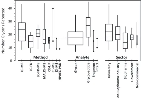
The number of glycan compositions reported by each lab- oratory ranged from 4 to 48. Most reports listed about the same number of glycan compositions for each of the two samples. Fig. 1 summarizes the number of unique glycan compositions reported for NISTmAb and/or mod-NISTmAb samples as a function of the laboratory’s analytical method, analyte, and organizational type. On average, more compo- sitions were reported by laboratories (1 using MS-based methods, (2 analyzing glycopeptides, and (3 that were uni- versity-based. However, the wide range in the number of compositions reported within most of the groups suggests that the technology is not the major determinant.
Fig. 2 summarizes the proportion of compositions that were reported with isomeric information as a function of identification method. On the average, laboratories that used exoglycosidases reported the greatest number of iso- meric information, followed by retention times, then by FD, then by MS/MS. Surprisingly, about one-fourth of the data sets that nominally used accurate mass (MS) identification identified some isomers.
Glycan Quantification—Laboratories were asked to report quantitative values for each glycoform as proportions relative to the sum of all glycoforms detected. Table II lists the con- sensus median abundances of glycan compositions and gly- FIG. 1.Number of unique glycan compositions reported, grouped by method, analyte, and sector.The boxes span the central 50% of reported values, 25% to 75%; the whiskers span the central 90%, 5% to 95%; the central line marks the median, 50%. Box widths are proportional to the number of reports. Groups within each category are presented in order of decreasing number of reports. Solid circles represent individual results within categories of fewer than six reports. The dotted line marks the median number of compositions reported in the 103 reports provided by 76 laboratories.
can structures on NISTmAb that were reported at least six times. The three compositions [h3n4f1], [h4n4f1] and [h5n4f1]
are the most commonly reported and the most abundant compositions; together, they account for more than 85% of the total signal intensity. Although the normalization factors (the sum of signals) for the different data sets are not based on the same compositions nor the same number of compo- sitions, the dominance of [h3n4f1] and [h4n4f1] ensures that the reported proportions are comparable across data sets.
However, the differences among the normalization factors are a source of variability. Other approaches such as normalizing to the most abundant few glycoforms could be pursued in future studies.
Laboratories were asked to report their results as percent abundances normalized such that they summed to 100% per sample.Supplemental Fig. S3shows a histogram of the sum of the unique glycan composition values for NISTmAb and mod-NISTmAb as reported by the laboratories (n⫽206, two samples per laboratory). Although results in most data sets summed to 100%, the sums ranged from 88% to 122%.
Some laboratories assigned percent abundances to uniden- tified glycans; the sum of these values was reported as one entry called “Unknown Glycans” and added to the abundance sum for that sample.
The number of replicate values per reported composition or structure per sample ranged from one to nine. The nature of these values ranged from purely technical replicates (multiple measurements of the same preparation) to process replicates (single measurements of multiple independent preparations).
In all cases where two or more replicate values were reported,
the values were summarized as their mean and standard deviation (SD).
Some data sets reported replicate values equal to zero, not detected (ND), and not quantified (NQ). These values can be ignored when there are no quantitative values in a set of replicates but cause numerical instability when there is at least one quantitative value in the set. Various options were explored for handling these situations in a uniform manner including: treating non-numerical results as zero, replacing zeros and non-numerical values with the data set’s minimum reported value (MRV, the smallest reported numerical value of a data set), or replacing them with the data set’s limit of reporting (LoR, the extrapolated smallest value of a data set).
Replacement with the LoR provided slightly smaller SDs than replacement by the MRV.Supplemental Fig. S4 shows the LoR for one set of results. Gray lines are traces of the unique non-zero values reported in each set of results, where the numbers are ordered by decreasing value. If the true amounts of the minor glycans are randomly distributed and all results reflect the same level of analytical effort, a best-fit line to the right-tail of the trace estimates the LoR for that set. As shown insupplemental Fig. S5, most of the LoRs agree well for MRVs above about 0.05%. Below this value, many of the sets con- tain a few values many-fold smaller than their LoR. This may reflect special interest in selected glycan components rather than reporting issues with the less-abundant glycans. The LoR values in the data set may be more representative of the ana- lytical sensitivity of a measurement system than is the MRV.
Derived Attribute Quantities in NISTmAb—Table III shows the degree of galactosylation, sialylation, fucosylation; levels of bisecting GlcNAc and high-mannose; and the sialic acid/
galactose ratio in NISTmAb. These values are estimated from the consensus median values of the glycan compositions. Cal- culations are based on previous works by Wuhrer (27) and are designed to reflect biosynthetic pathways (32) and, to some extent, enzyme activity. In addition, these glycosylation traits relate to differences in effector functions of monoclonal antibodies and circulation half-time for other therapeutic glyco- proteins.
All antennae were assumed to be available for galactosyla- tion by most galactosyltransferases. Antennae galactosylation may be a reasonable proxy for enzyme activity and may therefore reflect regulation of galactosylation process in a biological system. Thus, galactosylation levels were ex- pressed by calculating the number of galactosylated (occu- pied) antennae divided by the total number of antennae of the specific glycan. Only glycans identified with galactose resi- dues were included in the calculations. For biantennary gly- cans, the galactosylation levels are 0.0, 0.5, and 1.0 for 0, 1, and 2 galactoses, respectively. For triantennary glycans, the galactosylation levels are 0.0, 0.33, 0.67, and 1.0, reflecting the presence of 0, 1, 2, or 3 galactosylated antennae. For NISTmAb, the median degree of galactosylation is 36.2% with alpha-galactosylation at 3.8%, as shown in Table III.
FIG. 2.Proportion of glycan compositions reported as isomers.
The boxes span the central 50% of reported values, 25% to 75%; the whiskers span the central 90%, 5% to 95%; the central line marks the median, 50%. Box widths are proportional to the number of reports.
Categories are presented in order of increasing median proportion.
The dotted line marks the median proportion of compositions re- ported as isomers.
TABLEII
Community’s consensus abundances of glycans in NISTmAb PS 8670 reported by laboratories at least six times. Glycan compositions are arranged by decreasing number of values (N).Supplementary Table S2lists all glycan structures and names.Supplementary Table S3lists all
the community’s consensus values
TABLEII—continued
TABLEII—continued
n⫽number of values; consensus median⫽consensus median (50th percentile or 2nd quartile, expressed as percent of total composition) of the distribution of the reported results; MADE⫽median absolute deviation; Srep⫽a robust estimate of the expected repeatability, the median SD for glycan compositions with at least six results; CV⫽robust coefficient of variation (MADE/median); CVrep⫽(Srep/median).
For sialylation levels, the same principle is applied,i.e.the sialylation per antenna was calculated. For NISTmAb, NeuAc and NeuGc sialylation were observed at a medium value of 1.3% and 2.2%, respectively. The ratio of sialic acid per galactose was calculated as 0.1%. This value reflects the sialylation activity, i.e. whether available acceptor positions have been sialylated.
Monofucosylation was interpreted as core fucosylation.
NISTmAb had very high levels of core fucosylation (median of 104%; values exceeding 100% are artifacts of the variable normalization factors). The antenna fucosylation (manifested as difucosylation) at 0.38% was calculated separately be- cause the interaction between core and antenna fucosylation is assumed to be minimal.
Issues with Glycosylation Analysis Methods—Laboratories reported challenges in identifying and quantifying glycans.
Some laboratories reported their analysis at the composition level only and did not differentiate isomeric species; some laboratories analyzed at the glycan isomer level and had challenges in identifying co eluting or same mass species.
These issues are usually method dependent, as shown in supplemental Table 4. Some glycan structures were sup- ported by MS/MS and other structures were inferred from similar structures,e.g.triantennary structures. Consequently, some abundance values were assigned to triantennary struc- tures instead of bisecting glycans. The same laboratory ob- served a discrepancy for glycan G1FS N (NeuGc). Compared with quantitative data from subunit analysis, the glycopeptide abundance was higher than subunit abundance. Overall, the laboratory observed that glycopeptide abundances were in good accordance with the subunit data with slightly lower values for G0F and G1F in the glycopeptide analysis.
One laboratory analyzed protein fragments by LC MS that had masses up to 25 kDa. One or two nominal mass differ- ences were challenging to distinguish using their technique.
Another laboratory analyzed 2-AB glycans using HILIC LC FD with comparison to retention time of standards. It was difficult to distinguish glycans that coelute,e.g.G2 and Man6.
Moreover, the laboratory was unable to identify glycan peaks
present in the samples but absent in their lab-designated standard sample.
Supplemental Table S4 lists the advantages of certain methods as described by laboratories. Sialic acid specific derivatization of 2-AB or ethyl esterified glycans analyzed by LC FD or MALDI MS could confirm presence of terminal␣2 6 linked NeuGc in glycans.
Intact protein analysis could give the G0F/G1F, G1F/G2F, G0/G0F, G0F N/G0F, G2F⫹1aGal/G2F glycoforms present in the monoclonal antibodies. Due to the cleavage of glycopep- tides or glycans from the protein, analysis using these two analytes could not provide this specific information.
2-AB glycans analyzed by LC FD using glucose units and APTS labeled glycans analyzed by multiplexed capillary gel electrophoresis (xCGE) using migration time of standards and exoglycosidases could distinguish between isomers.
Additional Information on NISTmAb
Some laboratories performed unique analyses, resulting in additional information on the glycosylation of NISTmAb
Absolute Glycan Amounts—One laboratory determined ab- solute glycan amounts in the samples by employing isotopic dilution methods, using 13C-labeled N-glycans as internal standards followed by MALDI-TOF MS analysis. For example, the absolute amounts of three glycans in NISTmAb were reported to be:
G0Fa: (626.7⫾7.5) pmol per 100g NISTmAb G2F: (110.8⫾5.9) pmol per 100g NISTmAb G2: (19.0⫾3.8) pmol per 100g NISTmAb
Glycoforms in Intact Samples—One laboratory analyzed intact mAb samples using LC-MS and identified glycoforms on the two Fc portions that were analyzed. Example glyco- forms are G0/G0F, G0F/G1F, G1F/G2F, G0F-N/G0F, and G2F⫹1aGal/G2F. Abundance values for these glycoforms are shown insupplemental Table S3, bottom rows.
Unknown Modifications—One laboratory found an un- known delta mass of ⫹ 1856 Da at 0.40% abundance in NISTmAb by LC-MS. Another laboratory detected ⫹ 54 Da unidentified protein modification using1H-NMR and MS. The latter found a glycan present in⬍3% with no branching at the central-Mannose,i.e.there is only one arm present with a terminal NeuGc and a proximal Fuc.
Unglycosylated Forms—One laboratory used protein frag- ment analysis by C4-LC-MS and observed the unglycosylated form of the samples at 0.60% abundance in NISTmAb. An- other participant used glycopeptide analysis by C18-LC-MS and detected the unglycosylated form at 0.91% abundance in NISTmAb, as confirmed by high mass accuracy MS (⬍3 parts per million (ppmb) mass deviation).
aDefinition of naming convention of glycans is in the Experimental Procedures section.
bppm is a unit of mass measurement error (⌬m/z)/(m/z) * 1000000, expressed as unified atomic mass units (Daltons) divided by charge.
TABLEIII
Derived attribute quantities for NISTmAb PS 8670, estimated from the consensus median values of the glycan compositions
Features Number
of Labs 25% Median 75%
Galactosylation 32 31.78 36.21 43.30
alpha-Galactosylation 13 3.00 3.77 4.97
Sialylation 18 2.26 3.48 6.98
NeuAc sialylation 6 0.71 1.25 2.83
NeuGc sialylation 12 1.55 2.23 4.16
Core fucosylation only 37 92.36 103.95 118.23
Antenna fucosylation 3 0.25 0.38 0.73
Bisecting GlcNAc 7 1.49 2.17 3.83
High mannose 6 1.04 1.92 3.42
Sialic Acid/Galactose Ratio 0.07 0.10 0.16