Full Terms & Conditions of access and use can be found at
https://www.tandfonline.com/action/journalInformation?journalCode=ierm20
Expert Review of Clinical Immunology
ISSN: 1744-666X (Print) 1744-8409 (Online) Journal homepage: https://www.tandfonline.com/loi/ierm20
Advances in phototherapy for psoriasis and atopic dermatitis
Lajos Kemény, Emese Varga & Zoltan Novak
To cite this article: Lajos Kemény, Emese Varga & Zoltan Novak (2019) Advances in phototherapy for psoriasis and atopic dermatitis, Expert Review of Clinical Immunology, 15:11, 1205-1214, DOI:
10.1080/1744666X.2020.1672537
To link to this article: https://doi.org/10.1080/1744666X.2020.1672537
© 2019 The Author(s). Published by Informa UK Limited, trading as Taylor & Francis Group.
Published online: 01 Oct 2019.
Submit your article to this journal
Article views: 4100
View related articles
View Crossmark data
Citing articles: 4 View citing articles
REVIEW
Advances in phototherapy for psoriasis and atopic dermatitis
Lajos Kemény a,b,c, Emese Vargaaand Zoltan Novakd
aDepartment of Dermatology and Allergology, University of Szeged, Szeged, Hungary;bMTA-SZTE Dermatological Research Group, University of Szeged, Szeged, Hungary;cHCEMM-USZ Skin Research Group, Szeged, Hungary;dDepartment of Gynaecology, National Insitute of Oncology, Budapest, Hungary
ABSTRACT
Introduction: Phototherapy has long been used for the treatment of inflammatory skin diseases, such as psoriasis and atopic dermatitis. The most frequent treatment approach utilizes ultraviolet (UV) light, however, recently, different lasers and low-level light therapies (LLLT) emitting wavelengths in the spectrum of the visible light have also been tried for the treatment of inflammatory skin diseases with variable success.
Areas covered: This review provides an update on the different forms of phototherapy used for the treatment of psoriasis and atopic dermatitis. The proposed mechanism of action of the different phototherapeutical approaches are covered, including the immunosuppressive effect of UV light, the anti-inflammatory effect of vascular lasers and the LLLT induced photobiomodulation. The clinical efficacy of the different treatment options is also discussed.
Expert opinion: Based on the efficacy and safety, NB-UVB represents the gold standard for treating psoriasis and atopic dermatitis. The UVB excimer laser and excimer lamp might be the best option for clearing localized therapy-resistant lesions. Home UV phototherapy systems might promote treatment adherence and better compliance of the patients. Vascular lasers, IPLs and LLLT, however, can not currently be recommended for the treatment of inflammatory skin diseases because of the lack of well- controlled studies.
ARTICLE HISTORY Received 12 July 2019 Accepted 23 September 2019 KEYWORDS
Phototherapy; ultraviolet;
laser; apoptosis; cytokines;
immunosuppression;
psoriasis; atopic dermatitis
1. Introduction
Phototherapy is the use of ultraviolet (UV) radiation or visible light for the treatment of different diseases. The roots of photo- therapy can be traced back to 1500 BC when Hindus treated vitiligo, an autoimmune skin disorder, with photosensitizing plant extracts and subsequent sunlight exposure. For many cen- turies only natural sunlight (heliotherapy) was used for the treat- ment of different skin conditions, but even nowadays, it is still highly popular for psoriasis and atopic dermatitis in many geo- graphic areas in the World, especially in the Dead Sea [1]. As heliotherapy is only feasible in certain periods of the year with additional great dosing variables depending on the geographic locations, artificial light sources have been developed to emit selective wavelengths of the electromagnetic radiation.
Furthermore, the identification of photosensitizers from plant extracts with unique photochemical properties resulted in the development of the so-called photochemotherapeutic approaches, that were for a long time the most effective treatments for inflammatory skin diseases. Lasers and intense pulse light (IPL) are well established therapeutical tools to treat vascular congenital and acquired vascular lesions. As one of the hallmarks of skin inflammation is the erythema, targeting blood vessels with lasers or IPLs might be an alter- native option to treat inflammatory skin diseases as well.
Indeed, in the last decades vascular lasers have been shown to improve inflammatory skin diseases with variable success.
In recent years low-level light/laser treatments (LLLT) emitting low-intensity visible light were tried for psoriasis and atopic dermatitis, but their efficacy and the mechanism of action need further clarification.
When selecting the appropriate treatment for the patients, many different conditions, such as co-morbidities, age, and disease severity has to be considered [2,3]. Although there are many traditional agents and biological drugs for psoriasis and atopic dermatitis, phototherapeutical approaches are still widely utilized for the treatment of inflammatory skin diseases.
We performed a MEDLINE search via PubMed to review the recent advances in phototherapy for psoriasis and atopic der- matitis, and the most interesting papers based on the Expert’s selection were used for this review.
2. Phototherapeutical approaches
2.1. Common pathogenetic factors in immune-mediated skin diseases
Although psoriasis and atopic dermatitis are clinically, histolo- gically, and immunologically distinct diseases, both are char- acterized by activated T-cell infiltration in the skin that is followed by increased proliferation of keratinocytes resulting in the thickening of the skin. These T-cell mediated diseases differ in skin-infiltrating T-cell subpopulations [4]. In psoriasis, Th17, Th22 cells and type 3 innate lymphoid cells play a crucial
CONTACTLajos Kemény kemeny.lajos@med.u-szeged.hu Department of Dermatology and Allergology, University of Szeged, Szeged, Hungary EXPERT REVIEW OF CLINICAL IMMUNOLOGY
2019, VOL. 15, NO. 11, 1205–1214
https://doi.org/10.1080/1744666X.2020.1672537
© 2019 The Author(s). Published by Informa UK Limited, trading as Taylor & Francis Group.
This is an Open Access article distributed under the terms of the Creative Commons Attribution-NonCommercial-NoDerivatives License (http://creativecommons.org/licenses/by-nc-nd/4.0/), which permits non-commercial re-use, distribution, and reproduction in any medium, provided the original work is properly cited, and is not altered, transformed, or built upon in any way.
role in producing IL-17 and IL-22 and TNFα, whereas IL-23 is the key cytokine responsible for Th17 activation. Th1 cell- derived interferon-gamma (IFN-γ) is also elevated in psoriatic skin. In contrast, atopic dermatitis is characterized by the accumulation of Th2 cells, and their respective cytokine milieu IL-4, IL-13, and IL-31. In chronic atopic dermatitis lesions, Th1 cells-derived IFN-γ and Th22 cells-produced IL-22 might also have relevance [4].
2.2. UV-induced immunosuppression
Although there are Differences in the activated lymphocyte populations in psoriasis and atopic dermatitis, both topical (tacrolimus, pimecrolimus) and systemic (cyclosporine) immu- nosuppressive treatment approaches are highly effective for the treatment of these immune-mediated skin diseases. These immunosuppressive drugs exert their effect by inhibiting cal- cineurin, thereby suppressing activated T cells, independently on the subpopulation involved in the disease. The high effi- cacy of immunosuppressive treatments suggests that target- ing T cells might be a common therapeutical approach for psoriasis and atopic dermatitis.
Ultraviolet light from the sun or artificial light sources exert a profound immunosuppressive effect, that is mediated – at least partly – by inducing apoptotic cell death in activated T cells. The immunosuppressive effects of the UV light in the skin depend on many different variables, such as the wave- lengths, the radiation intensity and the treatment dose, the number of treatment sessions and on the optics of the human skin. Ultraviolet radiation can be classified to as UVB radiation, emitting wavelengths in between 280–320 nm, and UVA radia- tion that contains wavelengths in between 320–400 nm. In general, UVB light has a more profound immunosuppressive effect than UVA light. Psoralen plus UVA light (PUVA) is a form of photochemotherapy. Psoralens are small molecules, that enter cells and intercalate DNA. Esposure of cells to UVA light results in covalent binding of psoralens to the DNA, thereby blocks the proliferion of the cells, and modify their gene expression profile [5].
The most commonly used phototherapeutical devices that are in use today are broadband UVB (BB-UVB), narrowband UVB (NB- UVB), xenon chloride (XeCl) excimer laser and lamp, UVA1, and UVA for psoralen photochemotherapy (PUVA) (Table 1). The dif- ferent UV wavelengths have distinct photochemical and photo- biological properties, including differences in penetration depth and the molecules in the skin with which they interact (Table 2). As
a consequence, different UV light sources have unique properties regarding potency, side effects, and diseases in which they are effective. In general, the shorter UVB wavelengths have more powerful biological effects in vitro, however, in vivo the wave- lengths below 300 nm are absorbed in the upper layer of the skin, resulting in shorter penetration depth. Wavelengths below 300 nm are clinically less effective but more inflammatory.
Therefore, broadband UVB (BB-UVB) has been replaced by light sources filtering out wavelengths below 300 nm, such as in selec- tive UV phototherapy (SUP) devices, that emit wavelengths mainly in between 300–320 nm. Determination of the optimal wave- lengths in psoriasis resulted in the development of the narrow- band UVB (NB-UVB) lamp, that is nowadays the most frequently used treatment option for inflammatory skin diseases. The mono- chromatic 308 nm excimer laser and excimer lamp might be regarded as super narrow-band high-intensity UVB light sources, that are the best options for targeted phototherapy [6]. Recently UV light emitting diode (UV-LED) delivering high-intensity UV light in 300–312 nm wavelength, and a flat-type fluorescent UVB (F-UVB) lamps emitting UVB in the 311–313 nm range were devel- oped, that might provide novel options for targeted UV therapy [7,8]. The longer UV wavelengths of 320–400 nm (UVA) are biolo- gically less active and are used only for the treatment of mild atopic dermatitis. However, high-intensity UVA1, emitting wave- lengths in between 340–400 nm, is highly effective for acute atopic dermatitis.
Article Highlights
● UVB is a highly effective and safe treatment for psoriasis and atopic dermatitis
● Immunosuppression is the main mechanism of action of UV light
● Treatment with excimer lasers/lamps is an excellent new option for localized lesions
● Home UV phototherapy might represent a good alternative for office- based procedures
● The efficacy of vascular lasers, IPLs, and LLLT for psoriasis and atopic dermatitis are not convincing yet.
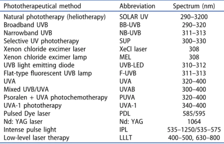
Table 1.Phototherapeutical approaches for the treatment of psoriasis and atopic dermatitis.
Phototherapeutical method Abbreviation Spectrum (nm) Natural phototherapy (heliotherapy) SOLAR UV 290–3200
Broadband UVB BB-UVB 290–320
Narrowband UVB NB-UVB 311–313
Selective UV phototherapy SUP 300–330
Xenon chloride excimer laser XeCl laser 308
Xenon chloride excimer lamp MEL 308
UVB light emitting diode UVB-LED 310–312
Flat-type fluorescent UVB lamp F-UVB 311–313
UVA UVA 320–400
Mixed UVB/UVA UVAB 300–400
Psoralen + UVA photochemotherapy PUVA 320–400
UVA-1 phototherapy UVA-1 340–400
Pulsed Dye laser PDL 585/595
Nd: YAG laser Nd: YAG 1064
Intense pulse light IPL 535–1250/535–575
Low-level laser therapy LLLT 400–500, 630–800
Table 2.Mechanism of action of UV phototherapy.
UV light-proposed effects References TARGET CELLS Apoptosis induction in T cells [28–31,39]
Apoptosis induction in keratinocytes [32]
Induction of regulatory T- cells [33–35,41–43]
Inhibition of NK cell function [37]
Improvement of the disturbed vasculature
[27]
Inhibition of Langerhans cells and macrophages
[21,36]
Inhibition of neutrophils [38]
INTRACELLULAR EFFECTS
DNA damage [21–24,26]
Cell membrane damage [21,24,25]
Isomerization of chromophores (urocanic acid)
[21]
Induction of cytoplasmic transcription factors
[21–24]
Modulation of cytokine production [39–43]
2.3. Targeting blood vessels
A hallmark of psoriatic skin is the remarkable transformation of the local microvascular system [9]. Pinpoint bleeding on the surface of psoriatic plaques after removing the scales (Aspitz sign) is a characteristic clinical phenomenon of psoriasis, that is due to regularly distributed dilated capillaries, and numerous, enlarged, superficialized dermal papillae containing dilated/tortuous capil- lary loops [10,11]. The increased dermal microvasculature facili- tates the trafficking of leukocytes from the circulation into the skin and therefore plays an essential role in the development and maintaining inflammation in psoriasis [12]. Therefore, inhibition of leukocyte trafficking via selective destruction of the dilated capillaries may be an effective therapeutic intervention in psoriasis.
Although the role of blood vessels and microcirculation in atopic dermatitis has not been extensively studied yet, similarly to other chronic inflammatory skin conditions, increased angio- genesis has also been suggested to be involved in the pathogen- esis of atopic dermatitis [13]. Therefore, targeting blood vessels might have therapeutic potential also in atopic dermatitis.
Congenital and acquired vascular lesions are treated with lasers with high efficacy. Among the various lasers used for treating vascular lesions, pulsed dye laser (PDL) emitting 585 nm or 595 nm monochromatic light, have the best effi- cacy in localized psoriasis [14,15], but also in localized lesions in atopic dermatitis [16]. There are also some reports on the use of Nd: YAG laser with emission in 1064 nm for psoriasis [17]. Intense pulsed light (IPL) devices produce flashes of light with high energy levels, with emitted wavelengths range of usually 400 nm to 1200 nm and the lower wavelengths can be eliminated by the various cut off filters, which usually range from 755–1000 nm. Therefore, targeting blood vessels by IPLs might also be a treatment option for psoriasis [18] but also for the reduction of facial redness in atopic dermatitis [19].
2.4. Photobiomodulation?
To avoid UV-induced increased risk of skin carcinogenesis, UV-free, low-intensity light sources have been developed for the treatment
of psoriasis and atopic dermatitis. These low-level light therapies include light emitting diodes (LEDs) operating in different wave- lengths usually in between 400–500 nm (blue light), and in 630–- 770 nm (red light), and up to 830 nm (near-infrared), but there are also many low-intensity lasers operating in these wavelengths, such as the Helium-Neon laser (He-Ne 632.5 nm). LLLT has been shown to exert a„photobiomodulation”effects, by targeting dif- ferent cells types and intracellular structures, but their exact mechanism of action is not known. Based on the high safety profile and the relatively low price of the LLLT devices, in recent years the LLLT treatments became popular for the treatment of many inflammatory skin conditions including psoriasis and atopic dermatitis.
3. Mechanism of action of phototherapy 3.1. UV phototherapy
UV phototherapy works through local rather than systemic effects, therefore without the side effect of profound systemic immunosuppression. This explains why it has still kept its place in the dermatologist’s arsenal even nowadays, in the era of the revolutionary novel biological agents.
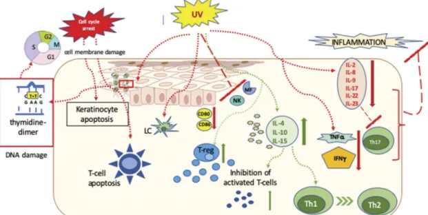
UV radiation (UVR) exerts a multitude of biological effects within the skin (Figure 1), different researches have already revealed many of them throughout the century, but still, it remains unclear which of these induce clearance of the lesions [20]. In general, the effects of UV radiation can be divided into two groups: immediate and delayed effects.
The immediate effects, such as membrane and DNA damage, induction of cytoplasmatic transcriptional factors and isomerization of chromophores (such as urocanic acid) have been known since 1967 [21], these induce instantly growth arrest and apoptosis [21].
There is a direct change of molecular structure in DNA due to photon absorption after UVB which inhibits the DNA transcription machinery and causes cell cycle arrest in human fibroblasts and epidermal cells (phototype I reaction) [22,23]. In the case of photo- chemotherapy, when the skin is treated with a photosensitizing
Figure 1.Mechanism of action of ultraviolet radiation in treating immune-mediated skin diseases.
EXPERT REVIEW OF CLINICAL IMMUNOLOGY 1207
drug (psoralen) subsequently followed by UVA radiation, the inter- action of photon-excited aromatic molecules with molecular oxy- gen generates reactive oxygen species, which cause many alterations in the cells, such as DNA damage, cell membrane damage via lipid peroxidation and interaction with signaling path- ways (phototype II reaction) [24–26].
The short-term effects are reversed 48 to 72 hours after irradiation, as the DNA repair starts within an hour after irradiaton, and the cells start then to proliferate again. UV radiation has the ability to reverse the skin’s disturbed vessel architecture as well, therefore in psoriatic lesions it normalizes the characteristical elongated capillary loops [27]. Delayed effects of UV radiation include the inhibition of both the adaptive and innate immune cells that results in immunosuppression.
Among the diverse effects on inflammatory cells, apoptosis induction in epidermal and dermal T cells seem to be the most important mechanism of action of UV radiation induced immunosuppression [28–31]. Although keratinocytes are more resistant to UV radiation compared to that of T cells, UVB induced apoptosis in keratinocytes also plays a key role in the clearance of psoriatic plaques [32]. Furthermore, UV-B and PUVA activate T regulatory (Treg) cells, and stimulation of Tregs results in the downregulation the overreactive immune response in psoriasis [33–35].
UV radiation affects antigen presenting cells as well. After 7 UV treatment sessions the number of Langerhans cells decreases by 90%, which alters the deviated antigen presenta- tion [21]. Furthermore, the photo-oxidative stress causes cytoskeleton damage in the remaining dendritic cells, which inhibits the further stimulation of T-cells. Cytokine secretion, as well as the number of macrophages, were also diminished by UV radiation [36]. Through the action of the reactive oxygen species UV light switches off the activity of neutrophils and inhibits the NK cell function as well [37,38].
These cellular effects are accompanied by altering the cytokine milieu, the suppression of inflammatory cytokines IL-2, IL-8, IL-9, IL-17, IL-22 and IL-23, TNF-a and IFN-g, and induction of the immunosuppressive cytokine IL-10 also con- tributes to the cessation of the inflammation [39–43].
3.2. Vascular lasers and IPLs
The effect of vascular lasers such as PDL or long pulsed Nd: YAG or IPLs is based on the selective absorption of light by oxyhemoglo- bin inducing photothermolysis of capillaries without causing damage to other skin structures [44]. The exact mechanism of action of PDL in psoriasis was investigated in detail by Racz et al.
[15]. They found that PDL treatment reduced the expression of VEGFR2 and VEGFR3 mRNA as early as 3 hours, whereas E-selectin expression was significantly reduced 24 hours after one single treatment. Interestingly, there was also a decreasing trend in the expression of IL-23p19 mRNA by PDL treatment. Long-term PDL treatment resulted in the decreased expression of TNF-αand IL- 23p19, and reduced β-defensin 2, keratin 17 and Bcl2. The decreased expression of the endothelial molecules VEGFR2 and E-selectin corresponds with the proposed primary target of the PDL. Furthermore, the decreased expression of VEGFR3 early after PDL treatment may also contribute to its efficacy in psoriasis.
These results suggest that selective targeting of blood vessels by PDL results in relevant biological changes in psoriatic plaques, calling attention to the role of blood vessels, as a previously less recognized target option in psoriasis.
3.3. Low-level light/laser therapy
The biological effect of low-intensity visible light/laser in the skin is not exactly clarified yet. The different wavelengths (400–800 nm), differences in intensity and penetration depth might result in contradictory data concerning the observed biological effects. In the blue range (400–500 nm), porphyrin-containing enzymes and flavoproteins are supposed to be the photoacceptors linking the mitochondrial respiratory chain to photostimulation [45–47]. In the visible-to-near-infrared spectral range, the cytochrome c oxidase might be the photoacceptor [48]. Red light at 660 nm exerts a so-called photobiostimulatory effect by producing oxygen radicals and inducing the proliferation of lymphocytes [49].
Liebmann investigated a panel of LEDs with distinct wavelengths ranging from near-UV to infrared (412–940 nm) in order to define wavelength-specific biological effects on cultured human skin cells [50]. They found that irradiation with wavelengths between 630 and 940 nm did not affect cell proliferation. Irradiation with wavelengths of 412, 419, and 426 nm at high fluences (66–100 J/
cm2) and 453 nm wavelength at very high fluences (4500 J/cm2) was cytotoxic for skin-derived endothelial cells as well as for keratinocytes. However, irradiation with nontoxic fluences reduced the proliferation of both endothelial cells and keratino- cytes by inducing differentiation [50]. These in vitro data might be relevant for LLLT therapy in patients with immune-mediated hyperproliferative skin diseases such as psoriasis or atopic dermatitis.
4. Clinical efficacy of phototherapy in psoriasis 4.1. UV light based treatments
4.1.1. Action spectrum of UV light in treating psoriasis: the traditional belief
UVB light (280–320 nm) is the most frequently used wave- length for the therapy of psoriasis, and there are a high num- ber of UV devices on the market. It is an important and clinically relevant question, which UV light sources are the most effective in treating psoriasis. Interestingly, there is only one study addressing this question, when Parrish and Jaenecke irradiated psoriatic plaques with different wave- lengths using a monochromator [51]. They found that wave- lengths below 300 nm were erythematogenic, but therapeutically not effective, whereas wavelength at 313 nm was less erythematogenic and at the same time was highly effective in improving the patients’skin lesions. These results served as the basis for the development of NB-UVB lamp, that is nowadays the most frequently used phototherapeutical option. Indeed, most of the clinical studies showed that NB- UVB produced a superior clinical and histopathological resolu- tion of moderate-to-severe psoriasis in patients compared with BB-UVB when used in suberythematogenic dosis [52].
However, in another randomized trial, the efficacies of the BB- UVB and NB-UVB light in treating psoriasis were compared,
and no difference was found between those treated with the two methods in terms of the median number of treatments to clear, proportions of patients achieving clearance and improvement in psoriasis severity scores [53]. These contra- dictory data suggest the necessity of the revision of the action spectrum of UVB for treating psoriasis published more than 35 years ago. The comparison of the therapeutical efficacies of the different UVB light sources in treating psoriasis is extre- mely difficult. The different variables (spectrum, irradiation dose, intesity, disease characteristics) will influence the ther- apeutical effect. Therefore, the general belief that NB-UVB is more effective than BB-UVB light might be questioned.
4.1.2. Action spectrum of UV light in treating psoriasis:
a new model
One of the major mechanisms of action of UVB light in the treatment of inflammatory dermatoses seems to be a cytotoxic effect on epidermal T cells, where the mechanism of cell death is most probably apoptosis [28]. Earlier, in a bilateral comparison study, NB-UVB cleared the psoriatic plaques more effectively and was a more potent inductor of T cell apoptosis than BB-UVB; therefore, the T cell apoptosis- inducing capacity of a UVB light source could be reflected by its clinical efficacy [29]. We also found that the Xenon chloride UVB laser was more effective in treating psoriasis and in indu- cing T cell apoptosis than NB-UVB [30]. Later, using polychro- matic UV light sources, the wavelength dependence of UVB light for the induction of T cell apoptosis was also determined.
The regression line of the action spectrum demonstrated a continuous decrease from 290 nm to 313 nm [31].
As the extent of T cell apoptosis induction and the penetration of UVB light into the dermis is wavelength-dependent, the wave- length spectrum of optimal dermato-phototherapy might depend
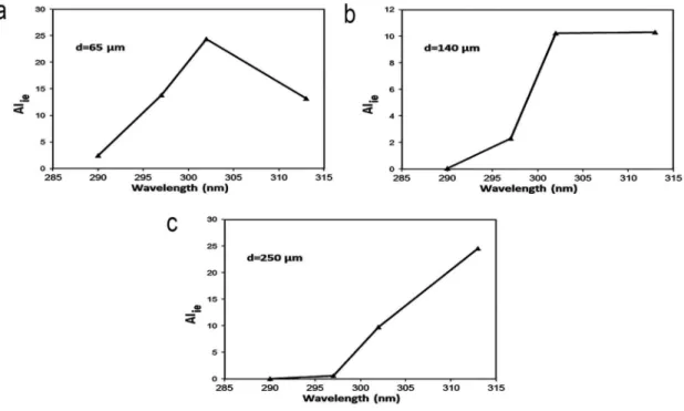
on the thickness of the skin lesion. We computed the theoretical action spectrum of UVB for the induction of intraepidermal T cell apoptosis. The formula created by Bruls et al. (1984) [54] was modified to compute the theoretical wavelength dependence of intraepidermal T cell apoptosis as a function of epidermal thick- ness. As the pathognomic T cells in psoriasis are localized along the dermo-epidermal junction and in the epidermis, we computed how the thickness of epidermis has to be transmitted by the UVB radiation to reach these intraepidermal The thickness of psoriatic human epidermis from the lower forearm is 180–360μm depend- ing on the extent of acanthosis [55]. In another work, the suprapa- pillar part of the stratum Malpighi seemed to be constantly 43μm thick, while the thickness of the rete pegs of the stratum Malpighi increased as the degree of acanthosis increased (160–420μm) [56].
In line with these observations, disease-mediating T cells are loca- lized at 65–460μm from the surface of the skin, depending on the degree of acanthosis. Using these data, we calculated the theore- tical extent of intraepidermal T cell apoptosis induction in the range 290–313 nm depending on the epidermal thickness of the irradiated skin. For T cells located at 65μm and 250μm from the surface, 302 nm and 313 nm seem to be the most efficient wave- lengths, respectively. For T cells localized from approximately 140μm from the skin surface, the wavelengths 302–313 nm are equally efficient in inducing apoptosis.Figure 2depicts these three theoretical action spectra.
The spectral distribution of a UVB light source influences the clinical results by having an effect on the apoptosis induction capacity and having an influence on the absorbance and trans- mittance of the light in the epidermis [57]. Although shorter wavelengths seem to induce T cell apoptosis more efficiently;
on the other hand longer wavelengths penetrate better into the epidermis. In our opinion, plaque thickness is to be con- sidered an important factor determining optimal phototherapy.
Figure 2.Action spectrum of ultraviolet B (UVB) to induce intraepidermal T cell apoptosis.
The efficacy of different UVB wavelengths to induce intraepidermal apoptosis in case of (a) thin epidermis (d = 65 um from the surface of the skin), (b) moderately acanthotic epidermis (d = 140 um from the surface of the skin), or (c) severily acanthotic epidermis (d = 250 um from the skin surface). AIie is the theoretical extent of intraepidermal T cell apoptosis induction computed by multiplying the extent of in vitro T cell apoptosis induction and the percentage of transmitted UVB light at each wavelength studied, plotted on a relative scale.
EXPERT REVIEW OF CLINICAL IMMUNOLOGY 1209
Gerber et al. (2003) [58] proposed a UVB excimer laser treat- ment using dosages that depend only on plaque erythema and ont he determination of epidermal thickness using ultrasound.
They found a correlation between the induction of erythema and the epidermal thickness of the plaque. With the introduc- tion of their phototherapeutic regimen, based on individual plaque thickness, the cumulative doses needed to achieve complete clearance were reduced by 40%.
Although UV induced T cell apoptosis is not the only mechanism underlying the efficacy of UVB phototherapy [59], we propose that plaque thickness is a major contributor to the success of UVB phototherapy. Based on our data, longer wave- length UVB phototherapy would be ideal for the treatment of thick psoriatic plaques, while short-wavelength UVB radiation would be optimal for the treatment of thin skin lesions. Further clinical studies are needed to address this question in a clinical setting.
4.1.3. UV phototherapy in the clinical practice
UV phototherapy represents a safe and highly effective treat- ment option for psoriasis. Nowadays, NB-UVB is the most com- monly used phototherapeutical approach and can be regarded as the gold-standard in phototherapy for moderate to severe psoriasis [60–62]. Although UV light was recently classified as a carcinogen, broad-band UVB and narrow-band UVB have not yet been linked to cutaneous carcinogenesis when treating psor- iasis. However, PUVA increases the risk for squamous cell carci- noma of the skin, especially after following 350 or more phototherapy sessions over a lifetime [63]. Therefore, PUVA is a less and less frequently used treatment option [60].
For mild psoriasis, affecting less than 10% of the body surface area, that do not respond well to conventional topical treatments, the targeted, localized therapy using XeCl excimer laser or the excimer lamp seems to be the best treatment approach [60].
These lasers and lamps emit monochromatic radiation with a wavelength of 308 nm, that is just in the middle of the antipsor- iatic spectrum. Moreover, using of excimer laser in treating thick, therapy-resistant lesions has the additional benefit of leaving the clinically uninvolved, healthy skin protected from UV, allowing high-intensity UV radiation to be delivered to the involved plaques that results in fast therapeutic response [6,64–66]. As the 308 nm excimer laser is expensive, and the maintenance costs are also high, cheaper monochromatic excimer lamps have been devel- oped. As there are no differences in the clinical efficacies in between the coherent 308 nm excimer lasers and the non- coherent 308 nm excimer lamps [67], in most of the clinics the excimer lamps are nowadays used for the treatment of localized psoriasis.
Office-based phototherapeutical approaches are highly effective, but the treatments are time-consuming and incon- venient. To overcome these difficulties, home phototherapeu- tical approaches represent a good alternative for the treatment of inflammatory skin diseases. Home UVB treat- ments include whole-body and portable handheld units for targeted phototherapy. Because of its excellent safety profile, NB-UVB is generally recommended for home phototherapy.
Great number of studies showed that home UVB photother- apy is safe and comparable efficacious as office-based treat- ment [68,69]. Although formal prescriptions are not required,
before starting a home-phototherapy regime, dermatologists should determine the dosing schedule, duration of treatment and how to address the potential side effects. Recently a lightweight, handheld UVB-LED has been developed for home phototherapy. This LED device with a smartphone appli- cation, a secure server with data storage, and a physician web portal might represent a highly precise modality for home phototherapy. Phototherapy administration guided by the smartphone application resulted in high patient’s compliance and satisfaction [70]. Theoretically, tanning beds could also represent a convenient way to obtain UV exposure when office phototherapy is not feasible. Indeed, about half of psor- iasis patients report using tanning beds, and most of them noted improvement [71]. However, the wavelengths of UVA and UVB irradiation from tanning beds are poorly defined;
they tend to emit primarily UVA irradiation. Therefore, recom- mending the use of tanning beds as a potential treatment should be carefully justified [69]. One of the physicians major fear of recommending self-administered home phototherapy is the overuse of the phototherapy units. With the improve- ment of knowledge of the physicians about home photother- apy and with careful patient selection, self-administered phototherapy could be a satisfactory option for psoriasis patients. Moreover, home phototherapy could also reduce the economic burden of health care institutes.
4.2. Vascular lasers and IPL
Although targeting blood vessels by PDL and Nd: YAG lasers, or with IPLs systems seem to be good alternatives for the 308 nm excimer lasers/lamps to treat localized psoriatic plaques, vascular lasers are not frequently used in the clinical practice to treat psoriasis due to their high cost and lack of reimbursement.
However, probably because of the insufficient well-controlled clinical data on the efficacy, the use of vascular lasers and IPLs are not mentioned for the treatment in the psoriasis guidelines.
However, clinial data suggest the efficacy for PDL, as several studies reported that psoriatic plaques were partly or completely cleared by PDL treatment [72–76]. However, PDL might serve an alternative option for difficult-to-treat areas, such as for palmo- plantar psoriasis [77], or nail psoriasis [78]. As treatment of iso- lated nail psoriasis is extremely difficult, new data on the efficacy of Nd: YAG laser or IPL in these special clinical conditions might be of clinical interest [17,18].
4.3. Low-level laser/light therapy (LLLT)
Recent attempts to use UV free devices for the treatment of inflammatory skin diseases resulted in the development of UV- free light sources emitting blue (400–500 nm) or red (600–- 800 nm) light. The clinical results using LLLT for psoriasis are, however, inconsistent. Blue light treatment of selected psor- iatic plaques three times weekly for four weeks did not result in any clinical improvement compared to untreated plaques [79]. On the other hand, in another study using higher irradia- tion dosages, blue as well as red light, three times weekly for four consecutive weeks, induced improvement of psoriasis [80]. This clinical improvement was identical for both light
sources. However, in this clinical study, there were no control, non-irradiated (or sham-irradiated) plaques, and 10% salicylic acid was allowed to use throughout the clinical trial. Therefore the interpretation of the results is difficult because just the removal of the scales by salicylic acid might result in a 30%
improvement of a single psoriatic lesion. On the other hand, in a prospective, randomized study, the UV-free blue light treat- ment improved the psoriatic plaques significantly compared to nontreated lesions [81]. Because of the safety of the UV-free phototherapy, this approach was designed for home treat- ment. Treatment barrier might be the lengths of the treat- ment, as irradiation of one single plaque in this study took 30 minutes, and the treatment was performed 5–7 times a week in the first four weeks, and three treatments for eight weeks. Although the safety is without any doubt is important in phototherapy, due to the conflicting results and the observed the low efficacy obtained with LLLT, further clinical trials are necessary before suggesting the use of LLLT for the treatment of psoriasis.
5. Clinical efficacy of phototherapy for atopic dermatitis
5.1. UV light based treatments
UV light based treatment is well documented and frequently used for mild to moderate atopic dermatitis. There are many treatment guidelines and systematic reviews on the use of the different UV light sources in atopic dermatitis [82–84]. Natural sunlight, BB-UVB, NB-UVB, UVB excimer laser, UVAB, UVA, UVA1, topical, and systemic psoralen plus UVA (PUVA) have all been shown to improve the clinical symptoms [85].
Although the comparison of the different therapeutical approaches is as difficult as in psoriasis, clinical trials suggest that NB-UVB is superior in clearing atopic dermatitis compared to BB-UVB or UVA1 [86,87].
Currently, the most commonly used or UV phototherapy in Europe is NB-UVB and UVA1, where NB-UVB may be preferred in chronic lesions and UVA1 for acute flares [84]. Although 308 nm UVB excimer laser might be a new treatment option for localized, therapy-resistant plaques [88], the recent European guideline does not recommend its use because of the limited number of reports [84]. All UV treatments, espe- cially PUVA, pose a long-term risk of development of skin cancer. Therefore careful evaluation is necessary before initiat- ing any UV phototherapy, especially PUVA treatment. Anyway, NB-UVB is generally the most commonly recommended phototherapeutical approach considering its low-risk profile, relative efficacy, and availability.
5.2. Vascular lasers and IPL
As there were some reports on the efficacy of vascular lasers in psoriasis, the efficacy of PDL was also tested in atopic derma- titis. In a pilot study, PDL treatment was effective in treating small areas of chronic localized eczema [16]. IPL treatment also reduced the facial redness caused by atopic dermatitis [19].
However, the data until now are sparse, therefore currently
PDL and IPL are not recommended for the treatment of atopic dermatitis [84].
5.3. Low-level laser/light therapy (LLLT)
As conventional UV irradiation bears the risk of developing skin cancer and promotes accelerated skin aging, attempts have been made to develop UV-free devices for the treatment of skin diseases. There are only a few reports on the efficacy of UV-free blue light irradiation in atopic dermatitis. Previously, blue light at wavelengths between 400 and 495 nm improved itch and skin lesions in patients with atopic dermatitis [89].
Full-body blue light irradiation was also effective in reducing the clinical symptoms of the patients [90]. However, in this clinical trial, the blue light was used as an „add on”therapy, patients could use topical corticosteroids as well. Therefore the results are not easy to interpret. The targeted, local UV- free blue light treatment was also found to improve the clinical symptoms in a small, randomized study [91].
Recently, a randomized controlled trial has been initiated using the full-body blue light device for the treatment of atopic dermatitis [92]. The results of this ongoing clinical study might answer the question whether blue light might be added to our therapeutical arsenal to treat atopic derma- titis. However, given the limited number and quality of reports, currently, the European guideline does not recom- mend LLLT for the treatment of atopic dermatitis [84].
6. Expert commentary
Based on the clinical observation that sunlight improves a great number of inflammatory skin conditions, different artificial UV light sources have been developed in the early 20th century for the treatment of skin diseases. Various attempts have been made to elucidate the exact mechanism of action and the clinically most adventurous wavelengths.
Initially, BB-UVB light sources were utilized that emit wave- lengths throughout the whole UVB spectrum. Action spectrum studies suggested that wavelengths between 304 and 313 nm were clinically most effective to clear psoriatic lesions [51].
These findings lead to the development of the NB-UVB light sources in the 1980s. We introduced the 308 nm excimer laser technology to treat mild to moderate psoriasis in 1996 [6]. This innovation opened a new way for the development of other targeted phototherapeutical approaches, such as for excimer lamps and UVB-LED devices. As office-based UV phototherapy is time-consuming, innovation in home UV phototherapy sys- tems implementing a smartphone application and web-based portal might promote adherence and better care for the patients [70].
Although UB phototherapy is a safe and cost-effective treat- ment for psoriasis and atopic dermatitis, previous data have shownd a decrease in the use of phototherapy [93]. However, a recent analysis based on the Healthcare Common Procedure Coding System (HCPCS) codes revealed that the overall volume of phototherapy services billed to Medicare from 2000 to 2015 increased annually by 5% in the US. The most commonly used phototherapy was the UVB comprising 77% of all phototherapy services. While there was a 9% decline in PUVA therapy, the
EXPERT REVIEW OF CLINICAL IMMUNOLOGY 1211
excimer laser services grew by 29% annually [94]. These data suggest that even in the era of highly effective biologics, UV phototherapy represent an important therapeutic tool in derma- tology. However, vascular lasers, IPLs, and LLLT cannot currently be recommended for the treatment of inflammatory skin dis- eases until more, well-controlled studies confirm their efficacy.
Funding
The authors were supported by the GINOP-2.3.2-15-2016-00015 research grant
Declaration of interest
The authors have no relevant affiliations or financial involvement with any organization or entity with a financial interest in or financial conflict with the subject matter or materials discussed in the manuscript. This includes employment, consultancies, honoraria, stock ownership or options, expert testimony, grants or patents received or pending, or royalties.
Reviewer disclosures
One peer reviewer has declared research, speaking and/or consulting support from Galderma, GSK/Stiefel, Almirall, Alvotech, Leo Pharma, BMS, Boehringer Ingelheim, Mylan, Celgene, Pfizer, Ortho Dermatology, Abbvie, Samsung, Janssen, Lilly, Menlo, Merck, Novartis, Regeneron, Sanofi, Novan, Qurient, National Biological Corporation, Caremark, Advance Medical, Sun Pharma, Suncare Research, Informa, UpToDate and National Psoriasis Foundation, and consultation through Guidepoint Global and Gerson Lehrman; the reviewer also declares financial interest in Causa Research, a company dedicated to enhancing patients’ adherence to treatment. Another reviewer declares research funds from Abbvie, Boehringer Ingelheim, Celgene, Eli Lilly, Incyte, Janssen/Johnson & Johnson, Leo Pharmaceuticals, Medimmune/Astra Zeneca, Novartis, Pfizer, Sciderm, Valeant, and ViDac, and consultancy for Allergan, Aqua, Boehringer-Ingelheim, LEO Pharma, Menlo, Mitsubishi, Promius and Theravance. Peer reviewers on this manuscript have no other relevant financial relationships or otherwise to disclose.
ORCID
Lajos Kemény http://orcid.org/0000-0002-2119-9501
References
Papers of special note have been highlighted as either of interest (•) or of considerable interest (••) to readers.
1. Matos TR, Ling TC, Sheth V. Ultraviolet B radiation therapy for psoriasis:
pursuing the optimal regime. Clin Dermatol.2016;34:587–593.
2. Kaushik SB, Lebwohl MG. Psoriasis: which therapy for which patient:
psoriasis comorbidities and preferred systemic agents. J Am Acad Dermatol.2019;80:27–40.
3. Kaushik SB, Lebwohl MG. Psoriasis: which therapy for which patient:
focus on special populations and chronic infections. J Am Acad Dermatol.2019;80:43–53.
4. Sabat R, Wolk K, Loyal L, et al. T cell pathology in skin inflammation.
Semin Immunopathol.2019;41:359–377.
5. Derheimer FA, Hicks JK, Paulsen MT, et al. Psoralen-induced DNA inter- strand cross-links block transcription and induce p53 in an ataxia-telangiectasia and rad3-related-dependent manner. Mol Pharmacol.2009;75:599–607.
6. Bonis B, Kemeny L, Dobozy A, et al. 308 nm UVB excimer laser for psoriasis. Lancet.1997;350:1522.
•• This is the first paper one use of excimer laser in dermatology.
7. Kemeny L, Csoma Z, Bagdi E, et al. Targeted phototherapy of plaque-type psoriasis using ultraviolet B-light-emitting diodes.
Br J Dermatol.2010;163:167–173.
8. Nishida E, Furuhashi T, Kato H, et al. Successful treatment of psoriasis vulgaris with targeted narrow-band ultraviolet B therapy using a new flat-type fluorescent lamp.
Photodermatol Photoimmunol Photomed. 2011;27:248–250.
9. Costa C, Incio J, Soares R. Angiogenesis and chronic inflammation:
cause or consequence? Angiogenesis.2007;10:149–166.
10. Archid R, Patzelt A, Lange-Asschenfeldt B, et al. Confocal laser-scanning microscopy of capillaries in normal and psoriatic skin. J Biomed Opt.2012;17:101511.
11. Nasca MR, Lacarrubba F, Caltabiano R, et al. Image gallery: repro- duction of the Auspitz sign by videodermatoscopy, confocal micro- scopy and horizontal histopathology. Br J Dermatol.2019;180:e178.
12. Heng MC, Allen SG, Haberfelde G, et al. Electron microscopic and immunocytochemical studies of the sequence of events in psoriatic plaque formation following tape-stripping. Br J Dermatol.
1991;125:548–556.
13. Varricchi G, Granata F, Loffredo S, et al. Angiogenesis and lymphan- giogenesis in inflammatory skin disorders. J Am Acad Dermatol.
2015;73:144–153.
14. Hacker SM, Rasmussen JE. The effect of flash lamp-pulsed dye laser on psoriasis. Arch Dermatol.1992;128:853–855.
15. Racz E, de Leeuw J, Baerveldt EM, et al. Cellular and molecular effects of pulsed dye laser and local narrow-band UVB therapy in psoriasis. Lasers Surg Med.2010;42:201–210.
16. Syed S, Weibel L, Kennedy H, et al. A pilot study showing pulsed-dye laser treatment improves localized areas of chronic atopic dermatitis. Clin Exp Dermatol.2008;33:243–248.
17. Kartal SP, Canpolat F, Gonul M, et al. Long-pulsed nd: YAG laser treatment for nail psoriasis. Dermatol Surg.2018;44:227–233.
18. Tawfik AA. Novel treatment of nail psoriasis using the intense pulsed light: a one-year follow-up study. Dermatol Surg.2014;40:763–768.
19. Oh SH, Bae BK, Kim TG, et al. Effective treatment of facial redness caused by atopic dermatitis using intense pulsed light systems.
Dermatol Surg.2010;36:475–482.
20. Schneider LA, Hinrichs R, Scharffetter-Kochanek K. Phototherapy and photochemotherapy. Clin Dermatol.2008;26:464–476.
21. Duthie MS, Kimber I, Norval M. The effects of ultraviolet radiation on the human immune system. Br J Dermatol.1999;140:995–1009.
22. Baden HP, Parrington JM, Delhanty JD, et al. DNA synthesis in normal and xeroderma pigmentosum fibroblasts following treat- ment with 8-methoxypsoralen and long wave ultraviolet light.
Biochim Biophys Acta.1972;262:247–255.
23. Walter JF, Kelsey WH, Voorhees JJ, et al. Psoralen plus black light inhibits epidermal DNA synthesis. Arch Dermatol.1973;107:861–865.
24. Wenk J, Brenneisen P, Meewes C, et al. UV-induced oxidative stress and photoaging. Curr Probl Dermatol.2001;29:83–94.
25. Kostovic K, Pasic A. Phototherapy of psoriasis: review and update.
Acta Dermatovenerol Croat.2004;12:42–50.
26. Gonzalez E. PUVA for psoriasis. Dermatol Clin.1995;13:851–866.
27. Staberg B. Psoriasis. Transcapillary and interstitial transport of plasma proteins, cutaneous blood, flow, and effect of phototherapy. Dan Med Bull.1985;32:295–308.
28. Schade N, Esser C, Krutmann J. Ultraviolet B radiation-induced immunosuppression: molecular mechanisms and cellular alterations. Photochem Photobiol Sci.2005;4:699–708.
•• This is the first report on the mechanism of UV-induced immunosuppression.
29. Ozawa M, Ferenczi K, Kikuchi T, et al. 312-nanometer ultraviolet B light (narrow-band UVB) induces apoptosis of T cells within psoriatic lesions. J Exp Med.1999;189:711–718.
30. Novak Z, Bonis B, Baltas E, et al. Xenon chloride ultraviolet B laser is more effective in treating psoriasis and in inducing T cell apoptosis than narrow-band ultraviolet B. J Photochem Photobiol B.
2002;67:32–38.
31. Novak Z, Berces A, Ronto G, et al. Efficacy of different UV-emitting light sources in the induction of T-cell apoptosis. Photochem Photobiol.2004;79:434–439.
32. Weatherhead SC, Farr PM, Jamieson D, et al. Keratinocyte apoptosis in epidermal remodeling and clearance of psoriasis induced by UV radiation. J Invest Dermatol.2011;131:1916–1926.
33. Kagen MH, McCormick TS, Cooper KD. Regulatory T cells in psoriasis. Ernst Schering Res Found.Workshop.2006; 193–209.
34. Aubin F, Mousson C. Ultraviolet light-induced regulatory (sup- pressor) T cells: an approach for promoting induction of opera- tional allograft tolerance? Transplantation.2004;77:S29–S31.
35. Kripke ML, Morison WL, Parrish JA. Systemic suppression of contact hypersensitivity in mice by psoralen plus UVA radiation (PUVA). J Invest Dermatol.1983;81:87–92.
36. Sethi G, Sodhi A. Role of p38 mitogen-activated protein kinase and caspases in UV-B-induced apoptosis of murine peritoneal macrophages. Photochem Photobiol.2004;79:48–54.
37. Weitzen ML, Bonavida B. Mechanism of inhibition of human natural killer activity by ultraviolet radiation. J Immunol.
1984;133:3128–3132.
38. Kasahara S, Aizawa K, Okamiya M, et al. UVB irradiation suppresses cytokine production and innate cellular immune functions in mice.
Cytokine.2001;14:104–111.
39. Krueger JG, Wolfe JT, Nabeya RT, et al. Successful ultraviolet B treatment of psoriasis is accompanied by a reversal of keratino- cyte pathology and by selective depletion of intraepidermal T cells.
J Exp Med.1995;182:2057–2068.
40. Teunissen MB, Piskin G, Di Nuzzo S, et al. Ultraviolet B radiation induces a transient appearance of IL-4+ neutrophils, which support the development of Th2 responses. J Immunol.
2002;168:3732–3739.
41. Singh TP, Schon MP, Wallbrecht K, et al. 8-methoxypsoralen plus ultraviolet A therapy acts via inhibition of the IL-23/Th17 axis and induction of Foxp3+ regulatory T cells involving CTLA4 signaling in a psoriasis-like skin disorder. J Immunol.2010;184:7257–7267.
42. Singh TP, Schon MP, Wallbrecht K, et al. 8-Methoxypsoralen plus UVA treatment increases the proportion of CLA+ CD25+ CD4+
T cells in lymph nodes of K5.hTGFbeta1 transgenic mice. Exp Dermatol.2012;21:228–230.
43. Schweintzger N, Gruber-Wackernagel A, Reginato E, et al.
Levels and function of regulatory T cells in patients with poly- morphic light eruption: relation to photohardening. Br J Dermatol.2015;173:519–526.
44. Anderson RR, Parrish JA. Selective photothermolysis: precise micro- surgery by selective absorption of pulsed radiation. Science.
1983;220:524–527.
45. Hockberger PE, Skimina TA, Centonze VE, et al. Activation of flavin-containing oxidases underlies light-induced production of H2O2 in mammalian cells. Proc Natl Acad Sci USA.
1999;96:6255–6260.
46. Ohara M, Fujikura T, Fujiwara H. Augmentation of the inhibitory effect of blue light on the growth of B16 melanoma cells by riboflavin. Int J Oncol.2003;22:1291–1295.
47. Lewis JB, Wataha JC, Messer RL, et al. Blue light differentially alters cellular redox properties. J Biomed Mater Res B Appl Biomater.
2005;72:223–229.
48. Karu TI, Afanas’eva NI. Cytochrome c oxidase as the primary photo- acceptor upon laser exposure of cultured cells to visible and near IR-range light. Dokl Akad Nauk.1995;342:693–695.
49. Stadler I, Evans R, Kolb B, et al. In vitro effects of low-level laser irradiation at 660 nm on peripheral blood lymphocytes. Lasers Surg Med.2000;27:255–261.
50. Liebmann J, Born M, Kolb-Bachofen V. Blue-light irradiation regu- lates proliferation and differentiation in human skin cells. J Invest Dermatol.2010;130:259–269.
51. Parrish JA, Jaenicke KF. Action spectrum for phototherapy of psoriasis. J Invest Dermatol.1981;76:359–362.
•• This paper served the basis for the development of NB-UVB therapy.
52. Coven TR, Burack LH, Gilleaudeau R, et al. Narrowband UV-B produces superior clinical and histopathological resolution of moderate-to-severe psoriasis in patients compared with broad- band UV-B. Arch Dermatol.1997;133:1514–1522.
53. Kirke SM, Lowder S, Lloyd JJ, et al. A randomized comparison of selective broadband UVB and narrowband UVB in the treatment of psoriasis. J Invest Dermatol.2007;127:1641–1646.
54. Bruls WA, Slaper H, van der Leun JC, et al. Transmission of human epidermis and stratum corneum as a function of thickness in the ultraviolet and visible wavelengths. Photochem Photobiol.
1984;40:485–494.
55. Welzel J, Bruhns M, Wolff HH. Optical coherence tomography in contact dermatitis and psoriasis. Arch Dermatol Res.
2003;295:50–55.
56. Chapman DM, Ross JB. Objective measurement of three epidermal parameters in psoriasis vulgaris and in dermatopathology in general. Br J Dermatol.1988;119:333–343.
57. Anderson RR, Parrish JA. The optics of human skin. J Invest Dermatol.1981;77:13–19.
58. Gerber W, Arheilger B, Ha TA, et al. Ultraviolet B 308-nm excimer laser treatment of psoriasis: a new phototherapeutic approach. Br J Dermatol.2003;149:1250–1258.
59. Nghiem DX, Kazimi N, Mitchell DL, et al. Mechanisms underlying the suppression of established immune responses by ultraviolet radiation. J Invest Dermatol.2002;119:600–608.
60. Pathirana D, Ormerod AD, Saiag P, et al. European S3-guidelines on the systemic treatment of psoriasis vulgaris. J Eur Acad Dermatol Venereol.2009;23(Suppl 2):1–70.
• European S3-guideline on the use of phototherapy in psoriasis.
61. Moseley H, Allan D, Amatiello H, et al. Guidelines on the measurement of ultraviolet radiation levels in ultraviolet phototherapy: report issued by the British Association of Dermatologists and British Photodermatology Group 2015. Br J Dermatol.2015;173:333–350.
62. Zhang P, Wu MX. A clinical review of phototherapy for psoriasis.
Lasers Med Sci.2018;33:173–180.
63. Hofbauer G. Phototherapy and carcinogenesis. Hautarzt.
2013;64:349–353.
64. Asawanonda P, Anderson RR, Chang Y, et al. 308-nm excimer laser for the treatment of psoriasis: a dose-response study. Arch Dermatol.2000;136:619–624.
65. Kemeny L, Bonis B, Dobozy A, et al. 308-nm excimer laser therapy for psoriasis. Arch Dermatol.2001;137:95–96.
66. Farkas A, Kemeny L. Applications of the 308-nm excimers lasers in dermatology. Laser Phys.2006;16:876–883.
67. Kollner K, Wimmershoff MB, Hintz C, et al. Comparison of the 308-nm excimer laser and a 308-nm excimer lamp with 311-nm narrowband ultraviolet B in the treatment of psoriasis. Br J Dermatol.2005;152:750–754.
68. Hum M, Kalia S, Gniadecki R. Prescribing home narrowband UVB phototherapy: a review of current approaches. J Cutan Med Surg.
2019;23:91–96.
69. Franken SM, Vierstra CL, Rustemeyer T. Improving access to home phototherapy for patients with psoriasis: current challenges and future prospects. Psoriasis (Auckl).2016;6:55–64.
70. Cline A, Unrue EL, Collins A, et al. Adherence to a novel home photo- therapy system with integrated features. Dermatol Online J.2019;25 (3).
71. Anderson KL, Huang KE, Huang WW, et al. Dermatology residents are prescribing tanning bed treatment. Dermatol Online J.2016;22 (7).
72. Katugampola GA, Rees AM, Lanigan SW. Laser treatment of psoriasis. Br J Dermatol.1995;133:909–913.
73. Ros AM, Garden JM, Bakus AD, et al. Psoriasis response to the pulsed dye laser. Lasers Surg Med.1996;19:331–335.
74. Hern S, Allen MH, Sousa AR, et al. Immunohistochemical evaluation of psoriatic plaques following selective photothermolysis of the superficial capillaries. Br J Dermatol.2001;145:45–53.
75. Zelickson BD, Mehregan DA, Wendelschfer-Crabb G, et al. Clinical and histologic evaluation of psoriatic plaques treated with a flashlamp pulsed dye laser. J Am Acad Dermatol.1996;35:64–68.
76. Erceg A, Bovenschen HJ, van de Kerkhof PC, et al. Efficacy of the pulsed dye laser in the treatment of localized recalcitrant plaque psoriasis: a comparative study. Br J Dermatol.2006;155:110–114.
• First controlled study on the efficacy of pulsed dye laser for psoriasis.
77. de Leeuw J, Tank B, Bjerring PJ, et al. Concomitant treatment of psoriasis of the hands and feet with pulsed dye laser and topical EXPERT REVIEW OF CLINICAL IMMUNOLOGY 1213
calcipotriol, salicylic acid, or both: a prospective open study in 41 patients. J Am Acad Dermatol.2006;54:266–271.
78. Youssef NY, Saleh HM, Abdallah MA. Pulsed dye laser in the treat- ment of psoriatic nails: a controlled study. J Eur Acad Dermatol Venereol.2017;31:e49–e50.
79. Maari C, Viau G, Bissonnette R. Repeated exposure to blue light does not improve psoriasis. J Am Acad Dermatol.2003;49:55–58.
80. Kleinpenning MM, Otero ME, van Erp PE, et al. Efficacy of blue light vs. red light in the treatment of psoriasis: a double-blind, randomized comparative study. J Eur Acad Dermatol Venereol.
2012;26:219–225.
81. Pfaff S, Liebmann J, Born M, et al. Prospective randomized long-term study on the efficacy and safety of UV-free blue light for treating mild psoriasis vulgaris. Dermatology.2015;231:24–34.
82. Sidbury R, Davis DM, Cohen DE, et al. Guidelines of care for the management of atopic dermatitis: section 3. Management and treatment with phototherapy and systemic agents. J Am Acad Dermatol.2014;71:327–349.
83. Boguniewicz M, Alexis AF, Beck LA, et al. Expert perspectives on management of moderate-to-severe atopic dermatitis:
a multidisciplinary consensus addressing current and emerging therapies. J Allergy Clin Immunol Pract.2017;5:1519–1531.
84. Wollenberg A, Barbarot S, Bieber T, et al. Consensus-based European guidelines for treatment of atopic eczema (atopic dermatitis) in adults and children: part I. J Eur Acad Dermatol Venereol.2018;32:657–682.
85. Patrizi A, Raone B, Ravaioli GM. Management of atopic dermatitis:
safety and efficacy of phototherapy. Clin Cosmet Investig Dermatol.
2015;8:511–520.
86. Reynolds NJ, Franklin V, Gray JC, et al. Narrow-band ultraviolet B and broad-band ultraviolet A phototherapy in adult atopic eczema: a randomised controlled trial. Lancet.
2001;357:2012–2016.
87. Legat FJ, Hofer A, Brabek E, et al. Narrowband UV-B vs medium-dose UV-A1 phototherapy in chronic atopic dermatitis.
Arch Dermatol.2003;139:223–224.
88. Baltas E, Csoma Z, Bodai L, et al. Treatment of atopic derma- titis with the xenon chloride excimer laser. J Eur Acad Dermatol Venereol.2006;20:657–660.
89. Morita H, Kohno J, Hori M, et al. Clinical application of low reactive level laser therapy (LLLT) for atopic dermatitis. Keio J Med. 1993;42:174–176.
90. Becker D, Langer E, Seemann M, et al. Clinical efficacy of blue light full body irradiation as treatment option for severe atopic dermatitis. PLoS ONE.2011;6:e20566.
91. Keemss K, Pfaff SC, Born M, et al. Prospective, randomized study on the efficacy and safety of local UV-free blue light treatment of eczema. Dermatology.2016;232:496–502.
92. Kromer C, Nuhnen VP, Pfutzner W, et al. Treatment of atopic dermatitis using a full-body blue light device (AD-Blue): protocol of a randomized controlled trial. JMIR Res Protoc.2019;8:e11911.
93. Housman TS, Rohrback JM, Fleischer AB, et al. Phototherapy utiliza- tion for psoriasis is declining in the United States. J Am Acad Dermatol.2002;46:557–559.
94. Tan SY, Buzney E, Mostaghimi A. Trends in phototherapy utilization among Medicare beneficiaries in the United States, 2000 to 2015.
J Am Acad Dermatol.2018;79:672–679.