For Peer Review. Do not distribute. Destroy after use.
1
Rac GTPase activating protein ARHGAP25 regulates leukocyte
1
transendothelial migration in mice
2
3 4
Running title: ARHGAP25 regulates leukocyte migration 5
6 7
Roland Csépányi-Kömi*,†,#, Éva Wisniewski*,#, Balázs Bartos*, Petra Lévai*, Tamás Németh*, 8
Bernadett Balázs*, Angela R. M. Kurz†, Susanne Bierschenk†, Markus Sperandio†,§, Erzsébet 9
Ligeti*,§
10 11
* Department of Physiology, Semmelweis University, Budapest, Hungary 12
† Walter Brendel Center of Experimental Medicine, Ludwig-Maximilians Universität, 13
Munich, Germany 14
#,§ these authors contributed equally 15
16 17 18 19
Corresponding author: Professor Erzsébet Ligeti 20
Department of Physiology, Semmelweis University 21
1094 Budapest, Tűzoltó u. 37-47., Hungary 22
Phone: +361 459 1500 ext. 60457; Fax: +361 266 7480 23
Email: ligeti.erzsebet@med.semmelweis-univ.hu 24
For Peer Review. Do not distribute. Destroy after use.
2 Abstract
25
ARHGAP25 is a Rac-specific GTPase activating protein that is expressed primarily in 26
hematopoietic cells. The involvement of ARHGAP25 in regulating the recruitment of 27
leukocytes to inflammatory sites was investigated in genetically modified mice. Using 28
intravital microscopy we show that Arhgap25-deficiency affects all steps of leukocyte 29
recruitment with a predominant enhancement of transendothelial migration of neutrophilic 30
granulocytes. Increased transmigration of Arhgap25-deficient leukocytes is demonstrated in 31
inflamed cremaster muscle venules, in a peritonitis model, and in an in vitro chemotaxis 32
assay. Using bone marrow chimeric mice lacking ARHGAP25 in the hematopoietic 33
compartment, we show that enhanced migration in the absence of ARHGAP25 is due to 34
defective leukocyte function. In search for potential mechanisms of ARHGAP25-regulated 35
migration of neutrophils, we detected an increase in the amount of active, GTP-bound Rac 36
and Rac-dependent cytoskeletal changes in the absence of ARHGAP25 suggesting a critical 37
role of ARHGAP25 in counterbalancing the Rac-activating effect of nucleotide exchange 38
factors. Taken together, using Arhgap25-deficient mice we identified ARHGAP25 as a 39
relevant negative regulator of leukocyte transendothelial migration.
40
For Peer Review. Do not distribute. Destroy after use.
3 Introduction
41
In inflammation, neutrophil recruitment to sites of injury is essential for the fast and effective 42
elimination of injurious agents. The first step of recruitment consists in the activation of 43
vascular endothelial cells, which leads to increased expression of several cell surface 44
molecules including selectins and integrin ligands (1, 2). These molecules are recognized by 45
circulating leukocytes enabling the stepwise recruitment and extravasation of leukocytes into 46
inflamed tissue. Capture to the inflamed endothelium is followed by rolling of leukocytes 47
along the endothelium. Both capture and rolling are mediated by endothelial selectins and 48
leukocyte expressed selectin ligands (3). During rolling, leukocytes get into intimate contact 49
with the endothelial surface which enables binding of endothelium-expressed chemokines to 50
their respective ligand on the leukocyte surface triggering firm arrest of leukocyte on the 51
endothelium. Thereafter, leukocytes begin to crawl along the vessel wall searching for an 52
appropriate exit point for transmigration into tissue (diapedesis) (2, 4, 5). Extravasated 53
leukocytes are directed by chemotactic agents to the pathogens to be eliminated (6). All these 54
different types of movements require a precise spatial and temporal organization of the actin 55
cytoskeleton (7-9). Although our knowledge on the involved receptors and signaling 56
pathways has increased tremendously in the last decade (10), differences in the molecular 57
organization of the actin cytoskeleton underlying the different types of movements are still 58
poorly understood.
59
Members of the Rac/Rho subfamily of small GTP-binding proteins are key regulators 60
of the actin cytoskeleton (11). Their prevalence in the active, GTP-bound state depends on the 61
balance between the three major regulatory proteins: guanine nucleotide exchange factors 62
(GEFs) that promote the active state, GTPase activating proteins (GAPs) that counteract it, 63
and guanine nucleotide dissociation inhibitors (GDI) that conserve the inactive state (12, 13).
64
In case of the Rac/Rho subfamily, the potential number of GEFs and GAPs expressed in a 65
For Peer Review. Do not distribute. Destroy after use.
4
specific cell is especially high (14). The majority of these GEFs and GAPs are large proteins 66
composed of several effector, interactive and regulatory domains that suggest multiple 67
functions (13, 14). In neutrophils, a specific involvement of certain GEFs has been 68
investigated for different neutrophil effector functions including chemotaxis and adhesion 69
(15-17). In contrast, similar data on potentially interacting GAPs are still scarce (18-20).
70
In a recent study, we have shown that ARHGAP25 is a Rac-specific GAP expressed 71
primarily in hematopoietic cells (21). We also demonstrated that ARHGAP25 serves as a 72
negative regulator of phagocytosis and related superoxide production (21, 22). The aim of the 73
present study was to reveal the role of ARHGAP25 in the complex process of leukocyte 74
recruitment during inflammation. We provide the first detailed description of the Arhgap25-/- 75
mice, and show that loss of ARHGAP25 affects several steps along the recruitment cascade 76
leading to a proinflammatory phenotype with elevated transmigration of neutrophils into 77
inflamed tissue which is accompanied by increased Rac activity in neutrophils.
78
For Peer Review. Do not distribute. Destroy after use.
5 Materials and Methods
79
Antibodies and reagents 80
Anti-CD11b-PE and anti-Ly-6G-Pacific Blue were purchased from BioLegend, rat IgG2bκ- 81
PE isotype control and anti-CD18-FITC from BD Biosciences, rat IgG2a-APC isotype 82
control, anti-CD11a-APC, anti-human Fcγ-biotin, Streptavidin-PECy5 and rat IgG2a-FITC 83
isotype control from eBioscience, anti-CXCR2-APC, recombinant murine (rm) TNFα, rmE- 84
selectin/CD62E-Fc chimera and rmICAM-1/CD54-Fc chimera from R&D Systems, 85
rmKC/CXCL-1 and rmCXCL12 from PreproTech, Ly-6G-PerCP-Cy5.5 and CD11b-PE from 86
BD Pharmingen, mouse anti-Rac antibody from BD Transduction Laboratories, 87
paraformaldehyde from Sigma-Aldrich. Anti-human ARHGAP25 polyclonal antibody was 88
prepared as described previously.(21) Cross-reactivity with mouse ARHGAP25 was tested 89
using the lysate of COS-7 cells transfected with human ARHGAP25-V5 and mouse Arhgap25- 90
V5 constructs (see Fig. S1 for details). All other reagents were of research grade.
91 92
Mice 93
The Arhgap25-/- mouse strain used for this research project was created from ES cell clone 94
(EPD0085_1_C10) obtained from the NCRR-NIH supported KOMP Repository 95
(www.komp.org) and generated by the CSD consortium for the NIH funded Knockout Mouse 96
Project (KOMP). Methods used on the CSD targeted alleles have been published in (23).
97
Arhgap25-/- mice had a C57BL/6 genetic background and were maintained in a homozygous 98
breeding colony. Genotyping was performed according to KOMP’s instructions using the 99
following primers: Common-loxP-F: 5’-GAGATGGCGCAACGCAATTAAT-3’; CSD- 100
Arhgap25-SR1: 5’- GCATGAGGCAGCTGTTCTTAGTTACC-3’; CSD-Arhgap25-GF4: 5’- 101
TGCACACGGTGGCATCTCTACTAAAG-3’. Analysis of blood parameters was carried out 102
with a haemocytometer. To reveal differences in body weight between wild type and 103
For Peer Review. Do not distribute. Destroy after use.
6
Arhgap25-/- mice, 5-week-old animals (3 males/genotype and 2 females/genotype) were 104
weighed for 14 weeks. Arhgap25-/- and control wild-type bone marrow chimeras were 105
generated using bone marrow cells from adult donors as described previously (24, 25).
106
Arhgap25-/- bone marrow cell suspensions were injected intravenously into lethally irradiated 107
(11.5-Gy) recipients carrying the CD45.1 allele on the C57BL/6 genetic background. An 108
equal number of control chimeras were also generated using Arhgap25-expressing 109
(Arhgap25+/+) bone marrow cells and will be referred to as wild-type chimeras. Efficiency of 110
repopulation of the hematopoietic compartment by donor-derived cells was more than 98%, 111
tested 4 weeks after transplantation by flow cytometry: we tested the expression of CD45.2 112
(donor) allele in the granulocyte gate determined by Ly-6G-staining, as described previously 113
(24, 25) (data not shown). Bone marrow chimeras were used 4-8 weeks after transplantation.
114
Mouse strain carrying the CD45.1 allele on the C57BL/6 genetic background (B6.SJL-Ptprca) 115
was purchased from The Jackson Laboratory (Bar Harbor, ME). Mice were kept in 116
individually sterile ventilated cages (Tecniplast, Buguggiate, Italy) in a conventional facility.
117
Age and gender-matched animals were used for all the experiments. Animal experiments were 118
approved by the Regierung von Oberbayern, Germany, AZ 55.2.1.54-2532-76-12, and by the 119
Governmental Office of Pest County, Hungary (22.1/S321/3/2011).
120 121
Intravital microscopy of the mouse cremaster muscle 122
Mice were pretreated with intrascrotal injection of 500 ng rmTNFα per mice. After 2 h, mice 123
were anesthetized and trachea and carotid artery were cannulated. Scrotum was opened, the 124
cremaster muscle exteriorized, spread over a cover glass and superfused with 35 °C 125
bicarbonate-buffered saline as described (26). Parameters of rolling, adhesion and crawling 126
were determined using an Olympus BX51WI intravital microscope equipped with a saline 127
immersion objective (40/0.8 NA, Olympus) and a CCD camera (model CF8/1, Kappa). All 128
For Peer Review. Do not distribute. Destroy after use.
7
scenes were recorded by the Virtual Dub software for later offline analysis. Systemic blood 129
samples (~ 50 µL) were collected through the carotid artery catheter before and during the 130
experiment and analysed using a haemocytometer. The offline analysis of venular diameter 131
and vessel segment length of postcapillary venules (between 20-40 µm in diameter) was 132
carried out with Fiji software (27). Leukocyte rolling flux fraction was calculated from the 133
number of rolling cells that crossed a perpendicular line through a given vessel within 1 min 134
in relation to the total number of circulating leukocytes (28). Velocities of rolling and 135
crawling were measured using MTrackJ plugin of Fiji software. Other experimental 136
parameters (centerline blood flow velocity, shear rate, systemic cell counts) are shown in 137
Table SI.
138 139
TNFα-induced peritonitis model 140
Mice were treated with intraperitoneal injection of 5 µg rmTNFα in a final volume of 100 µL.
141
Three hours after treatment, mice were sacrificed and peritoneal cavity was washed with 5 mL 142
ice-cold PBS supplemented with 20 mM HEPES and 10 mM EDTA. Ly-6G+ infiltrated cells 143
were analysed with BD FACSCalibur device. Cell counts were determined using Flow-Count 144
Fluorospheres (Beckman Coulter).
145 146
Histology 147
Three hours after intrascrotal injection of rmTNFα (500 ng in 200 µL/mouse) or sterile PBS 148
(200 µL/mouse), cremaster muscles were exteriorized, mounted on adhesive slides 149
(Superfrost, Thermo Scientific) and fixed in 4% (w/v) paraformaldehyde for at least 48 h at 150
4°C. Then samples were washed 3x5 min in 0.1 M Phosphate buffer (0.1 M NaH2PO4, 0.1 M 151
Na2HPO4 mixed in 81:19 ratio, pH 7.4) supplemented with 5% (v/v) ethanol and stained with 152
Giemsa’s azure eosin methylene blue solution (Merck) for 4 min. After a rinse with water, 153
For Peer Review. Do not distribute. Destroy after use.
8
slices were differentiated with 0.03% (v/v) acetic acid for 10 min, and immersed in ascending 154
alcohol series from 70% (v/v) to absolute alcohol, in each for 3 min. Draining was carried out 155
with xylol (2x5 min), followed by sealing with rectangular coverslips using Eukitt (Sigma- 156
Aldrich). Whole mounts were analyzed with a Zeiss microscope equipped with an oil 157
immersion objective (100x/1.4 NA, Zeiss). Whole mounts of bone marrow chimeras were 158
analyzed with a Leica DMI 6000 B microscope equipped with an oil immersion objective 159
(63x/1.25 NA, Leica).
160
After 3 hours of TNFα challenge peritoneal tissue samples were taken and fixed in 4%
161
(w/v) phosphate-buffered paraformaldehyde for 48 hours. The tissue samples were paraffin- 162
processed, embedded, and 4 µm sections cut with a Microm HM340E rotary microtome 163
(Thermo Fisher Scientific). Cut sections were then used for hematoxylin and eosin (H&E) 164
staining. Representative pictures were captured with a Nikon ECLIPSE Ni microscope 165
equipped with 10x/0.30 NA and 40x/0.75 NA dry objectives (Nikon) and a Nikon DS-Ri2 166
camera. Images were processed with NIS Elements v4.50 Imaging Software (Nikon).
167 168
Ex vivo flow chamber assay 169
Glass capillaries (Rectangular Boro Capillaries, 0.04x0.40mm, VitroCom) were coated 170
overnight with rmE-selectin (CD62E Fc chimera, 20 µg/mL) or a combination of rmE- 171
selectin and rmICAM-1 (ICAM-1 Fc chimera, 15 µg/mL) or a combination of rmE-selectin 172
and rmICAM-1 and rmKC/CXCL-1 (15 µg/mL) at 4°C followed by blocking with 5% (w/v) 173
casein (Sigma-Aldrich) in PBS for 2h. Carotid artery catheter was connected directly to one 174
end of the chamber; while the other end was left open to regulate blood flow (shear stress 175
level was at 3-4 dyn/cm2). One representative field was recorded for 5 min using an Olympus 176
BX51WI intravital microscope equipped with a water immersion objective (40/0.8 NA, 177
For Peer Review. Do not distribute. Destroy after use.
9
Olympus) and a CCD camera (model CF8/1, Kappa). Rolling velocity was determined using 178
MTrackJ plugin of Fiji software.
179 180
Transwell migration assay 181
In vitro migration of neutrophils was tested using a Transwell (Corning) assay with inserts of 182
3 µm pore size coated with 10% fetal bovine serum (FBS) for 1 hour at 37°C. Isolated cells 183
were pretreated with 50 µg/mL TNFα for 10 min in a 37°C incubator humidified with 5%
184
CO2. For chemoattractant, 50ng/mL CXCL12 was used per well, containing 1x10⁶ cells. After 185
1 hour incubation at 37°C, transmigrated cells were counted using an acid phosphatase assay 186
(29).
187 188
Determination of leukocyte adhesion proteins and filamentous actin 189
Neutrophils were isolated from bone marrow with percoll gradient centrifugation as described 190
previously (30). To determine cell surface expression of several receptors involved in 191
neutrophil migration, 100 µL whole blood was obtained retro-orbitally from wild type and 192
knock out mice pretreated with 500 ng TNFα intrascrotally for 2 h. Alternatively, 193
transmigrated cells were collected from the Transwell plate. Then, whole blood or 194
transmigrated neutrophil samples were transferred into 5 mL centrifuge tubes. Samples were 195
washed once with 3 mL HBSS+ medium (Hank’s Balanced Salt Solution (Sigma-Aldrich) 196
supplemented with 1 mM CaCl2, 1 mM MgCl2, 0.1% (w/v) glucose, 10 mM HEPES and 197
0.25% (w/v) Bovine Serum Albumin (BSA), pH 7.4) and centrifuged with 350g for 5 min at 198
RT. Cells were stained with the indicated antibodies diluted in FACS buffer (PBS containing 199
1% (w/v) BSA), for 20 min at 4°C. After staining, 1 mL FACS Lysing Solution (BD 200
BioSciences) was added to the samples and cells were fixed on ice for 10 min. Then cells 201
were centrifuged with 350 g for 5 min at RT, resuspended in 300 μL FACS buffer and 202
For Peer Review. Do not distribute. Destroy after use.
10
analyzed by flow cytometry (Beckman Coulter Gallios). For actin-staining, 1x10⁶ bone 203
marrow derived neutrophils were fixed with 4% (w/v) paraformaldehyde for 20 min at RT 204
and centrifuged with 500 g for 5 min at RT. Cells were permeabilized with 0.1% (v/v) Triton- 205
X-100 for 5 min at RT, then stained with Alexa-488-phalloidin (Life Technologies) in 1:500 206
dilution for 20 min at RT. Filamentous actin amount was analyzed with BD FACSCalibur 207
device. To investigate actin-polymerization in time, 1x10⁶ bone marrow derived neutrophils 208
were stimulated with 50 ng/mL TNFα from 0 to 15 min at 37 °C. After stimulation, cells were 209
fixed and labelled with Alexa-488-phalloidin as detailed above.
210 211
Soluble ICAM-1 binding assay 212
For each sample, 1.5x10⁶ cells were resuspended in 30 μl HBSS+ and prewarmed at 37°C for 213
1 min. Pre-complexed master mix containing rmICAM-1-human Fc in 20 μg/mL, anti-human 214
IgG1-biotin in 10 μg/mL, Streptavidin-PE-Cy5 in 1:100 dilution and the indicated stimulus 215
were also prewarmed for 10 min at 37°C. Then, 10 μL pre-complexed master mix was added 216
to 30 μL cell suspension and incubated for 3 min at 37°C. Reaction was stopped with 900 μL 217
ice-cold FACS Lysing Solution, samples were transferred on ice and fixed for 10 min. Cells 218
were washed with 2 mL HBSS+ and centrifuged with 350 g for 5 min at 4 °C. Then cells were 219
stained with anti-Ly-6G-Pacific Blue in 1:600 dilutions for 20 min at 4°C. After a washing 220
step (350 g, 5 min at 4°C in 2 mL HBSS+), cells were resuspended in 300 μL HBSS+ and 221
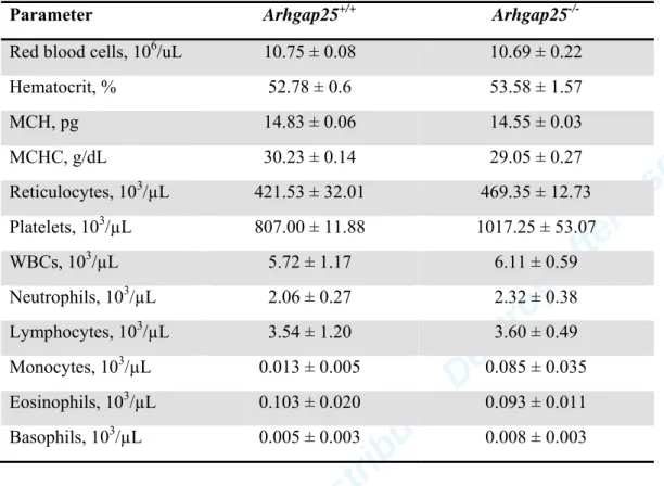
analyzed by flow cytometry (Beckman CoulterGallios).
222 223
Measurement of the amount of active Rac 224
The cellular levels of GTP loaded Rac were determined with pull-down assay using GST 225
fusion proteins containing the GTPase-binding domain of p21-activated kinase (PAK) (GST- 226
PBD) as described (31, 32). GST-PBD has been expressed in Escherichia coli. For pull-down, 227
For Peer Review. Do not distribute. Destroy after use.
11
bone marrow-derived neutrophils were activated with 50 ng/mL TNFα in HBSS+ medium at 228
37 °C for 10 min. Basal Rac-activation was determined from resting cells. Whole cell lysates 229
were run on SDS-PAGE, blotted onto nitrocellulose (33) and stained with anti-Rac antibody 230
in 1:5000 dilution. Bound antibody was detected with enhanced chemiluminescence using 231
horseradish peroxidase-conjugated anti–mouse-Ig (from sheep) secondary antibody (GE 232
Healthcare) used in 1:5000 dilution. ImageJ software was used for densitometry analysis.
233 234
Statistical analysis 235
All data were analyzed and plotted using SigmaPlot 11.0 Software (Systat Software, Inc.).
236
Pairwise comparison of experimental groups was carried out with paired t-test or Mann- 237
Whitney Rank Sum Test or two way ANOVA followed by a Tukey post-hock test, depending 238
on the condition. All P-values<.05 were considered statistically significant.
239
For Peer Review. Do not distribute. Destroy after use.
12 Results
240 241
Arhgap25 knockout mice 242
Arhgap25 knockout mice were generated by the CSD consortium for the NIH funded 243
Knockout Mouse Project (KOMP) inserting the L1L2_Bact_P cassette upstream of the 6th 244
exon of the Arhgap25 gene. The cassette contains the following sites and sequences in the 245
given order: FRT, lacZ, loxP, neomycin (under the control of the human beta-actin promoter), 246
SV40 polyA, FRT, loxP. A third loxP site was inserted downstream of the 6th exon (23).
247
Fertile homozygous mice (Arhgap25-/-) were obtained with the expected Mendelian ratios 248
(data not shown) and did not show any obvious phenotype. No ARHGAP25 protein could be 249
detected in either bone marrow derived neutrophils or in the spleen of Arhgap25-/- mice (Fig.
250
S1A, B). Blood panel (e.g. circulating cell counts, hematocrit, mean corpuscular hemoglobin, 251
mean corpuscular hemoglobin concentration) of Arhgap25-/- mice did not differ from the wild 252
type (Arhgap25+/+) (Table I, Table SII). We assessed the body weight of male and female 253
mice during a 130 days period and in 3 independent experiments. Body weight of male 254
Arhgap25-/- mice was decreased compared to wild type but in the case of female mice, no 255
difference was observed (Fig. S2).
256 257
Reduced leukocyte rolling velocity and prolonged crawling in the absence of 258
ARHGAP25 259
Using intravital microscopy, we first investigated leukocyte rolling, adhesion and crawling in 260
TNFα-stimulated cremaster muscle venules of WT (Arhgap25+/+) and Arhgap25-/- mice in 261
vivo. Microvessel diameters, wall shear rates, centerline blood flow velocities and circulating 262
leukocyte counts were similar between wild type and Arhgap25-/- mice (Table SI). While we 263
observed no difference in leukocyte rolling (Fig. 1A), mean leukocyte rolling velocity was 264
For Peer Review. Do not distribute. Destroy after use.
13
markedly decreased in the absence of ARHGAP25 (Fig. 1B). Furthermore, we analyzed the 265
number of adherent leukocytes and found no difference in adhesion between Arhgap25-/- and 266
WT mice (Fig. 1C). Next, we investigated leukocyte crawling along the inflamed 267
endothelium. Individual crawling paths of >140 cells were analyzed per group (Fig. 1D and 268
E). We did not observe any difference in crawling directionality or accumulated distance 269
between WT and ARHGAP25-/-mice (Fig. 1D-F). However, we found a significant increase in 270
crawling velocity and Euclidean distance in Arhgap25-/- mice compared to WT mice (Fig. 1G 271
and H) suggesting that ARHGAP25 is regulating leukocyte crawling in vivo.
272 273
Lack of ARHGAP25 augments transendothelial migration in vivo.
274
Next, we studied leukocyte extravasation in TNFα-stimulated cremaster muscle whole mount 275
preparations of WT and Arhgap25-/- mice. As shown in Fig. 2A-B, Arhgap25+/+ leukocytes 276
were found mainly in the vessels and the extravasated cells were scattered in the tissue. In 277
contrast, a large number of Arhgap25-/- leukocytes lined up around the vessel from which they 278
extravasated (Fig. 2C-D). Transendothelial migration was quantified and we found a 279
significant increase in leukocyte extravasation in Arhgap25-/- compared to WT mice (Fig. 2E).
280
Intrascrotal injection of PBS as a control caused no significant difference between WT and 281
Arhgap25-/- mice (P= 0.194, data not shown). Further analysis of the different leukocyte 282
populations extravasated into the inflamed cremaster muscle tissue revealed that the major 283
component of extravasated leukocytes were neutrophilic granulocytes (PMN), followed by 284
monocytes and lymphocytes (marked as “Others”) and eosinophils (Fig. 2F). Increased 285
leukocyte extravasation upon TNFα stimulus was confirmed in an acute peritonitis model.
286
Analyzing the H&E stained sections of inflamed peritoneal tissue, elevated leukocyte 287
infiltration was observed in Arhgap25-/- mice compared to WT (Fig. 3A). Specific analysis of 288
For Peer Review. Do not distribute. Destroy after use.
14
Ly-6G+ neutrophil count in peritoneal lavage revealed a significant increase in case of 289
Arhgap25-/- neutrophils compared to WT (Fig. 3B).
290 291
Leukocyte rolling and adhesion under ex vivo conditions and in vitro Transwell 292
migration assay.
293
As Arhgap25-/- mice are complete knockout mice, the question arose, whether the observed 294
alterations are due to functional changes in leukocytes or endothelial cells. To investigate the 295
contribution of leukocytes on the recruitment phenotype observed in Arhgap25-/- mice, we 296
performed ex vivo flow chamber assays and assessed rolling and adhesion of leukocytes in 297
flow chambers coated with adhesion relevant proteins. In flow chambers coated with 298
recombinant murine (rm)E-selectin, we saw a 2.5-fold increase in the number of rolling 299
Arhgap25-/- leukocytes compared to wild type leukocytes (Fig. 4A). Next, we analyzed 300
leukocyte adhesion in flow chambers coated with rmE-selectin alone, with rmE-selectin and 301
rmICAM-1, and with rmE-selectin, rmICAM-1 and rmCXCL-1. Similar to the in vivo results 302
we found no difference in the number of adherent cells between the different groups (Fig.
303
4B). However, when we analyzed leukocyte rolling velocities, lack of ARHGAP25 resulted in 304
a significant decrease in rolling velocity (Fig. 4C) in flow chambers coated with rmE-selectin 305
or with rmE-selectin and rmICAM-1 surface (Fig. 4C).
306
Taken together, we were able to reproduce under ex vivo conditions the pattern of 307
rolling and adherence observed in ARHGAP25-deficient animals under in vivo conditions 308
suggesting that loss of ARHGAP25 in leukocytes accounts for the observed pro-inflammatory 309
phenotype.
310
In order to test the role of ARHGAP25 in cell migration under in vitro conditions, we 311
determined neutrophil migration toward CXCL12 in a Transwell assay. As shown in Fig. 4D, 312
For Peer Review. Do not distribute. Destroy after use.
15
Arhgap25-/- neutrophils pretreated with TNFα for 10 min showed a significant increase in 313
transmigration compared to WT.
314 315
Verification of altered leukocyte function in bone marrow chimeras 316
In view of the decisive role of the adhesive surface provided in vivo by the endothelial cells 317
we wanted to verify our flow chamber data in bone marrow chimeric mice. These animals 318
express the CD45.1 allele and carry Arhgap25-/- or Arhgap25+/+ hematopoietic cell 319
populations that express CD45.2. Using these animals, we investigated the extravasation of 320
leukocytes in cremaster muscle whole mounts after 3 h local stimulation with TNFα. CD45.2- 321
expressing Arhgap25-/- leukocytes were able to transmigrate more efficiently in CD45.1- 322
expressing WT recipients than Arhgap25+/+ cells (Fig. 5A-D). Similar to the results presented 323
in Fig. 2, we found a threefold increase in leukocyte extravasation in chimeric mice with 324
Arhgap25-/- hematopoietic cells compared to chimeric mice where WT hematopoietic cells 325
had been transferred (Fig. 5E). Similar to the results obtained in complete knock-out animals, 326
mainly neutrophilic granulocytes were responsible for the increase in extravasation followed 327
by mononuclear cells (marked as “Others”) and eosinophils (Fig. 5F).
328
These results substantiate that the alteration of leukocyte transendothelial migration 329
observed in the absence of ARHGAP25 is due to primary changes in the hematopoietic 330
compartment but not in endothelial or other non-hematopoietic cell compartment.
331 332
Potential mechanism of altered leukocyte function 333
To examine, whether ARHGAP25 has a regulatory role in the expression of adhesion relevant 334
molecules and signaling events during neutrophil recruitment, we investigated cell surface 335
expression and ligand binding ability of receptors and molecules involved in leukocyte- 336
endothelial cell interactions. As shown in Fig. 6A, ARHGAP25 deficiency did not affect the 337
For Peer Review. Do not distribute. Destroy after use.
16
expression of β2 integrins (CD18, CD11a and CD11b), L-selectin (CD62L), PSGL-1, 338
chemokine receptor CXCR2 or CD44. In vitro analysis of adhesion molecule expression after 339
direct chemotactic migration of neutrophils did not reveal any difference between WT and 340
Arhgap25-/- cells either (data not shown). Lack of ARHGAP25 also did not result in any 341
difference in rmICAM-1 binding to LFA-1 in resting cells. As stimulation of bone marrow- 342
derived neutrophils leads to integrin activation and increased ligand binding (34), we also 343
investigated binding of rmICAM-1 to neutrophils stimulated with CXCL1 or PMA.
344
Compared to unstimulated controls, PMA caused a significant increase in rmICAM-1 binding 345
to LFA-1 on both Arhgap25-/- and wild type neutrophils. However, ARHGAP25-deficiency 346
did not influence rmICAM-1 binding to stimulated neutrophils. (Fig. 6B).
347
Our previous study indicated that ARHGAP25 has a regulatory role in neutrophilic 348
functions through its GAP activity on Rac1 (21). In addition, our in vitro studies demonstrate 349
that it has a GAP activity on Rac2 as well (data not shown). Therefore, we investigated the 350
presence of active Rac in bone marrow-derived neutrophils. Interestingly, we observed no 351
difference in Rac-activity between ARHGAP25-/- and wild type cells in the resting state (Fig.
352
6C). In contrast, treatment of neutrophils with TNFα resulted in a marked decrease of active 353
Rac in the presence of ARHGAP25, while the lack of ARHGAP25 completely abolished this 354
alteration (Fig. 6C).
355
As Rac is known to be a key regulator of actin-polymerization during leukocyte 356
migration (35, 36), we measured filamentous actin (F-actin) in Arhgap25-/- and wild type BM 357
neutrophils. In resting ARHGAP25-deficient cells, increased F-actin was observed compared 358
to wild type cells (Fig. 6D). Similar difference could be revealed upon stimulation with TNFα 359
(Fig. 6E). Taken together, we suggest that ARHGAP25 affects actin-polymerization and 360
depolymerization through its GTPase activating effect on Rac.
361
For Peer Review. Do not distribute. Destroy after use.
17 Discussion
362
The present study provides a detailed characterization of leukocyte recruitment during 363
inflammation in vivo in ARHGAP25-deficient mice. Alterations have been uncovered for 364
several steps along the recruitment cascade, indicating a role for the protein in those 365
processes. Most remarkable is the increase of transmigrating neutrophils observed both in the 366
inflamed cremaster muscle and the inflamed peritoneal cavity. No striking changes were 367
found in circulating leukocyte counts between wild type and Arhgap25-/- animals excluding 368
differences in circulating leukocyte numbers for the observed alterations in leukocyte 369
recruitment in the absence of ARHGAP25.
370
Alteration of leukocyte migration may be the result of primary changes in circulating 371
leukocytes, the endothelial cells, or both. Based on the following observations, we believe that 372
in case of ARHGAP25-deficient animals, the altered migration is caused by leukocytes: i) 373
ARHGAP25 was shown to be expressed primarily in hematopoietic cells (21) ii) all the 374
trafficking alterations observed in living animals could be reproduced with isolated cells 375
under ex vivo or in vitro condition iii) the difference between the movements of Arhgap25+/+
376
and Arhgap25-/- cells was reproduced in bone marrow chimeric animals in vivo, where the 377
deficiency affected only the hematopoietic but not the endothelial or other peripheral cells.
378
In control experiments it was verified that ARHGAP25-deficiency had no influence on 379
the expression of the major leukocyte adhesive proteins and receptors or ligand binding of β2 380
integrins. On the other hand, stimulation of neutrophils with TNFα resulted in a significant 381
decrease in measurable GTP-bound Rac which was abolished by absence of ARHGAP25. The 382
observed increase in the amount of filamentous actin indicates the biological relevance of 383
enhanced Rac activity (Fig. 6). Deficiency in various RacGEFs was reported to result in 384
decreased amount of GTP-bound Rac and a decrease in phagocyte migration (16, 37, 38), i.e.
385
changes opposite to our findings in animals lacking ARHGAP25. We thus ascribe the 386
For Peer Review. Do not distribute. Destroy after use.
18
alteration of leukocyte trafficking in ARHGAP25-deficient animals to cytoskeletal 387
reorganization due to elevation of RacGTP concentration. In human endothelial cells 388
(HUVECs), TNFα was shown to induce actin-rearrangement through activation of Rho family 389
small G-proteins (39) and several studies reported the role of TNFα in neutrophil priming and 390
its involvement in neutrophil effector functions and inside-out signaling (40-43). Our findings 391
strongly suggest that the leukocyte-specific RacGAP ARHGAP25 is a critical player in 392
TNFα-induced, Rac-mediated actin reorganization in neutrophils. Two recent reports provide 393
important information on its physiological role: ARHGAP25 was shown to be required for 394
actin depolymerization in the course of phagocytosis (21, 44), and it was demonstrated to 395
undergo significant changes in its phosphorylation pattern and GAP activity upon biological 396
stimulation (45). TNFα-initiated modulation of the phosphorylation pattern of ARHGAP25 397
with subsequent alterations of its GAP function may provide the link between the cytokine 398
effect and the actin cytoskeleton rearrangement.
399
Taken together, our data indicate that ARHGAP25 is a critical negative regulator of 400
Rac activity and leukocyte transmigration. This qualifies ARHGAP25 as an interesting drug 401
target in autoimmune disorders (e.g. rheumatoid arthritis and multiple sclerosis) where 402
leukocyte recruitment is unwanted.
403 404
Acknowledgements 405
The authors are indebted to Professor Attila Mócsai for helpful suggestions and critical 406
reading of the manuscript and to Ms. Regina Tóth-Kun and Nadine Schmidt for expert 407
technical assistance.
408 409 410 411
For Peer Review. Do not distribute. Destroy after use.
19 Authorship contributions
412
R. Csépányi-Kömi and É. Wisniewski carried out the majority of experiments and prepared 413
writing of the manuscript; B. Bartos, P. Lévai, A. Kurz and S. Bierschenk carried out part of 414
the experiments; T. Németh carried out the bone marrow transplantation; B. Balázs carried 415
out the histology on murine peritoneums; M. Sperandio and E. Ligeti supervised, coordinated 416
and financed the experimental work and had a major role in writing of the manuscript.
417 418
Disclosure of Conflict of Interest 419
The authors have no conflict of interest to disclose.
420 421
For Peer Review. Do not distribute. Destroy after use.
20 References
422
1. Rao, R. M., L. Yang, G. Garcia-Cardena, and F. W. Luscinskas. 2007. Endothelial-dependent 423
mechanisms of leukocyte recruitment to the vascular wall. Circ Res 101: 234-247.
424
2. Ley, K., C. Laudanna, M. I. Cybulsky, and S. Nourshargh. 2007. Getting to the site of 425
inflammation: the leukocyte adhesion cascade updated. Nat Rev Immunol 7: 678-689.
426
3. McEver, R. P. 2002. Selectins: lectins that initiate cell adhesion under flow. Curr Opin Cell Biol 427
14: 581-586.
428
4. Luster, A. D., R. Alon, and U. H. von Andrian. 2005. Immune cell migration in inflammation:
429
present and future therapeutic targets. Nat Immunol 6: 1182-1190.
430
5. Zarbock, A., and K. Ley. 2009. Neutrophil adhesion and activation under flow.
431
Microcirculation 16: 31-42.
432
6. Borregaard, N. 2010. Neutrophils, from marrow to microbes. Immunity 33: 657-670.
433
7. Insall, R. H., and L. M. Machesky. 2009. Actin dynamics at the leading edge: from simple 434
machinery to complex networks. Dev Cell 17: 310-322.
435
8. Meyer, W. H., and T. H. Howard. 1987. Actin polymerization and its relationship to 436
locomotion and chemokinetic response in maturing human promyelocytic leukemia cells.
437
Blood 70: 363-367.
438
9. Vicente-Manzanares, M., and F. Sanchez-Madrid. 2004. Role of the cytoskeleton during 439
leukocyte responses. Nat Rev Immunol 4: 110-122.
440
10. Futosi, K., S. Fodor, and A. Mocsai. 2013. Neutrophil cell surface receptors and their 441
intracellular signal transduction pathways. Int Immunopharmacol 17: 638-650.
442
11. Hall, A. 1998. Rho GTPases and the actin cytoskeleton. Science 279: 509-514.
443
12. Csepanyi-Komi, R., M. Levay, and E. Ligeti. 2012. Rho/RacGAPs: embarras de richesse? Small 444
GTPases 3: 178-182.
445
13. Ligeti, E., S. Welti, and K. Scheffzek. 2012. Inhibition and termination of physiological 446
responses by GTPase activating proteins. Physiol Rev 92: 237-272.
447
14. Csepanyi-Komi, R., D. Safar, V. Grosz, Z. L. Tarjan, and E. Ligeti. 2013. In silico tissue- 448
distribution of human Rho family GTPase activating proteins. Small GTPases 4: 90-101.
449
15. Kunisaki, Y., A. Nishikimi, Y. Tanaka, R. Takii, M. Noda, A. Inayoshi, K. Watanabe, F.
450
Sanematsu, T. Sasazuki, T. Sasaki, and Y. Fukui. 2006. DOCK2 is a Rac activator that regulates 451
motility and polarity during neutrophil chemotaxis. J Cell Biol 174: 647-652.
452
16. Lawson, C. D., S. Donald, K. E. Anderson, D. T. Patton, and H. C. Welch. 2011. P-Rex1 and 453
Vav1 cooperate in the regulation of formyl-methionyl-leucyl-phenylalanine-dependent 454
neutrophil responses. J Immunol 186: 1467-1476.
455
17. Gakidis, M. A., X. Cullere, T. Olson, J. L. Wilsbacher, B. Zhang, S. L. Moores, K. Ley, W. Swat, T.
456
Mayadas, and J. S. Brugge. 2004. Vav GEFs are required for beta2 integrin-dependent 457
functions of neutrophils. J Cell Biol 166: 273-282.
458
18. Costa, C., G. Germena, E. L. Martin-Conte, I. Molineris, E. Bosco, S. Marengo, O. Azzolino, F.
459
Altruda, V. M. Ranieri, and E. Hirsch. 2011. The RacGAP ArhGAP15 is a master negative 460
regulator of neutrophil functions. Blood 118: 1099-1108.
461
19. Cho, Y. J., J. M. Cunnick, S. J. Yi, V. Kaartinen, J. Groffen, and N. Heisterkamp. 2007. Abr and 462
Bcr, two homologous Rac GTPase-activating proteins, control multiple cellular functions of 463
murine macrophages. Mol Cell Biol 27: 899-911.
464
20. Gambardella, L., K. E. Anderson, C. Nussbaum, A. Segonds-Pichon, T. Margarido, L. Norton, T.
465
Ludwig, M. Sperandio, P. T. Hawkins, L. Stephens, and S. Vermeren. 2011. The GTPase- 466
activating protein ARAP3 regulates chemotaxis and adhesion-dependent processes in 467
neutrophils. Blood 118: 1087-1098.
468
21. Csepanyi-Komi, R., G. Sirokmany, M. Geiszt, and E. Ligeti. 2012. ARHGAP25, a novel Rac 469
GTPase-activating protein, regulates phagocytosis in human neutrophilic granulocytes. Blood 470
119: 573-582.
471
For Peer Review. Do not distribute. Destroy after use.
21
22. Lorincz, A. M., G. Szarvas, S. M. Smith, and E. Ligeti. 2014. Role of Rac GTPase activating 472
proteins in regulation of NADPH oxidase in human neutrophils. Free Radic Biol Med 68: 65- 473
71.
474
23. Testa, G., J. Schaft, F. van der Hoeven, S. Glaser, K. Anastassiadis, Y. Zhang, T. Hermann, W.
475
Stremmel, and A. F. Stewart. 2004. A reliable lacZ expression reporter cassette for 476
multipurpose, knockout-first alleles. Genesis 38: 151-158.
477
24. Nemeth, T., K. Futosi, C. Hably, M. R. Brouns, S. M. Jakob, M. Kovacs, Z. Kertesz, B. Walzog, J.
478
Settleman, and A. Mocsai. 2010. Neutrophil functions and autoimmune arthritis in the 479
absence of p190RhoGAP: generation and analysis of a novel null mutation in mice. J Immunol 480
185: 3064-3075.
481
25. Kovacs, M., T. Nemeth, Z. Jakus, C. Sitaru, E. Simon, K. Futosi, B. Botz, Z. Helyes, C. A. Lowell, 482
and A. Mocsai. 2014. The Src family kinases Hck, Fgr, and Lyn are critical for the generation of 483
the in vivo inflammatory environment without a direct role in leukocyte recruitment. J Exp 484
Med 211: 1993-2011.
485
26. Frommhold, D., A. Ludwig, M. G. Bixel, A. Zarbock, I. Babushkina, M. Weissinger, S.
486
Cauwenberghs, L. G. Ellies, J. D. Marth, A. G. Beck-Sickinger, M. Sixt, B. Lange-Sperandio, A.
487
Zernecke, E. Brandt, C. Weber, D. Vestweber, K. Ley, and M. Sperandio. 2008.
488
Sialyltransferase ST3Gal-IV controls CXCR2-mediated firm leukocyte arrest during 489
inflammation. J Exp Med 205: 1435-1446.
490
27. Schindelin, J., I. Arganda-Carreras, E. Frise, V. Kaynig, M. Longair, T. Pietzsch, S. Preibisch, C.
491
Rueden, S. Saalfeld, B. Schmid, J. Y. Tinevez, D. J. White, V. Hartenstein, K. Eliceiri, P.
492
Tomancak, and A. Cardona. 2012. Fiji: an open-source platform for biological-image analysis.
493
Nat Methods 9: 676-682.
494
28. Sperandio, M., J. Pickard, S. Unnikrishnan, S. T. Acton, and K. Ley. 2006. Analysis of leukocyte 495
rolling in vivo and in vitro. Methods Enzymol 416: 346-371.
496
29. Mocsai, A., M. Zhou, F. Meng, V. L. Tybulewicz, and C. A. Lowell. 2002. Syk is required for 497
integrin signaling in neutrophils. Immunity 16: 547-558.
498
30. Mocsai, A., E. Ligeti, C. A. Lowell, and G. Berton. 1999. Adhesion-dependent degranulation of 499
neutrophils requires the Src family kinases Fgr and Hck. J Immunol 162: 1120-1126.
500
31. Ren, X. D., and M. A. Schwartz. 2000. Determination of GTP loading on Rho. Methods 501
Enzymol 325: 264-272.
502
32. Benard, V., and G. M. Bokoch. 2002. Assay of Cdc42, Rac, and Rho GTPase activation by 503
affinity methods. Methods Enzymol 345: 349-359.
504
33. Levay, M., B. Bartos, and E. Ligeti. 2013. p190RhoGAP has cellular RacGAP activity regulated 505
by a polybasic region. Cell Signal 25: 1388-1394.
506
34. Nishida, N., C. Xie, M. Shimaoka, Y. Cheng, T. Walz, and T. A. Springer. 2006. Activation of 507
leukocyte beta2 integrins by conversion from bent to extended conformations. Immunity 25:
508
583-594.
509
35. Lammermann, T., and R. N. Germain. 2014. The multiple faces of leukocyte interstitial 510
migration. Semin Immunopathol 36: 227-251.
511
36. Park, H., M. M. Chan, and B. M. Iritani. 2010. Hem-1: putting the "WAVE" into actin 512
polymerization during an immune response. FEBS Lett 584: 4923-4932.
513
37. Herter, J. M., J. Rossaint, H. Block, H. Welch, and A. Zarbock. 2013. Integrin activation by P- 514
Rex1 is required for selectin-mediated slow leukocyte rolling and intravascular crawling.
515
Blood 121: 2301-2310.
516
38. Watanabe, M., M. Terasawa, K. Miyano, T. Yanagihara, T. Uruno, F. Sanematsu, A. Nishikimi, 517
J. F. Cote, H. Sumimoto, and Y. Fukui. 2014. DOCK2 and DOCK5 act additively in neutrophils 518
to regulate chemotaxis, superoxide production, and extracellular trap formation. J Immunol 519
193: 5660-5667.
520
39. Wojciak-Stothard, B., A. Entwistle, R. Garg, and A. J. Ridley. 1998. Regulation of TNF-alpha- 521
induced reorganization of the actin cytoskeleton and cell-cell junctions by Rho, Rac, and 522
Cdc42 in human endothelial cells. J Cell Physiol 176: 150-165.
523
For Peer Review. Do not distribute. Destroy after use.
22
40. Elbim, C., S. Chollet-Martin, S. Bailly, J. Hakim, and M. A. Gougerot-Pocidalo. 1993. Priming of 524
polymorphonuclear neutrophils by tumor necrosis factor alpha in whole blood: identification 525
of two polymorphonuclear neutrophil subpopulations in response to formyl-peptides. Blood 526
82: 633-640.
527
41. Elbim, C., S. Bailly, S. Chollet-Martin, J. Hakim, and M. A. Gougerot-Pocidalo. 1994.
528
Differential priming effects of proinflammatory cytokines on human neutrophil oxidative 529
burst in response to bacterial N-formyl peptides. Infect Immun 62: 2195-2201.
530
42. Jablonska, E., M. Kiluk, W. Markiewicz, and J. Jablonski. 2002. Priming effects of GM-CSF, IFN- 531
gamma and TNF-alpha on human neutrophil inflammatory cytokine production. Melanoma 532
Res 12: 123-128.
533
43. Bouaouina, M., E. Blouin, L. Halbwachs-Mecarelli, P. Lesavre, and P. Rieu. 2004. TNF-induced 534
beta2 integrin activation involves Src kinases and a redox-regulated activation of p38 MAPK. J 535
Immunol 173: 1313-1320.
536
44. Schlam, D., R. D. Bagshaw, S. A. Freeman, R. F. Collins, T. Pawson, G. D. Fairn, and S.
537
Grinstein. 2015. Phosphoinositide 3-kinase enables phagocytosis of large particles by 538
terminating actin assembly through Rac/Cdc42 GTPase-activating proteins. Nat Commun 6:
539
8623.
540
45. Wang, L. D., S. B. Ficarro, J. N. Hutchinson, R. Csepanyi-Komi, P. T. Nguyen, E. Wisniewski, J.
541
Sullivan, O. Hofmann, E. Ligeti, J. A. Marto, and A. J. Wagers. 2016. Phosphoproteomic 542
profiling of mouse primary hematopoietic stem and progenitor cells reveals new regulators 543
of HSPC mobilization. Blood.
544 545 546
For Peer Review. Do not distribute. Destroy after use.
23
Footnotes: 1 Experimental work was financially supported by the Hungarian Research Fund 547
(OTKA K108382) to E.L., by the European Community's Seventh Framework Programme 548
[FP7] under grant agreement n°HEALTH-F4-2011-282095 to M.S. and by Deutsche 549
Forschungsgemeinschaft SFB914, project B1 to M.S.
550
For Peer Review. Do not distribute. Destroy after use.
24 Figure legends
551 552
Fig.1. Measurement of leukocyte rolling and crawling in TNFα-stimulated mouse 553
cremaster muscle venules. Intravital microscopy was conducted to investigate leukocyte 554
recruitment in the mouse cremaster muscle 2 hours after injection of rmTNFα (500 ng/mouse 555
intrascrotally). (A) Leukocyte rolling flux fraction (%) is presented as mean+SEM of 33 556
vessels from 10 wild type mice and 42 vessels of 12 Arhgap25-/- mice. Mean rolling velocity 557
of leukocytes (µm/s) (B) was quantified and is shown as bar chart (mean+SEM of 166 558
Arhgap25+/+ cells and 190 Arhgap25-/- cells), ***: P< .001 compared to Arhgap25+/+. (C) 559
Number of adherent cells per mm2 vessel wall is given as mean+SEM of 10 (Arhgap25+/+) 560
and 12 (Arhgap25-/-) separate experiments. (D, E) Leukocyte crawling paths of individual 561
leukocytes of Arhgap25+/+ (n=163 cells) and Arhgap25-/- (n=154 cells) mice. Direction of 562
blood flow is indicated by arrows. (F) Accumulated distance of leukocytes. Mean+ SEM of 8 563
wild type and 7 Arhgap25-/- mice. (G) Mean crawling velocity presented as mean+SEM of 564
163 Arhgap25+/+ and 154 Arhgap25-/- cells. **: P< .01 compared to Arhgap25+/+. (H) 565
Euclidean distance determines the length of section between starting and end points of 566
crawling pathways. Mean+SEM of 8 (Arhgap25+/+) and 7 (Arhgap25-/-) separate experiments 567
are shown.*: P< .05 compared to Arhgap25+/+. 568
569
Fig. 2. Transmigration of leukocytes under in vivo conditions. (A-D) Representative 570
images of Giemsa-stained cremaster muscle whole mounts from Arhgap25+/+ and Arhgap25-/- 571
mice 3 hours after 500 ng intrascrotal rmTNFα injection. Images were captured with a Leica 572
DMI 6000 B microscope equipped with a 10x/0.30 NA dry objective (Leica) and a Leica DFC 573
480 camera. ROIs with high leukocyte infiltration (rectangles) are captured with 40x objective 574
and shown in the right side of the panel (B,D). Bars represent 100 µm. (E) Quantification of 575
For Peer Review. Do not distribute. Destroy after use.
25
total number of extravasated cells per mm2 microvessel wall surface. (F) Distribution of 576
extravasated cell types (PMN: neutrophilic granulocytes, Eos: eosinophilic granulocytes, 577
Others: lymphocytes, macrophages and basophilic granulocytes). Mean+SEM of 3 578
(Arhgap25+/+) and 7 (Arhgap25-/-) separate experiments.***: P< .001 compared to 579
Arhgap25+/+. 580
581
Fig. 3. TNFα-induced leukocyte infiltration into the peritoneal cavity.
582
(A) Hematoxylin and eosin staining of peritoneal tissues 3 hours after intraperitoneal injection 583
of TNFα. Left side of the panel indicates 2 representative images from Arhgap25+/+ and 2 584
from Arhgap25-/- mice captured with a 10x objective. ROIs with high leukocyte infiltration 585
(rectangles) are captured with 40x objective and shown in the right side of the panel. Bars 586
represents 50 µm. Results shown are representatives of multiple experiments and of multiple 587
sections and fields. (B) Ly-6G+ cell count measured from peritoneal lavage of Arhgap25+/+
588
and Arhgap25-/- mice 3 hours after TNFα administration. Data represent mean+SEM of 6 589
separate experiments. *: P< .05 compared to Arhgap25+/+. 590
591
Fig. 4. Leukocyte rolling and adhesion under flow conditions and transmigration in a 592
Transwell assay. (A-C) Ex vivo flow chamber assay. Blood cells of Arhgap25+/+ and 593
Arhgap25-/- mice were perfused through glass capillaries coated with different cell surface 594
molecules as indicated in panel B. Number of rolling (A) and adherent (B) leukocytes per 595
field of view (FOV) and mean rolling velocity (C) of wild type and Arhgap25-/- animals are 596
shown. Rolling was assessed in E-selectin coated chambers (A). E: rmE-selectin; I: rmICAM- 597
1; CXCL1: rmKC/CXCL1. Mean+SEM of 4 separate experiments. *: P< .05, **: P< .01, ***:
598
P<.001 compared to Arhgap25+/+. (D) In vitro transmigration of Arhgap25+/+ and Arhgap25-/- 599
bone marrow neutrophils pretreated with TNFα toward CXCL12 in an FBS-coated Transwell 600
For Peer Review. Do not distribute. Destroy after use.
26
system. Data represent mean+SEM of 4 independent experiments. *: P< .05 compared to 601
Arhgap25+/+. 602
603
Fig. 5. Leukocyte transmigration in bone marrow chimeric mice carrying Arhgap25-/- or 604
Arhgap25+/+ hematopoietic cells. (A-D) Representative images of a Giemsa-stained 605
cremaster muscle whole mount. Microscopic analysis was performed 3 hours after 500 ng 606
intrascrotal rmTNFα injection. Images were captured with a Leica DMI 6000 B microscope 607
equipped with a 10x/0.30 NA dry objective (Leica) and a Leica DFC 480 camera. ROIs with 608
high leukocyte infiltration (rectangles) are captured with 40x objective and shown in the right 609
side of the panel (B,D). Bars represent 100 µm. (E) Quantification of total number of 610
extravasated cells per mm2 microvessel wall surface. (F) Distribution of transmigrated cell 611
types (PMN: neutrophilic granulocytes, Eos: eosinophilic granulocytes, Others: lymphocytes, 612
macrophages, basophilic granulocytes). Mean+SEM of 3 separate experiments. *: P< .05 613
compared to Arhgap25+/+. 614
615
Fig. 6. Investigation of the potential mechanism of altered migration in Arhgap25-/-cells.
616
(A) Cell surface expression of molecules relevant in leukocyte-endothelial cell interactions 617
during recruitment. Mean fluorescence intensity relative to isotype control is presented.
618
Mean+SEM of 5 separate experiments. Panel B shows binding of ICAM-1 to LFA1. Bone 619
marrow-derived neutrophils were co-incubated with fluorescently labeled rmICAM-1. Bound 620
ICAM-1 was detected with flow cytometry. Data are presented as mean fluorescence intensity 621
ratio relative to unstimulated cells. CCXL1: rmKC/CXCL1, PMA: Phorbol 12-myristate 13- 622
acetate. Mean+SEM of 4 separate experiments is shown. *: P< .05, **: P< .01. (C) GTP- 623
bound active Rac amount in resting and stimulated bone marrow-derived neutrophils.
624
Stimulation was carried out with 50 ng/mL TNFα in HBSS+ medium at 37 °C for 10 min.
625
For Peer Review. Do not distribute. Destroy after use.
27
After lysis, active Rac was pulled down with PBD-GST-glutathione-sepharose beads. Bar 626
chart presents densitometric analysis of 6 (unstimulated) and 3 (TNFα-treated) separate 627
western blot experiments. Under the graph, a representative western blot experiment is shown.
628
Active and total Rac were decorated with anti-Rac antibody in 1:1000 dilution. (D, E) 629
Filamentous actin amount of bone marrow-derived neutrophils. Actin was stained with Alexa- 630
488-Phalloidin in 1:500 dilution and measured with flow cytometry. Pane D shows F-actin 631
content in resting neutrophils from Arhgap25-/- mice as mean fluorescence intensity of 632
phalloidin relative to Arhgap25+/+. Mean+SEM of 4 separate experiments is present. **: P<
633
.01 compared to Arhgap25+/+. (E) Changes in relative F-actin content of neutrophils treated 634
for 5, 10 and 15 minutes with 50 ng/mL TNFα. Mean fluorescence intensity of phalloidin is 635
expressed relative to unstimulated (0 min) control in each genotype. Mean±SEM of 6 separate 636
experiments is shown. *: P< .05 compared to Arhgap25+/+. 637
For Peer Review. Do not distribute. Destroy after use.
Figure1.
B C
A
D E
G H
F
Mean crawling velocity (Pm/sec) 0.00 0.05 0.10 0.15 0.20 0.25 0.30 0.35 0.40
**
Arhgap25+/+ Arhgap25-/- Euclidean distance (Pm)
0 5 10 15 20 25
*
Arhgap25+/+ Arhgap25-/-
Accumulated distance (Pm)
0 10 20 30 40 50
Arhgap25+/+ Arhgap25-/- Arhgap25+/+
x axes (Pm)
-40 0 40 80
y axes (Pm)
-40 -20 0 20 40
Arhgap25-/-
x axes (Pm)
-40 0 40 80
y axes (Pm)
-40 -20 0 20
direction of blood flow 40 direction of blood flow
Leukocyte rolling flux fraction (%)
0.00 0.02 0.04 0.06 0.08 0.10 0.12 0.14
Arhgap25+/+ Arhgap25-/- Mean rolling velocity (Pm/sec)
0 1 2 3 4 5 6 7
***
Arhgap25+/+ Arhgap25-/- Adherent cells (cells/mm2 )
0 200 400 600 800 1000
Arhgap25+/+ Arhgap25-/-
For Peer Review. Do not distribute. Destroy after use.
A B
Arhgap25-/-
Figure 2.
C D
Perivascular cells (cells/mm2 ) 0 100 200 300 400 500 600 700 800
***
Arhgap25+/+Arhgap25-/- PMN Eos Others Perivascular cells (cells/mm2 )
0 100 200 300 400 500
600 Arhgap25+/+
Arhgap25-/-
***
***
C D
E F
Arhgap25+/+
10x 40x
For Peer Review. Do not distribute. Destroy after use.
Arhgap25-/- Arhgap25+/+
10x 40x Figure 3.
A
Ly6g+ cells/peritoneal cavity (%, rel. to Arhgap25+/+ ) 0 100 200 300 400 500
B
Arhgap25+/+Arhgap25-/-
*
For Peer Review. Do not distribute. Destroy after use.
Figure 4.
Arhgap25+/+ Arhgap25-/-
Rolling cells per FOV
0 10 20 30 40 50 60
70
**
P
A
E E+I E+I+CXCL1
Adherent cells per FOV
0 5 10 15 20 25 30
Arhgap25+/+
Arhgap25-/-
B
P
B
E E+I E+I+CXCL1 Mean rolling velocity (Pm/sec)
0.0 0.2 0.4 0.6 0.8 1.0
1.2 Arhgap25+/+
Arhgap25-/-
***
C
***
* * C
Transmigrated cells (%, rel. to Arhgap25+/+ ) 0 20 40 60 80 100 120 140 160 180
Arhgap25+/+ Arhgap25-/-
*
D
For Peer Review. Do not distribute. Destroy after use.
Perivascular cells (cells/mm2 ) in BM chimera
0 500 1000 1500 2000 2500
Arhgap25+/+Arhgap25-/-
*
PMN Eos Others Perivascular cells (cells/mm2 ) in BM chimera
0 200 400 600 800 1000 1200 1400
1600 Arhgap25+/+
Arhgap25-/-
*
Arhgap25-/-Arhgap25+/+
10x 40x
A B
Figure 5.
C D
E F
For Peer Review. Do not distribute. Destroy after use.
Unstim. +CXCL1 +PMA ICAM-1 binding to LFA-1 (MFI ratio, rel. to unstimulated)
0.0 0.5 1.0 1.5 2.0
2.5 Arhgap25+/+
Arhgap25-/-
B
**
n.s.
*
n.s.Total Rac Active Rac
TNFα - + - +
C
GTP-bound Rac rel. to total (%)
0 10 20 30 40
50 Unstim.
+TNFD
*
Arhgap25+/+ Arhgap25-/- CD18CD11aCD11bCD62LPSGL
-1
CXCR2CD44
MFI ratio (rel. to isotype)
0 50 100 150 200 250
300 Arhgap25+/+
Arhgap25-/-
A
E
Time (min)
0 2 4 6 8 10 12 14 16 F-actin amount (MFI ratio, rel. to 0 min)
90 100 110 120 130 140 150 160
Arhgap25+/+
Arhgap25-/-
* *
D
F-actin amount (MFI ratio, rel. to Arhgap25+/+ ) 0 50 100 150 200 250
Arhgap25+/+ Arhgap25-/-