PhD THESIS
LÁSZLÓ PONGRÁCZ
UNIVERSITY OF WEST HUNGARY
FACULTY OF AGRICULTURAL AND FOOD SCIENCES MOSONMAGYARÓVÁR
2002
UNIVERSITY OF WEST HUNGARY
FACULTY OF AGRICULTURAL AND FOOD SCIENCES Institute of Animal Breeding and Husbandry
Program leader, subprogram leader and supervisor Prof. Dr. Dr. h.c. János Iváncsics †
DSc in Agriculture
SOME ASPECTS OF UDDER HEALTH RELATED TO MILK QUALITY AND GRAVITY OF MASTITIS
Written by
LÁSZLÓ PONGRÁCZ
MOSONMAGYARÓVÁR 2002
SOME ASPECTS OF UDDER HEALTH RELATED TO MILK QUALITY AND GRAVITY OF MASTITIS
értekezés doktori (PhD) fokozat elnyerése érdekében Írta:
PONGRÁCZ LÁSZLÓ
Készült a Nyugat-Magyarországi Egyetem AZ ÁLLATI TERMÉK ELŐÁLLÍTÁS BIOLÓGIAI ÉS ÖKONÓMIAI KÉRDÉSEI program, Szarvasmarha termékek előállítása és feldolgozása alprogramja keretében.
Témavezető: Prof. Dr. Dr. h.c. Iváncsics János †
Elfogadásra javaslom (igen / nem): Kovácsné Prof. Dr. Gaál Katalin
……….……….
(aláírás) A jelölt a doktori szigorlaton ….…... % -ot ért el.
Sopron/Mosonmagyaróvár, …...………
a Szigorlati Bizottság elnöke
Az értekezést bírálóként elfogadásra javaslom (igen /nem)
Első bíráló (Dr. …... …...) igen /nem
(aláírás) Második bíráló (Dr. …... …...) igen /nem
(aláírás) (Esetleg harmadik bíráló: Dr. …... …...) igen /nem
(aláírás) A jelölt az értekezés nyilvános vitáján……...% - ot ért el.
Sopron/Mosonmagyaróvár, ...…….
a Bírálóbizottság elnöke A doktori (PhD) oklevél minősítése…...…….
az EDT elnöke
ABSTRACT
Somatic cell count (SCC) and milk yield from the Livestock Performance Testing Ltd., Gödöllő (Hungary) of Hungarian Spotted and red Holstein Friesian cows were used for statistical analyses. Means of 305 days milk production and SCC of lactations were calculated. Herd-year-season-calving age at first calving effects were not taken into consideration. Biometric calculation were processed by Microsoft Excel 97, BIO MATE and Statistica softwares. The maximum milk yield can be obtained at nlact=3.88. It reflects the importance of longevity and lifetime performance. Correlation of milk yield and number of lactation was rf=0.88, correlation of transformed somatic cell count (called SCS) and number of lactation was rf=0.93 while correlation of milk yield and somatic cell count was rf=-0.12.
Then the bacteriological status and some screening methods were studied.
Flow cytometric technique may develop as a good alternative or supplementary tool to evaluate udder health. Further studies are required to establish discrimination limits.
Research increasingly demonstrates that selection for mastitis resistance is simultaneously necessary and potentially effective.
A TŐGYEGÉSZSÉGÜGY NÉHÁNY VETÜLETE, KÜLÖNÖS TEKINTETTEL A TEJMINŐSÉGRE ÉS A
TŐGYGYULLADÁS MÉRTÉKÉRE (KIVONAT)
A szerző dolgozatában az Állattenyésztési Teljesítményvizsgáló Kft.
(Gödöllő) adatai alapján értékeli magyar tarka és vöröstarka holstein-fríz tehenek 305 napos tejtermelését valamint a termelt tej (logaritmikus transzformációval nyert) szomatikus sejt(pont)számát. Az állomány-év-évszak-első ellési kor hatásait figyelmen kívül hagyták. A biometriai számítások a Microsoft Excel 97, BIO MATE és Statistica programok használatával történtek. Maximális tejtermelést átlagosan a 3,88. laktáció körül várhatunk, ami a hosszú hasznos élettartam fontosságát hangsúlyozza. A 305 napos tejtermelés és a laktációk száma között rf=0,88, míg a szomatikus sejtszám (illetve a logaritmikus transzformációval nyert sejtpontszám) és a laktációk száma között rf=0,93 volt a korreláció. A tejtermelés és szomatikus sejtszám között ugyanez rf=-0,12.
A továbbiakban a bakteriológiai állapotot és néhány gyakorlati vizsgálati módszert tanulmányoztak. Az áramlásos sejtanalízis alkalmazása kiegészítő eljárás lehet és a tőgyegészségügyi helyzet javításához vezethet. E téren még további vizsgálatokra van szükség.
Habár a tőgygyulladás visszaszorítására történő szelekció csekély mértékű,
LIST OF SYMBOLS AND ABBREVIATIONS
Symbol or
abbreviation Quantity or term Unit
AMS automatic milking system BLUP best linear unbiased prediction
BTSCC bulk tank somatic cell count cells/ml CMT California Mastitis Test
CNS Coagulase Negative Staphylococci CV% coefficient of variation
DCC differential cell count DCS differential cell stain
DMSCC direct microscopic somatic cell count ESCC electronic somatic cell count
F1 first generation
FCM flow cytometry
g gravitation
HF Holstein Friesian
IDF International Dairy Federation IMI intramammary infection FL (HL) front (hind) left (quarter) FR (HR) front (hind) right (quarter) LSCS lactation somatic cell score
log logarithm
LP lactoperoxidase
M. Mycoplasma
NMC National Mastitis Council
NS not significant
P level of significance %
PTA predicted transmitting ability PTASCS predicted transmitting ability for
somatic cell score
R1 , R2 ... first, second ... re-cross generation
R2 correlation coefficient
REL reliability
rf phenotypic correlation
rpm rotation per minute
S. Staphylococcus
SCC somatic cell count of milk cells/ml SCS somatic cell score
SD standard deviation Str. Streptococcus spp.
TPC total plate count
x mean
~ approximately
= is equal to
> (<) more than (less than)
≥ (≤) is equal or more (less) then
1. CONTENTS
ADATLAP 3 ABSTRACT 4 KIVONAT 4
LIST OF SYMBOLS AND ABBREVIATIONS 5
1. CONTENTS 7
2. INTRODUCTION 10
3. LITERATURE REWIEV 13
3.1. FUNDAMENTAL PRINCIPLES OF MASTITIS 13
3.1.1. Definition of mastitis 13
3.1.2. Impact of mastitis 13
3.2. COMPLEXITY OF MASTITIS 16
3.2.1. Environment: keeping system and technology 16 3.2.2. Milking: technique and hygiene 18
3.2.3. Conformation 19
3.2.4. Genetic background 20
3.2.5. Other factors 21
3.3. PHYSIOLOGY OF MASTITIS 22
3.3.1. Infection 22
3.3.2. Defence mechanisms 24
3.3.3. Inflammation 27
3.3.3.1. Physiological inflammation 28 3.3.3.2. Invasion of leukocytes to the mammary gland 28
3.3.3.3. Infection - inflammation 29
3.3.4. Changes in the composition of milk 30 3.3.4.1. The effect of mastitis on milk yield 31
3.3.4.2. Physical changes 31
3.3.4.3. Changes in the chemical composition 31
3.3.4.4. Microbiological changes 33
3.4. DIAGNOSTICS OF MASTITIS 33
3.4.1. Examination 33
3.4.2. Detection of inflammatory changes in milk 34
3.4.2.1. Compositional changes 35
3.4.2.2. Bacteriological changes 43
3.4.3. Other substitute markers 43
4. MATERIALS AND METHODS 44 4.1. BREED AND GENOTYPE DIFFERENCES 44 4.1.1. Quantity – quality: Simmental and crossbreed cows 44
4.1.1.1. Farm selection 44
4.1.1.2. Statistical analysis 45
4.1.2. Quantity – quality: red Holstein Friesian cows 46
4.1.2.1. Farm selection 46
4.1.2.2. Statistical analysis 47
4.2. GENOTYPE AND ENVIRONMENT 47
4.2.1. Hygiene 47
4.2.1.1. Farm selection 47
4.2.1.2. Sample analysis 49
4.2.2. Resistance 50
4.2.2.1. Farm selection 50
4.2.2.2. Examination 50
4.2.2.3. Statistical analysis 50
4.2.3. Herd mastitis control program 52
4.2.3.1. Examination 52
4.2.3.2. Sample analysis 54
4.3. TOTAL AND DIFFERENCIAL CELL COUNT 55 4.3.1. Differential cell stain (DCS) 56
4.3.1.1. Farm selection 56
4.3.1.2. Sample preparation 56
4.3.1.3. Sample analysis 57
4.3.1.4. Statistical analysis 57
4.3.2. Differential cell count (DCC) 57
4.3.2.1. Farm selection 58
4.3.2.2. Sample preparation 58
4.3.2.3. Microbiological analysis 59
4.3.2.4. Statistical analysis 59
5. RESULTS AND DISCUSSION 60
5.1. MILK PRODUCTION: YIELD AND CELL COUNT 60 5.1.1. Milk yield and somatic cell count of Simmental and
Holstein Friesian F1 cows in different lactations 60 5.1.2. Milk yield and somatic cell count of red Holstein
Friesian cows in different lactations 66 5.2. INFECTION, DEFENCE MECHANISMS AND
DETECTION 75
5.2.1. Bacteriological environment and milking 75
5.2.1.1. “Environmental” microorganisms 76 5.2.1.2. “Contagious” microorganisms 77
5.2.1.3. Proper milking order 77
5.2.2. Defence system 80
5.2.2.1. Genotype differences (red and black) 80 5.2.2.2. Unusual correlations (“breakers”) 81 5.2.3. The use of a few screening methods 85
5.2.3.1. Clinical examination 85
5.2.3.2. Strip test 86
5.2.3.3. The California Mastitis Test 86 5.2.3.4. The Electronic Somatic Cell Count 89
5.2.3.5. High SCC cows 90
5.2.3.6. Evaluation of the herd mastitis control
program based on SCC (SCS) 91
5.2.3.7. Treatment - antibacterial mastitis therapy 92
5.2.3.8. Follow-up 93
5.3. SOMATIC CELL COUNT AND DISTRIBUTION 95 5.3.1. Differential staining of milk somatic cells 95 5.3.2. DCC: flow cytometric analysis of milk samples 97
6. CONCLUSIONS AND SUGGESTIONS 100
7. SUMMARY 104
8. ACKNOWLEDGEMENTS 106
9. REFERENCES 107
APPENDICES
2. INTRODUCTION
Despite considerable research on bovine mastitis the disease still remains a relevant problem to the dairy industry. Losses are estimated due to reduced production, increased replacement costs, discarded milk, drug costs, veterinary fees and labour costs (DeGraves and Fetrow, 1993). Additional costs that are seldom mentioned are incurred by the processing industry in terms of reduced cheese yields and consumer acceptance (Heeschen et al., 1985; Barbano et al., 1991; Barbano, 1999).
While it has done a great job in mastitis control in producer herds the need to control mastitis is increasingly driven by the consumers of milk and milk products. Large numbers of consumers from all countries of the world are demanding dairy products that are wholesome, nutritious, safe and produced from healthy cows. The international trade in dairy products will likely continue to increase and governments will need to know that the quality and safety of imported products meets or exceeds their internal requirements. These quality and safety demands of the international trade also continue to apply pressure on producers in many countries to control mastitis (Rasmussen, 1999;
Smith and Hogan, 1999).
Furthermore, the concept that mastitic animals are diseased people believe that mastitis is an animal welfare issue, too (Rasmussen, 1999; Smith and Hogan, 1999).
Dairy products possess a wide range of beneficial nutritional and sensory properties but this changes due to the composition of milk. Because correlation was found between some properties of milk and prevalence/gravity of mastitis a number of tests and methods have been developed for detecting the disease.
Most tests estimate the somatic cell count (SCC) of a milk sample (Kitchen, 1981).
Milk somatic cells are primarily leukocytes coming from the blood. Some epithelial cells are shed from the lining of the mammary gland. Leukocytes accumulate at the inflamed site to combat invading bacteria. This indigenous antibacterial effect of the healthy udder is activated by the inflammatory and immune system. The defence system of the udder consists of not only the
chemical-cellular but also the anatomical-mechanical mechanisms (Korhonen and Sandholm, 1985).
According to an internationally accepted standard the cell count for
“normal” milk is nearly always less than 200,000 cells/ml. Higher counts are considered “abnormal” and indicate probable infection, and are also associated with decreased production. Factors such as late lactation, age, environmental stress, milking technique and hygiene or genetical disposition may cause elevations of SCC. However, such increases are inconsequential when compared to the elevation that results from infection (Harmon, 1994).
SCC is widely used to predict the mammary health status of quarters and cows as a measure of the prevalence of mastitis in a dairy herd, safety and suitability of raw milk for human consumption, and is also used by regulatory agencies as an indicator of the wholesomeness and monetary losses to producers due to mastitis (DeGraves and Fetrow, 1993; Heeschen, 1996;
Barkema et al., 1999; Hillerton, 1999; Smith and Hogan, 1999). Nearly all developed countries have adopted upper regulatory limits for SCC in milk.
Hungary, the EU and Nordic countries, Switzerland, New Zealand, Australia etc. all accepted 400,000 cells/ml as the upper limit but already discussing lowering it to 300,000 or perhaps even 250,000 cells/ml. Canada has agreed on 500,000 cells/ml throughout all of the provinces and is discussing the possibility of going to 400,000 cells/ml. The limit for SCC in the USA is 750,000 cells/ml but this will be also officially reduced.
Many countries are able to determine a national average SCC based on all registered/evaluated producers in the country. These averages have been in general declining over the past 10 years and indicate considerable progress in control of subclinical mastitis or increased ability to control/manage the SCC of the herd bulk milk. In countries it is less than 200,000 cells/ml clearly indicates that producers can control the technical, environmental and hygienical, biological and genetical effects caused subclinical or clinical mastitis (Dohy, 1984; Iváncsics et al, 1996; Dohy, 1999).
The demand of basic and advanced research on mastitis shows an upward tendency worldwide. The National Mastitis Council (NMC) in the USA established a National Mastitis Research Foundation (NMRF) because of:
• a lack of funds for needed mastitis and udder health research,
• few research groups in the USA and the world focusing on mastitis,
• scientists being lost to other research areas, and mastitis not attracting enough new, capable people due to inadequate funding,
• a rapidly changing dairy industry being adversely affected because current efforts do not produce answers to problems which arise under modern management methods,
• consumer and consumer advocate concerns about what is in food.
The goal is to provide consumers with consistently high-quality, good tasting dairy products free of chemical and pathogenic contaminants (www.nmconline.org/nrmf/nmrfinfo.htm, 2001). This phenomenon can be accounted for people working in different sectors of the Hungarian dairy industry, too.
As a result of the facts discussed above, this Dissertation meets a long-felt want since reports on similar experiments were carried out in areas affecting udder and cow health and milk quality in order to protect the image of milk as one of nature’s most complete foods.
Aims
This work aims to study the genetic and bacteriological aspects of udder health and to carry out applied research related to milk quality.
Special attention follows:
• the role of some factors (environment and sire) on milk production and quality of milk (somatic cell count) in different stocks (Holstein Friesian and Hungarian Spotted),
• evaluation the effect of genotype – resistance (within and across breeds),
• examination of the bacteriological status,
• the comparison of some screening methods,
• the applicability of flow cytometry.
Thereafter, experiments will be conduct to identify the substances responsible for the observed effects according to regular and advanced technology of cattle breeding and milk production.
3. LITERATURE REVIEW
3.1. FUNDAMENTAL PRINCIPLES OF MASTITIS 3.1.1. Definition of mastitis
As it is known, mastitis is an inflammation of the mammary gland caused by injuries or microorganisms. These are usually bacteria that invade the udder, multiply and produce toxins. Based on duration the four different forms of mastitis are generally defined as peracute, acute, subacute and chronic.
According to severity it can be clinical, subclinical and latent.
Acute mastitis is easily distinguished by its generalised, often life- threatening effect upon the individual. Chronic mastitis is characterised by continued clinical signs over a period of weeks or months, sometimes separated by apparently normal periods. Clinical mastitis is distinguished by visual abnormalities. A cow with subclinical mastitis does not have a swollen, painful udder or abnormal looking milk. The presence of subclinical mastitis only becomes obvious when milk is closely examined. Unfortunately, the apparently healthy cow can harbor subclinical mastitis, which creates tremendous loss in milk production.
The dairyman is generally aware of clinical mastitis because it can be seen as changes in the milk, swollen udder and other signs exhibited by the cow. Compared with subclinical mastitis, clinical mastitis is much less costly, tends to be an individual cow problem, and is detected without special tests (Schalm et al., 1971; Kitchen, 1981; Horváth, 1982; Giesecke, 1983; Klastrup, 1985; IDF, 1987a; Shuster et al., 1991; IDF, 1999a; IDF, 1999c).
3.1.2. Impact of mastitis
Mastitis is one of the most costly diseases of dairy cattle reported DeGraves and Fetrow (1993), Báder (1996), Fleischer et al. (2001) and many others. It has been estimated that mastitis costs about 150 to 300 dollars per cow per year, totally 1.5 to 3.0 billion dollars annually in the USA (Smith and Hogan, 1999). In herds without an effective mastitis control program, approximately 40% of cows are infected in an average of two quarters. Ózsvári
et al. (2001) reported that because of high somatic cell count losses only in production cost ~8,500 HUF and 25,000 HUF in Hungary by first lactation and older cows respectively.
The prevalence of mastitis ranges from 18 to 80%, in general about 40%
in Hungary (Markus, 1994; Unger, 1996; Markus, 1999 personal communication; Baltay and Jánosi, 2001). This means that udder health is worth than it presented by the quality of milk processed in milk plants (Iváncsics et al., 1996). Based on the National Mastitis Council (NMC, 1987), Smith and Hogan (1999) and Ózsvári et al. (2001) practically it results approximately 20% decrease in production (Table 1). This may increase unless dairy producers can achieve a reduction in prevalence of the disease.
Table 1. Losses in milk production associated with elevated BTSCC (NMC, 1987)
BTSCC
(1,000 cells/ml) Infected quarters
(%) Production loss (%)
200 6 0 500 16 6 1,000 32 18 1,500 48 29 The most substantial losses due to mastitis result from decreased milk production (approximately 70% of the total losses), changes in the composition (reduced milk quality), discarded milk, increased treatment and management cost (drugs, veterinarian, labour), risk of contamination if antibiotics are used, culling or death, and decreased genetic advancement. Subclinical mastitis is the primary cause of these losses because it is quite difficult to recognise that less milk was produced (Afifi, 1968; Hansen et al., 1979; Eberhart et al., 1982;
Craven, 1987; Hogan et al., 1989a; Browning et al., 1990; Emanuelson and Funke, 1991; Cullor, 1992; DeGraves and Fetrow, 1993; Ózsvári et al., 2001).
Genetic studies have found that single-trait selection of dairy cattle for higher milk production brings with it slightly higher rates of reproductive failures, mastitis and other diseases (Brochart et al., 1985 (cit.: Janke and
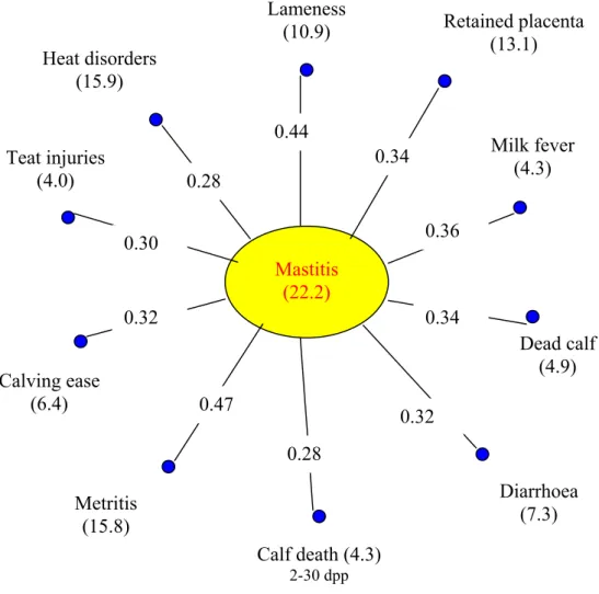
Funke, 1989); Oltenacu et al., 1990; Jánosa et al., 1999; Fleischer et al., 2001) (Figure 1).
However, not all high-production bulls sire high rates of mastitis (Dunklee, 1991; Dohy 1999).
Figure 1. Prevalence of some diseases and its correlations
(n=3216 cow in 90 heard, Brochart et al., 1985; cit.: Janke and Funke, 1989) Mastitis
(22.2) Lameness
(10.9) Heat disorders
(15.9)
Teat injuries (4.0)
Calf death (4.3)
2-30 dpp
Metritis (15.8) Calving ease
(6.4)
Diarrhoea (7.3) Dead calf
(4.9) Milk fever
(4.3) Retained placenta
(13.1)
0.44
0.34
0.36
0.34
0.32 0.28
0.47 0.32
0.30
0.28
3.2. COMPLEXITY OF MASTITIS
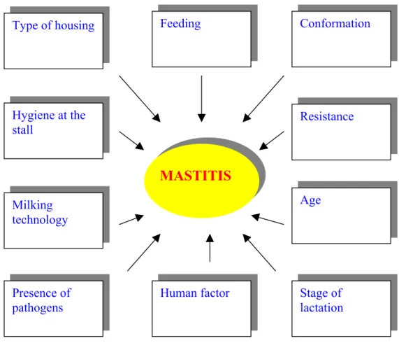
Many authors reported that numerous factors influence the prevalence and increase of mastitis (Figure 2) (Saloniemi, 1980; Horváth, 1982; Dohy, 1985;. Cassell, 1988; Janke and Funke, 1989; Dunklee, 1991; Erskine, 1993;
Süpek, 1994; Süpek, 1995; Gulyás and Iváncsics, 1999; Iváncsics and Gulyás, 1999; Baltay and Bedő, 2000; Baltay et al., 2000; Busato et al., 2000).
Figure 2. Complexity of mastitis (Iváncsics and Gulyás, 1999)
3.2.1. Environment: keeping system and technology
Several studies on the impact of the production environment on udder health have been published in the last years reported Woolford et al. (1998). In
MASTITIS Type of housing
Resistance Conformation
Hygiene at the stall
Milking technology
Human factor Presence of
pathogens
Feeding
Stage of lactation Age
the most recent studies epidemiological methods were employed and the numerous factors influencing the increase in mastitis accurately measured.
Thus, the knowledge on the impact of the production environment on udder health is considerable. Identification and elimination of individual environmental factors that threaten udder health reduces the overall stress on the cow, and so udder health improves. However, it is important to remember that elimination of a single and obvious predisposing factor is not always sufficient to maintain the herd below the disease threshold (Neave et al., 1969;
Saloniemi, 1980; Radostits and Blood, 1985; Rasmussen, 1999).
In many comparison of cow house types fewer cases of mastitis were treated in loose houses than in tie-stall housing because:
• movements of cows getting up and down are less restricted,
• the lying area is usually covered and therefore soft,
• cows are milked at a milking parlour.
However, health differences among the herds was large with both types of housing (Oltenacu et al., 1990; Báder, 1996; Kertész et al., 2001).
A concrete floor gives poor support to the hoof as the cow rises. A stall without a bedding and messy with dung can be very slippery and teat tramp is frequent. Injuries to the skin of the hock are common both in slippery and hard stalls. Damaged skin is a favourable environment for some pathogenic microbes of the udder. Using sufficient straw lessens the incidence of such injuries considerably and also protects the udder from cold and from rubbing on the concrete.
However, population of coliform bacteria increase rapidly in straw and sawdust bedding dirtied with faeces. In dirty bedding streptococci (and some staphylococci) are abundant therefore the risk of (environmental) mastitis increases. Cases of coliform mastitis are associated with dirty loose houses (Golodez, 1985; Hogan et al., 1989b; Oltenacu et al., 1990).
The effect of a night light in reducing the frequency of teats tramp is based on the natural instinct of cows. Cows, in common with other artiodactyls are lived in open areas. In a dark barn they do not equate strange noises with harmless situations and will invariable get up to escape the presumed danger, often tramping their teats as they do so (Seabrook, 1984).
High humidity in the cow house and a draught on the udder increase susceptibility to mastitis. Wetness of the udder due to moist stall floor or due to
frequent washing of the udder increases the deleterious effect of draughts by increasing heat loss from its skin (Seabrook, 1984; Barkema et al., 1999).
In North America, the practice of tail docking dairy cattle is on the increase. Producers cite a number of reasons for docking cows, including improved ease of milking, cow cleanliness and reduced SCC because reducing contact with bacteria via the tail.
Tucker et al. (2001) monitored milking cows after half of the animals in a herd were docked to determine whether tail docking would influence cow cleanliness and udder health in a free-stall system. No treatment differences were found in four measures of cow cleanliness, two measures of udder cleanliness, or udder health. However, analysis of a subsample of cows illustrated individual differences in cleanliness.
The correlation between mastitis and environment is often difficult to interpret as it is difficult to estimate the impact of the herdsman. The incidence of mastitis in the herd may be much less then would be expected in the presence of several potentially deleterious factors if the herdsman is professional and manages milking, feeding, cleaning and other duties with care. When the potential etiological component of the production environment is estimated during investigation of a mastitis herd, both the range of causes of mastitis and the important role of the herdsman must be kept in mind.
It has been estimated that a large part of the variability in occurence of mastitis between herds is explained by differences between the care herdsman manage their herds (Neave et al., 1969; Miller, 1984; Seabrook, 1984; Radostits and Blood, 1985; IDF, 1987b; NMC, 1987; Lawstuen et al., 1988; Markus, 1994; Iváncsics et al., 1996; Barkema et al., 1999; Rasmussen, 1999; Hogeveen et al., 2001).
3.2.2. Milking: technique and hygiene
Milking machine (and nowadays milking robot) was developed to ease the heavy work of manual milking (Moxley et al., 1978; Mein and Thompson, 1993; Justesen and Rasmussen, 2000; Hogeveen et al., 2001). However, Weaver (1982) reported that machine milking can be so rough that the natural defence system of the udder fails to prevent the ingress of bacteria, significantly increasing the risk of mastitis (teat injuries because of vacuum fluctuation and incorrect pulsation). On the other hand, milking machines can transport
pathogens both passively from the skin of one cow to another and actively by dispersing them inside the udder (contagious mastitis) reported O’Shea and Meaney (1979), Grindal and Hillerton (1991), IDF (1994a) etc.
The cow and the machine are linked by the milker. No matter how good the machine is. A careless operator can cause serious teat injuries and active dispersal of pathogens inside the udder. Making the upper part of the udder wet is not recommended because the dripping water carries bacteria down to the teats (IDF, 1994a; Smith and Hogan, 1999). Recovery of the teat from machine milking takes more than half an hour whereas suckling by the calf or with manual milking the teat becomes slightly thinner and returns to normal within half an hour. For this reason it is suggested to provide food directly after milking. The more the teat swells during machine milking the more sensitive the quarter is to infection wrote Nickerson in 1992.
In addition, proper preparation increases milk yield by approximately 10% in comparison with poor preparation. In the design and use of milking machines the starting points must be the demands set by the cow and the milk, techniques must be compatible with the conditions set by biology (Pankey et al., 1984; Hamann, 1990; Hamann and Stanitzke, 1990; Hogan et al., 1990;
Erskine and Eberhart, 1991; Pankey and Drechsler, 1993; Myllys et al., 1994).
Rasmussen et al. (2001) reported that robotic milking (AMS) had a negative influence on udder health measured by an increase in acutely elevated cell counts during the first year compared with the previous year with conventional milking. The number of cows with elevated SCC decreased slowly after 3 months. They did not have a conclusive reason for the increase but similar to IDF (2000), Justesen and Rasmussen (2000) and Hogeveen et al.
(2001) suggested that more focus is needed on the introductory period.
Attention should pay on farms try milk production through organic ways, too (Chamings, 1984; Murdough and Pankey, 1993; Vaarst and Enevoldsen, 1997; Busato et al., 2000; Vorst and Hogeveen, 2000).
3.2.3. Conformation
The conformation of the cow significantly influences predisposition to mastitis reported Hámori (1974), Horváth (1982), Dohy (1985), Seykora and McDaniel (1985) etc. Because of the favourable relationship between some udder traits and the SCC of milk, screening on udder characteristics may have
slowed the genetic increase in LSCS (Rogers et al. 1991; Boettcher et al. 1998).
Rogers et al., (1991) reported genetic correlation of LSCS with udder depth, fore udder attachment and front teat placement to be –0.35, -0.32 and –0.22 respectively. Gulyás et al. (1998) and Gulyás and Iváncsics (1999) also reported the importance of udder morphology and the role of pigmented teat ends. Dohy (1985), Seykora and McDaniel (1986), Schutz et al. (1993) and Dohy (1999) suggested that sire analysts from AI organizations should screen perspective bull-dams for udder conformation traits and should eliminate cows with deep udders or wide front teats from consideration.
The structure of legs and hoofs is an also important predisposing factor.
Movements involved getting feed and lying down are naturally necessary. Leg injuries in a herd are indicative of likely increase in udder diseases. However, long and untreated hoofs are often a question of mismanagement rather than a conformation fault of the cow (Markus, 1999 personal communication).
Culling cows with poor udder shape and leg injuries results in improved udder health.
3.2.4. Genetic background
Several authors pointed out that prevalence of mastitis is genetically determinated (Table 2). Depending on the level of environment it ranges h2=0.02-0.2. The overall mean is about h2=0.1 (Shook and Schutz, 1994).
Breed also has an impact on mastitis. Ayrshire cows have a significantly lower LSCS (Schutz et al., 1994).
Many studies have reported genetic correlation of LSCS and milk yield ranged from –0.2 to 0.48 but most values were closer to the mean of 0.12 (Kennedy, 1982; Kennedy et al., 1982; Ruabertas and Shook, 1982; Monardes and Hayes, 1985; Emanuelson et al., 1988; Banos and Shook, 1990; Boettcheret al., 1992). Kennedy et al. (1982) and so Monardes and Hayes (1985) reported a mean correlation of 0.28 between SCS and milk yield for first lactation, -0.15 for second lactation and 0.05 for third and later lactations. Schutz et al. (1990) suggested that mastitis as indicated by LSCS is more common during first lactations of cows with sires that transmit higher milk yield, perhaps because of the stress from producing more milk.
Table 2. Heritability of mastitis (Pongrácz and Iváncsics, 2001)
Author Year Heritability (h2) Remarks Ward (cit.: Schutz,
1994) 1938
Lush 1950
? susceptibility
Miller 1984 0.12 (0 – 0.5)
Emanuelson et al. 1988 0.01-0.02
Simianer et al. 1991 0.05
Weller et al. 1992 0.01 (field study)
cases of treated cows
O’Bleness et al. 1960 0-0.07 Norman and Van Vleck 1972 0-0.07
Lawstuen et al. 1988 0.03
marks of resistance
Hansen et al. 1979 0.18
Miller 1984 0.11
Weller et al. 1992 0.04
bacteriological status Shook and Schutz 1994 0.1 (0.02-0.2)
Mean correlation of LSCS and fat yield is about 0.2 while the genetic relationship of LSCS and protein yield found estimates of correlations ranged from –0.14 to 0.54 and had a mean of 0.17 (Kennedy et al., 1982; MacMillin et al., 1983; Monardes and Hayes, 1985; Weller et al., 1992). It means that genetic gain can be achieved in selection for lower SCS only by decreasing selection emphasis on milk and protein yields.
3.2.5. Other factors
Influences of age and season of calving on LSCS are documented (Norman and Van Vleck, 1972;. Coffey et al., 1986; Miller et al., 1991; Schutz et al., 1994). Solutions for LSCS increases with age and rates of increases are steeper after about 36 months of age (Laevens et al., 1997). Effects of month of calving are smaller than age at calving but LSCS is higher for cows calving during midsummer.
Smith et al. (1984), Thomas et al. (1990), Tucker et al. (1992), Erskine (1993), Block (1994) and Hogan et al. (1996) reported the role of feeding, mainly through the minerals and vitamin supply.
3.3. PHYSIOLOGY OF MASTITIS
3.3.1. Infection
When bacteriological examination of single quarter milk samples and careful analysis of the inflammation are carried out in parallel bacteria are found in approximately 80% of the inflamed quarter milk samples. Some 20%
of the inflammations are such that the irritant is something other than bacteria sequestered at the moment in the udder, or the number of bacteria in milk might be too low to be detected, or the host has already eliminated the infection (IDF, 1975a; Piddock, 1990; Huszenicza and Albert, 2000; Egyházi and Hargitai, 2001).
Incorrect milking causes small traumas in the teat ends and they become predisposed to bacterial colonization, easily followed by infection of the
quarter. The milker may also act as an infection source.
According to Fox and Gay (1993) contagious mastitis appears as high cell counts in milk. In a herd infected with contagious mastitis the causal
bacteria are usually S. aureus. In some herds the Coagulase Negative Staphylococci (CNS) predominate. Furthermore, Str. agalactiae and Str.
dysgalactiae can cause contagious mastitis, too (Anderson, 1982; Harmon and Langlois, 1989; Devriese, 1990; IDF, 1994b).
A characteristic of staphylococcal mastitis is that the number of bacteria in the milk varies greatly between repeated samples. The somatic cell count closely paralells the number of bacteria in milk (Tolle, 1982; Sanchez, 1988). S.
aureus infections often turn chronic (Schukken et al., 1993). S. aureus (and Str.
agalactiae) can survive on the mucous membrane. Therefore, milk from infected cows not allowed/suggested to (heifer) calves (Saperstein et al., 1988;
Sandholm et al., 1990; Nickerson et al., 1993). Vaccination against staphylococci is not a practical tool nowadays (Watson, 1992; Markus, 2000 personal communication).
Environmental mastitis is associated with a relatively high incidence of clinical (related to subclinical) mastitis in the herd that may fluctuate according to season (Smith et al., 1985a; Smith et al., 1985b; IDF, 1987b; Smith and Hogan, 1993). The bacteria isolated from the clinical cases are usually gram
negative or Str. uberis (Hogan et al., 1988). Cases of E. coli mastitis seldom become chronic (Golodez, 1985; Hogan et al., 1989a; Hogan et al., 1992).
With herds suffering from Str. uberis infections that often occur during the dry period the bacteria can be isolated from nearly all infected quarters. The sources of infection are generally the cow and its immediate environment, particularly the bedding (Golodez, 1985; Hogan et al., 1989b; Oltenacu et al., 1990;Williamson et al., 1995).
Klebsiella infections are typically very difficult to treat, and the infected animals usually have to be culled. Before the mastitis problem is found to be caused by Klebsiella, a few cows may already have been lost. Humid sawdust and chippings favour the growth of Klebsiella. The problem is often solved when sawdust storage is improved (dry storage) or preferably sawdust is replaced with some other bedding material.
With Pseudomonas infections, contaminated water is the most likely infection source. The detergents used for cleaning the milking equipment are potential sources as Pseudomonas can grow in the detergents.
/Summer mastitis was first described at the beginning of the twentieth century.
However, a precise definition of summer mastitis, which all researchers agree upon, has not yet been made. This mastitis type also goes under other names: heifer mastitis, dry cow mastitis, fly mastitis and pyogenes mastitis.
Summer mastitis occurs mainly in heifers and dry cows, and is typically associated with strong clinical symptoms with a suppurative and foul-smelling secretion from the quarter. Despite treatment the affected quarter is usually lost from milk production. Summer mastitis is most common towards the end of summer when Hydrotea irritans, the fly considered to be the main vector of the disease, is abundant.
The causing microbe is Actinobacillus (Corynebacteria) pyogenes. The distribution of typical summer mastitis correlates with that of the fly; the disease is found in northern Europe and Japan, but never in Hungary.
A syndrome resembling summer mastitis is also found during the indoors season, affecting dry cows in particular, but also lactating cows. The disease develops following a teat injury. Summer mastitis is a mixed infection caused by aerobic and anaerobic bacteria and thus a comprehensive name for it would be anaerobe-aerobe- mastitis.
Mycoplasmas are pleomorphic bacteria which lack a cell wall. They are smaller and structurally more simple than other bacteria. Approximately 70 species of
mycoplasmas are known, of which at least five cause bovine mastitis, the most important being Mycoplasma bovis and M. bovigenitalium (Erno and Perreau, 1985).
A mycoplasmal mastitis infection in the herd typically results in a fast spreading epidemic. The infection easily spreads during milking and is often widespread before the first symptoms are noticed. Morbidity in the herd depends on how fast the disease is recognized (and milking hygiene). Cows of all ages and at all stages of lactation are sensitive to mycoplasmal infections. Mycoplasmal mastitis may affect 50-60% of the cows.
This type of mastitis is characterized by a sharp drop in milk production and extremely swollen udders that are not painful. In the early stages the milk secretion becomes watery in appearance and contains fibrin flakes. Characteristically the milk, on standing e.g. in a test tube, rapidly separates into a flaky deposit and a clear supernatant. Later the milk becomes purulent. The quantity of leukocytes is usually high (106-103/ml) and the milk contains high numbers of mycoplasmas (up to 109/ml).
A definitive diagnosis of mycoplasmal mastitis requires culture of the organism and species identification. Mycoplasmas require complex media containing sterols for growth. Therefore, they are not detected in routine mastitis culture of milk specimens. If mycoplasmal mastitis is suspected on the basis of clinical symptoms, it must be mentioned when the milk sample is sent to the laboratory (Rickhard et al., 1980; Horváth, 1982; Bushnell, 1984; Huszenicza and Albert, 2000)./
3.3.2. Defence mechanisms
The teat canal represents a physical barrier to the penetration of bacteria. It remains open after milking for approximately two hours. The cow should be prevented lying down during this critical period (the role and importance of feeding). The idea of post-milking teat dipping is to disinfect the teat canal and reduce the risk of ascending infection (Guidry, 1985; Nickerson, 1985; Nickerson, 1992).
Epithelial desquamation and milk flow are mechanisms of the host to decrease local bacterial colonization. The keratin layer contains basic antibacterial proteins and antibacterial fatty acids (Dohy, 2000). Specific immunological factors also play a role in the defence of the teat canal.
Lymphocytes and plasma cells accumulate beneath and between the epithelium of the teat canal wall, particularly around the Fürstenberg’s rosette. This indicates local immunological activity. Neutrophil phagocytes directly penetrate the teat wall to the infected and inflamed teat canal (Nickerson, 1985;
Korhonen and Sandholm, 1995).
Besides the anatomical-physical defence mechanism of the teat canal, the immune system of the mammary gland consists of both humoral and cellular components (Davidson et al., 1982; Sears, 1984; Craven and Williams, 1985; Guidry, 1985; Sheldrake and Husband, 1985; IDF, 1991a; IDF, 1991b;
Knight, 1991; Tyler et al., 1993; Sandholm and Korhonen, 1995).
Immunoglobulins have specific antibody activity against antigenic stimuli form the humoral component. The cellular components consist of several different cell groups.
The total amount of immunoglobulin varies during the stage of lactation, as does the relative proportion of the different Ig-subclasses (isotypes). In colostrum the immunoglobulin content is as high as ~100 mg/ml but falls within the first week of lactation to less than l mg/ml. This low level is hardly of any importance in defence of the udder. This concentration is very low in comparison with that in human or sow milk. However, the antibacterial factors in milk have a concerted action and it is difficult to judge the relative importance of an individual factor like lactoferrin, transferrin, lysosyme, lactoperoxidase or complement (Reiter, 1978; IDF; 1985; Saeman et al., 1987;
Sandholm and Korhonen, 1995).
The most important feature of antibodies in milk is their opsonizing ability: alien antigens become labelled and the phagocyting granulocytes and macrophages are directed to their targets. In addition, the antibodies neutralise toxins and are occasionally directly bactericidal (Andrews, 1983). An antibody can kill directly via the combined effect of the antibody and the complement, or via the concerted action of complement and lysozyme.
Bacterial elimination, which takes place through phagocytosis, is however considered as the most important antibacterial mechanism of the udder (Sandholm and Korhonen, 1995).
Lymphocytes
During lactation normal milk contains a small number of lymphocytes.
Approximately 50% of the lymphocytes in normal milk belong to various T-cell subsets and 20% to B-cells. The remaining cells belong to so-called null-cells.
The milk lymphocytes have been found to respond to antigen and mitogen stimulation in vitro. It can be presumed that most of the interaction between the lymphocytes and the macrophages occurs locally in the supramammary lymph nodes. Some lymphocyte activity can be observed in the Fürsterberg’s rosette and beneath the mammary gland epithelium. Antigens stimulate the subepithelial B-cells to multiply and differentiate into plasma cells producing secretory antibodies
Phagocytes
Macrophages
Macrophages are phagocytic cells that serve to remove tissue debris and bacteria. Milk from a healthy udder contains approximately 105 cells/ml, most of which are macrophages. Macrophages are the first cells to encounter bacteria and process information to other cells involved in host resistance. These cells function as recogniser and alarm cells in initiating an inflammatory reaction and immunity. They phagocyte and destroy bacteria, process antigens for the immune system, regulate the function at the lymphocytes and regulate the inflammatory cascade by secreting various cytokines and other mediators.
Macrophages accumulate during involution of the mammary gland phagocyting residual milk, tissue debris as well as bacteria (Sanchez, 1988).
Polymorphonuclear neutrophils (PMN)
During inflammation the SCC of the lactating gland can increase to more than 106 cells/ml. Most of this increase is due to neutrophils that move from blood into milk and milk gets a pus-like appearance (Kehrli et al., 1989).
The function of the granulocytes is based on their ability to adhere to endothelium at the site of inflammation and to find their way from the blood to the site of the inflammatory focus to phagocyte and destroy bacteria and to remove damaged tissue. The reaction of the neutrophils is considered to be the most important defence and cleaning mechanism of the udder. If the cells of the granulocyte series are destroyed, the cow is incapable of clearing an infection within the mammary gland.
Intramammary inoculation with graded doses of bacteria has shown that the udder is significantly protected against ascending infections when the somatic cell count of milk is 106/ml or higher. In this case > 90% of cells belong to PMN. There has been a lot of interest in increasing the number of
PMN in milk in the hope that the udder would clear infections without the need for antimicrobials. In order to increase the number of PMN in milk, mechanically irritating intramammary devices were developed, e.g. a plastic loop to be inserted in the milk cistern. Another approach is to enhance production and stimulate transfer of PMN to the site of inflammation. This can be done by colony-stimulating factors and those cytokines which enhance the adhesion and transfer process.
The phagocytes kill bacteria by activating oxygen (oxygen burst) through the NADPH-oxidase – myeloperoxidase system. Therefore, the antibacterial mechanism is analogue to LP. Several lysosomal enzymes are involved in the lysis of the bacterial cell wall. The defensins produced by the phagocytes have recently aroused interest. They are small proteins (MW <
4000) that have an antibiotic-like effect on bacteria.
Phagocytes do not function effectively in the milk compartment of the udder. The neutrophils of milk phagocyte and destroy less effectively than the corresponding cells in blood. The phagocytic effect of the granulocytes is relatively poor in milk due to low energy reserves (the glucose content of milk is low) and low opsonin content (antibodies, complement). In the milk compartment, the granulocytes waste their capacity by phagocyting casein and fat globules. A steady transfer of fresh neutrophils from blood into milk is required for local defence against new infections.
3.3.3. Inflammation
Inflammation is defined as the response of the body to an alien substance (e.g. bacteria, bacterial metabolite or toxin), or tissue injury. Once
activated, the body’s inflammation mechanisms can combat microbial infections and pave the way for resolution and restoration of normal function (Guidry, 1985; Gallin et al., 1988; Knight, 1991; Sandholm, 1995; Sandholm
and Korhonen, 1995; IDF, 1996b).
The microcirculation plays a crucial role in the initial events of the inflammatory reaction. The inflammation response consists of three stages:
1) Inflammation begins with endothelial reactions at the responding tissue site.
During this acute, transient phase the local capillary circulation increases and the permeability of the capillaries increases as the endothelial cells contract. Inter-endothelial gaps are formed through which plasma and its
proteins leak in the interstitium causing oedema. Blood leukocytes begin to adhere to endothelium.
2) During the subacute phase, phagocyting cells migrate from the circulation to the infection site.
3) Tissue degeneration, regeneration and formation of fibrotic tissue is characteristic to the chronic proliferative phase.
Sometimes the inflammation fails to eliminate the causal microbe; in subclinical mastitis the udder maintains the inflammation without being able to eradicate the bacteria sequestered in the milk compartment.
3.3.3.1. Physiological inflammation
As milk production ceases towards drying-off the udder involutes and the body initiates a sterile inflammation in the udder which prepares it for repair and cleaning. Oestrogens play an essential role in the drying-off of the udder and in creating a sterile inflammation. The alveolar epithelial cells disappear by an apoptotic process. Udder macrophages are particularly aggressive during this period when they clean milk ducts of milk and other debris. The udder efficiently clears latent infections during involution. As a consequence of the inflammation milk plasmin is activated which leads to degradation of casein and absorption of the products. The fibrotic tissue developing in the udder during involution is considered as part of the inflammatory reaction (Afifi, 1968; Ali and Shook, 1980; Andrews, 1983; Guidry, 1985; Gallin et al., 1988;
Browning et al., 1990; Knight, 1991; Sandholm, 1995; Sandholm and Korhonen, 1995; IDF, 1996b; IDF, 1996c).
3.3.3.2. Invasion of leukocytes to the mammary gland
According to Afifi (1968), Ali and Shook (1980), Andrews (1983), Guidry (1985), Gallin et al. (1988), Browning et al. (1990), Knight (1991), Sandholm (1995), Sandholm and Korhonen (1995), IDF (1996b) and IDF (1996c) leukocytes migrate extensively throughout the body to mediate immune surveillance and to mount inflammatory responses to foreign antigens.
Neutrophils are rapidly recruited in large numbers from blood to the site of inflammation. Other circulating cells such as lymphocytes, platelets and eosinophils may also retained at the inflammatory area. Lymphoid T-cells are
recruited later and more selectively than neutrophils to sites of inflammation where they have antigen-restricted functions .
Normal milk contains some 105 somatic cells/ml, most of which are macrophages. These function as recogniser and alarm cells in the udder. During an emergency, these cells begin to secrete substances which attract neutrophils from blood to the inflammation site. Most of the somatic cells in mastitic milk are neutrophil granulocytes. The purpose of the invading granulocytes is to clear the inflammation area of foreign matter including bacteria and tissue debris (Nickerson, 1985; Sandholm, 1995).
To reach the inflammation site from the blood compartment the leukocytes have to evade the circulatory system. The leukocyte migration into tissue is regulated by adhesion to endothelium. The endothelium of the postcapillary venules at the inflammatory site become adhesive to various leukocytes; the leukocytes within the blood flow initially come into brief contact with the vessel wall, slowing their movement, and roll on the endothelium. Over the next few minutes the cells undergo diapedesis and migrate between endothelial cells into tissue.
Endothelial cells also express immunoglobulin superfamily adhesion proteins. The selectivity of various endothelial adhesion proteins towards specific leukocyte subsets and chronological expression of various adhesion proteins results in selective granulocyte infiltration in the acute phase and mononuclear cell (lymphocytes, monocytes) infiltration in the chronic phase of inflammation.
3.3.3.3. Infection - inflammation
It is widely accepted that the predominant cause of mastitis is intramammary infection by a microorganism, usually bacteria. The infection, which results from bacteria entering the udder through the teat has traditionally been considered the primary process in mastitis (Afifi, 1968; Ali and Shook, 1980; Andrews, 1983; Guidry, 1985; Gallin et al., 1988; Browning et al., 1990;
Grindal and Hillerton, 1991; Knight, 1991; Sandholm, 1995; Sandholm and Korhonen, 1995; IDF, 1996b; IDF, 1996c).
Whatever is the reason for inflammation, the change in the composition of milk makes it favourable for the growth of certain bacteria. Mastitis pathogens grow faster in milk from inflamed quarters than in normal milk,
although the levels of the endogenous antibacterial factors of milk (phagocytes, antibodies, complement factors, lysozyme, lactoferrin etc.) are elevated in mastitic milk. When foremilk from hand-milked and machine-milked quarters within cows are compared, machine milking induce a change in foremilk to support growth of pathogens. This means that current machine milking technique is traumatizing and induces faint inflammation within the teat.
Infection and inflammation are dynamic processes (Figure 3). The cow’s response to the infection is inflammation. Clinical mastitis is, in most cases, short-lived and becomes subclinical, latent mastitis, and the inflammation response is suppressed subsequent. Analysis of samples taken from consecutive milking shows that short term subclinical infections are surprisingly common. The bacteria are eliminated quickly in most cases, but the inflammation takes longer to disappear (Guidry, 1985; Sandholm, 1995; IDF, 1996b; IDF, 1996c).
Figure 3. Dynamic of mastitis (Sandholm, 1995)
3.3.4. Changes in the composition of milk
Inflammatory reactions change the composition of milk in terms of quantity and quality reported many authors like Kitchen (1981), Harmon
(1994), IDF (1996a) etc.
Several phenomena connected with inflammation occur simultaneously:
1) the permeability of the blood vessels increases resulting in the passage of ions and proteins from blood to milk,
good health
clinical mastitis subclinical mastitis
2) inflammatory cells move from blood to milk,
3) the epithelial cells, which produce milk, become less efficient; cells break down and enzymes are released.
3.3.4.1. The effect of mastitis on milk yield
Reduction in milk yield is one of the clearest symptoms of mastitis.
Reduction in yields depends on the degree of inflammation. This can be estimated from the somatic cell count in milk (Table 1). As the somatic cell count exceeds 100,000/ml, milk quantity begins to decrease linearly in relation to the logarithmic value of the cell count reported Eberhart et al. (1982), NMC (1987), Cullor (1992), DeGraves and Fetrow (1993), Smith and Hogan (1999)
and Ózsvári et al. (2001).
Somatic cell count also reflects the changes that occur in the composition of milk (Griffin et al., 1977; Ali and Shook, 1980; Kitchen, 1981;
Munro et al., 1984; Monardes and Hayes, 1985; Mattila, 1985; Mattila and Sandholm, 1986;.McFadden et al., 1988; Harmon, 1994; Korhonen and
Kaartinen, 1995; IDF, 1996a etc.).
3.3.4.2. Physical changes
Along with the yield changes (and the chemical and bacteriological properties), the physical traits of mastitic milk change, too (Szakály, 1982;
Politis and Ng-Kwai-Hang, 1988a; Korhonenand Kaartinen, 1995; IDF, 1997;
Woolford et al., 1998; Szakály, 2001). Conductivity, pH and viscosity increases while density decreases and buffering capacity and titratable acidity shows no changes. The reduction ability increases.
3.3.4.3. Changes in the chemical composition
Mastitis affects both the quantity and the quality of milk. As the degree of inflammation increases, the chemical composition of milk approaches more and more that of blood because the components filter from the blood circulation into the mammary gland (Munro et al., 1984; Monardes and Hayes, 1985;
McFadden et al., 1988; Jensen and Knudsen, 1991; Korhonen and Kaartinen, 1995). The changes in quantities of individual components vary.
Milk synthesis diminishes when the udder tissue is inflamed.
Consequently the quantities of the major components of milk decrease and the total dry matter drops by 5-15%. There is a significant negative correlation
between the somatic cell count and the dry matter content of milk (Kitchen, 1981; Mattila, 1985; Mattila and Sandholm, 1986).
The results of changes in the fat content of milk caused by mastitis are diverse. According to the results of most investigations, the fat content decreases by less than 10%. The fat composition, however, changes considerably, lowering the quality of milk products. The total amount of fatty acids remains unchanged, but the quantity of free fatty acids increases. On the other hand, the amount of phospholipids diminishes due to the decrease in the amount and size of fat globules. The membrane matter of fat globules decreases by approximately 10% and its composition changes in comparison with that of a healthy cow’s milk. The composition of fatty acids changes so that the amount of short-chained fatty acids (C4-C12) increases slightly and the amount of the long-chained fatty acids (C16-C18) drops correspondingly. The quantity of unsaturated long-chained fatty acids is, however, higher in mastitic milk than in normal milk. The changes in the lipid phase increase the lipolytic sensitivity in mastitic milk. This is intensified by the increased lipase activity.
The total quantity of milk proteins does not decrease clearly until the SCC exceeds 1,000,000/ml. The ratios between the different proteins, however, change at a much lower SCC.
A highly significant negative correlation exists between lactose content and SCC (Seelemann, 1964).
The changes of mineral and trace element contents of milk have considerable importance both for processing properties and its nutritive value (Szakály, 2001).
The quantity of water soluble vitamins fall by 10-50%. The changes affect bacteriological fermentation process and lower the quality of sour milk products reported Szakály (1982).
Mastitis generally increases the enzymatic and biochemical activity in milk. Some of these characteristics have for many years been used to detect mastitis. Increased biochemical activity in milk may in particular cause faulty fermentation of sour milk products and induce various quality problems (Szakály, 1982; Renner, 1983; Heeschen et al., 1985; Politis and Ng-Kwai- Hang 1988b; Ma et al., 2000; Szakály, 2001).
3.3.4.4. Microbiological changes
Normal milk contains several kind of bacteria, too. Based on the update regulations the legal limit of total plate count is less than 100,000 cells/ml at first class (highest quality, called "extra") milk in Hungary. Sometimes it can detect as much as 106 cells/ml in infected milk samples. However, 79% of the milk that have been sold to dairy plants contained less than 50,000 cells/ml because of the strict monitoring system. This reduction is a very important point of quality requirements (Hargitai et al.,1989; Unger, 1996; Markus, 2000).
The majority of mastitic cases are caused by the so called contagious S.
aureus, Str. agalactiae, sometimes Corynebacterium bovis and perhaps Mycoplasma bovis and other Mycoplasmas. The microbiological background of environmental mastitis is usually E. coli, Str. uberis and other Str., Bacillus and Nocardia spp., Pasteurella, Actinobacillus and Klebsiella spp. and some yeasts.
CNS spp. and Str. dysgalactiae are frequently isolated and sometimes Pseudomonas aeruginosa and Prototheca zopfii can be detected, too. These species can be either contagious or environmental based on the circumstances (IDF, 1975a; Horváth, 1982; Hargitai et al., 1989; Huszenicza and Albert, 2000; Egyházi and Hargitai, 2001).
3.4. DIAGNOSTICS OF MASTITIS
From the several possibilities of diagnosing mastitis we just deal with the few most important and practically used methods. However, many other tools were developed that vary in accuracy (IDF, 1975a; Kitchen, 1981;
Brolund, 1985; Hoare et al., 1980; Klastrup, 1985; Mattila, 1985; Sandholm and Mattila, 1985; IDF, 1987a; Swets, 1988; Sears et al., 1993).
3.4.1. Examination
A preliminary diagnosis is based on the health record of the cow and its clinical signs. The focus is on the individual cow/quarter but the case must always be seen in a broader perspective, i.e. the herd.
It is important to check the individual health record and earlier treatments. The exact calving date should be available.
According to Markus (1996, personal communication) the clinical examination must proceed systematically. Starting from a carefully documented case history is useful. The general examination should include assessment of the posture, behaviour, body condition, general condition, respiratory rate, pulse frequency, rumen motility and body temperature. The general examination of the cow will indicate how much the general health of the cow is affected by the udder disease.
Then the udder itself is examined by inspection, palpation and examination of quarter milk secretion and milk appearance. Inspection takes account the size, shape and symmetry of the udder and teats by viewing it from behind and each side.
The anatomical/clinical unit of the udder is the quarter. Therefore, diagnostic methods must be applied to each quarter if mastitis is present. Inter- quarter comparison is helpful in recognizing abnormal quarters.
Asymmetry of the udder is usually due to atrophy of one quarter, or on the other hand, enlargement caused by oedema (Al-Ani and Vestweber, 1986;
Nestor et al., 1988). Skin of the udder and teats is inspected for injuries, discoloration or other abnormalities. Special attention should be paid to the teat orifices. Palpation includes teat canal and cistern, udder cistern, glandular tissue and skin, and supramammary lymph nodes. The udder is best palpated immediately after milking.
Milk sample can be examined first physically, then chemically and microbiologically if necessary. The CMT is a practical cow-side test for detecting mastitis in milk. The milk should be inspected for clots, discoloration or wateriness before adding the CMT reagent.
3.4.2. Detection of inflammatory changes in milk
Obviously, a trait is allowed to substitute for another if it is genetically correlated with the other trait, if recording is less expansive or easier, if measurement is earlier in life or, if heritability is higher.
Because of a strong relationship between some of the inflammatory or compositional changes in milk and the presence of infection, the measurement of certain components is used to monitor udder health, and so milk quality (Griffin et al., 1977; Ali and Shook, 1980; Kitchen, 1981; Munro et al., 1984;
Brolund, 1985; Monardes and Hayes, 1985; Mattila, 1985; Sandholm and
Mattila, 1985; Mattila and Sandholm, 1986; McFadden et al., 1988; Hutton et al., 1990; Harmon, 1994; Korhonen and Kaartinen, 1995; Sandholm et al., 1995; Hillerton, 1996; IDF, 1996a etc.). So, several tests are available to determine the presence or absence of clinical and subclinical (unseen) udder infection. These range in difficulty and sensitivity from the very simple strip test to sophisticated laboratory procedures which detect the presence of microorganisms or some changes of composition. All of these tests serve a need and can be useful if conducted properly and interpreted correctly.
3.4.2.1. Compositional changes
Some proposed screening tests for monitoring the course of infections include the measurement of catalase, NAGase, antitripsin, chloride, sodium, serum albumin, and SCC in milk (Mattila, 1985; Woolford et al., 1998). The majority of these tests primarily indicate inflammation in the udder. They do not measure infection or bacterial presence.
Somatic cell count (SCC) has been most widely used as a measure of milk quality, indicator of mastitis, and a management tool to control mastitis worldwide (IDF, 1975b; Griffin et al., 1977; Ali and Shook, 1980; Kitchen, 1981; Dahoo et al., 1981; Horváth, 1982; Bramley and Dodd, 1984; Munro et al., 1984; Brolund, 1985; Mattila, 1985; Monardes and Hayes, 1985; Sandholm and Mattila, 1985; Mattila and Sandholm, 1986; Harmon, 1994; Korhonen and Kaartinen, 1995; Sandholm et al., 1995; IDF, 1996a; Barkema et al., 1999; IDF, 1999b).
Genetic correlation between SCC and clinical mastitis or bacterial status is moderately high. Young et al. (1960) estimated the correlation of SCC and clinical mastitis to be 0.8 or 0.98 from two methods. Afifi (1968) reported a correlation of 0.83. Using more appropriate statistical techniques, Emanuelson et al. (1988) found a genetic correlation of 0.46 for Swedish Black and White cattle and 0.78 for Swedish Red and White cattle from a field study. Weller et al. (1992) found a smaller genetic correlation between SCC and clinical mastitis of 0.3 but the genetic correlation estimated to be near 1 of SCC with bacterial infections status. Thus, selection for lower SCC would apparently reduce subclinical as well as clinical mastitis (Afifi, 1968; McDaniel, 1984; Dohy, 1985; Emanuelson et al., 1988; Powell, 1992; Weller et al., 1992; McDanielet al., 1993; Vági, 1996; Vági, 1998; Dohy, 1999; Dohy, 2000; Iváncsics et al., 2001).
Nearly all developed countries have adopted upper regulatory limits for SCC in milk (Model, 1994; IDF, 1996c; Savelle et al., 2000; Hogeveen et al., 2001). Hungary, the EU and the Nordic countries, Switzerland, New Zealand, Australia etc. all accept 400,000 cells/ml as the legal limit for a high quality bulk tank milk sold in the commercial market place but further reductions likely will occur (EEC 92/46, amend 94/71). The EU is already discussing lowering the regulatory SCC limit to 300,000 or perhaps even 250,000 cells/ml reported Hillerton (2001) and Schukken (2001). Canada has now agreed on 500,000 cells/ml throughout all of the provinces and is discussing the possibility of going to 400,000 cells/ml. The limit for SCC in the USA is 750,000 cells/ml but also will be officially reduced (Pongrácz and Iváncsics, 2001).
The following discussion presents the most commonly used/known tests to indicate elevated SCC including the strip test, the California Mastitis Test (CMT), the direct microscopic somatic cell count (DMSCC), the electronic somatic cell count (ESCC), the differential cell count (DCC) and the differential cell stain (DCS) mainly according to relevant literature (IDF, 1981;
Kitchen, 1981; Sandholm and Mattila, 1985).
Strip Test
The strip cup or strip plate is indispensable in the milking parlour for determining the presence of clinical mastitis. The milking machine operator visually examines the foremilk for gross abnormalities by squirting a few streams of milk onto the strip cup. The test is rapid and can easily be adapted as a part of the normal milking routine.
California Mastitis Test (CMT)
The California Mastitis Test (CMT) is a simple, inexpensive and rapid screening test that estimates the number of somatic cells present in milk.
It is conducted by mixing the test reagent with an equal quantity of milk. The reagent reacts with the DNA of the somatic cells in the milk to form a gel. The reaction is then visually scored as 0 (or N, negative), T (Trace), 1, 2, or 3, depending upon the consistence or amount of gel that forms (Table 3). The more viscous the gel, the higher the score. This indicates the presence of a higher number of somatic cells (Schalm et al., 1971; IDF, 1975b; IDF, 1981;
NMC, 1987).
Table 3. Approximate ranges in SCC for CMT scores (NMC, 1987) CMT score SCC range
(1000 cells/ml)
N (0) 0-200
T 200-400 1 400-1200 2 1200-5000 3 5000- A simplified method of scoring is: Negative (N), Suspect (S) and Positive (P). Negative corresponds to 0 on the traditional system while Suspect corresponds to Trace and 1. Scores of 2 and 3 on the traditional method are scored Positive in the simplified system.
This procedure can be used in several ways. Bulk tank, composite samples from individual cows and individual quarter samples can all be examined using the CMT procedure. Each is valuable in monitoring udder infection; however, the interpretation is different depending upon the type of sample.
Direct microscopic somatic cell counting (DMSCC)
The direct microscopic somatic cell counting (DMSCC) is the most accurate of the mastitis screening tests when conducted properly. For this reason, regulatory agencies generally use this test for confirmation of high somatic cell counts based on other tests. This test is also the standard by which all other tests are calibrated.
Studies identifying cell types in milk have shown that somatic cells in milk are primarily (75%) leukocytes which include macrophages, lymphocytes and polymorphonuclear neutrophil leukocyte (Sandholm et al., 1995).
Leukocytes increase in milk in response to infection (or injury). They are the body's primary defence against microorganisms and disease.
Epithelial cells (25%) that are secretary and lining cells, on the other hand, increase as a result of injury (or infection). They indicate that damage to body tissue, particularly udder tissue, has occurred. They are in fact dead cells that have been sloughed from the alveoli and canals within the udder.
