METAGENOMIC ANALYSIS OF BAT GUANO SAMPLES REVEALED THE PRESENCE OF VIRUSES POTENTIALLY
CARRIED BY INSECTS, AMONG OTHERS BY APIS MELLIFERA IN HUNGARY
Brigitta ZANA1,2, Gábor KEMENESI1,2, Péter URBÁN2,3, Fanni FÖLDES1,2, Tamás GÖRFÖL4,5, Péter ESTÓK6, Sándor BOLDOGH7, Kornélia KURUCZ1
and Ferenc JAKAB1,2*
1Virological Research Group, Szentágothai Research Centre, University of Pécs, Ifjúság út 20, H-7624 Pécs, Hungary; 2Institute of Biology, Faculty of Sciences, University of Pécs, Pécs, Hungary; 3Microbial Biotechnology Research Group, Szentágothai Research Centre, University of Pécs, Pécs, Hungary; 4Institute for Veterinary Medical Research, Centre for Agricultural Research, Hungarian Academy of
Sciences, Budapest, Hungary; 5Department of Zoology, Hungarian Natural History Museum, Budapest, Hungary; 6Department of Zoology, Eszterházy Károly College,
Eger, Hungary; 7Aggtelek National Park Directorate, Jósvafő, Hungary (Received 13 November 2017; accepted 12 February 2018)
The predominance of dietary viruses in bat guano samples had been de- scribed recently, suggesting a new opportunity to survey the prevalence and to de- tect new viruses of arthropods or even plant-infecting viruses circulating locally in the ecosystem. Here we describe the diversity of viruses belonging to the order Picornavirales in Hungarian insectivorous bat guano samples. The metagenomic analysis conducted on our samples has revealed the significant predominance of aphid lethal paralysis virus (ALPV) and Big Sioux River virus (BSRV) in Hunga- ry for the first time. Phylogenetic analysis was used to clarify the relationship to previously identified ALPV strains infecting honey bees, showing that our strain possesses a close genetic relationship with the strains that have already been de- scribed as pathogenic to honey bees. Furthermore, studies have previously con- firmed the ability of these viruses to replicate in adult honey bees; however, no signs related to these viruses have been revealed yet. With the identification of two recently described possibly honey bee infecting viruses for the first time in Hungary, our results might have importance for the health conditions of Hungari- an honey bee colonies in the future.
Key words: Aphid lethal paralysis virus, Big Sioux River virus, Rhopalosiphum padi virus, Apis mellifera, honey bee, bat guano
Besides representing the second largest group of mammals, bats are dis- tributed all around the world with the exception of the two arctic areas (He et al.,
*Corresponding author; E-mail: jakab.ferenc@pte.hu
152 ZANA et al.
2013; Calisher et al., 2006). In recent years, bats have received increasing atten- tion in two perspectives: they possess major ecological impact and they serve as natural hosts for a large variety of viruses that may pose a threat to human and animal health (Li et al., 2010; Dacheux et al., 2014; Wang et al., 2015). Howev- er, recent studies analysing the virus assemblages of insectivorous bat guano samples have demonstrated the predominance of dietary viruses, suggesting that next generation sequencing techniques enable us to obtain approximate infor- mation on arthropod or even plant-infecting viruses circulating locally in the eco- system (Donaldson et al., 2010; Li et al., 2010; Ge et al., 2012; He et al., 2013;
Dacheux et al., 2014; Kemenesi et al., 2016).
Rhopalosiphum padi virus (RhPV), aphid lethal paralysis virus (ALPV) and Big Sioux River virus (BSRV) are members of the Dicistroviridae family (Moon et al., 1998; Runckel et al., 2011). Rhopalosiphum padi virus is an aphid pathogen, which is maintained in nature by horizontal transmission through plants as passive reservoirs (Moon et al., 1998). The two latter viruses (ALPV and BSRV) had been first linked with honey bees during a comprehensive meta- genomic survey on honey bee colonies conducted in the USA in 2011. The fre- quent detection of the virus in samples originating from different locations and collection times raised the possibility that ALPV was not just of forage (pollen or nectar) origin and presumably caused infection (Runckel et al., 2011). Later stud- ies proved its infective potential to honey bees along with its capability of verti- cal transmission (Ravoet et al., 2015). The presence of ALPV in European honey bee colonies was first reported in Spain in 2013. This new strain showed high sequence similarity to viruses previously detected in the USA (Granberg et al., 2013). Soon thereafter, a study identified the presence of the virus in Belgium where the detected ALPV nucleotide sequences showed the highest identity to the Spanish and American honey bee related ALPV strains (Ravoet et al., 2013).
In this study, we first describe the presence of honey bee infecting viruses in bat guano samples in Hungary.
Materials and methods
Sample collection
In this study, we investigated randomly selected guano samples collected in multiple localities of Hungary. The study was approved by The National In- spectorate for Environment, Nature and Water (No. 14/2138 – 7/2011); no ani- mals were invasively sampled or harmed during collection. Samples were col- lected as described by Kemenesi et al. (2014). Until laboratory processing, all samples were stored in 500 µl RNAlater Stabilization Reagent (Qiagen) and kept on dry ice.
Sample preparation and viral nucleic acid extraction
Following homogenisation in 600 μl PBS, bat guano samples were centri- fuged at 17,000 g at room temperature for 10 min. Before library preparation for Ion Torrent PGM (Thermo Fisher Scientific) platform, 200 µl supernatant of each sample was submitted to a viral enrichment protocol as described by Con- ceição-Neto et al. (2015). Briefly, the supernatants were filtered through a 0.8- µm Sartorius™ Vivaclear™ centrifugal (PES) filter (Fisher Scientific) and cen- trifuged at 2,000 g for 10 min. After filtration, the samples were treated with a mixture of 1 µl micrococcal nuclease (NEB), 2 µl of benzonase (Millipore) and 7 µl of buffer at 37 °C for 2 h. Thereafter, the samples underwent an RNA ex- traction procedure conducted by the use of a DiaExtract Total RNA Extraction kit (Diagon) according to the manufacturer’s instructions.
Semiconductor sequencing and bioinformatics
Library preparation for semiconductor sequencing was performed as de- scribed by Bányai et al. (2016). The cDNA libraries were loaded onto Ion- Torrent 316 chip and sequenced following the protocol recommended for the Ion PGM™ OT2 and Sequencing Kit. Bioinformatics analysis consisted of the map- ping of reads longer than 40 bases against ~1.7 million viral sequences down- loaded from GenBank using moderately rigorous mapping parameters (length fraction, 0.6; similarity fraction, 0.8). The CLC Genomics Workbench (http://
www. clcbio.com/) was used for de novo sequence assembly and reference map- ping of the Ion Torrent reads.
Polymerase chain reactions
In the case of ALPV and RhPV, the most representative sample was cho- sen to amplify the missing gaps within the assembled contigs. PCR primers were designed using OligoExplorer 1.2 to fill the gaps. PCRs were performed with Superscript® III One-Step RT-PCR system with Platinum® Taq High Fidelity DNA Polymerase (Invitrogen) in 25 µl reaction mixture according to the manu- facturer’s instructions. Amplicons were purified with Gel/PCR DNA Fragments Extraction Kit (Geneaid). Sequencing of the purified PCR products was per- formed with BigDye Terminator v1.1 Cycle Sequencing Kit (Applied Biosys- tems) on ABI-PRISM 310 Genetic Analyzer sequencing platform. Basic se- quence manipulations and verifications were performed using GeneDoc v2.7 software. The longer (> 1,500 bp) amplified fragments were sequenced on Ion PGM™ System. SPADES 3.6.1 Genome Assembler software was used for the assembly of the PCR products that were used for filling the gaps in the genome sequences.
154 ZANA et al.
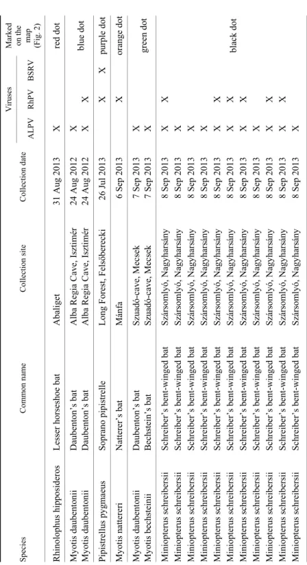
Table 1 Overview of the examined bat species and the isolated viruses Species Common name Collection site Collection date
Viruses Marked on the map (Fig. 2) ALPVRhPV BSRV Rhinolophus hipposideros Lesser horseshoe bat Abaliget 31 Aug 2013X red dot Myotis daubentonii Daubenton’s bat Alba Regia Cave, Isztimér24 Aug 2012X blue dot Myotis daubentonii Daubenton’s bat Alba Regia Cave, Isztimér24 Aug 2012X X Pipistrellus pygmaeus Soprano pipistrelle Long Forest, Felsőberecki 26 Jul 2013X X purple dot Myotis nattereri Natterer’s bat Mánfa 6 Sep 2013X orange dot Myotis daubentonii Daubenton’s bat Szuadó-cave, Mecsek7 Sep 2013X green dot Myotis bechsteinii Bechstein’s bat Szuadó-cave, Mecsek7 Sep 2013X Miniopterus schreibersii Schreiber’s bent-winged bat Szársomlyó, Nagyharsány 8 Sep 2013X X black dot
Miniopterus schreibersii Schreiber’s bent-winged bat Szársomlyó, Nagyharsány 8 Sep 2013X Miniopterus schreibersii Schreiber’s bent-winged bat Szársomlyó, Nagyharsány 8 Sep 2013X Miniopterus schreibersii Schreiber’s bent-winged bat Szársomlyó, Nagyharsány 8 Sep 2013X Miniopterus schreibersii Schreiber’s bent-winged bat Szársomlyó, Nagyharsány 8 Sep 2013X X Miniopterus schreibersii Schreiber’s bent-winged bat Szársomlyó, Nagyharsány 8 Sep 2013X X Miniopterus schreibersii Schreiber’s bent-winged bat Szársomlyó, Nagyharsány 8 Sep 2013X X Miniopterus schreibersii Schreiber’s bent-winged bat Szársomlyó, Nagyharsány 8 Sep 2013X Miniopterus schreibersii Schreiber’s bent-winged bat Szársomlyó, Nagyharsány 8 Sep 2013X X Miniopterus schreibersii Schreiber’s bent-winged bat Szársomlyó, Nagyharsány 8 Sep 2013X X Miniopterus schreibersii Schreiber’s bent-winged bat Szársomlyó, Nagyharsány 8 Sep 2013X
Phylogenetic analysis
Reference viral sequences were obtained from the GenBank database. Nu- cleotide sequence alignments were generated using Muscle Alignment (Edgar, 2004). Aligned sequences were trimmed to match the genomic regions of the vi- ral sequences obtained in our study and phylogenetic trees generated by MEGA6 (Tamura et al., 2013), using the Maximum Composite Likelihood method, based on the general time reversible model (GTR+G+I). The number of bootstraps for simulations was 1,000. The GenBank accession numbers of the viral sequences used in the phylogenetic analyses are indicated in the trees.
Results
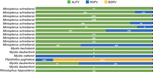
Metagenomic analysis resulted in a high proportion of Picornavirales- related sequences in all examined samples. The total number of reads for each identified virus of this study is summarised in Fig. 1. In addition, several other viruses were identified in samples of this study, which results were discussed elsewhere previously (Kemenesi et al., 2015a,b).
R h i n o l o p h u s h i p p o s i d e r o s M y o t i s d a u b e n t o n i i M y o t i s d a u b e n t o n i i P i p i s t r e l l u s p y g m a e u s M y o t i s n a t t e r e r i M y o t i s d a u b e n t o n i i M y o t i s b e c h s t e i n i i M i n i o p t e r u s s c h r e i b e r s i i M i n i o p t e r u s s c h r e i b e r s i i M i n i o p t e r u s s c h r e i b e r s i i M i n i o p t e r u s s c h r e i b e r s i i M i n i o p t e r u s s c h r e i b e r s i i M i n i o p t e r u s s c h r e i b e r s i i M i n i o p t e r u s s c h r e i b e r s i i M i n i o p t e r u s s c h r e i b e r s i i M i n i o p t e r u s s c h r e i b e r s i i M i n i o p t e r u s s c h r e i b e r s i i M i n i o p t e r u s s c h r e i b e r s i i
183 9 79
6 3 285
275 380 475 801
60 184
1648 1283 636
73
134 1695
2
453 530
9 43
9 111
9635 ALPV RhPV BSRV
Fig. 1. Total read number of the identified honey bee related viruses in bat guano samples of the examined bat species
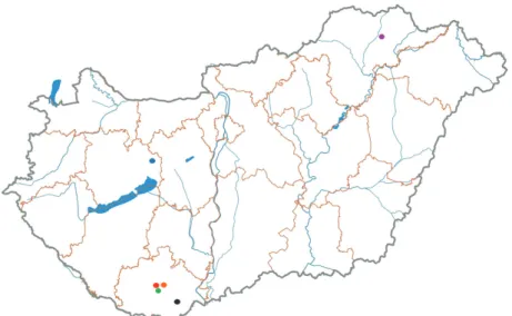
Of the selected 18 samples, we identified ALPV-related sequences in 16 samples collected in different locations of the country (Fig. 2, Table 1). The as- sembled contigs from the obtained sequences segregated in separated clusters covering different genome regions of different lengths. The PCRs conducted to close the gaps (or to amplify the missing fragments) within the contigs obtained in total a 8979-nt-long sequence of ALPV (GenBank: MF535297) encompassing
Miniopterus schreibersii Miniopterus schreibersii Miniopterus schreibersii Miniopterus schreibersii Miniopterus schreibersii Miniopterus schreibersii Miniopterus schreibersii Miniopterus schreibersii Miniopterus schreibersii Miniopterus schreibersii Miniopterus schreibersii Myotis bechsteinii Myotis daubentonii Myotis nattereri Pipistrellus pygmaeus Myotis daubentonii Myotis daubentonii Rhinolophus hipposideros
ALPV RhPV BSRV
156 ZANA et al.
the complete coding region and covering 91% of the complete genome. Using a reference ALPV genome sequence (GenBank: KJ817182) we estimated that ap- proximately 940 nt were missing at the 5’ UTR region. Of the two ORFs that were identified, one encodes the 1896-aa-long nonstructural polyprotein and is flanked by IRES at both ends. The second ORF encodes the 801-aa-long capsid protein. The 3’ UTR Poly (A) tail was successfully amplified and sequenced.
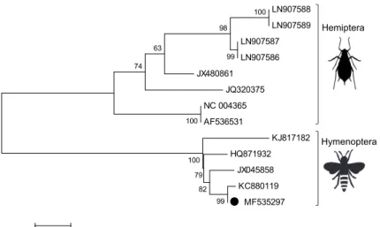
BLASTn search showed 98% identity of our sequence to the honey bee related Belgian strain (GenBank: KC880119). The phylogenetic analysis of ALPV strains deposited in GenBank revealed two firmly separated clusters of ALPVs. Our strain segregated into the cluster containing ALPV strains previously associated with honey bees (GenBank: HQ871932, JX045858 and KC880119) and branched together with the Belgian isolate (GenBank: KC880119), while strains associated with aphid species segregated into the other cluster (Fig. 3).
Fig. 2. Locations of bat sampling sites. Red dot: Abaliget; blue dot: Alba Regia Cave, Isztimér;
orange dot: Mánfa; green dot: Szuadó-Cave, Mecsek; black dot: Szársomlyó, Nagyharsány;
purple dot: Long forest, Felsőberecki. Each dot is also indicated in the last column of Table 1
BSRV-related sequences (GenBank: MF928582) were present in only one bat guano sample collected in Felsőberecki (Fig. 2, Table 1). The alignment of the assembled contigs showed 87% sequence identity to the American honey bee associated BSRV strain (GenBank: JF423195) and 99% sequence identity to a Kenyan strain (GenBank: KY826434) isolated from black bean aphid (Aphis fa- bae). As the obtained sequences covered the same region of the genome, the de- sign of specific primer pairs to amplify further fragments or even the complete genome proved to be unfeasible.
LN907588 LN907589 LN907587 LN907586 JX480861
JQ320375 NC 004365 AF536531
KJ817182 HQ871932
JX045858 KC880119
MF535297 99
82 79 100 100
100
99 98 63
74
0.02
Fig. 3. Phylogenetic analysis of aphid lethal paralysis virus strains detected in bat guano samples in Hungary, 2012–2013. The phylogenetic tree was constructed using the Maximum Composite Like-
lihood method, based on the general time reversible model (GTR+G+I). The number of bootstraps for simulations was 1000. The ALPV strain characterised in the present study is indicated by a
black dot. Silhouettes represent the two firmly separated clusters of ALPV
Sequences displaying similarity to RhPV were present in 8 samples (Fig. 2;
Table 1) and the assembled contigs were segregated throughout the genome cov- ering its different regions. Compared to our sequence (GenBank: MF535298) with a reference RhPV strain (GenBank: AF022937) approximately 340 nt were missing at the 5’ UTR region of the genome. Further PCRs were performed to fill the gaps. Thus we obtained a 9667-nt-long sequence covering approximately 96% of the complete genome. The new sequence contained the complete coding region, which consists of two ORFs. The first ORF that encodes the 1998-aa-long nonstructural polyprotein, is bordered by IRESs at both ends in the same way as in ALPV. The 3’ UTR poly (A) tail of the genome was successfully amplified and sequenced too. The BLASTn search with our sequence showed 97% se- quence identity to an American RhPV strain deposited in GenBank (AF022937).
Discussion
In this study, we examined the viral assemblages of selected bat guano samples. Similarly to previous research, this study revealed the dominance of di- etary viruses (Donaldson et al., 2010; Li et al., 2010; Ge et al., 2012; He et al., 2013; Dacheux et al., 2014; Kemenesi et al., 2016). Interestingly, geographical segregation of the detected viruses could be observed between the sampling sites
Hemiptera
Hymenoptera
158 ZANA et al.
(Table 1). Our findings may suggest distinct viral load in the local insect com- munity, which is typical of the area from which the samples originated. Referring to the findings of previous studies (Donaldson et al., 2010; Li et al., 2010; Ge et al., 2012; He et al., 2013; Dacheux et al., 2014; Kemenesi et al., 2016), the meta- genomic screening of bat guano samples may represent an alternative and indi- rect surveillance method to predict or indicate new threats to the local honey bee colonies. The necessity of this new screening procedure is confirmed by many factors. In the last decades, unusually large losses of honey bees were reported mainly from the USA and Europe. Many theories exist, suggesting different con- tributors in the decline of colonies including pathogens, pesticides, nutrition and limited genetic diversity but the main contributor in this phenomenon is not fully elucidated yet (Ravoet et al., 2013). Colony Collapse Disorder (CCD), another negative contributor, is associated with extensive honey bee loss mainly in the USA (Vanengelsdorp et al., 2009; McMenamin and Genersch, 2015). Although CCD is defined by clear aspects, nowadays elevated pathogen levels are also characteristic of CCD colonies (McMenamin and Genersch, 2015).
In this study, we identified and characterised two recently described virus- es infecting honey bees in Hungary for the first time. Our study revealed the presence of an aphid lethal paralysis virus strain in bat guano samples sharing the highest nucleotide identity with ALPV strains previously isolated from honey bees (GenBank: HQ871932, JX045858 and KC880119). Furthermore, phyloge- netic analysis of our strain confirmed the phylogenetic relationships among ALPV strains reported previously (Liu et al., 2014) and indicated the existence of two major lineages of ALPVs. One lineage is composed of ALPV-AP and ALPV-AM strains described from pea aphid (Acyrthosiphon pisum = AP) and honey bees (Apis mellifera = AM) respectively, while the other group consists of ALPV strains found in other aphid species, as well as in bat faeces and western corn rootworm (Diabrotica virgifera virgifera), such as a previously described Hungarian ALPV strain. Therefore, our study suggests that the different ALPV strains presumably represent different virus species. Furthermore, ALPV species may be subject to a strong selective pressure, which may result in host range dif- ferences and altered infectivity of the virus. According to these findings, the presence of an ALPV strain in Hungary that is capable of infecting honey bees may have adverse consequences for the Hungarian honey bee colonies along with BSRV (Runckel et al., 2011). The significant prevalence of ALPVs in hon- ey bees has been suggested to be strongly associated with the presence of Nose- ma apis and Nosema ceranae spores (Runckel et al., 2011; Ravoet et al., 2013).
Our metagenomic analysis, unfortunately, did not produce any results that would indicate the presence of these spores in the samples. However, previous studies have reported their presence in Hungarian apiaries (Bakonyi et al., 2002; Tap- aszti et al., 2009).
The confirmed ability of these viruses to replicate and the presence of ma- ture virions in adult honey bees may suggest their role in honey bee health, alt- hough no signs of the infection have been revealed yet (McMenamin and Genersch, 2015; Ravoet et al., 2015). Only two of the identified viruses (ALPV and BSRV) have been reported previously as possible infective agents of honey bees (Lee et al., 2015; Ravoet et al., 2015). We cannot declare with complete cer- tainty the infective capability of RhPV, since spillover events have been demon- strated and virus transmission between hosts is influenced by the elevated inter- actions between insect species sharing the same resources (Mordecai et al., 2016). Even though the BSRV strain described in this study showed stronger homology to an aphid-related sequence, because of the above statements the pathogenic capability of our strain cannot be totally excluded.
Therefore, as it has been suggested by many studies, the importance of monitoring honey bee pathogens is based on the elevated number of detectable pathogens which have significant adverse consequences for the health and winter mortality of the colony (Vanengelsdorp et al., 2009; Cornman et al., 2012; Ravoet et al., 2013). Moreover, since the colonies affected by CCD are characterised by elevated susceptibility to different pathogens, the increased virus and Nosema spp. load and the synergism of pathogens with which honey bees are co-infected lead to rapid and extensive worker honey bee depletion (Vanengelsdorp et al., 2009; Cornman et al., 2012).
Comprehensive studies have already summarised the persisting pathogenic agents of honey bees in Hungary (Forgách et al., 2008; OMME, 2017). The pre- sent study gives additional information about the presence of possible pathogens of honey bees in the country. Furthermore, as it has been suggested previously, examining bat guano samples proved to be an efficient way to detect potentially emerging infectious diseases of humans or animals, including economically im- portant species (Wu et al., 2016). This study presents the first genetic data re- garding ALPV and BSRV from Central Europe, and contributes to the general understanding of the genetic diversity and geographic distribution of these virus- es. These data may provide a basis for further surveillance studies, especially on the economically important pathogens described here, potentially infecting hon- ey bee populations.
Acknowledgements
The research activity of F. J. was supported by TÁMOP (4.2.4.A/2-11-1-2012- 0001) – National Excellence Programme, K. K. was supported by the Szentágothai Tal- ent Programme (awarded by the Szentágothai Research Centre, University of Pécs), G.
K. and F. J. were supported by the ÚNKP-17-3-III and ÚNKP-17-4-III – New National Excellence Programme of the Ministry of Human Capacities. The project was supported by the European Union, co-financed by the European Social Fund: Comprehensive De- velopment for Implementing Smart Specialization Strategies at the University of Pécs
160 ZANA et al.
(EFOP-3.6.1.-16-2016-00004), and by the University of Pécs in the framework of the
‘Viral Pathogenesis’ Talent Centre programme. The present scientific contribution is dedicated to the 650th anniversary of the foundation of the University of Pécs, Hungary.
S. B. was supported by the SH/4/13 Multipurpose assessment serving forest biodiversity conservation in the Carpathian region of Hungary (SAB).
References
Bakonyi, T., Farkas, R., Szendröi, A., Dobos-Kovács, M. and Rusvai, M. (2002): Detection of acute bee paralysis virus by RT-PCR in honey bee and Varroa destructor field samples:
rapid screening of representative Hungarian apiaries. Apidologie 33, 63–74.
Bányai, K., Kemenesi, G., Budinski, I., Földes, F., Zana, B., Marton, S., Varga-Kugler, R., Oldal, M., Kurucz, K. and Jakab, F. (2016): Candidate new rotavirus species in Schreiber’s bats, Serbia. Infect. Genet. Evol. 48, 19–26.
Calisher, C. H., Childs, J. E., Field, H. E., Holmes, K. V. and Schountz, T. (2006): Bats: important reservoir hosts of emerging viruses. Clin. Microbiol. Rev. 19, 531–545.
Conceição-Neto, N., Zeller, M., Lefrère, H., De Bruyn, P., Beller, L., Deboutte, W., Yinda, C. K., Lavigne, R., Maes, P., Van Ranst, M., Heylen, E. and Matthijnssens, J. (2015): Modular approach to customise sample preparation procedures for viral metagenomics: a reproduci- ble protocol for virome analysis. Sci. Rep. 5, 16532.
Cornman, R. S., Tarpy, D. R., Chen, Y., Jeffreys, L., Lopez, D., Pettis, J. S., vanEngelsdorp, D.
and Evans, J. D. (2012): Pathogen webs in collapsing honey bee colonies. PLoS ONE 7, e43562.
Dacheux, L., Cervantes-Gonzalez, M., Guigon, G., Thiberge, J. M., Vandenbogaert, M., Maufrais, C., Caro, V. and Bourhy, H. (2014): A preliminary study of viral metagenomics of French bat species in contact with humans: identification of new mammalian viruses. PLoS ONE 9, e87194.
Donaldson, E. F., Haskew, A. N., Gates, J. E., Huynh, J., Moore, C. J. and Frieman, M. B. (2010):
Metagenomic analysis of the viromes of three North American bat species: viral diversity among different bat species that share a common habitat. J. Virol. 84, 13004–13018.
Edgar, R. C. (2004): MUSCLE: multiple sequence alignment with high accuracy and high throughput. Nucl. Acids Res. 32, 1792–1797.
Forgách, P., Bakonyi, T., Tapaszti, Z., Nowotny, N. and Rusvai, M. (2008): Prevalence of patho- genic bee viruses in Hungarian apiaries: situation before joining the European Union. J. In- vertebr. Pathol. 98, 235–238.
Ge, X., Li, Y., Yang, X., Zhang, H., Zhou, P., Zhang, Y. and Shi, Z. (2012): Metagenomic analysis of viruses from bat fecal samples reveals many novel viruses in insectivorous bats in Chi- na. J. Virol. 86, 4620–4630.
Granberg, F., Vicente-Rubiano, M., Rubio-Guerri, C., Karlsson, O. E., Kukielka, D., Belák, S. and Sánchez-Vizcaíno, J. M. (2013): Metagenomic detection of viral pathogens in Spanish honeybees: co-infection by Aphid Lethal Paralysis, Israel Acute Paralysis and Lake Sinai Viruses. PLoS ONE 8, e57459.
He, B., Li, Z., Yang, F., Zheng, J., Feng, Y., Guo, H., Li, Y., Wang, Y., Su, N., Zhang, F., Fan, Q.
and Tu, C. (2013): Virome profiling of bats from Myanmar by metagenomic analysis of tissue samples reveals more novel mammalian viruses. PLoS ONE 8, e61950.
Kemenesi, G., Dallos, B., Görföl, T., Boldogh, S., Estók, P., Kurucz, K., Kutas, A., Földes, F., Oldal, M., Németh, V., Martella, V., Bányi, K. and Jakab, F. (2014): Molecular survey of RNA viruses in Hungarian bats: discovering novel astroviruses, coronaviruses, and calici- viruses. Vector Borne Zoonotic Dis. 14, 846–855.
Kemenesi, G., Dallos, B., Görföl, T., Estók, P., Boldogh, S., Kurucz, K., Oldal, M., Marton, S., Bányai, K. and Jakab, F. (2015a): Genetic diversity and recombination within bufaviruses:
Detection of a novel strain in Hungarian bats. Infect. Genet. Evol. 33, 288–292.
Kemenesi, G., Földes, F., Zana, B., Kurucz, K., Estók, P., Boldogh, S., Görföl, T., Bányai, K., Oldal, M. and Jakab, F. (2016): Genetic characterization of Providence virus isolated from bat guano in Hungary. Genome Announc. 4, e00403-16.
Kemenesi, G., Zhang, D., Marton, S., Dallos, B., Görföl, T., Estók, P., Boldogh, S., Kurucz, K., Oldal, M., Kutas, A., Bányai, K. and Jakab, F. (2015b): Genetic characterization of a novel picornavirus detected in Miniopterus schreibersii bats. J. Gen. Virol. 96, 815–821.
Lee, K., Steinhauer, N., Travis, D. A., Meixner, M. D. and Deen, J. (2015): Honey bee surveillance: a tool for understanding and improving honey bee health. Curr. Opin. Insect Sci. 10, 37–44.
Li, L., Victoria, J. G., Wang, C., Jones, M., Fellers, G. M., Kunz, T. H. and Delwart, E. (2010): Bat guano virome: predominance of dietary viruses from insects and plants plus novel mamma- lian viruses. J. Virol. 84, 6955–6965.
Liu, S., Vijayendran, D., Carrillo-Tripp, J., Miller, W. A. and Bonning, B. C. (2014): Analysis of new aphid lethal paralysis virus (ALPV) isolates suggests evolution of two ALPV species.
J. Gen. Virol. 95, 2809–2819.
McMenamin, A. J. and Genersch, E. (2015): Honey bee colony losses and associated viruses. Curr.
Opin. Insect Sci. 8, 121–129.
Moon, J. S., Domier, L. L., McCoppin, N. K., D’Arcy, C. J. and Jin, H. (1998): Nucleotide se- quence analysis shows that Rhopalosiphum padi virus is a member of a novel group of in- sect-infecting RNA viruses. Virology 243, 54–65.
Mordecai, G. J., Brettell, L. E., Pachori, P., Villalobos, E. M., Martin, S. J., Jones, I. M. and Schroeder, D. C. (2016): Moku virus; a new Iflavirus found in wasps, honey bees and Var- roa. Sci. Rep. 6, 34983.
OMME – Országos Magyar Méhészeti Egyesület [National Hungarian Beekeeping Association]
(2017): Magyar Méhészeti Nemzeti Program, Környezetterhelési Monitoringvizsgálat 2016–
2017 [Hungarian National Beekeeping Programme, Environmental Impact Monitoring Sur- vey 2016–2017]. Accessed 22 January 2018. Retrieved from: http://www. omme.hu/?p=9982 Ravoet, J., De Smet, L., Wenseleers, T. and de Graaf, D. C. (2015): Vertical transmission of honey
bee viruses in Belgian queen breeding program. BMC Vet. Res. 11, 61.
Ravoet, J., Maharramov, J., Meeus, I., De Smet, L., Wenseleers, T., Smagghe, G. and de Graaf, D.
C. (2013): Comprehensive bee pathogen screening in Belgium reveals Crithidia mellificae as a new contributory factor to winter mortality. PLoS ONE 8, e72443.
Runckel, C., Flenniken, M. L., Engel, J. C., Ruby, J. G., Ganem, D., Andino, R. and DeRisi, J. L.
(2011): Temporal analysis of the honey bee microbiome reveals four novel viruses and seasonal prevalence of known viruses, Nosema, and Crithidia. PLoS ONE 6, e20656.
Tamura, K., Stecher, G., Peterson, D., Filipski, A. and Kumar, S. (2013): MEGA6: Molecular Evo- lutionary Genetics Analysis Version 6.0. Mol. Biol. Evol. 30, 2725–2729.
Tapaszti, Z., Forgách, P., Kővágó, C., Békési, L., Bakonyi, T. and Rusvai, M. (2009): First detec- tion and dominance of Nosema ceranae in Hungarian honeybee colonies. Acta Vet. Hung.
57, 383–388.
Vanengelsdorp, D., Evans, J. D., Saegerman, C., Mullin, C., Haubruge, E., Nguyen, B. K., Frazier, M., Frazier, J., Cox-Foster, D., Chen, Y., Underwood, R., Tarpy, D. R. and Pettis, J. S.
(2009): Colony collapse disorder: a descriptive study. PLoS ONE 4, e6481.
Wang, J., Moore, M. E., Murray, Z. L., McInnes, K., White, D. J., Tompkins, D. M. and Hall, R. J.
(2015): Discovery of novel virus sequences in an isolated and threatened bat species, the New Zealand lesser short-tailed bat (Mystacina tuberculata). J. Gen. Virol. 96, 2442–2452.
Wu, Z., Yang, L., Ren, X., He, G., Zhang, J., Yang, J., Qian, Z., Dong, J., Sun, L., Zhu, Y., Du, J., Yang, F., Zhang, S. and Jin, Q. (2016): Deciphering the bat virome catalog to better under- stand the ecological diversity of bat viruses and the bat origin of emerging infectious dis- eases. ISME J. 10, 609–620.