Assessment
Developing a Novel Anatomical Phantom and Performance Assessment Method for Laparoscopic Prostatectomy Training
L´aszl´o Jaksa, Ill´es Nigicser, Bal´azs Szab´o, D´enes ´Akos Nagy, P´eter Galambos, and Tam´as Haidegger
Abstract The rising domain of Cognitive infocommunications investigates the link between the research areas of infocommunications and cognitive sciences , of which probably the most important is the human factor. The systematic assessment and de- velopment of human learning capabilities and skill acquisition is one of the biggest challenges in applied sciences. It can be well observed within the medical domains, how evidence-based paradigms are gradually gaining space. The methods and ap- proaches offered by coginfocom and usability engineering for surgical skill assess- ment have been put into practical application in our research. In this chapter, the development process of a custom laparoscopic box trainer is introduced. A cog- nitive surgical simulator including an upgraded phantom for prostatectomy is de- scribed, which feeds into medical staff training and skill assessment. As a first step, an extensive overview of laparoscopic surgical simulators is provided, ranging from physical constructions to complex computer-integrated systems, with a special fo- cus on their cognitive skill assessment capabilities. Based on the state of the art, and our previous experience, a clear need was formulated to develop a hybrid—partially physical, partially computer-integrated— simulator. From the viewpoint of coginfo- com, such a system would be a step forward regarding the monitoring and analysis of human learning capabilities in laparoscopic surgical education. In order to im- prove the slope of learning curves, high fidelity tissue models and anatomy were implemented in the new design. The upgraded phantom and the box trainer was L. Jaksa·B. Szab´o·D. ´A. Nagy·P. Galambos·T. Haidegger are with the
Antal Bejczy Center for Intelligent Robotics, ´Obuda University, 82 Kiscelli Street, 1032 Budapest, Hungary
e-mail: {laszlo.jaksa,balazs.szabo,denes.nagy,szilvia.barcza,peter.
galambos,tamas.haidegger}@irob.uni-obuda.hu T. Haidegger is also with the
Austrian Center for Medical Innovation and Technology, 2 Viktor Kaplan Street, 2700 Wiener Neustadt, Austria
I. Nigicser is also with the
University of Surrey, GU2 7XH Guildford, United Kingdom e-mail:nigicseri@gmail.com
1
tested on 8 laymen and 6 clinicians resulting in an overall subjective rating of 4.43 out of 5 and 4.21 out of 5 in the suitability of the box trainer for laparoscopic envi- ronment simulation and of the fidelity of the phantom, respectively. To gain a better understanding of the cognitive load and relevant physical stress, fierce measurement capabilities were introduced into the test environment. Load cells were placed in the training box, under the anatomical phantom. This way the reaction forces become measurable during surgical practice. The force and time data are used to evaluate the performance of the participant. During testing a new assessing metric, the Time- Force Product (TFP) was introduced that indicates swift (little time) and safe (little force) procedures. This proved to be a comprehensive performance indicator having the evidence of an average 60% TFP decrease of residents/specialists compared to laymen. The assessment method can be used to point out the weak aspects of sur- gical technique, and the participant can do this on his own. In addition, the method is algorithmic, which opens new possibilities for the further development of hy- brid laparoscopic simulators, and also points out the peak stress points of a subject.
Computer-integrated assistive technologies for surgical education are believed to rapidly become the gold standard for human training on a global scale.
Key words: Cognitive skill training, Surgical simulation, Laparoscopy training, Surgical education and skill assessment
Acknowledgement
The research was supported by the workers and laboratory facilities of Antal Be- jczy Center for Intelligent Robotics, ´Obuda University Budapest, Hungary and the workers of the Oncology Center of the Semmelweis University, Budapest, Hun- gary, who were the professional subjects of the measurements. Special thanks to Tivadar Garamv¨olgyi who supervised the phantom development part of the project, and to Szilvia Barcza, who established the basics of the skill assessment metrics.
This work has been partially supported by ACMIT (Austrian Center for Medical In- novation and Technology), which is funded within the scope of the COMET (Com- petence Centers for Excellent Technologies) program of the Austrian Government.
The work of T. Haidegger was partially supported through the New National Excel- lence Program of the Ministry of Human Capacities and by the Hungarian OTKA PD 116121 grant.
Introduction
The improvement of assistive technologies in surgical device development has taken a rapid turn where high level support systems like the da Vinci system are gaining importance. The foundation of these were established by the emergence of cognitive
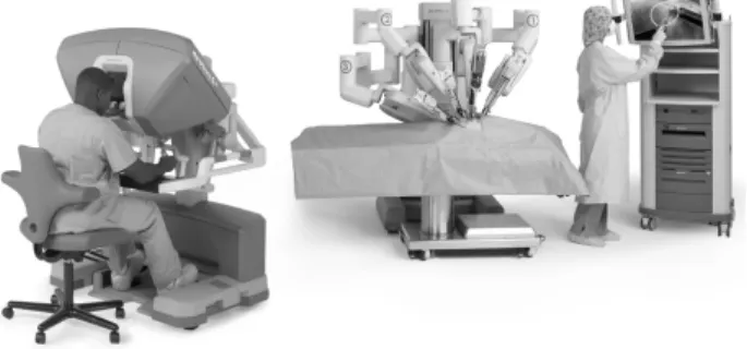
sciences and human factor engineering. In the process of surgical device develop- ment, minimizing invasiveness has become a central focus since it reduces tissue damage, improves surgery and recovery time, increases accuracy and expands the scope of feasible surgeries. A first level, there has been the introduction of laparo- scopic surgery where small incisions are sufficient to introduce the tools, and an endoscopic camera is employed as a digital sensor. The second level exceeds the first with the invention of Computer Assisted Laparoscopic Robot [4], most signif- icantly the da Vinci System, where an artificial entity is introduced that bridges the cognitive control of the doctor and the execution of procedures on a surgery for higher accuracy, safety and efficiency. This then serves as a ground for a whole new area of development of natural and artificial cognitive systems communication in the medical field. The development of assistive technologies for surgery has reached a point where even the cognitive abilities of surgeons can be supported and enhanced by artificial cognitive systems. Decision support systems are routinely employed in image-guided surgeries, and low level autonomous functions are also on the devel- opment horizon. Currently one of the most developed assistive technologies of such is embodied by the da Vinci Surgical System. The da Vinci System is a teleoperation system where the surgeon navigates the surgery from a distance through a refined system as a medium resulting in a very high level support system (Figure 1).
Fig. 1 The da Vinci Si Surgical System consisting of the Surgeon Console, a Patient-side Cart with Endo-wrist Instruments and the Vision System. Photo courtesy by Intuitive Surgical, Inc.
Nevertheless, the current real need in Hungary and the region is more towards low level support systems and teaching tools, which can already make a big differ- ence in the skill acquisition and maintenance of junior medical staff. For this reason this paper focuses on the first level, the enhancement of the manual laparoscopic surgery, as a central and crucial step in this process of surgical device development.
More particularly it focuses on how an increasing level of fidelity of training equip- ment develops the cognitive learning process of doctors on laparoscopic surgeries.
For assessing this an own laparoscopic trainer box was further developed and im- plemented consisting a prostatectomy phantom with high fidelity tissue models and anatomy.
Theory
Performing laparoscopic surgery requires high level of cognitive capability where dexterity and fine psychomotor skills are indispensable for the surgeon. Fortunately these skills can be and must be developed and therefore Hospitals and Medical Cen- tres have started applying different learning tools. Assessing the efficiency of the learning process can be done through observing the invested time and energy in a particular learning environment against the improvement of skills. This can be de- picted by so called learning curves that graphically display the time to complete a task against practice mainly used in the field of cognitive psychology. The results of tests on performing a wide range of both mental or cognitive and practical tasks that require dexterity show that these learning curves generally follow the “power law of practice”. This pattern is characterized by a steep start that reaches a plateau after exceeding certain amount of practice (Figure 2) [22].
Fig. 2 A learning curve that shows the “power law of practice” where the initial steep slope grad- ually reaches a plateau, also it represents an ideal surgical learning curve [1]
The derivative i.e. the slope of the curve before the plateau indicates the qual- ity of the learning process and thus the steeper the slope the faster the learning.
Learning curves are derived from industry applications and is significantly used in economics relating unit costs to accumulated volume [28]. The term then began to gain significance in medical training and education and more importantly in min- imally invasive surgeries as it is a technically demanding learning process [1]. An article review about high-fidelity medical simulations conducted a thorough litera- ture search (109 journal articles) on the features and aspects of simulation that lead to effective learning. These included providing feedback, as the most important ed- ucational feature, room for repetitive practice, curriculum integration into medical school education, providing a range of difficulty level, individualized learning and defined outcomes among others [26]. The aspect of providing feedback including visual, auditory or haptic, indicates the fact that the more senses are involved in
a learning process the steeper the learning curve will get. Research shows that at laparoscopic surgical training the presence of the haptic force feedback as a sense of touch resulted in improved training transfer [13]. Visual fidelity and more accu- rate haptic fidelity of tissues on laparoscopic simulators may further improve learn- ing outcomes although a couple of research articles could not find evidence of a linear correlation of improved fidelity of models versus training effectiveness [13]
[18]. However a paper states that a perfect laparoscopic training model should be anatomically and physiologically analogous to a human patient thus fidelity is of great importance [35].
Background
The best way to gain proficiency in laparoscopic operations is to observe, assist and then perform surgeries on real patients. However this involves high risk factor for the patient in the skill acquisition of junior medical staff and thus moving from observer to assistant and finally to performing surgeon is a slow and regulated process. In order to accelerate this skill acquisition, outside-operating theater learning facility was required.
Nowadays laparoscopic surgery is widely spread in the clinical practice which is why appropriate training and assessment of skills in laparoscopic procedures be- came an important issue. These coordination skills significantly differ from those of traditional open surgeries. Due to the flat screen the depth perception is reduced and the tactile information is lost [17]. To the development of these appropriate skills there is a clearly defined learning curve associated, where the teaching meth- ods used in conventional surgery cannot guide the students with full satisfaction and without risk [2]. Surgical simulation may be the solution to this problem, the us- ability of which is confirmed by several studies completed in laparoscopic training [19]. A review article about low cost, suitable-for-home-use laparoscopic simula- tors highlighted many very low cost, not on the market, prototype solutions which can be built easily. These devices obviously did not undergo any validation thus their practicality is opposed by their dubious educational value [17]. A study ex- amined the maintainability of laparoscopic surgical skills of inexperienced subjects.
After a short training on a simulator followed by a test the gained skills were still significantly present and maintained upon re-examination even after one year [10].
The simulation grants the possibility for the student to practice laparoscopic surgi- cal technique outside the operating theatre with less stress and without the risk of damaging the patient [12]. A number of approaches and solutions to such a simula- tion were developed ranging from simple and cheap, but low fidelity box trainers to high fidelity virtual reality simulators. Box trainers are usually simply constructed, portable, allowing for basic laparoscopic skills development: for example manoeu- vring and manipulating with the tools, cutting and suturing. The computerized surgi- cal simulators use computer graphics and sometimes even haptic feedback to create a virtual anatomic environment enabling the practice of specific surgical procedures
in addition to general surgical skills [8]. According to a study the laparoscopic vir- tual reality simulators improve the surgical skills more than box trainers. It is not clear however how this additional development will appear in everyday surgical practice [36]. The virtual reality simulators are expensive which makes it impossi- ble for many educational institutions to purchase them making it difficult to access for students [27],[5]. Nowadays box trainers are much more commonly used than virtual reality simulators [2]. The educational integration of such simulation equip- ment vastly varies for different disciplines, for example there is much less emphasis on practice on simulators in urology where there is more emphasis on practice with senior supervision, as opposed to in other endoscopic surgical disciplines [34].
In the past for learning traditional open surgeries medical student and staff used wax models for centuries until autopsies were allowed in medical schools [25]. Hu- man cadaver dissection has been widely used for learning purposes although their limited availability and high cost led to first using slightly more accessible animal cadaver. Laparoscopic surgeries have been performed on animal models that repre- sented in vivo training environment that could provide skill acquisition opportunity of hand-eye coordination, depth perception and knot-tying [35]. Mostly pigs were chosen for these animal models since they resemble the human anatomy very well thus they are specially widely used for laparoscopic cholecystectomy training with success [16]. However animal cadaver have been still only limitedly accessible so it left the need for creating artificial simulation environment and tools. Hospitals and medical schools have started using low-cost simple laparoscopic box trainers that can provide useful training for hand-eye co-ordination, triangulation and depth queuing. What is more these laparoscopic trainers can be built at home and be used for individual practice and thus restrictions on medical working hours on practice may be avoided [25], [9], [20]. These solutions are very simple, cheap and practical although they do not resemble realistic anatomy by far.
For simulating more accurate anatomy especially visually, augmented reality (AR) and virtual reality (VR) simulators have been developed. VR creates a 3D virtual environment where various can be repeatedly simulated without any need of external assistance, although this tool does not provide haptic or tactile feedback of the procedure thus limiting the slope of the learning curve. AR differs from VR mainly in providing real time force feedback thus improving the quality of training [25]. However both VR and AR software are very expensive and thus its price effi- ciency makes these products unaffordable for some institutions [4]. Because of the limitations of all the above training tools and environments, physical laparoscopic box trainers with anatomical phantoms may fill a market gap and provide a practi- cal, affordable but high quality training tool with realistic haptic and tactile feedback and anatomy. Quality box trainers can already be found on the market although with limited scope. Such a phenomenon is visible in other surgical disciplines that use la- paroscopic instruments. A recent study [15] presents a training dummy for pediatric Nissen fundoplication.
A thorough overview of commercially available box trainers was conducted. Dur- ing the research for mapping the laparoscopic trainers the Google search engine was used with the following key words: ”laparoscopic simulator”, ”laparoscopic
trainer”, ”laparoscopic box trainer”, ”virtual reality surgical simulator”, ”laparo- scopic surgical simulator”. Additionally, overview articles and case studies were searched in Google Scholar with a similar method.
The chosen skill assessment method will greatly affect the “qualifying” surgery skill level of students so the choice of the appropriate method of measurement is key in ensuring the quality of surgical education [32]. For subjects with minimal laparo- scopic experience, neither did the box trainer practice nor did the LapVR virtual reality simulator practice show any significant correlation with the scores achieved in the GOALS (Global Assessment of laparoscopic Operative Skills) evaluation sys- tem. This is a warning sign that these methods are not necessarily suitable for rapid skill assessment or selection [29]. There is a program aiming to unify laparoscopic skill assessment which is called the FLS program (Fundamentals of Laparoscopic Surgery) which is established the SAGES (Society of American Gastrointestinal and Endoscopic Surgeons). Their practical skill enhancement and assessment module consists of five tasks: peg transfer, cutting out a pattern, loop suturing, suturing with intracorporeal knot and suturing with extracorporeal knot [25]. The RFLS (Funda- mentals of Robotic Surgery, laparoscopic) is the surgical robotic FLS embodiment.
The features of the laparoscopic box trainers found during the research are de- tailed in Table 1 and Table 2. In case of both tables, in the ”Curriculum” column there is no difference in content between the ”FLS” exercises and the ”basics”. This is because such products that do not have validated FLS compatibility the term
”FLS” could not be used but an alternative term had to be referenced.
Table 1 Laparoscopic box trainers I.
Manufacturer Product Price range
FLS
compliancePortability Forceps Curriculum Resource (webpage)
3-Dmed
LapTab Trainer
400–
1000 $ yes excellent included FLS
3d-med.com T3 Plus 2500–
3000 $ yes good no data
FLS, camera handling
T5 2500–
3500 $ yes good no data
basics, camera handling, ultrasound
T9 3000–
3500 $ no data good no data
FLS, camera handling
T12 4000–
5000 $ yes acceptable no data
FLS, camera handling ForceSense 8500–
9000 $ no data undefined undefined force mea- surement
CamTronics LapStar no data yes good not
included basics laparoscopic- trainer.com Delletec Laparoscopy
Simulator no data no data acceptable not included
basics, camera handling
delletec.com eoSurgical eoSim 700–
4000 $ no data excellent included basics eo- surgical.com Ethicon TASKit 400–
600 $ yes excellent not
included FLS ethicon.com Grena
Laparoscopic Trainer System
200–
300 $ no data good no data basics grena.co.uk
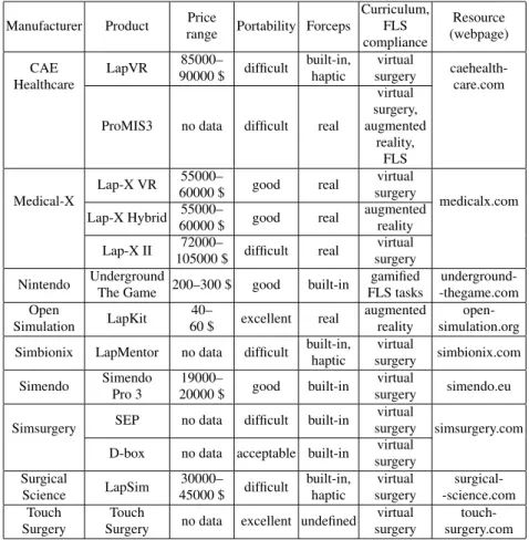
The virtual reality simulators are playing an increasingly important role in surgi- cal education. The features and details of the virtual reality simulators available on the market are summarized in Table 3. The application of force feedback through built-in haptic forceps is one of the most significant features of virtual reality simu- lators since enabling the sense of tactile properties of only virtually existing tissues greatly enhances the fidelity and the training value.
In recent years the rapid development of telerobotic systems made it possible for such robots to not only be used in industrial settings but in clinical applica- tions too. This way the surgeon is able to perform an operation distanced from the patient controlling a robotic interface. The medical teleoperation systems are very successful in increasing precision. Today, each year, more than 1.5 million patients benefit from surgery performed by telerobotic systems worldwide [30]. The design and development of such medical teleoperating systems apart from establishing full motion control it also facing not-everyday-challenges because of the strict regula- tions stemming from the operational environment and due to the unique mechanical
Table 2 Laparoscopic box trainers II.
Manufacturer Product Price range
FLS
compliancePortability Forceps Curriculum Resource (webpage) Hospiinz Endo
Trainer no data no data good included basics hospi- inz.com Inovus
Surgical Solutions
Pyxus 500–
2200 $ no data acceptable included basics inovus.org iSurgicals iSim2 3000–
4800 $ no data excellent included basics isurgi- cals.com iSim
SMART
1500–
2000 $ no data excellent included basics Lagis
EndoSurgical
Laparoscopy
Simulator no data no data excellent not
included basics lagis.
com.tw Limbs
&Things
FLS System
1000–
6200 $ yes good no data FLS limbsand-
things.com Helago HD
Laparoscopic Trainer
8500–
9300 $ no data acceptable no data basics Nahl
Medical
HY-01 Laparoscopic
Trainer box
no data no data good included basics nahl- med.com
Pharmabotics
BodyTorso Laparoscopic
Trainer BTS300D
600–
1000 $ no data acceptable not included
basics, camera handling
pharma- botics.com Samed Laparoscopie-
Trainer no data no data acceptable no data basics samed- -dresden.com Simulab LapTrainer 2000–
2200 $ yes excellent not included
FLS, camera handling
simu- lab.com
properties of soft tissues [31]. Apart from the engineering challenges the education of the appropriate use of such robotic systems is a complex issue. The robotic surgi- cal simulators (Table 4) greatly facilitate the acquisition of movement coordination skills needed for robotic surgery. Additionally, the simulators aiming to enhance cooperation skills of assisting medical staff is a new branch of surgical education (Table 4).
Apart from the robotic surgical simulators, none of the mentioned simulators contain 3D image display. According to a study [11] the 3D image display results in a significant improvement over 2D visualization in the skills for beginner and experienced laparoscopic surgeons. The skills of a beginner surgeon viewing a 3D image would compete with an experienced surgeon’s skills viewing a 2D image only. Another article stated that students using laparoscopic simulators with 3D vi- sualization are able to perform specific tasks significantly quicker with the same amount of practice compared to students practicing on simulators with 2D visuali- sation with a flat screen [23]. The laparoscopic surgical simulators are not only good for education but also for warm-up before surgery as this type of warm-up improves
Table 3 Laparoscopic virtual reality simulators Manufacturer Product Price
range Portability Forceps
Curriculum, FLS compliance
Resource (webpage) CAE
Healthcare
LapVR 85000–
90000 $ difficult built-in, haptic
virtual
surgery caehealth- care.com ProMIS3 no data difficult real
virtual surgery, augmented
reality, FLS Medical-X
Lap-X VR 55000–
60000 $ good real virtual
surgery
medicalx.com Lap-X Hybrid 55000–
60000 $ good real augmented reality Lap-X II 72000–
105000 $ difficult real virtual surgery Nintendo Underground
The Game 200–300 $ good built-in gamified FLS tasks
underground- -thegame.com Open
Simulation LapKit 40–
60 $ excellent real augmented reality
open- simulation.org Simbionix LapMentor no data difficult built-in,
haptic
virtual
surgery simbionix.com Simendo Simendo
Pro 3
19000–
20000 $ good built-in virtual
surgery simendo.eu Simsurgery SEP no data difficult built-in virtual
surgery simsurgery.com D-box no data acceptable built-in virtual
surgery Surgical
Science LapSim 30000–
45000 $ difficult built-in, haptic
virtual surgery
surgical- -science.com Touch
Surgery
Touch
Surgery no data excellent undefined virtual surgery
touch- surgery.com
the time of performing particular tasks [7]. According to a study, learned and prac- ticed skills on laparoscopic simulators also impact and improve arthroscopic skills [3]. Another rising discipline within laparoscopic training is gamification [6]. A re- sent study [24] indicates that playing with specific video games is an effective way fro surgeons to warm up before actual surgeries. Devices that are designed for home use may add another dimension to skill training, as forming training curriculums for home circumstances is a specific challenge [33]. Pure mental training also proved to be an effective and cheap supplement to regular skill training regarding laparo- scopic cholecystectomy [14]. These results can indicate the future directions of the development of laparoscopic skills training simulators.
Table 4 Robotic surgery and operation room simulators
Manufacturer Product Price range Resource (webpage) BBZ Medical
Technologies Actaeon 30000–
32000 $
bbzmedical- technologies.com Intuitive
Surgical
da Vinci Skills
Simulator no data intuitivesurgical.com Mimic
Simulation
dV-Trainer 60000–
100000 $ mimicsimulation.com FlexVR no data
ORZONE ORCAMP –
MIS Setup no data orzone.com Simbionix RobotixMentor no data
simbionix.com TeamMentor no data
SimSurgery SEP Robot no data simsurgery.com Simulated
Surgicals
Robotic Surgery
Simulator no data simulatedsurgicals.com Surgical
Science TeamSim no data surgical-science.com
Motivation and Design Requirements
A box trainer design for simulating radical prostatectomy was chosen since it is one of the most common routine surgeries performed both laparoscopically and with robotic assistance by the da Vinci System. For this reason an educational need for laparoscopic prostatectomy is clearly on demand. This project was a continuation of a first prototype box trainer, created by Szilvia Barcza, M.D. [4]. For creating the second prototype the aspects of development focused on the pelvic phantom which included the full remodelling of the Rectum, the Pelvic Floor Muscles (PFM) and improving fidelity of the Bladder and the Connective Tissues.
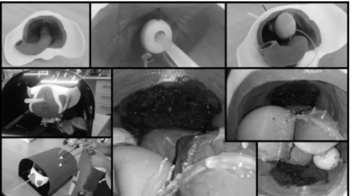
The first prototype used craftsman solutions for modelling both the Rectum and the PFM. The Rectum was made making cuts on a hollow insulation foam tube of appropriate diameter to form a realistic rectum shape. This was then poured off with transparent silicone after which the foam tube was removed and the inner side of the hollow shape was painted red to give a realistic appearance. The PFM was fabricated using smaller thin foam sheets taping them to the inner cavity of the pelvis to form a continuous surface which was then painted red.
The modelling of both the Rectum and the PFM proved to be very tedious and time consuming preventing efficiency in replication. Although the Bladder already had a mould for silicone moulding in the first prototype, but since the Bladder had to be hollow the mould had to be constantly rotated until the given amount of two- component silicone solidified. This, however, resulted in a very uneven surface for the Bladder. Finally the Connective Tissue was just indicated with painted gauze pieces which fell very far from realistic both in appearance and texture (Figure 3).
Therefore the motivation for creating the second prototype was to find a com- pletely new and better design technology that yields more realistic models for both appearance and texture that may be easily replicable for mass production. It was
Fig. 3 The first prototype of the laparoscopic trainer box with the pelvic phantom
aimed to take the robotic surgical training environment into consideration since a design requirement was set to achieve high dexterity training environment that sim- ulates real life surgery where cutting, gripping and tearing tissues are realistic with limited bleeding authenticity included.
Anatomical Phantom Design and Implementation
The new concept of the Rectum design consisted of first creating a 3D model of the Rectum using Solid Edge and Blender 3D modelling software. The idea was to create an outer mould for silicone fill-up and an inner thinner mould for a paraffin core. The thinner paraffin body would then be inserted and fixed in the outer mould leaving a gap between the paraffin and the outer mould with an equivalent wall thickness of the real Rectum. This gap would then be filled up with silicone after which the paraffin could be melted out and removed from the model resulting in a hollow realistic Rectum body.
The 3D model was first created in Solid Edge based on anatomical appearance and parameters. The smooth muscle like wavy patterned surface of the Rectum model was sculpted on it in Blender. Using Solid Edge again the negative of the Rectum was modelled giving the mould which was then 3D printed out with PLA in the lab. A thinner mould for the paraffin core was created similarly. The assembled
outer mould with the paraffin core was then filled up with two component Rubosil SR-20 silicon. Paint and silicone oil was added for the desired colour and stiffness (Figure 4).
Fig. 4 The design steps and the final result of the Rectum model
The Pelvic Floor Muscle (PFM) was similarly designed. Our main goals was for the new concept of the PFM that it should be easily manufactured and replaced while it fits as tight as possible. Therefore the base of the model was the existing 3D model of the Pelvis. The model of the Pelvis was complemented in Blender based on reference pictures, then the unnecessary parts of the model were deleted leaving only the ones that determine the PFM. Considering the possibility of dis-, and reassembly of the mould, it had to been cut up. The surfaces used to cut up the mould was also made in Blender. Then the PFM model was imported into SolidWorks as a solid model, then it was subtracted from a solid cuboid and cut into 3 separate bodies, with the help of the previously generated surfaces, and optimised for 3D printing.
After the mould had been assembled the silicone PFM was created using the same method as before (Figure 5).
Fig. 5 The design steps and the final result of the Pelvic Floor Muscle
The design of the Bladder consisted of an improvement of the first prototype, where the same paraffin core technological concept was added as for the Rectum.
The Connective Tissue was established as an extra feature compared to the first pro- totype. A composite was created made from gel candle and painted gauze resulting is a both visually and texturally plausible model that simulates realistic dissection experience. The other extra feature created was the bleeding simulation by a sim- ple silicone tube filled with food coloring and held under pressure by a syringe.
Here it was applied to the plexus santorini solely because of its size although during real surgical procedure surgical knotting is applied to prevent bleeding. Bleeding would be more of a concern dissecting blood vessels around the pediculus that has to be prevented by electrosurgical instruments but this was not on the scope of this project. Finally the second prototype was also improved by installing load cells on the base of the pelvic phantom for force sensing in x,y and z directions. The final re- sult became a precise and anatomically realistic phantom that is simple to replicate and assemble hence may be suitable for mass production (Figure 6).
Fig. 6 The new, second prototype of the pelvic phantom with extra features of higher fidelity of Connective Tissue and bleeding effect when cutting the Plexus Santorini
Experimental Setup
In the box trainer there are three TAL220 load cells thus at the attachment of the pelvic phantom the forces in three perpendicular directions could be measured (Fig- ure 7). The load cells were not suitable to measure the forces exerted by each laparo- scopic tool during the procedure but only suitable to measure the sum of the reac- tion forces caused by the laparoscopic tools together. The signals from the load cells were sampled at a sampling frequency of 10 Hz and converted using three analog- digital converters which was then fed into the Arduino Nano microcontroller. This was screened and tracked by Arduino IDE serial monitor as the data arrived from the microcontroller to the PC. The calibration of the load cells was achieved by placing
five different masses on top of the phantom reading off the signal of the load cell in the Z direction parallel to the gravitational direction. Knowing the masses and the signal strengths the sensor constant was determined showing the unit change of the signal corresponding to a given force.
Fig. 7 The force measurement system under the phantom
The quality and improvement of the created second prototype was assessed by experimental surgery simulation of the device. The experimental tests were con- ducted identically to the tests conducted by Barcza, M.D. in her thesis [4]. The participants were asked to perform a simplified prostatectomy on the laparoscopic box trainer that consisted 5 tasks: the cutting of plexus santorini, the right pediculus, the left pediculus, the urethra at the prostate base and the urethra at the prostate apex (Figure 8). The measured variables were the force data during the experiment, the time of completion if each task and the time of the entire task. The survey, used by Barcza, M.D. in her thesis testing the first prototype [4], was also filled out by the participants that assessed the previous experience, and questioned subjective eval- uation of the fidelity and usability of the box trainer and phantom for laparoscopic surgery simulation and training. The phantom was prepared for repeated tests with the meltable gel candle and the various sizes of dissectable silicone tubes cut to unit lengths. This way after each trial only a few minutes were required to save all the data and to reset the phantom for the next test.
Results
The tests were conducted on a total of number of 13 participants: 7 laymen, 3 resi- dents and 3 specialists. In the first prototype tests a total of 23 participants, 9 laymen, 7 medics, 5 residents and 2 specialists were tested thus the results of both tests are comparable [4]. The averaged time expired for each task and for the entire test by categories of experience are summarized in Table 5 below with the overall times from the first prototype tests of Barcza, M.D. included for comparison. The aver-
Fig. 8 The measurement setup
aged values of the resident / specialist group excludes one of the specialists time results as it was extremely high due to known experimental setup error.
Table 5 Average time (s) and standard deviation of the tasks in groups (Avg. +/- Dev.) Cutting of
plexus santorini
Cutting of right pediculus
Cutting of left pediculus
Cutting of urethra at the apex of prostate
Cutting of urethra at the base of prostate
Overall time of procedure
Laymen 95.9 80.3 111.4 127.8 90.0 506.9
(+/- 197.9) Residents /
Specialists 48.8 96.8 90.2 90.8 70.2 395.2
(+/- 143.9) Summarization of
the second prototype
75.7 87.4 103.2 111.9 81.5 450.1
(+/- 43.8) (+/- 41.1) (+/- 57.6) (+/- 80.1) (+/- 47.7) (+/- 175.1) Summarization of
the first prototype
71.1 80.8 150.2 131.6 84.2 517.9
(+/- 50.2) (+/- 44.5) (+/- 143.9) (+/- 87.9) (+/- 45.7) (+/- 281.1)
Here it is clearly seen that the overall time of procedure decreases as the medical experience of the participant increases resulting in a 29% difference between the time of the laymen and of the residents and specialists. It is also seen that the average time reduced for the second prototype at each individual task except for the first 2, thus cutting of the plexus santorini and cutting of right pediculus took slightly more time in case of the second prototype. The compared overall time of procedure on groups of layman and resident and specialist for both tests are illustrated on Figure 9 below.
This shows that the average time of procedure generally decreased for the second prototype, although this increase was less significant for participants with increased experience. The subjective evaluations of the fidelity and usability of the box trainer and phantom for laparoscopic surgery simulation and training are summarized in Table 6 below including the results from the first prototype tests for comparison.
This shows that the weakest fidelity remained for the preparation in the second prototype, but the overall suitability of the trainer box remained reasonably high scoring 89% on the 1-5 scale. It was also observed that the all the individual scores
Fig. 9 Compared overall time results of the first and second prototype tests
Table 6 Subjective evaluations of the laparoscopic box trainer averaged over the entire population of experimental participants. Evaluations taken on a scale of 1-5 where 5 represents the best score.
Question Average score of
the second prototype
Average score of the first prototype Suitableness of the experiment
for laparoscopic training 4.43 4.85
Similarity of
- task for the camera navigation 4.36 4.50
- task for the preparation 4.04 4.21
- task for the cutting of plexus santorini 4.43 4.50 - task for the cutting of pediculus 4.29 4.28
- task for the cutting of urethra 4.07 4.57
Fidelity of the phantom 4.21 4.43
Suitability of the box trainer for
laparoscopic environment simulation 4.43 4.64
generally decreased for the second prototype but not significantly since nine of the scores went below 4 out of 5. A summarized force values with respect to time data obtained are represented in Table 7. Here one of the participants produced outlying results, which was excluded from the table due to known experimental setup error.
Once the measured force data was imported into Microsoft Office Excel for every subject the respective three directional force and the resultant force pattern (calcu- lating with the constant found in the calibration phase) with respect to time was available. These maximum force values gave a range of 4.26-12.46N. Knowing the force and time results the next task was to objectively analyse the subjects’s perfor- mance. For this such a measuring variable was needed to be introduced that would be proportional to the time data and the measured highest force data. The lower the force (smooth movement) and the shorter the time expired during the tasks the performance of the subject will be considered to be better. Based on this the Time- Force Product (TFP) was introduced which is the multiplication of the completion time [s] and the measured highest force [N] which gives units of Ns. The physical significance of the TFP value is not interpreted but used as a performance indicator only. Since it is beneficial to be quick and apply as little force as possible during the procedure, the lower the TFP the higher the operation performance. Here the
Table 7 The results of the force measurements Participant Medical
ExperienceTime (s) Maximal Force (N)
Time-Force Product [TFP] (Ns)
TFP (Ns) average
GT layman 334 8.71 2907.92
3614.66
T ´A layman 452 6.52 2945.44
JL layman 381 7.90 3010.20
TM layman 814 4.26 3466.05
ER layman 783 4.67 3657.61
NI layman 355 12.46 4422.73
NT layman 580 8.44 4892.71
JK resident 268 4.29 1148.41
2253.91
NDA resident 394 4.37 1723.59
R ´A resident 481 6.47 3109.79 SZJ specialist 314 5.02 1576.56 MS specialist 339 10.95 3711.18
average of TFPs for different groups showed that the laymen had a 60% higher TFP compared to the residents / specialists in average (Table 7). In the dataset a two sampled t-test and Welch test was applied too. By 95% significance level both tests’
null hypothesis was discarded and concluded that the 60% difference of the average TFP value of the layman and the experienced medical staff was significant. Based on the measurement results and the statistical analysis the TFP proved to be applica- ble for reflecting the level of training of medical staff and thus may even be used for exam evaluation and marking. However the fine-tuning of the weights used should be done following advice from experienced surgeons.
Discussion
Based on the experimental results the new anatomical phantom proved to be suc- cessful reaching very high scores on the subjective evaluations since none of the aspects of the box trainer with the phantom fell below a score of 4 out of 5. This shows that not just the laymen but the more experienced medical staff also agreed that the device is suitable for laparoscopic surgery simulation. The fidelity of the phantom received a score of 4.21 out of 5 which proves the success of one of the main focus of the design (Table 6). The significant 29% reduction of the overall procedure time for the residents and specialists compared to the laymen shows that the medical knowledge and laparoscopic experience significantly affected perfor- mance (Table 5). This clearly validates the suitability of the design since it shows that the laparoscopic experience and skills were transferable to the box trainer en- vironment. This firmly supports the statement and assumption that practice on this box trainer will provide transferable skills to real laparoscopic surgeries. The newly introduced measure of TFP proved to be a comprehensive performance indicator since successful surgery requires both swift (little time) and safe (little force) pro-
cedures. The TPF results of the laymen were were average 60% higher compared to the residents/specialists which is a clear and significant difference that stands as firm evidence that the new box trainer is suitable for skill assessment with this feature (Table 7).
Comparison of the results of the first and second prototype:
Comparing the subjective evaluations of the second prototype with the first pro- totype a slight overall decrease was observed, although this decrease was not sig- nificant (Table 5). Validation of this comparison result is not reliably quantifiable since the participants were not the same for both testing and thus the participants executed absolute judgement and could not give comparison feedback which would have been more reliable. The conclusion of these results were more about the fact that the fidelity of the new design brought similar results holding the quality while a new replicable for mass production prototype was delivered compared to the first craftsman, hardly replicable prototype. The compared time results suggested the ef- fect of improved fidelity of the anatomical phantom. All the individual tasks were completed on average slightly quicker on the second prototype except for the first two tasks: cutting of the plexus santorini and cutting of right pediculus (Table 5).
This was because the increased fidelity of the Connective Tissue mostly affected these first two tasks where preparation was mostly necessary. The other effect of the improved fidelity was on the average overall time of procedure for the laymen and residents/specialists (Figure 9). It showed that improved fidelity affects laymen sig- nificantly more than medical staff with experience. This result indicates that in the initial stages of learning, i.e. at the beginning of the learning curve, fidelity plays a more important role while for experienced medical staff fidelity after a certain point does not make a significant difference [13], [18]. However, this study did not consist of repeated tests of the same individuals thus an analysis of learning curve differences between the models could not be examined.
Discussion of improvement outlooks:
Since the development of the box trainer and the prostatectomy phantom is still in the research phase there are a number of improvement goals and outlooks. The sec- ond prototype made significant advancement on mass reproducibility and anatom- ical fidelity giving research directions of improvement. During testing clinicians reported the need to improve the quality of the endoscopic camera suggesting that it would significantly improve the quality of the simulation experience. They have also noted that the design of the bladder needs to be developed. Firstly in order to es- tablish secure connection to the urethra the implementation of urinary catheters was suggested. Secondly the modelling of a deflated or collapsed bladder was phrased
which may be achieved by using softer silicone composition. Additionally the fi- delity of the bladder could be further approximated by modelling a wavy, smooth muscle surface. The postate gland preparation, separation from other tissues and dis- section of blood vessels and nerves may be better modelled with potentially adding extra tasks of clipping, suturing and using electrical surgical tools suspending bleed- ing. The modelling of membrane like connective tissue may also be added for im- proved preparation simulation. Beyond these simpler modifications a more accurate improvement in anatomical shape fidelity would be the use of 3D models from CT scans with the appropriate software but using these technologies may be beyond the design requirements of this product. Finally after having created a third prototype the research testing and validation process needs to be more in depth and exten- sive. This could be achieved by significantly increasing the number of participants of professional clinicians in the experiment, including the previous prototypes in the testing for comparison feedback and conducting repeated experiments on par- ticipants for the examination of learning curves. Lastly additional metrics may be included for surgical performance testing such as visual 3D tracking and observa- tions of tissue deformation apart from the already implemented force sensing.
Evaluation of force data:
It is important to mention that the TFP does not show which component of the sub- ject’s skills are missing in case of bad performance. In this case it is advisable to observe the subject’s time and force data independently (Figure 10). From the mea- surements there are three main categories the subjects can grouped into independent of the level of surgical training: exerting big forces but working quickly, exerting low forces but also being slow, or exerting low forces and being quick. The latter is the ultimate goal of the practice since a surgeon needs smooth movements and quick performance within sensible limits.
Here a few individual cases will be analysed. Analysing the data of a subject grouped in the first category (exerting big forces but working quickly ) it can be seen that the highest force mostly determining the high TFP value is only observable in a few occasions (Figure 11). This indicates shock errors. Based on the time data associated to the force values and based on the video recording it can be found in which part of the simulated surgery did the shock error happen that so significantly determined the TFP results. This shock error is dangerous because in real surgical settings such an abrupt movement can likely cause injury to the patient. Based on this information the student can identify their areas of weakness by themselves and work on improving them.
Analysing the results of another subject from the second category (exerting low forces but also being slow) it is seen that the patient would not be exposed to danger of injury but the slow work can lead to fatigue of the surgeon which can increase the probability of the occurrence of an unwanted mistakes (Figure 12).
Fig. 10 The completion time and maximal resulting force of each participant
Fig. 11 Case analysis I. - fast completion, great forces
Examining the results of another subject from the third category (exerting low forces and being quick) it is clear that the subject achieved the lowest maximum force result and performed the procedure the quickest (Figure 13). It can be seen that even in this case the TFP value is determined by a few spiking and instantaneous force values.
Based on these it can be stated that in ideal cases the measured highest force would minimally deviate from the average force values. This would mean complete absence of any abrupt movement. The real significance of this and the possibility of achieving this would be a subject of future measurements and a question of ar- gument. In ideal cases the subject would perform the tasks as quick as possible.
Further question can arise whether the abrupt movements would decrease the over-
Fig. 12 Case analysis II. - slow completion, low forces
Fig. 13 Case analysis III. - fast completion, low forces (ideal case)
all procedure time or not. Further analysis of this question is not detailed in this study.
Developmental context:
The box trainer may meet a number of clinical, educational and surgical techno- logical needs that feeds into the research of inter-cognitive communication between human and artificial cognitive systems in the field of surgical robotics [21]. First of all it meets educational needs since medical students could gain relevant practice and dexterity using the box trainer. Secondly it may be used for skill assessment, for which primarily the product would need to be validated. This validation is aimed to
be achieved by analysing and quantifying the performance of specialists of laparo- scopic surgeries. This analysis and performance quantification on the box trainer can be obtained from visual data of the endoscopic camera and from force data of the load cells (e.g. TFP values (Table 7)). Once the performance of a number of specialists are quantified the validation process can start and be completed. Skill assessment can be then achieved with the box trainer using objective standards that may become a crucial educational asset for standardized examination. Finally once a quantified successful surgical procedure is already achieved it can feed into an- other technological advancement namely the robotic surgery automation for the da Vinci System. Therefore the finalized box trainer can potentially lead to the future of medical robotics where the da Vinci System with increasingly automated surgical capabilities embodies the artificial cognitive system in surgical technology.
Conclusion
In this project a new laparoscopic box trainer was created which has been designed to accommodate several types of procedures, and therefore can be used in medical education to practice different types of procedures. One of these procedures is radi- cal prostatectomy, for which we presented an anatomical phantom with high fidelity silicone tissue models. With the model the goal was to create a low cost, easily re- producible phantom which can be mass produced. The development is still in the research phase but early results from tests with clinicians proved that the phantom can be used for medical training and could become an important platform for surgi- cal education.
Apart from developing a new anatomically relevant pelvic phantom a new mea- surement method was introduced with which objective performance may be mea- sured. In case of insufficient performance the in-depth analysis of the force and time results can be used to identify the causing errors. This error diagnosis can be independently conducted by the subject which increases the efficiency of practice without the need of supervision. The next step of the development may be to place the force sensors at the tip of the laparoscopic tools so that the force data of each tool may be analysed independently. From coginfocom aspects, this would allow a more detailed characterisation of the user’s insufficiently developed skills. The TFP could also be introduced in haptic virtual-reality systems, where it would be easy to measure the forces within each instrument separately. A virtual environment would also eliminate the need for single-use organ phantoms. However, it would be much more expensive to develop a sufficiently realistic virtual reality software.
The separate analysis of individual tasks may also be edifying. An algorithm for the introduced method can easily be developed so that in the future the assessment methods may be accompanied with a developed software which will supposedly enhance the assessment of the performance of residents and specialists practising laparoscopic surgical procedures. The extension of this method to other surgeries
may increase the effectiveness of the system which would however require the de- velopment of further anatomical phantoms.
The phantom is planned to be updated to move from anatomical correctness to the exact modelling of the surgical field. For this the surgical procedure needs to be better examined and understood. It is planned that the surgical environment will be better involved in the phantom, for which one example is the use of urinary catheters, and the modelling of softer collapsible bladder. Further research is planned to exam- ine the procedure and phases of radical prostatectomy, find metrics to measure surgi- cal performance and evaluate the progress of surgical skill development for surgery residents. The first step towards this goal was to implement force sensing into the phantom, but later research intends to expand the range of measured parameters with 3D tracking for example and observations on tissue deformation. Lastly The presented box trainer and phantom will later be validated on the daVinci surgical robot, examining the performance of robotic surgery and developing a curriculum for robotic surgery education.
References
[1] M H Jamison A N Hopper and W. G. Lewis. “Learning curves in surgical practice”. In:Postgraduate Medical Journal83.986 (2007), pp. 777–779.
[2] R. Aggarwal, K. Moorthy, and A. Darzi. “Laparoscopic skills training and assessment”. en. In:Br J Surg91.12 (Dec. 2004), pp. 1549–1558.ISSN: 1365- 2168.DOI:10.1002/bjs.4816.
[3] Kash Akhtar et al. “The Transferability of Generic Minimally Invasive Sur- gical Skills: Is There Crossover of Core Skills Between Laparoscopy and Arthroscopy?” English. In:Journal of Surgical Education73.2 (Mar. 2016), pp. 329–338.ISSN: 1931-7204, 1878-7452. DOI:10 . 1016 / j . jsurg . 2015.10.010.
[4] Szilvia M.D. Barcza. “Surgical skill assessment with robotic technology”.
PhD thesis. Budapest University of Technology, Economics, Faculty of Elec- trical Engineering, and Informatics, 2016.
[5] Bernier, Greta.Surgical simulation: the value of individualization. 2016.
[6] B. Cowan et al. “SCETF: Serious game surgical cognitive education and training framework”. In:2011 IEEE International Games Innovation Con- ference (IGIC). Nov. 2011, pp. 130–133. DOI:10 . 1109 / IGIC . 2011 . 6115117.
[7] Jos´e Arnaldo Shiomi da Cruz et al. “Does Warm-Up Training in a Virtual Re- ality Simulator Improve Surgical Performance? A Prospective Randomized Analysis”. English. In: J Surg Educ73.6 (Nov. 2016), pp. 974–978. ISSN: 1931-7204, 1878-7452.DOI:10.1016/j.jsurg.2016.04.020.
[8] B. Dunkin et al. “Surgical simulation: a current review”. en. In: Surg En- dosc21.3 (Mar. 2007), pp. 357–366.ISSN: 0930-2794, 1432-2218.DOI:10.
1007/s00464-006-9072-0.
[9] BH van Duren and GI van Boxel. “Use your phone to build a simple laparo- scopic trainer”. In:ournal of Minimal Access Surgery10.4 (2014), pp. 219–
220.
[10] J. E. F. Fitzgerald and B. C. Caesar. “The European Working Time Directive:
A practical review for surgical trainees”. In:International Journal of Surgery 10.8 (2012), pp. 399–403. ISSN: 1743-9191.DOI: 10 . 1016 / j . ijsu . 2012.08.007.
[11] Semiu Eniola Folaranmi et al. “Does a 3D Image Improve Laparoscopic Mo- tor Skills?” In: Journal of Laparoendoscopic & Advanced Surgical Tech- niques26.8 (July 2016), pp. 671–673.ISSN: 1092-6429.DOI:10 . 1089 / lap.2016.0208.
[12] D. M. Gaba. “The future vision of simulation in health care”. en. In:Qual Saf Health Care13.suppl 1 (Oct. 2004), pp. i2–i10. ISSN: 2044-5423.DOI: 10.1136/qshc.2004.009878.
[13] David W. Rattner & Mandayam A. Srinivasan Hyun K. Kim. “Virtual-reality- based laparoscopic surgical training: The role of simulation fidelity in haptic feedback”. In:Computer Aided Surgery9.5 (Jan. 2004), pp. 227–234.DOI: 10.3109/10929080500066997.
[14] Marc Immenroth et al. “Mental training in surgical education: a randomized controlled trial”. eng. In:Ann. Surg.245.3 (Mar. 2007), pp. 385–391.ISSN: 0003-4932.DOI:10.1097/01.sla.0000251575.95171.b3.
[15] Takahiro Jimbo et al. “A new innovative laparoscopic fundoplication training simulator with a surgical skill validation system”. en. In:Surg Endosc31.4 (Apr. 2017), pp. 1688–1696.ISSN: 0930-2794, 1432-2218.DOI:10.1007/
s00464-016-5159-4.
[16] Waldron R Kirwan WO Kaar TK. “Starting laparoscopic cholecystectomy–the pig as a training model”. In: 10.4 (1991), pp. 219–220.
[17] Mimi M. Li and Joseph George. “A systematic review of low-cost laparo- scopic simulators”. en. In:Surg Endosc(May 2016), pp. 1–11.ISSN: 0930- 2794, 1432-2218.DOI:10.1007/s00464-016-4953-3.
[18] Friedman Zeev MD & Siddiqui Naveed MD & Katznelson Rita MD & Devito Isabella MD & Bould Matthew D. MB & Naik Viren MD. “Clinical Impact of Epidural Anesthesia Simulation on Short- and Long-term Learning Curve:
High- Versus Low-fidelity Model Training”. In: 34.3 (2009), pp. 229–232.
[19] Myura Nagendran et al. “Virtual reality training for surgical trainees in la- paroscopic surgery”. eng. In:Cochrane Database Syst Rev8 (Aug. 2013), p. CD006575.ISSN: 1469-493X.DOI:10.1002/14651858.CD006575.
pub3.
[20] Lee M. & Savage J. & Dias M. & Bergersen P. and Winter M. “Box, cable and smartphone: a simple laparoscopic trainer”. In:Clin Teach12.6 (July 2015), pp. 384–388.DOI:10.1111/tct.12380.
[21] Ad´am Csap´o P´eter Baranyi. “Definition and Synergies of Cognitive Infocom-´ munications”. In:Acta Polytechnica Hungarica9.1 (2012), pp. 67–83.
[22] & Schooler L. J. Ritter F.E. “The learning curve”. In:International Encyclo- pedia of the Social and Behavioral Sciences(2002), pp. 8602–8605.
[23] Sujey Romero-Loera et al. “Skills comparison using a 2D vs. 3D laparoscopic simulator”. en. In:Cirug´ıa y Cirujanos (English Edition)84.1 (Jan. 2016), pp. 37–44.ISSN: 24440507.DOI:10.1016/j.circen.2015.12.012.
[24] James C. Rosser et al. “Impact of Super Monkey Ball and Underground video games on basic and advanced laparoscopic skill training”. eng. In:Surg En- dosc31.4 (Apr. 2017), pp. 1544–1549.ISSN: 1432-2218.DOI:10.1007/
s00464-016-5059-7.
[25] J S´andor et al. “Minimally invasive surgical technologies: Challenges in ed- ucation and training”. en. In:Asian Journal of Endoscopic Surgery3.3 (Aug.
2010), pp. 101–108.ISSN: 1758-5910.DOI:10.1111/j.1758- 5910.
2010.00050.x.
[26] S. Barry Issenberg & William C. Mcgaghie & Emil R. Petrusa & David Lee Gordon & Ross J. Scalese. “Features and uses of high-fidelity medical simu- lations that lead to effective learning: a BEME systematic review”. In:Medi- cal Teacher27.1 (2005), pp. 10–28.
[27] M. Schijven and J. Jakimowicz. “Virtual reality surgical laparoscopic simu- lators - How to choose”. In:ResearchGate17.12 (Jan. 2004), pp. 1943–50.
ISSN: 1432-2218.DOI:10.1007/s00464-003-9052-6.
[28] A. Michael Spence. “The Learning Curve and Competition”. In:The Bell Journal of Economics12.1 (1981), pp. 49–70.
[29] Sarah N. Steigerwald et al. “The Fundamentals of Laparoscopic Surgery and LapVR evaluation metrics may not correlate with operative performance in a novice cohort”. eng. In:Med Educ Online20 (2015), p. 30024.ISSN: 1087- 2981.
[30] Arp´ad R. Tak´acs et al. “Origins of surgical robotics”. English. In:´ Acta Poly- technica Hungarica13.1 (2016), pp. 13–30.ISSN: 1785-8860.
[31] Arp´ad Tak´acs et al. “Models for force control in telesurgical robot systems”.´ In:Acta Polytechnica Hungarica12.8 (2015), pp. 95–114.
[32] Ebbe Thinggaard et al. “Ensuring Competency of Novice Laparoscopic Surgeons- Exploring Standard Setting Methods and their Consequences”. eng. In:J Surg Educ73.6 (Dec. 2016), pp. 986–991.ISSN: 1878-7452.DOI:10.1016/j.
jsurg.2016.05.008.
[33] Ebbe Thinggaard et al. “Take-home training in a simulation-based laparoscopy course”. eng. In:Surg Endosc31.4 (Apr. 2017), pp. 1738–1745.ISSN: 1432- 2218.DOI:10.1007/s00464-016-5166-5.
[34] Shabnam Undre and Ara Darzi. “Laparoscopy Simulators”. In:Journal of Endourology 21.3 (Mar. 2007), pp. 274–279.ISSN: 0892-7790. DOI: 10 . 1089/end.2007.9980.
[35] Hoffmann P van Velthoven RF. “Methods for laparoscopic training using an- imal models”. In:PubMed7.2 (2006), pp. 114–9.
[36] Benjamin Zendejas et al. “State of the Evidence on Simulation-Based Train- ing for Laparoscopic Surgery A Systematic Review”. In:ResearchGate257.4
(Apr. 2013), pp. 586–93.ISSN: 1528-1140.DOI:10.1097/SLA.0b013e318288c40b.
![Fig. 2 A learning curve that shows the “power law of practice” where the initial steep slope grad- grad-ually reaches a plateau, also it represents an ideal surgical learning curve [1]](https://thumb-eu.123doks.com/thumbv2/9dokorg/1400640.117309/4.918.280.628.429.654/learning-practice-initial-reaches-plateau-represents-surgical-learning.webp)