Psychological side-effects of immunotherapies in the treatment of malignant melanoma
Ph.D. Dissertation
Kovács Péter
School of PhD Studies, Semmelweis University School of Mental Health Sciences
Supervisor: Dr. Gabriella Juhász M.D., Ph.D.
Official reviewers: Dr. Zsuzsanna Lengyel M.D., Ph.D.
Dr. Mária Hoyer Ph.D.
President of the Final Examination Committee:
Dr. Gábor Faludi M.D., D.Sc.
Members of the Final Examination Committee:
Dr. Csaba L. Dégi Ph.D.
Dr. Ágnes Csikós M.D., Ph.D.
Dr. Gábor Csukly M.D., Ph.D.
Budapest
2016
1
The body has its stock, the mind its treasures.1 (Beckett, 1957, pg. 67)
Quand vous avez la chance de ne pas comprendre quelque chose, il ne faut pas la laisser échapper.2
(Ajar, 1979, pg. 193)
1 "A testnek állaga van, a szellemnek kincse." (Beckett, 2003, 123.o. - Tandori Dezső
fordítása)
2 “Ha van szerencsénk valamit nem érteni, azt nem szabad kihagyni.” (Ajar, 2014, 173.o. - Bognár Róbert fordítása)
2
Table of contents
Table of contents ... 2
Abbreviations ... 5
1. Introduction ... 7
1.1. Malignant melanoma ... 7
1.1.1. The “black cancer” – the history of melanoma... 7
1.1.2. The incidence of melanoma ... 8
1.1.3. Risk factors ... 11
1.1.4. Stagingof the disease ... 12
1.2. Treatment of melanoma ... 16
1.2.2. Chemotherapy of melanoma ... 20
1.2.3. Vaccine therapy of melanoma ... 21
1.2.4. Targeted therapy of melanoma ... 21
1.2.5. Immunotherapies of melanoma ... 22
1.2.4.1. The role of cytokines in the treatment of melanoma ... 23
1.2.4.2. The role of interferon in the treatment of melanoma ... 24
1.2.4.3.Ipilimumab – new therapy in the treatment of melanoma ... 26
1.2.4.4. Other immunotherapies in the treatment of melanoma ... 28
1.3. Side effects of immune therapies in melanoma ... 29
1.3.1. General symptoms ... 30
1.3.2. Psychological side effects ... 30
1.3.3. Differences in psychological side effects according to pharmacodynamics of drugs ... 31
1.3.4. Psychological distress and cancer ... 33
1.3.4.1. Depression ... 34
1.3.4.2. Anxiety ... 35
1.3.5. Vulnerability factors for psychological side effects ... 35
1.3.6. Screening and prevention of psychological side effects ... 35
1.4. Potential pathophysiology behind psychological side effect of immune therapies in melanoma ... 36
1.4.1. The inflammation theory of depression ... 36
3
1.4.2. Theory of HPA-axis hyperactivity in depression ... 38
1.4.3. Serotonin and depression ... 39
1.5. Percevied social support and psycho-neuro-immunomechanism ... 40
1.6. Synthesis of theories... 42
1.7. Gap in the knowledge ... 43
2. Hypothesis and aims ... 44
2.1. Interferon-induced depression- first study ... 44
2.2. Ipilimumab vs. interferon - second study ... 45
3. Methods ... 46
3.1. Interferon-induced depression – first study ... 47
3.1.1. Participants ... 47
3.1.2. Questionnaires ... 47
3.1.3. Other measures ... 48
3.1.4. Statistics ... 48
3.2. Ipilimumab vs. interferon – second study ... 49
3.2.1. Participants ... 49
3.2.2. Questionnaires ... 49
3.2.3. Statistics ... 50
4. Results ... 51
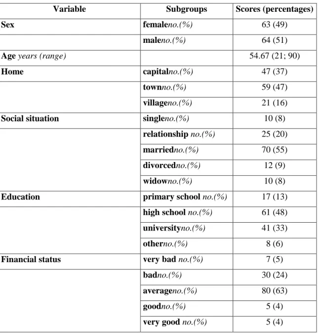
4.1. Interferon-induced depression - first study ... 51
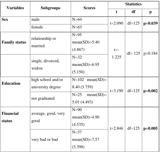
4.1.2. Baseline differences in psychometric scores according to background variables ... 53
4.1.3. Longitudinal effect of interferon treatment on depression ... 57
4.1.4. Effect of high versus low social support on depressogenic side effects of interferon treatment ... 59
4.1.5. Longitudinal effect of interferon treatment on anxiety ... 61
4.2.Ipilimumab vs. interferon – second study ... 65
4.2.1. Baseline ... 65
4.2.2. Drug effect in time on depression ... 68
4.2.3. Drug effect in time on anxiety... 69
5. Discussion ... 71
5.1. Depression in melanoma patients ... 72
5.1.1. Potential pathophysiological effects of interferon of mood ... 73
4
5.1.2. Possible interaction between interferon treatment and social support ... 74
5.2. Differences betweenipilimumab and interferon treatment ... 75
5.3. Implication of the results for the clinical practice ... 77
5.4. Limitations ... 79
6. Conclusion ... 80
7.a. Summary ... 82
7.b. Összefoglaló ... 83
References ... 84
List of own publications related to the thesis ... 104
Acknowledgement ... 105
List of tables and figures ... 107
5
Abbreviations
5-HT – 5-hidroxytriptamine (serotonin)
5-HTTLPR – serotonin-transporter-linked polymorphic region AJCC – American Joint Committee on Cancer
ANOVA – Analysis of variance ANCOVA – Analysis of covariance
anti-CTLA-4 – anticytotoxic T-lymphocyte-associated antigen 4 APC – antigen presenting cell
BC – Before Christ
BCG – Bacillus Calmette–Guérin BDI – Beck Depression Inventory CD8 – cluster of differentation 8 CTL – cytotoxic T lymphocytes
CRH – corticotropin releasing hormone DNA - deoxyribonucleic acid
DTIC -Dimethyl triazeno imidazole carboxamide FDA – U.S. Food and Drug Administration HDI – high-dose interferon
HPA – hypothalamic–pituitary–adrenal(axis) HLA – human leukocyte antigen
IFN – interferon IL – interleukin
IL-1RA – interleukin-1 receptor antagonist irAE – immune related adverse effects LAK – lymphokine activated killer
6 LDH – lactate dehydrogenase
MHC – major histocompatibility complex MIU – million international units
MM – malignant melanoma
NCCN -National Comprehensive Cancer Network NK – natural killer (cell)
PD-1 – programmed death-1 (protein)
PD-L1 – programmed death ligand-1 (protein) SD – standard deviation
SDS – Zung Self-Rating Depression Scale
SPSS – Statistical Package for the Social Sciences SSRI – selective serotonin reuptake inhibitors STAI – State-Trait Anxiety Inventory for Adults TCR – T-cell receptor
Th1 – T-helper 1
TNF – tumour necrosis factor
TNM – Tumour-Node-Metastases classification system UV – ultraviolet
XP - Xeroderma pigmentosum
7
1. Introduction
1.1. Malignant melanoma
Malignant melanoma is a type of cancer arising from melanocytes. Melanocytes in the epidermis of the skin produce the pigment melanin, which occurs in several forms that variably protect the skin from ultraviolet (UV) radiation. Environmental insults followed by proto-oncogene activation coupled with suppression of tumour suppressor genes, and defects in DNA repair mechanism further exacerbated by the inability of the immune system to contain these insults results in melanoma (Kraemer et al. 1994).
Melanomas typically occur in the skin but may rarely occur in the mouth, intestines, or eye. Melanoma often affects a relatively younger population and it metastasizesat an early stage. Early detection leads to a cure rate of over 90% in low-risk melanomas but advanced melanomas respond poorly to current therapies (Ehret, 2014).Immunotherapies and their combinations are one of the new ways of treatments in advanced melanoma. Immunotherapy is a general term referring to artificial activation of the immune system to induce objective responses and/or disease stabilization (Drake et al. 2014). Immunotherapies can cause an early neurovegetative syndrome characterised by depression, anxiety, fatigue, anorexia, pain and psychomotor slowing which pathophysiological background are not fully understood yet (Lotrich, 2013; Lin, 2014; Kovács et al. 2015).
1.1.1. The “black cancer” – the history of melanoma
In Greek “melas” means dark and “oma” means tumour. The first description of melanoma dates back to Hippocrates in the 5th century BC. Melanoma can be diagnosed from skeletons with osteolytic bone metastases. The earliest physical evidence of the disease comes from the metastases of melanoma found in skeletons of Pre-Columbian mummies (Vito et al. 2012). Between 1650 and 1760 the medical literature referred the disease as “fatal black tumours with metastases and black fluid in the body” (Vito et al.
2012, pg 2.). The first surgical removal of a melanoma was performed by John Hunter in 1787 and he labelled it as “cancerous fungous excrescence” (Gorantla and Kirkwood,
8
2014). Rene Laennec (the inventor of stethoscope) was the first who realized that melanoma is a distinct disease – he used the term melanose to describe the tumour.In 1966 Clark developed a standard five-level scale to assess the prognosis of melanoma based on histological examination (Clark’s levels): deeper invasion of tumour cells (epidermis, dermis, subcutaneous tissue) means worse prognosis (Clark, 1969).
Alexander Breslow defined the tumour thickness as prognostic factor of malignant melanoma in 1970 (Breslow thickness) (Breslow, 1970). Clark’s standard scale and the tumour thickness are currently still relevant prognostic factors (Balch et al. 2001).
1.1.2. The incidence of melanoma
The incidence of melanoma has been increasing in the past few decades (Petrella et al. 2012). Worldwide the incidence of melanoma continues to rise and despite advances in local and systemic therapy, mortality continues to rise with 80% of skin cancer-related deaths attributable to melanoma (WHO, 2013). Melanoma accounts for less than 5% of all skin cancers, but is the leading cause of skin cancer mortality.
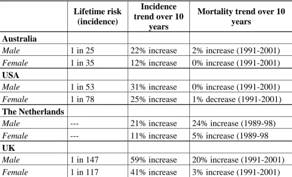
In the United States from 1973 to 2004 the incidence of melanomas has risen from 8 to 34 per 100.000 in males and 7 to 25 per 100.000 in females and the worldwide incidence rates vary significantly: to 0.2-0.5 per 100.000 in India, to 12-15 per 100.000 in Germany and to 40-50 per 100.000 in Australia (Roesch and Volkenandt, 2009). Data of 10-years incidence in four countries were shown in Table 1.
9
Table 1. Lifetime risk, incidence and mortality trends in melanoma based on statistics of Australia, USA, The Netherlands and UK (adapted from Thompson et al. 2005)
Lifetime risk (incidence)
Incidence trend over 10
years
Mortality trend over 10 years
Australia
Male 1 in 25 22% increase 2% increase (1991-2001) Female 1 in 35 12% increase 0% increase (1991-2001)
USA
Male 1 in 53 31% increase 0% increase (1991-2001) Female 1 in 78 25% increase 1% decrease (1991-2001)
The Netherlands
Male --- 21% increase 24% increase (1989-98)
Female --- 11% increase 5% increase (1989-98
UK
Male 1 in 147 59% increase 20% increase (1991-2001) Female 1 in 117 41% increase 3% increase (1991-2001)
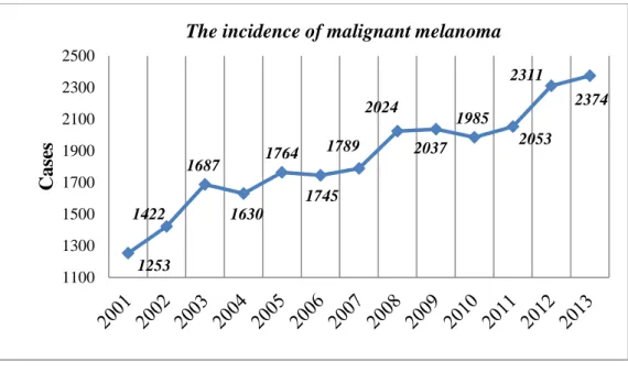
Also in Hungary the incidence of melanoma has been progressively rising in the past decade. According the data of Hungarian National Cancer Registry and Center of Biostatistics in the last 10 years the number of melanoma cases has doubled in Hungary (Figure 1., Table 2.).
10
Figure 1. The incidence of malignant melanoma (without melanoma in situ) in Hungary (2001-2013) (National Cancer Registry and Center of
Biostatistics, Hungary)
Table 2. Incidence data from Hungary (2001-2013). The incidence of in situ melanoma and melanoma malignum in females and males in Hungary (2001-2013). National Cancer Registry and Centre of Biostatistics, Hungary.
MM: malignant melanoma; in situ: in situ melanoma.
Year Male Female
In situ MM In situ MM
2001 91 569 100 684
2002 119 626 147 796
2003 123 742 209 945
2004 159 741 228 889
2005 187 807 261 957
2006 196 817 264 928
2007 134 847 204 942
2008 176 936 207 1088
2009 187 956 248 1081
2010 194 914 242 1071
2011 210 1014 265 1039
2012 234 1072 272 1239
2013 295 1116 324 1258
1253 1422
1687 1630
1764 1745
1789 2024
2037 1985
2053 2311
2374
1100 1300 1500 1700 1900 2100 2300 2500
Cases
The incidence of malignant melanoma
11 1.1.3. Risk factors
Major risk factors of melanoma are the following (NCCN Guidelines, 2015):
1. Exposure to ultraviolet (UV) rays is a major risk factor for most melanomas. This exposure can be characterised by the cumulative solar exposure and sunburn events (UV-B). UV rays originated from sunlight, tanning beds and sun lamps damage the DNA of skin cells and from the point when this damage affects the DNA of genes that control skin cell growth skin cancers develop. The nature of the UV exposure may be important in melanoma development. For example, the development of melanoma on the trunk (chest and back) and legs has been linked to frequent sunburns (especially in childhood).
2. Benign pigmented naevus are dense areas of melanin. Naevuses are not usually seen on babies they often begin to appear in children and young adults. Most naevuses will never cause any problems, but withincreasing number of naevuses the risk to develop melanoma is also increased. Thus many and/or large and/or atypical naevuses could mean higher risk for the disease.
3. The white colour of the skin carries a much higher risk for melanoma.
Whites with red or blond hair, blue or green eyes, or fair skin that freckles or burns easily are at increased risk.
4. If one or more first-degree relatives (parent, brother, sister, or child) has had melanoma the risk of melanoma is greater. Around 10% of all people with melanoma have a family history of the disease (Thompson et al.
2005).
5. Already having a melanoma increase the risk to of getting it again. About 5% of people with melanoma will develop a second one at some point.
People who have had basal or squamous cell skin cancers are also at increased risk of getting melanoma. Xeroderma pigmentosum (XP) is a rare, inherited condition that affects skin cells ability to repair damage to their DNA caused by ultraviolet lights which is also associated with increase occurrence of melanoma.
12
6. Melanoma is more likely to occur in older people, but it is also found in younger people. In fact, melanoma is one of the most common cancers in people younger than 30 (especially younger women).
7. People with weakened immune systems (from certain diseases or medical treatments) are more likely to develop many types of skin cancer, including melanoma.
In summary both increased exposure to risk factors and decreased defence system of the individual could lead to melanoma which further emphasize the role of the immune system in the pathogenesis of this disorder.
1.1.4. Stagingof the disease
Despite the fast development of molecular medical approaches,at present the most important information to determine the most appropriate evidence-based treatmentfor a given patient is thestage of the disease.According to the guideline of the American Society of Clinical Oncologythe staging is based on the TNM Classification System, which is explained below (NCCN Guidelines, 2016).
1.1.4.1.Overview of the TNM Classification System of melanoma
The TNM staging system is widely used in clinical practice to classify tumours.
TNM staging of melanoma describes the thickness of the melanoma and whether there is any spread to lymph nodes or other parts of the body.
The letters (T-N-M) describes the different areas of cancer growth:
T-Tumour: how far it has grown within the skin and other factors
N-Node: bean-sized collections of immune system cells, to which cancers often spread first
M-Metastases: spread to distant organs and which organs it has reached
This staging system describes the size of a primary tumour (T), whether any lymph nodes contain cancer cells (N) and whether the cancer has spread to another part
13
of the body (M). The T part of the TNM describes the thickness of the melanoma (primary tumour) according to the Breslow scale.
The T part of the TNM system is further divided into two groups a and b, depending on whether the melanoma is ulcerated or not. Ulcerated means that the covering layer of skin over the tumour is broken. Ulcerated melanomas have a higher risk of spreading than those which are not ulcerated.
The N part of the stage is further divided into groups a, b and c. If the cancer in the lymph node can only be seen with a microscope (micrometastasis) it is classed as a.
But if there are obvious signs of cancer in the lymph node (macrometastasis) it is classed as b.
The letter c means that there are melanoma cells in small areas of skin very close to the primary melanoma or in the skin lymph channels. These groups of melanoma cells in the skin are called satellite metastases. Melanoma cells in the lymph channels are called in transit metastases.
M0 means the cancer has not spread to another part of the body. M1 means the cancer has spread to another part of the body.
This staging system is the only internationally accepted classification system, and it includes also sentinel node staging (AJCC: Americal Joint Comittee on Cancer).
1.1.4.2.Treatment-relevant stages of melanoma
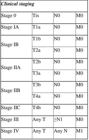
The T, N, and M groups can be combined to create overall stages for melanoma, which are denoted with the combination of Roman numerals I to IV (1 to 4) and capital letters. Thisclinical staging system (Table 3.) is a standard way to describe how far the disease has spread and will determine the treatment.Patients with lower stage melanomahave a better outcome regarding the long-term survival.
14
Table 3. The clinical staging of melanoma (AJCC) Clinical staging
Stage 0 Tis N0 M0
Stage IA T1a N0 M0
Stage IB
T1b N0 M0
T2a N0 M0
Stage IIA
T2b N0 M0
T3a N0 M0
Stage IIB
T3b N0 M0
T4a N0 M0
Stage IIC T4b N0 M0
Stage III Any T ≥N1 M0
Stage IV Any T Any N M1
Stage 0 melanomas have not grown deeper than the top layer of the skin (the epidermis). They are usually treated by surgery (wide excision) to remove the melanoma. Stage I and Stage II melanomas are treated also by wide excision. If the melanoma is stage IB or has other characteristics that make it more likely to have spread to the lymph nodes, sentinel lymph node biopsy is recommended. Lymph node dissection is recommended if cancer cells were found on the biopsy (Stage II). Adjuvant therapy after surgery may advise in Stage II melanoma. Stage III cancers have already reached the lymph nodes when the melanoma is first diagnosed. Surgery and adjuvant treatment and/or radiation therapy are recommended. Stage IV melanomas are very hard to cure, as they have already spread to distant lymph nodes or other areas of the body.
Metastases that cause symptoms but cannot be removed may be treated with radiation, immunotherapy, targeted therapy, or chemotherapy. The treatment of widespread melanomas has changed in recent years as newer forms of immunotherapy (known as
15
immune checkpoint inhibitors) and targeted drugs have been shown to be more effective than chemotherapy.
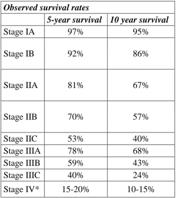
Same stage means the same or similar prognosis. Observed survival rates shown below for the different stages (Table 4.).
Table 4. Observed survival rates in melanoma (adapted from the AJCC Melanoma Staging Database, 2008.). Observed survival rates based on data of 60.000 patients *:The outlook is better if the spread is only to distant parts of the skin or distant lymph nodes rather than to other organs, and if the blood level of lactate dehydrogenase (LDH) is normal.
Observed survival rates
5-year survival 10 year survival
Stage IA 97% 95%
Stage IB 92% 86%
Stage IIA 81% 67%
Stage IIB 70% 57%
Stage IIC 53% 40%
Stage IIIA 78% 68%
Stage IIIB 59% 43%
Stage IIIC 40% 24%
Stage IV* 15-20% 10-15%
16 1.2. Treatment of melanoma
1.2.1. The history of melanoma therapy
The intent of treatment for advanced melanoma which was once considered incurable has changed in the last decades from palliative to potentially curative.
Treatment and outcomes for advanced melanoma have improved over the past twenty years on the basis of rapid advances in the fields of tumour cell biology, immunology, and surgical techniques, radiosurgery (Garbe et al. 2012).
In the beginning of the use of medical treatments mostly palliative (and largely ineffective) treatments were used.
Since Ehrlich in 1909 first proposed the idea that nascent transformed cells arise continuously in the body and that the immune system scans for and eradicates these transformed cells before they are manifested clinically, immune surveillance has been a controversial topic in tumour immunology (Ehrlich, 1909).
Tumour transplantation models show the experimental evidence that tumours could be repressed by the immune system. These findings suggested the existence of tumour-associated antigens and formed the basis of “immune surveillance” (Burnet and Thomas, 1957). “Immune surveillance’ implied surveillance of the host for malignant cells, which were presumed to be recognized and destroyed as they emerged. This idea was supported by the observation of spontaneous regression in human melanoma, the lymphocytic/dendritic infiltrates documented pathologically in and around primary melanoma tumours, and the increased incidence of melanoma among immunosuppressed patients (Gorantla and Kirkwood, 2014).
The idea of cancer immune surveillance resisted widespread acceptance until the 1990s. The central roles of immune effector cells, such as B, T, natural killer (NK) and natural killer T (NKT) cells, and interferon (IFN) have been clarified in cancer immune surveillance (Dunn, 2004; Kim et al. 2007).
17
Golub et al. demonstrated in vitro increased cytotoxic activity of melanoma patient-derived lymphocytes against melanoma cell lines (Golub, 1974). The discovery of interleukin 2 (IL-2) as a T cell growth factor helped in vivo studies of immune modulation and its role in advanced melanoma (Gorantla, 2014). Supporting this observationin metastatic melanoma the use of anticytotoxic T-lymphocyte-associated antigen 4 (anti-CTLA-4 antibody) immunotherapy (ipilimumab) seems to be effective (2011 FDA approval) (Lee et al. 2013). Thus, a better understanding of the mechanisms of immunoediting during tumour progression may provide new insights for improving cancer immunotherapy.
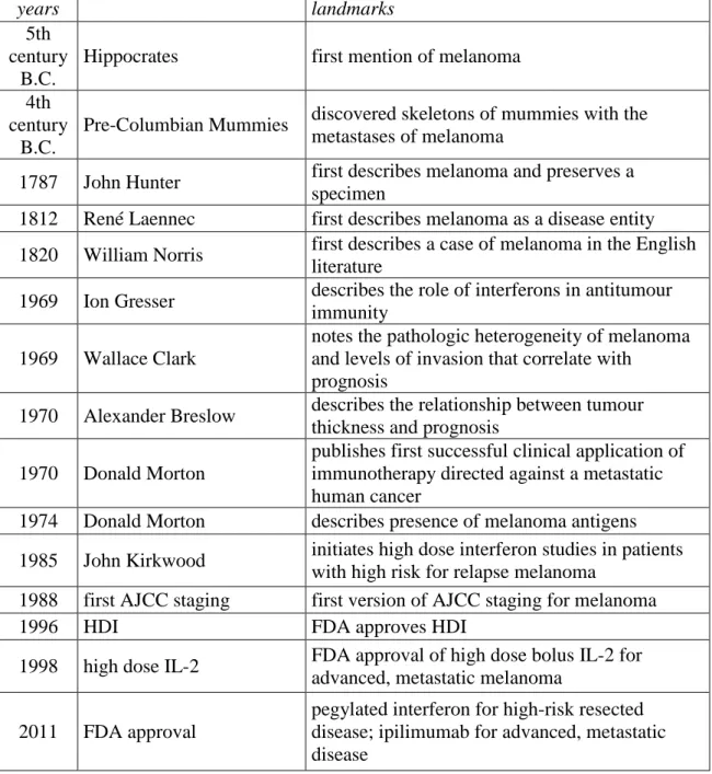
The most important milestones in the history of melanoma immunetreatment are shown in Table 5.
18
Table 5. Major steps in the immunetreatment of melanoma (adapted from Lee et al. 2013). FDA: Food and Drug Administration; HDI, high-dose interferon; IL, interleukin; LAK, lymphokine activated killer.
years landmarks
5th century
B.C.
Hippocrates first mention of melanoma 4th
century B.C.
Pre-Columbian Mummies discovered skeletons of mummies with the metastases of melanoma
1787 John Hunter first describes melanoma and preserves a specimen
1812 René Laennec first describes melanoma as a disease entity 1820 William Norris first describes a case of melanoma in the English
literature
1969 Ion Gresser describes the role of interferons in antitumour immunity
1969 Wallace Clark
notes the pathologic heterogeneity of melanoma and levels of invasion that correlate with
prognosis
1970 Alexander Breslow describes the relationship between tumour thickness and prognosis
1970 Donald Morton
publishes first successful clinical application of immunotherapy directed against a metastatic human cancer
1974 Donald Morton describes presence of melanoma antigens 1985 John Kirkwood initiates high dose interferon studies in patients
with high risk for relapse melanoma
1988 first AJCC staging first version of AJCC staging for melanoma
1996 HDI FDA approves HDI
1998 high dose IL-2 FDA approval of high dose bolus IL-2 for advanced, metastatic melanoma
2011 FDA approval
pegylated interferon for high-risk resected disease; ipilimumab for advanced, metastatic disease
90% of melanomas are diagnosed as primary tumours without metastasis and for patients with early-stage non-metastatic disease surgical management remains the mainstay of therapy (Garbe et al. 2012; Mocellin et al. 2013). Adjuvant immunotherapy is offered to those melanoma patients who have no evidence of metastases but at high
19
risk for further tumour spread, e.g. tumours thicker than 1.5 mm, or in stage II and III melanoma. Specifically, interferon alpha is the fundamental therapy as it was the first substance in the adjuvant treatment of melanoma to have shown a significant improvement of disease-free survival (Garbe et al. 2008; Friebe et al. 2010). However, adjuvant interferon treatment is frequently accompanied by psychological side effects including depression, fatigue, irritability, anxiety, or suicide.
The treatment of melanomas with distant metastases are still not satisfactory with the median survival of patients with melanoma is less than 1 year (Schaefer et al.
2002) and the expected 2-year survival rate is 10-20% (Rychetnick et al. 2012).
Although the prognosis for melanoma is improving because lesions are diagnosed at a much earlier point, the therapy of disseminated melanoma has been unsolved for decades. Median survival with distant metastasis in the fourth stage based on former data is less than 1 year (Gray-Schopfer et al. 2007; Mocellin et al. 2010). Survival increased as a result of new therapies of recent years, but the mortality rate of the disease is still significant. In the event of localised diseases, surgical intervention plays a leading role. However, following excision further therapies may be needed due to the parameters of primer tumour (ulceration, tumour thickness, lymph node status) (Liszkay et al. 2003; Kasparian et al. 2009).
Systemic pharmacotherapies are widely used in the treatment of melanoma, especially if malignant cells spread beyond the skin to distant organs. Broad range of drugs are available including chemotherapies, targeted therapy and vaccine therapy. If distant metastases emerged, promising opportunities have been arising recently in the treatment of the disease with the progress of molecular pathology and immunology (Garbe et al. 2011). New products, primarily developed for other medical conditions, are retargeted for application in melanoma treatment by utilising their potentialto strengthen the immune response of the organism against foreign or altered antigens, such as tumour cells (Bhatia and Thompson, 2014).
20 1.2.2. Chemotherapy of melanoma
Surgery and radiation therapy remove, kill, or damage cancer cells in a certain area, but chemotherapy can work throughout the whole body. Chemotherapy is a medication-based, systemic therapy to treat many types of cancer, including melanoma, by destroying melanoma cells throughout the body. This means chemotherapy can kill cancer cells that have spread (metastasized) to parts of the body far away from the original (primary) tumour (Fuchs-Tarlovsky, 2013).Chemotherapy drugs kill the fast- growing cells, also cancer cells and also normal cells. Systemic chemotherapy uses anticancer drugs that are usually injected into a vein or given by mouth. These medications travel through the bloodstream to all parts of the body, where they attack cancer cells that have already spread beyond the skin to involve lymph nodes and other organs. Usually the combination of more than one drug is used. Many other chemotherapy agents are being evaluated for their use in the treatment of Stage IV melanoma as both single agents and in combination with other chemotherapy, targeted therapy and immunotherapy agents.
Although chemotherapy is usually not as effective in melanoma as in some other types of cancer, it may relieve symptoms or extend survival of some patients with stage IV melanoma (Middleton et al. 2000).
Chemotherapy drugs often used to treat melanoma include:
Dacarbazine (DTIC) is the only FDA-approved chemotherapy agent for the treatment of Stage IV melanoma. It is administered as an intravenous infusion.
Cisplatin, vinblastine, and DTIC is another chemotherapy combination for treating melanoma.
Temozolomide is a drug that works like DTIC, but it can be given in the form of a pill.
Conventional chemotherapy with dacarbazine and temozolomide has yielded poor response rates of 7%–20% and a median survival of nine months, with mild toxicity profiles (Chapman et al. 1999).
21
These chemotherapy drugs may also be combined with immunotherapy drugs, such as interferon alpha and/or interleukin 2.
1.2.3. Vaccine therapy of melanoma
Melanoma vaccines are experimental therapies that are being tested in patients with stage III or stage IV melanoma. Anti-melanoma vaccines are similar to the vaccines used to prevent diseases caused by viruses. Antivirus vaccines usually contain weakened or killed viruses or parts of a virus that cannot cause the disease. The vaccine stimulates the body's immune system to destroy the more harmful type of virus.
In the same way, weakened melanoma cells or parts of melanoma cells called antigens can be injected into a patient in an attempt to stimulate the body's immune system to destroy melanoma cells. Usually, the melanoma cells are mixed with substances that help stimulate the body's immune system.
1.2.4. Targeted therapy of melanoma
Targeted therapy is a form of treatment in which drugs (or other substances) are developed with the goal of destroying cancer cells while leaving normal cells intact.
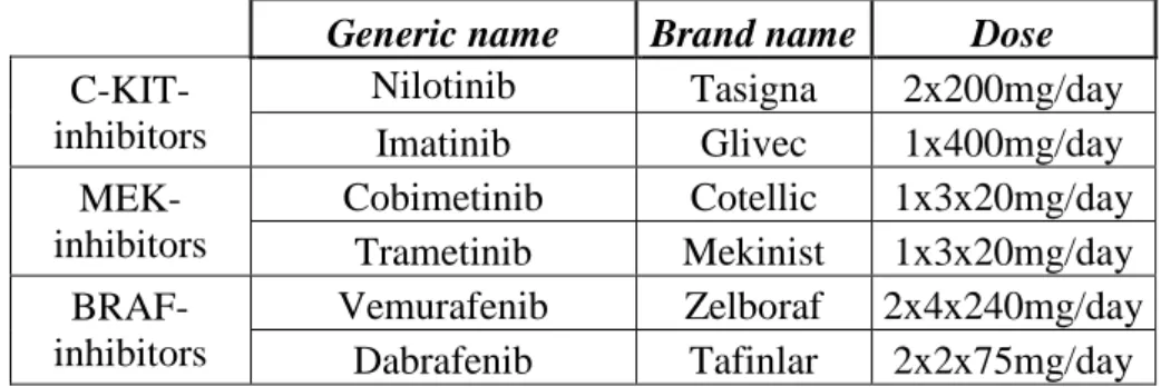
These drugs are designed to interfere with the specific molecules that are driving the growth and spread of the tumour. Because they are “targeted” to the tumour, these therapies may be more effective and associated with fewer side effects compared to chemotherapy and radiation therapy (Davey et al. 2016).Targeted therapy drugs for melanoma see on Table 6.
Molecular targeted therapies have shown promise in the management of various malignancies, including melanoma, with lower toxicity profiles and better overall survival as compared with conventional therapy (Chakraborty et al. 2013).
22
Table 6. Targeted therapy drugs for melanoma(adapted from the NCCN Guidelines, 2016). KIT- receptor tyrosine kinase - inhibition of KIT leads to defects in melanocyte migration, survival, proliferation, and differentiation; MEK - allostetic mitogen-activated protein/extracellular signal-regulated kinase - MEK inhibition blocks cell proliferation and induced apoptosis;BRAF - serine/threonine protein kinasewich is involved in sending signals inside cells which are involved in directing cell growth.
Generic name Brand name Dose C-KIT-
inhibitors
Nilotinib Tasigna 2x200mg/day
Imatinib Glivec 1x400mg/day
MEK- inhibitors
Cobimetinib Cotellic 1x3x20mg/day Trametinib Mekinist 1x3x20mg/day BRAF-
inhibitors
Vemurafenib Zelboraf 2x4x240mg/day Dabrafenib Tafinlar 2x2x75mg/day 1.2.5. Immunotherapies of melanoma
Immunotherapy is a general term referring to artificial activation of the immune system to induce objective responses and/or disease stabilization (Drake et al. 2014).
Immunotherapy enhances and encourages a patient's immune system to recognize and destroy cancer cells more effectively. Several types of immunotherapy are used in treating patients with melanoma. Some are being studied as adjuvant treatment. Immunotherapy (also called biological therapy) is a treatment that increases the activity of immune system. These drugs improve the ability of the body to find and destroy cancer cells. Immunotherapeutic interventions offer the hope for effective treatment against melanoma as it is considered traditionally as immunogen tumour cancer because of its ability to undergo spontaneous regression (Thumar and Kluger, 2010). The most convincing evidence that melanoma can be immunogenic is derived from preclinical research on the fundamental aspects of T-cell biology and antigen recognition (Komenaka et al. 2004). Although the fundamentals of tumour immunology can be broadly applied to all types of tumours, many of these principles were first demonstrated in melanoma, and the clinical application of immunotherapy for cancer has been most widely studied in melanoma (Gogas et al. 2006).
23
1.2.4.1. The role of cytokines in the treatment of melanoma
Cytokines are proteins that activate the immune system in a general way. Two cytokines, interferon alpha and interleukin 2, can help boost immunity in patients with melanoma. Both drugs can help to shrink metastatic (stage III and IV) melanoma in about 10% to 20% of patients (Verma et al. 2006; Bhatia et al. 2009).
Interleukin 2, particularly in high doses, can cause fluid to accumulate in the body, so the person swells up and can feel quite sick. Some patients may need to be hospitalized because of this problem (Komenaka et al. 2004; Agarwala, 2009).
Patients with deeper melanomas often have cancer cells that break away from the primary melanoma and travel to other parts of the body. Interferons are immune substances produced by the body in response to infection. Interferon alpha 2b can be used as an adjuvant therapy (Garbe et al. 2008). Side effects include fever, chills, aches, and severe tiredness. Interferon alpha 2b can also affect the heart and liver, and patients should be followed by an oncologist who is experienced with this treatment (Schaefer et al. 2002). Interferon alpha 2b given to patients with stage III melanoma following surgery can delay the recurrence of melanoma but may not prolong patients' lives.
Decisions about adjuvant therapy by patients and their doctors should take into account the potential benefits and side effects of this treatment.
Another cytokine is called tumour necrosis factor (TNF). This is a naturally occurring substance that seems to kill tumours. It is particularly effective when the TNF is given as an infusion directly into a tumour or a part of the body containing the tumour. This is called regional perfusion (Krementz et al. 1994).
Cytokines (interleukins and interferons) are part of the immune system and exist naturally in the body stimulating immune cells. Synthetically manufactured cytokines are also applied in treatment of melanoma. According to treatment protocols in Hungarypatients without distant metastases, with aprimarytumour parameter thicker than 1.5 mm adjuvant immunotherapyshould be given (Egészségügyi Közlöny, 2008).
At present, interferon is the most widely used adjuvant therapy to increase asymptomatic survival after the removal of primer tumour. It is applied for almost 20
24
years usually at the early phase of the disease in various doses, as supported by clinical evidence (Garbe et al. 2008; Friebe et al. 2010).
1.2.4.2. The role of interferon in the treatment of melanoma
Interferons are pleiotropic molecules that share a number of biologic effects such as antiviral, antiproliferative and immunomodulatory actions. The alpha interferons are produced by leukocytes or lymphoblastoid cells.
Interferon-alpha has a general inflammatory action which skews the immune response towards a Thelper 1 (Th1) profile (Belardelli and Gresser, 1996). Type Th1 cells produce interferon-gamma, interleukin (IL)-2, and tumour necrosis factor (TNF)- beta, which activate cell-mediated immunity. When interferon-alpha are investigated in vitro they activate Th1 immune responses. In in vitro studies subtype alpha2 increased the expression of human leukocyte antigen (HLA) molecules wich correlate with interferon-alpha mediated activation of memory cluster of differentiation 8(CD8) cells and increased cytolytic action against tumour cells (Finter, 1991).
Interferon-alpha enhances the proliferation of human B cells as well as strongly activates natural killer (NK)-cells (Ortaldo and Herberman, 1984; Hibbert and Foster, 1999). The interferon-alpha2 also affect human T cell mobility and dendritic cell activation (Foster et al. 2004).
Effects of interferon linked to cell membrane include stimulation of the activity of macrophages and lymphocytes in addition to direct cytostatic effect.
Multi-subtype interferons such as human leukocyte interferon-alpha might be more appropriate for melanoma treatment than interferon-alpha2 given its wider spectrum of immunological activities. Another difference between human leukocyte interferon-alpha and recombinant interferons is the lack of glycosylation of specific subtypes in the recombinant formulations (Kontsek, 1994).
All interferon-alpha bind to the same receptor and are expected to have the same biological functions. Human leukocyte interferon-alpha consists ofsix major subtypes, and all subtypemay exert differentanti-viral or anti-tumour effects. For example,
25
recombinant interferon-alpha2b has been investigated in a recently published paper by Ruuth et al. (Ruuth et al. 2007). In this study three melanoma lines were treated with both types of interferon, and the effect on proliferation and survival was estimated both after short-term and prolonged treatment. The results indicated that the melanoma cell lines were sensitive to the antiproliferative effects of both interferon-alpha species during short-term treatment (Ruuth et al. 2007).
The induction of a characteristic Th1 response in melanoma patients appears to be an important indicative factor for best disease prognosis. The results of Gogas et al.
(Gogas et al. 2006) indicate a clear correlation between the potential of strong interferon-alpha induced Th1 inflammatory response and longer disease free periods and overall survival in malignant melanoma patients (Go gas et al. 2006). Th1 inflammatory response has also been correlated with tumour regression. In studies with subcutaneous interleukin-2 and interferon-alpha circulating activated cytotoxic T lymphocytes (CTL) increased NK cells in the blood correlated with tumour growth retardation (Atzpodien et al. 1993; Schneeklothet al. 1993; Jorkov et al. 2003).
The therapeutic significance of multiple interferon-subtypes is still unclear. This multiplicity of activity may be a result of different specific biological activities, altering diffusability, tissue distribution and pharmacokinetics. Another possibility is that multiple interferon-subtypes may compete for receptor binding resulting in a natural selection process and possibly antagonistic effects. Some results suggest that particular subtypes, such as interferon alpha2 and alpha8 may be more effective than other subtypes at protecting cells from viral infection, and that combinations of these two subtypes or natural interferon alpha preparations containing such combinations appear to be optimal for treatment of melanoma (Hino et al. 1993).
Interferons affect many organs and cause multiple side effects in most of the treated patients (Schaefer et al. 2002). The most common, 1 out of 10-100 patients, includes anorexia, mild depression, headache and concentration disturbances, cough, dermatitis, fever and arthralgia. Inthe first period of treatmentanaemia, low white cell count and thrombocytopenia can occur, whichlevels usually are closelymonitored.
These latter findings often result in dose reduction (Garbe et al. 2008). Among the rare side effects, 1 out of 1000-10000 patients can be mentioned: psychosis, suicide,
26
thyroiditis, erythema multiforme, retinal bleeding, endocarditis and liver failure (Kasparian et al. 2012). Their spectrum of toxicity is well documented with a ‘flu-like syndrome’ occurring universally in patients upon initiation of therapy but disappearing with repeated drug administration.
1.2.4.3.Ipilimumab – new therapy in the treatment of melanoma
The fast progress in understanding of immunobiology made significant break- throughs in immunotherapies that have radically changed and renewed the treatment of malignant melanoma (Anderton and Fillatreau, 2015; Atherton et al. 2016). The approval of anti-cytotoxic T-lymphocyte antigen 4 (CTLA-4) antibody ipilimumab by US FDA in 2011, as well as the new drugs including antibodies to programmed cell death 1 (PD-1) such as pembrolizumab and nivolumab (both approved in 2014) have extended the potential of immunotherapy for advanced melanoma (Eggermont et al.
2008; Zhu et al. 2010). They have shown a significant increase in progression free and overall survival rate with long-term benefits in a proportion of patients compared with chemotherapy (Hodi et al. 2010; Wolchok et al. 2010; Zhu et al. 2010). Applied immunotherapies for stage III and IV melanoma exert different mechanisms of action that manifest in different adverse events (Ma and Armstrong, 2014). Each of the drugs used to treat metastatic melanoma exert particular mechanisms of action.
The anti-CTLA-4antibody ipilimumab was the first immunotherapy that showed a benefit for overall survival in two controlled trials in metastatic melanoma (Hodi et al.
2010). Ipilimumab is a human anticytotoxic T-lymphocyte-associated antigen 4 (anti- CTLA-4) monoclonal antibody which blocks the inhibitory signal of CTLA-4 molecule having an effect of “natural brake” to the immune system thereby stimulating T-cell activation and proliferation that leads to cancer cell death (Robert et al. 2015).
Ipilimumab binds to the cytotoxic T-lymphocyte-associated antigen 4 (CTLA-4). This T-cell molecule suppresses the immune response. Ipilimumab in melanoma patients has an indirect effect through T-cell mediated anti-tumour immune response. Ipilimumab is a T-cell potentiator that blocks the inhibitory signal of CTLA-4 (Fong and Small 2008;
Fecher et al. 2013). Suppression of CTLA-4 can augment the immune system's T-cell
27
response in fighting disease. T-cell-mediated antitumour therapies have played an important role in the treatment of advanced melanoma in last years. Tumour-associated antigens attached to the major histocompatibility complex (MHC) on specialized antigen-presenting cells (APC) bind with T-cell receptors (Chmielowski, 2013). CTLA- 4 is a surface protein expressed on activated and regulatory T cells and is upregulated in malignancy. It functions as a negative regulator of T-cell function, binding to B7 antigen-presenting cells and inducing cell-cycle arrest. On the T cells, surface protein CD28, is a positive regulator of T-cell function and binds B7 with less affinity (Schatton et al. 2010). The goal of CTLA-4 blockade is to break immune tolerance to a cancer and trigger a prolonged tumour-specific immune attack (Fecher et al. 2013). Ipilimumab was registered in 2011 by the FDA.
During ipilimumab therapy, most often autoimmune side effects were reported (colitis, thyreoiditis, hepatitis) as well as depression, confusion, insomnia, change in mental condition, tiredness (fatigue) are mentioned in the literature (Pardoll, 2012).Its most common adverse events related to the study drugs were immune-related events.
Severe autoimmune reactions commonly skin rash, colitis, thyreoiditis, hepatitis, hypophysitis may develop in some patients. Depression, confusion, insomnia, mental status changes are named as expected adverse events in ipilimumab treatment (Garbe et al. 2012), but there are only few data in the literature. Immune-related adverse events associated with the use of ipilimumab were already evident in randomized trials (Hodi et al. 2010;Quirk et al. 2015; Robert et al. 2015). Immune-mediated adverse effects are common, but not severe in most patients. Skin-related adverse events can occur 2–3 weeks after the first dose of ipilimumab, whereas liver and gastrointestinal events typically occur 6–7 weeks after treatment initiation, and endocrinopathies are usually observed 9 weeks after the initial drug administration (Di Giacomo et al. 2010). Less frequent, but well-documented and potentially serious or irreversible immune-related adverse events can involve the liver or endocrine or nervous systems, including sensory, motor or ocular manifestations (Quirk et al. 2015).
28
1.2.4.4. Other immunotherapies in the treatment of melanoma
Besides antibodies against CTLA-4, most experimental and clinical information was gathered on the inhibition of programmed death (PD)-1-route in connection with the treatment of advanced diseases. The aim is not to kill cancer cells directly but to block a pathway that shields tumour cells from immune system components able and to fight cancer.The pathway includes two proteins:
programmed death-1 (PD-1), which is expressed on the surface of immune cells
programmed death ligand-1 (PD-L1), which is expressed on cancer cells When PD-1 and PD-L1 connect they form a biochemical barrier protecting tumour cells from being neutralised by the immune system.PD-1 is important mainly at the later phase of implementation of T-cell activation, in inhibiting inflammatory reactions and autoimmunity in peripheral tissues. The autoimmune side effects of the anti-PD-1 therapy are much lighter and develop later as compared to anti-CTLA-4. On the other hand, its interaction with the ligands of PD-1 receptor proved to be an important immune-resistant mechanism of tumours (Dong et al. 2002; Ladányi, 2002; Topalian et al. 2012), and therefore it is a promising objective of the therapy aimed at
“accessibility” to antitumour immune response. Anti-PD-1 antibody is a fully human antibody presently in clinical use. At the moment, two medicinal products are registered: pembrolizumab and nivolumab. A test performed using higher number of cases confirmed that the ratio of objective response given to the treatment was 28% in the case of melanoma and the effect proved to be durable (Topalian et al. 2012). 6% of grave side effects were potentially immune associated. Most frequent side effects described in connection with the treatment were tiredness, apathy, loss of appetite, diarrhoea, nausea, cough, constipation, skin rash, fever and headache (Topalian et al.
2012; Robert et al. 2014).
A number of clinical trials aimed at blocking PD-1-route are pending in melanoma and other tumours especially as monotherapy with PD-1-antagonist antibodies and also in various combinations (Sznol and Chen, 2013). Therapy
29
combining anti-CTLA-4 and anti-PD-1 may further increase the effectiveness of treatments.
In PD-1 therapies generalized symptoms, including fatigue and asthenia, fever and chills, myalgias, and headaches, were reported frequently but were of low grade in more than 95% of the cases (Hamid et al. 2013). The most common adverse events, regardless of causality, were fatigue, decreased appetite, diarrhea, nausea, cough, dyspnea, constipation, vomiting, rash, pyrexia, and headache (Topalian el al. 2012).
Fatigue is by far the most common symptom reported by patients and is often difficult to treat (Ribas, 2012; Hamid et al. 2013). Fatigue (56.3%), nausea (25%), diarrhea, xerostomia, and pruritus (18.8% each) were reported by anti-PD1 therapy (Freemann- Keller and Weber, 2015). In the comparative study of Robert et al. (Robert et al. 2014) the most common drug-related adverse events were fatigue (33%-37%), pruritus (19- 26%) and rash (18%). Fatigue is among the most common side effects seen, with an estimated overall frequency of 16 to 24 percent for the anti-PD-1 and anti-PD-L1 agents and approximately 40 percent in those treated with ipilimumab (Horvat et al. 2015;
Naidoo et al. 2015; Postow et al. 2015).
The two new immunotherapeutic agents, anti-CTLA-4 and anti-programmed cell death 1, show promise as potentially effective therapies with manageable side effect profiles in metastatic melanoma (Chakraborty et al. 2013).
1.3. Side effects of immune therapies in melanoma
As more-effective therapies become available based on modulation of the immune system in order to trigger or enhance anti-tumour immune responses, clinicians will need to become familiar with recognizing and controlling the adverse effects arising from immune therapy (Gangadhar and Vonderheide, 2014).
30 1.3.1. General symptoms
A new category of side effects, so called “immune-related adverse events”
(irAE) could complicate cancer immunotherapy. It is mainly due to the inflammation of off-target organ system, such as well described immune-related dermatitis, hepatitis, colitis, and hypophysitis (Postow et al. 2015). The most common and earliest onset of irAE is dermatologic toxicity. Adverse effects are usually reversible, although early recognition and intervention are essential. In this context, patient awareness, close monitoring and good communication between the care provider and patient are imperative.
Side effects are unplanned physical or emotional conditions caused by treatment.
Each treatment for melanoma has and can cause different side effects. The side effects of immunotherapies depend on:
the drug
how it is given
the amount taken
the length of treatment
the person
1.3.2. Psychological side effects
In the long run, adjuvant interferon treatment has the most common and also clinically relevant psychological side effects. Fatigue, anhedonia, social isolation, psychomotor slowness is reported during treatment and is frequently accompanied by psychological side effects including depression, irritability, anxiety, or suicide(Gogas et al. 2006). These psychological problems may lead to a significant deterioration in quality of life and often result in premature suspension or discontinuation of treatments.
Not only physical side effects presenting serious subjective symptoms, if any, may imply distress but the effects of immunotherapy in the central nervous system may also
31
cause grave anxiety and clinically significant depressive symptoms (Schaefer et al.
2002). Depression of clinically significant degree developing during interferon therapy varies between 20 and 40% (Schaefer et al. 2002) which at the same time is one of the most frequent reasons for premature discontinuation of the therapy (Reyes-Vazquez et al. 2012).
Besides early neurovegetative symptoms which manifest in the majority of patients during the first weeks of IFN-alpha treatment as fatigue, pain and anorexia, long-term IFN-alpha treatment often causes a wide variety of psychiatric side-effects, such as depression, fatigue, insomnia, anxiety, and cognitive disturbances (Capuron et al. 2002). 10%–40% of patients additionally develop a full depressive disorder syndrome that can include suicidal ideation, aboulia, lack of motivation, social withdrawal, guilt, anhedonia, irritability, anxiety, and crying (Lotrich, 2013). Mania, delirium, and psychosis are further but less common side effects of IFN-alpha treatment. Approximately 30–70% of hepatitis C virus-infected patients treated with IFN-alpha experience different degrees of depression. Most of them suffer from mild or moderate depressive symptoms, while severe major depression occurs in about 15%
(Schaefer et al. 2012).
1.3.3. Differences in psychological side effects according to pharmacodynamics of drugs
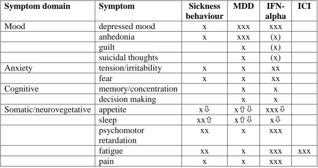
In addition to these similarities, the symptom profile (Table 7.) of treatment- emergent depression and naturally occurring major depressive episode show some distinctions (Pala et al. 2016). Namely, during long-term IFN-alpha treatment patients reported more severe weight loss and decreased activity, while feeling of guilt was less prominent compared to medically healthy depressed subjects (Capuron et al. 2009).
This observation was supported by a recent finding which suggested that risk genetic variant in the IL-6 gene more specifically increased depressive symptoms measured by the Zung Self-rating Depression Scale compared to the Brief Symptom Inventory, suggesting that inflammatory risk mechanisms are more responsible for
32
somatic/neurovegetative symptoms than cognitive-emotional signs of depression (Kovacs et al. 2015). Furthermore, newly developed immune checkpoint inhibitors, such as anti-cytotoxic T-lymphocyte antigen 4 (CTLA-4) antibodies or humanised immunoglobulins against programmed death 1/ligand 1 (PD-1/PD-L1) which also enhance tumour-specific immune activity are associated with a new category of side effects called “immune-related adverse events” (irAE), in which the most frequent symptom is fatigue (Postow et al. 2015).
Although both IFN-alpha and immune checkpoint inhibitors increase tumour- specific immune response their psychological side-effect profiles are strikingly different. It has been recently demonstrated that CTLA-4 antibodies (e.g. ipilimumab) decrease the number of regulatory T cells through non-classical monocytes (Romano et al. 2015). Nonclassical or patrolling monocytes are responsible for clearing up the consequences of inflammation at the vascular endothelium and maintaining the integrity of the BBB thus they might decrease expansion of the inflammation into the central nervous system (Ribas et al. 2015). PD-1/PD-L1 antibodies (e.g. nivolumab and pembrolizumab, both approved in 2014) increase effector T cell activity within tissues or tumours where cells express PD-1/PD-L1 which tends to be low in the brain (Postow et al. 2015; Ribas et al. 2015).
33
Table 7. Symptom profile of sickness behaviour, major depressive disorder, IFN-alpha induced depression, and psychological side effects of immune checkpoint inhibitors. MDD: major depressive disorder, IFN-alpha: interferon alpha treatment, ICI: immune checkpoint inhibitor treatment, x: symptom is present, number of x:
dominance of symptoms
Symptom domain Symptom Sickness
behaviour
MDD IFN- alpha
ICI
Mood depressed mood x xxx xxx
anhedonia x xxx (x)
guilt x (x)
suicidal thoughts x (x)
Anxiety tension/irritability x x xx
fear x x xx
Cognitive memory/concentration x x
decision making x x
Somatic/neurovegetative appetite x x xxx
sleep xx x x
psychomotor retardation
xx x xxx
fatigue xx x xxx xxx
pain x x xxx
1.3.4. Psychological distress and cancer
Prevalence of clinically relevant psychological distress among patients with melanoma (all stages) is approximately 30% (Kasparian et al. 2012; Rychetnick et al.
2012). Several of the diagnostic criteria for major depressive disorder are related to symptoms resulting from malignant and chronic diseases or their treatment: among them the most prevalent are low energy, poor appetite and impaired concentration.
Other cardinal psychological symptoms must be also present to diagnose major depressive disorder, such as low mood, loss of interest, rumination on negative emotions, grief, hopelessness, demoralization. Meyer et al. gave a thorough summary of cancer associated psychiatric problems (Meyer et al. 2009). Meyer et al. highlights that psychological problems depends on: premorbid psychiatric status, biological effect of cancer, biological effect of treatment, effect of coping style/psychological factors on cancer (quality of life, defense mechanisms, phases of illness). The most commonly
34
occurring psychiatric disease, according to the review of Meyer et al. are: depression (25%), anxiety disorders (panic disorder – 4.8%, generalized anxiety disorder – 3.2%, posttraumatic stress disorder – 4%), delirium (28-44%), fatigue (80%), bipolar disorders (0.4-1.6%), character disorders (2-3%), schizophrenia (0.5-1.5%), substance use disorders (~28%) (Meyer et al. 2009). The metaanalysis of Satin et al. showed that depression predicts mortality, but not progression, in cancer patients. Based on data from 25 studies, mortality rates were up to 25% higher in patients with depressive symptoms (Satin et al. 2009).
1.3.4.1. Depression
The incidence of clinically relevant depression during interferon therapy varies between 20% and 40% (Schaefer et al. 2002) making it the most common side effect and one of the main reasons for early discontinuation of treatment (Garbe et al. 2008).
Depression has a broad list of symptoms. Typically, physical problems (like change in bodyweight, difficulty in sleeping, fatigue, motor agitation or inhibition, etc.) also often develop in somatic conditions requiring hospitalisation and so they provide for less solid points of reference in differential diagnosis of depression. Therefore, it is particularly important to identify and also to considerpsychosomatic symptoms according to the condition. Depressed mood, anhedonia, loss of interest, sense of guilt, self-accusation, thinking of death frequently, mentioning of thoughts of committing suicide may be the chief symptoms of depression (Schaefer et al. 2002).
35 1.3.4.2. Anxiety
Anxiety is often an expression of the sense of helplessness which may be generalised in all areas of life (Erim et al. 2013). Physical and emotional components also must be taken into consideration when making diagnosis. Somatic palpitation, muscle stiffness, dizziness, stomach pain, dry mouth that can be felt; otherwise, patients can be characterised by restlessness, sleep difficulty (nightmares), irritability and sensitivity (Stark and House, 2000). Nonverbal signals (look, pose, loudness, tone etc.) of patients may also convey important messages. It is particularly important to observe them in order for precise assessment of the condition (Traeger et al. 2012).
1.3.5. Vulnerability factors for psychological side effects
Depressive or anxiety disorders in the psychiatric history increase the risk of psychiatric side effects during treatment. In addition, female sex, younger age, lower education, and lack of social support were investigated as risk factors for evolving psychiatric side effects, but conclusive biological or psychological markers predicting psychological side effects of interferon treatment are lacking (Kasparian et al. 2009).
Thus, routine monitoring of melanoma patients remains to identify clinically relevant psychological distress and possible protective factors (Friebe et al. 2010).
1.3.6. Screening and prevention of psychological side effects
Development of depression is a relatively slow process when immuntherapies are used (Friebe et al. 2010; Hanaizi et al. 2012). Prevention can be started early on by means of screening tests. It is necessary to evaluate the psychological state multi- dimensionally in order to establish individualised psychological care related to medical interventions. In addition to the knowledge based on the impressions of personal meetings and everyday practical experiences, psychological symptoms can be measured
36
by questionnaires developed internationally and validated for the given country or population.
1.4. Potential pathophysiology behind psychological side effect of immune therapies in melanoma
Below are listed some possible biological mechanisms that likely to mediate immunotherapy induced psychological adversities.
1.4.1. The inflammation theory of depression
Interferon alpha enhances production of proinflammatory cytokines which can create symptoms of psychiatric diseases through psychoneuroimmunological effects in the central nervous system (Sano et al. 1999; Chalise et al. 2013).
The inflammation theory of major depression hasbeen proposed several decades ago based on observations that unipolar major depression is paralleled by alterations in several immune parameters indicating chronic albeit low grade inflammation as well as cell-mediated immune activation (Maes et al. 1990; Maes et al. 1992a). These initial observations were later on followed by replication studies and new observations related to inflammation-associated alterations during major depression (Leonard and Maes, 2012) as well as metaanalyses also supporting the presence of inflammation and t-cell activation in depression (Dowlati et al. 2010; Liu et al. 2012).
The inflammatory response consists of several components including cellular, cytokine and complement reactions as well as an acute phase reaction. During the process, primary inflammatory mediators such as interleukin 1beta (IL1β) and TNFα lead to increased production of interleukins 6 and 8, as well as IFNγ. IL1β and TNFα also induce the production of such acute-phase proteinsas c-reactive protein,and decreased production of negative acute phase proteins including transferrin and albumin. These processes lead to the body reaction called anti-inflammatory response
37
syndrome including symptoms highly overlap with depression(Leonard and Maes, 2012).
The activation of cell-mediated immune response and other inflammatory pathways have been shown by several studies to be associated with the onset and pathophysiology of major depression (Leonard and Maes, 2012). Most studies focused on the role of such consequences of cell-mediated immune activation as increased IFNγ and IL2 levels, and the increased productions of pro-inflammatory citokins including IL1β, IL6 and TNFα and their association with several features and symptoms of depressive illness including melancholic symptoms and anhedonia, anxiety, somatic symptoms, neurocognitive symptoms and fatigue (Leonard and Maes, 2012). Most important observations supporting that depression is an inflammatory illness is
a consistent elevation of brain or blood inflammatory cytokine levels including IL1β, IL6 and TNFα reported in depressed patients
the presence of acute phase response in depression reflected in an increase in such positive acute phase protein serum levels as α1 antitrypsin, α1 acidglycoprotein, coeruloplasmin and haptoglobin paralleled by decreased negative acute phase protein levels such as transferrin and albumin
higher concentrations of complement C3 and complement C4 in the plasma of depressed patients
and increasedinterleukin-1 receptor antagonist (IL-1RA) synthesis (Maes et al. 1990; Maes et al. 1991; Maes et al. 1992a; Maes et al. 1992b; Maes et al. 1994; Leonard and Maes, 2012).
Besides the observations of the above parallel immunological and inflammatory parameter changes during depression, proinflammatory cytokines were directly found to be associated with induction of depressive symptoms. Administration or increase in IL6 levels is associated with anxiogenic and depressogenic behavioural effects as well as psychomotor retardation in various animal models (Sakic et al. 2001; Brydon et al.
2009). Increase in IL1β plasma levels also showed association with increased anxiety and depressive-like as well as melancholic behaviours, anhedonia, fatigue and neurocognitive deficits such as impaired memory (Anisman et al. 2008). TNFα increase
38
leads to somatic symptoms, anxiety, anorexia as well as autonomic symptoms also observable during depression (Anisman et al. 2005). Similarly, t-cell derived cytokines, such as IL2 and IL12 or IFNγ were also associated with depressive symptoms in various studies in humans and animal models including anorexia, anhedonia, cognitive dysfunction and reduced psychomotor activation (Capuron et al. 2001; Anisman et al.
2005; Little et al. 2006). Thus, the role of altered immune processes and responses in the development of depression and depressive symptoms is not questionable. In addition, increased cytokine levels observable during depression may contribute to the emergence of depressive symptoms in various ways.
1.4.2. Theory of HPA-axis hyperactivity in depression
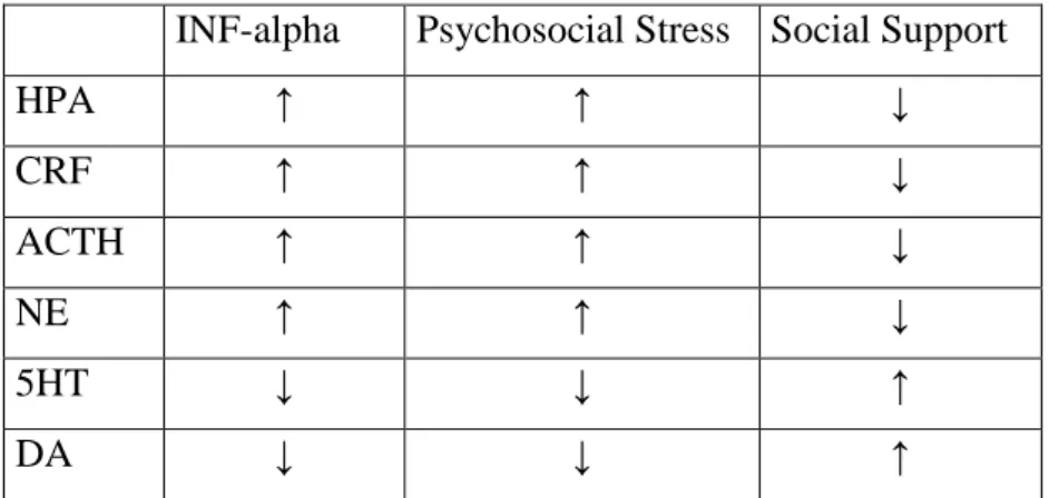
The hypothalamic–pituitary–adrenal (HPA) axis is one of the main biological systems mediating the effects of stress in the body and in the central nervous system (CNS): the activity is significantly heightened in patients suffering from depression, compared with healthy controls (Murri et al. 2014). During experimental manipulation of HPA axis can lead to the occurrence of depressive-like behaviour. Also risk factors for depression (e.g. early life trauma, repeated psychosocial stress) are characterized by hyperactivity of the HPA axis (Pariante and Lightman, 2008).
The increase in HPA axis activity isfrequently observed in depressive mood changes (Nijm et al. 2007; Höhne et al. 2014). Proinflammatory cytokines may cause HPA axis hyperactivity by disturbing the negative feedback inhibition of circulating corticosteroids on the HPA axis (Raison et al. 2010).
As a further possible background mechanism, prolonged psychological stress and related HPA-axis hyperactivity may play an important role in the development of mood disorders (Müller and Schwarz, 2007). The inflammatory cytokines such as interferon (interleukin-1, interleukin-6) by increasing the activity of the HPA-axis generate sickness-behaviour in animals and depression in human beings. Interferon treatment also creates similar effects by decreasing monoamine activation (Capuron et al. 2001;
Reiche et al. 2004). Hyperactivity of corticotrophin releasing hormone (CRH) signals