CARDIAC MUSCLE CELL CULTURE AS A PHARMACOLOGICAL TOOL;
THE EFFECT OF CORONARY DILATORS ON THE METABOLISM OF ADENOSINE
S. JAMAL MUSTAFA
Department of Pharmacology University of South Alabama
College of Medicine Mobile, Alabama
I. INTRODUCTION
During the last decade a number of efforts have been made to characterize the contractile phenomena of the mammalian myocardium and to study the biochemistry of its physiological response to nutritional and pharmacological agents (16). Investigators have related their findings to the physiological status of the myocar- dium using the spontaneous, rhythmic contractibility typical of the mammalian heart as a parameter of its function. However, studies of the in vivo heart are difficult to assess because of the complex and sometimes confusing influence of humoral and neuronal interactions which effect the performance and response of the myocardium (42), m vitro or isolated heart preparations are useful only for short term experiments but undesirable for long
729
term experiments because of their inability to maintain stable function for more than a few hours due to the development of ischemia and necrosis C42Î, Similarly, the organ culture tech- nique developed by Wildenthai C42) for pre-natal rat hearts is not useful in maintaining long term myocardial function at physio- logical levels. Subsequently methods were developed for separat- ing heart tissue into its component cells and culturing them for long periods of time (8, 14, 15, 17, 19, 38). Cultured heart cells offer some advantages over other heart preparations, and some of these advantages are:
Ca) The direct effect of drugs or other chemical agents can be studied without equivocation since the cells are denervated;
Cb) Pure myocardial muscle cell cultures can be prepared;
thus, studies can be made without contamination by other cell types present in the myocardium, such as endothelial or vascular smooth muscle cells;
(c) The problem of diffusion lag in the interstitial fluid space of the intact heart is eliminated;
(d) Cytochemistry, autoradiography, and flurorescence mi- croscopy are facilitated, because sectioning is not necessary, and the cells can be examined under living conditions; and
(c) cultured cells can be maintained for long-term experi- ments.
In the present communication, cultured chick embryonic cardi- ac muscle cells were chosen as a model for mammalian myocardium
(there are no significant differences between the adult and em- bryonic cardiac cells) in order to answer some of the criticisms of the adenosine hypothesis for the regulation of coronary blood flow. According to this hypothesis, first proposed by Berne C3) in 1963, a reduction in myocardial oxygen tension associated with a negative oxygen balance in response to conditions such as h y r - poxia, reduced coronary blood flow, or increased myocardial oxy- gen demand leads to the breakdown of adenine nucleotides to aden-
CARDIAC MUSCLE CELL CULTURE 731 osine, This adenosine may enter the interstitial fluid space and produce anteriolar dilation thereby increasing coronary blood
flow to match the oxygen needs of the heart. Adenosine has been shown to be one of the factors responsible for this type of vaso- dilatory response (30),
Most of the studies concerning adenosine hypothesis for the regulation of coronary blood flow C3, 19, 22, 30, 32) have been conducted with either in situ hearts or isolated perfused hearts.
These approaches are open to certain criticisms since; Ca) it cannot be determined with certainty that adenosine is released by muscle cells or other cell types present in the myocardium; Cb) the observed amounts of adenosine released into the perfusates of the isolated perfused hearts may not reflect the in situ amounts since during its passage across the capillary membrane some of the adenosine is degraded to inosine and hypoxanthine C33); and
Cc) the extracellular concentrations of adenosine cannot be di- rectly measured. Thus, the first objective of this study was to use cultured chick embryonic cardiac muscle cells as a model in order to avoid some of these problems associated with the study of adenosine release due to hypoxia.
Drugs such as dipyridamole and aminophylline have been em- ployed in attempts to elucidate the mechanism of coronary blood
flow regulation. The interpretation of the results with these drugs is quite controversial with respect to the role played by adenosine. For example, the vasodilator effect of dipyridamole is primarily attributed to inhibition of cellular uptake of aden- osine C4). However, Kubier and co-workers C24) have suggested that dipyridamole blocks the release of adenosine from myocardial cells, thus decreasing its extracellular concentration, a situa- tion that would not be consistent with the adenosine hypothesis, Aminophylline attenuates the vasodilation produced by adenosine
C2), but the exact mechanism of its action is not clear; there- fore, a second object of the present investigation was to study
the effects of dipyridamole and aminophylline on the metabolism of cultured cardiac cells,
II. MATERIALS AND METHODS
A. Preparation of Heart Cell Cultures
Cardiac cells were isolated from 16 day old chick embryonic hearts using essentially the same procedure as described by Mustafa and co-workers (31) with slight modification. The hearts were dissected with sterile techniques from six dozen chick em- bryos and placed in N-16 Pucks medium (obtained from Microbio- logical Associates, Bethesda, Md.). Each heart was cut into four pieces to facilitate removal of the intracardiac blood. The heart
segments were left at room temperature in N-16 Pucks medium until all the hearts were harvested (about 1 hour). After one rinsing, the hearts were minced into pieces about 1-1.5 mm while immersed in N-16 Pucks medium. These pieces were then transferred to a 50 ml sterile conical flask. About 10-12 ml of 0.1% trypsin solu-
tion (100 mg of trypsin dissolved in 50 ml of N-16 Pucks medium and 50 ml of normal modified Hanks medium containing 3.06 mM Ca++
and 0.81 mM Mg++) was added to the flask and stirred for 5 minutes at low speed with a small magnetic stirring bar. In some experi- ments a mixture of 0.1% collagenase (Type I, Sigma Chemical Co., St. Louis, Missouri) and 0.2% hyaluronidase (Type I, Sigma Chemi- cal Co., St. Louis, Missouri) was used to improve the yield of the cardiac cells. The first supernatant fraction which con- tained broken cells and blood was discarded. Fresh trypsin solu- tion (maintained at 37°C) was added to the remaining tissue in the flask and agitated with a stirring bar for 10 minutes at low speed. When agitation was stopped, the supernatant fraction which contained cardiac cells was decanted into a 50 ml sterile precooled centrifuge tube. Immediately thereafter, an equal vol- ume of growth medium having 15% horse serum (obtained from Micro-
CARDIAC MUSCLE CELL CULTURE 733 biological Associates, Bethesda, Md,l, 40% N^16 Pucks medium, 45%
modified Hanks solution and 1% penicillin^streptomycin (obtained from Microbiological Associates, Bethesda, Md.) pH 7.2 was added to dilute the action of trypsin Cor other proteolytic enzymes), and the tube was placed in ice. More fresh trypsin solution was added to the remaining tissue and this process was repeated 5-6 times to get a sufficient number of cardiac cells. The collected supernatant fractions were centrifugea at 250 g for 8 minutes at room temperature. The pellet was resuspended, washed once with growth medium, and resuspended again in about 80 ml of the growth medium. This suspension was transferred to 15 x 65 cm culture dishes (5 ml to each dish) and sealed under sterile conditions.
Some experiments were carried out on freshly isolated cardiac cells for comparison of ATP values with those of the cultured cells. In a few experiments the isolated cells were layered over a 3% Ficoll (obtained from Sigma Chemical Co., St. Louis, Mo.) solution according to the method of Glick and his co-workers (12) in order to separate the contracting cells (myocytes) from non- contracting cells. The cultures were then kept in a water jack- eted C 02 incubator, and the following day the growth medium re-
-4
placed with fresh medium containing 1 x 10 M adenosine. Incu- bation was then carried out for about 24 hours to restore (Refer- ence 36, and our observations) cell ATP values of the cultures to control values (hearts removed from chick embryos and immediately frozen with liquid nitrogen and analyzed).
The viability of the preparation was assessed by examining the monolayer cultures with a phase contrast microscope. The cul- tures, observed after 24 hours of plating, beat spontaneously, and about 85% of the cultured cells were beating cardiac muscle cells C261. Cultures older than 48 hours were not used because the ratio of fibroblasts to muscle cells increased as the culture aged.
Â. Determination of ATP in Freshly Isolated and Cultured Cardiac Muscle Cells
The isolated cardiac muscle cells after separation (and also the cells isolated after Ficoll treatment) were thoroughly washed with modified Hanks solution to remove the broken cells and con- taminating blood. The cells were then homogenized in 0.5 Ν per- chloric acid, centrifuged, and processed for ATP and protein de- termination according to the methods described below in detail.
Cultured cardiac muscle cells were treated in a similar manner.
The experimental protocol for the various experiments de- scribed further below is outlined on the next page.
C. Release of Adenosine from Cultured Cardiac Muscle Cells Due to Hypoxia
Cultures obtained after 2 days of incubation were washed several times with modified Hanks solution to remove all the ad- hering growth medium and cell debris without disturbing the thin film of cells attached to the bottom of the culture dishes. The assay was carried out in the culture dishes. The incubation mix- ture consisted of 1.0 ml of modified Hanks solution containing phosphate buffer but without glucose. The dishes were swirled several times to distribute the medium evenly over the cell lay- ers, and the dishes incubated at 37°C for 15 minutes in an atmos- phere of 95% N2 + 5% C 02. A control culture with 95% 02 + 5% C 02 was run simultaneously. At the end of the incubation, the dishes were removed from the incubator and the thin layer of cultured cells scraped from the dishes with a plastic spatula. The con- tents of the dishes were immediately transferred to polyethylene centrifuge tubes and immersed in ice to stop the enzymatic reac- tions. Two dishes were used in each assay and each was washed once with 1 ml of Hanks solution, and the washings added to the original tubes. Immediately thereafter, the tubes were centri- fuged at 0°C at 10,000 g for 5 minutes. After centrifugation, as much supernatant as possible was decanted into a separate tube.
ο
eu
1
ë H
W PH X
w
O φ
û CQ
i O
rd •
• H Ό
U rd
T J φ
• P r H U
CQ
g *
• H - P M - H rd "
>
> φU · - H C J Φ tT> - P
Ö B . o fi
- H • P rd
U Φ
Ό Ö - H
C n CQ
Ό Ό CQ
« 4 H Ö Ö rd
• H Ό C - P
* 4 Τ * • H 0 - P rd rd C! - P ft
Ö CQ T J û ng 0 an
Ό - H 0
fi •fi CQ Ö
rd CQ Ό rd • P • H fi
Ό £ fi ÷ 0
• H rd 0 • H û Ö - P - P rd •fi rd rd
• P C
û U
• H T l Ö 4 - 1
U C ft • H
0 rd
H CQ • Η • P
2 û T l Ö Ö CQ CQ φ fi
U . f i Λ rd û φ CQ En • Ρ ft rd
in co - ci
•iH -iH Φ - P O
Ό ft Φ U
" ^ &
> CQ
O
O >i
ft Λ
+ J φ
'd >
T J Ö 8
Ö H Ν
φ Ν Φ
• Η * fi
Φ 3 r - J Φ Λ G û CQ φ
¹
ft
rd T J fi rd
U Φ rH
ft Ï 3 MH CQ . Ό Φ T J
Φ Ν Ν > ι
• Η ΓΗ r H rd
rd fi
U rd - P 3 Φ
Φ Λ fi Φ r H r H
r f i - P
• H Φ • P
• H û CQ rd
O O fi - H
• H T <
- U rd φ C Ό
• H fi CQ rd
ß U U o
φ < R
ft 3 T J « CQ φ Τ Ι > ι Ν
Φ Ν
• Η r H rd U
• Ρ Φ fi
EH
>i
- Η - Ρ
>
Ρ4- Η - Ρ
- · Η ï
04 Τ » EH rd
*C 5 Ç
735
The pellet was resuspended and washed once with. 0.5 ml of Hanks solution, then centrifuged and again, the washings added to the supernatant fraction. The fraction will be referred to as "medi- um" and the pellet as "cells." Perchloric acid was added to both fractions in a final concentration of 0.5 Ν for precipitation of the proteins. The separated cells and medium were then immedi- ately homogenized with a Polytron homogenizer for 90 seconds at a speed of 4000 rpm and then centrifuged for 10 min at 10,000 g in the cold (4°C). The supernatant fractions were removed and the pellet of the protein precipitate was resuspended and washed once with 0.5 ml of 0.5 Ν perchloric acid, and the washings were added to the previous supernatant fraction. The protein precipitate obtained from the cell fraction was used for protein measure- ments by the method of Lowry and co-workers (28), and the re-
sults expressed on the basis of cellular protein concentrations.
The supernatant fractions were then brought to pH 7.0 by the addition of potassium hydroxide, placed in the refrigerator over- night, and the Perchlorate precipitate was removed by centrifuga- tion. The cell fractions were then analyzed for ATP by the luci- ferase method (13) and ADP and AMP by a spectrophotometric method
(1). The medium fractions were analyzed for adenosine, inosine, and hypoxanthine by enzymatic assay (11). In two experiments the cells were analyzed for nucleosides, and the medium was analyzed for nucleotides, but none were detected.
D. Effect of Dipyridamole and Aminophylline on the Release of Radioactive Adenosine from Cultured Cardiac Muscle Cells
During the change of growth medium 24 hours after plating, 0.5 ml of U-C14-adenosine 0-4.08 ñ moles; 15,000 CPM; obtained from Amersham/Searle Corp., Arlington Heights, 111.) in addition to the unlabeled adenosine, was added to each culture dish in order to label the nucleotide pool Csl of the cells. After a total of 48 hours Cor lessl of growth these cultures were used for measuring the release of radioactivity under hypoxic condi-
CARDIAC MUSCLE CELL CULTURE 737 tions. The medium was decanted and the adhering cell layer washed several times with modified Hanks solution to remove cell debris and growth medium which also contained labeled adenosine and its metabolites. The assay mixture contained 0.1 ml drug (dipyrida- mole, obtained from Boehringer Ingelheim, Elmsford, New York, at a final concentration of 1 x 1 0 ~6 M or aminophylline, obtained from Sigma Chemical Co., St. Louis, Mo. at a final concentration of 1 x 10"5 M) and 0.9 ml modified Hanks solution. In the con- trol experiments the drug was replaced by modified Hanks solution.
The dishes were swirled to mix the contents thoroughly and then incubated at 37°C for. 15 minutes in an incubator under 95% N2 + 5% C 02 atmosphere to accelerate adenine nucleotide degradation.
The cells and medium fractions were isolated as described in the previous section and analyzed for adenine nucleotides and nucleo- sides. An aliquot from each fraction was removed for measurement of radioactivity in a Packard liquid scintillation counter. The scintillation cocktail consisted of 5.5 tm PPO, 0.1 gm POPOP, 333 ml Triton X-100, and 667 ml toluene.
E. Effect of Dipyridamole and Aminophylline on the Uptake of Radioactive Adenosine in Cultured Cardiac Muscle Cells
The cultures (grown for 48 hrs or less) were washed thorough- ly without disturbing the cell layer in the manner described ear- lier. The assay was carried out in the culture dishes. The incu- bation mixture consisted of 0.05 ml U-C14-adenosine (14.08 ñ moles; 15,000 CPM), 0.1 ml of drug (dipyridamole at a final con- centration of 1 x 10"^ M or aminophylline at a final concentra- tions of 1 x 10~5 M) with the addition of modified Hanks solution containing phosphate buffer to a final volume of 1.0 ml. A con- trol with vehicle (modified Hanks solutionI in place of the drug was run simultaneously. The incubation mixture was added to the dishes and the latter were swirled several times to distribute the medium evenly over the cell layers. The dishes were then in- cubated at 37°C for 15 minutes in an atmosphere of 95% 02 + 5%
CQ2. The cells and medium fractions were isolated and processed for nucleoside and adenine nucleotide analyses as described. An aliquot from each fraction was counted for radioactivity,
III. RESULTS
A. Levels of ATP in Various Preparations of Isolated Chick Embryonic Cardiac Muscle Cells
A common problem with the freshly isolated cardiac cells was the low concentration of ATP on the basis of per mg protein. The results of such an experiment are presented in Table I.
Therefore, freshly isolated cells were prepared according to the present method and according to the method of Glick and co- workers (12) in which the cells were isolated using conventional enzymatic methods and then layering them over a 3% Ficoll solu- tion and at low centrifugation. Both isolation procedures re- sulted in low ATP values (together with low ADP and AMP) . The present method did result in 65% higher ATP values than the meth- od of Glick and co-workers (12). These isolated cells (with and without the Ficoll step) were placed in growth medium for 24 hours and then replaced with the same growth medium having 1 x
-4
10 M adenosine and kept for another 24 hours in an incubator resulted in ATP values which are more comparable to control hearts
(Table I ) . The ATP values of the cultured cardiac cells are 3-4 times higher than the freshly isolated cells (present method) from 16 day old chick embryonic hearts. Burns and Reddy (6) had reported higher ATP values for the freshly isolated cells from rat heart (using the Ficoll method) which we were unable to reproduce.
In another experiment with cultured cardiac cells, we found that inclusion of 1 * 10^4 M adenosine in the growth media for 24 hours increases the ATP values by 35% (unpublished observation!P In our experience we have noticed no significant difference in
CARDIAC MUSCLE CELL CULTURE 739 TABLE I Levels of ATP in Various Preparations of Isolated
Cells from a 16 Day Old Chick Embryonic Heart (Values Expressed as Ν Moles/mg Cellular Protein)
Control heartsa 33.8 ±2,23
Cultured cardiac cellsb 23,59 ± 4.49 Freshly isolated cells
(a) Present methodb 7.32 ± 1.31 (b) According to Glick, et 4.43 ± 0.79 al. (12)c
aHearts removed from the embryo and homogenized immediately in 0.5 Ν PCA are taken as controls.
bA mixture of .2% hyaluronidase +.1% collagenase + 50% N-16 medium + 50% Hanks was used as a dispersion solution. The isola-
tion procedure is described in the text.
cCells were isolated (present method) and layered over 3% Ficoll Solution. The muscle cells were then isolated by low centrifuga- tion and washed with Hanks solution.
the levels of ATP by using the earlier described methods where cells were isolated by trypsin or by a combination of collagenase and hyaluronidase (unpublished observation). Incubating the freshly isolated cells (present method) with growth media for 90 minutes at 37°C and then removing the media by thorough washings with modified Hanks solution did not seem to increase the ATP values. As a further note, we were not very successful using i- solated adult rat heart muscle when compared with chick embryonic cardiac muscle cells.
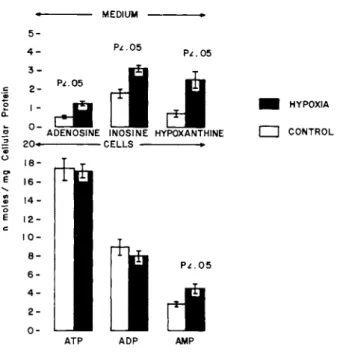
B. Levels of Nucleosides and Adenine Nucleotides in Cultured Cardiac Muscle Cells Due to Hypoxia
Hypoxia did not cause a significant change in the levels of ATP and ADP in the cultured cells compared to cells incubated in the presence of 95% 02 + 5% C 02 (Fig. H . However, AMP in the cells and adenosine, inosine, and hypoxanthine in the medium were significantly increased by hypoxia and were 1,6, 2,2, 1,8 and 3,6 fold greater respectively than the corresponding controls.
«· MEDIUM
5-
ATP ADP AMP
FIGURE 1 The cells in the control and hypoxic groups were incubated with 95% 02 + 5% C02 and 95% N2 + 5% C02 respectively for 15 minutes at 37°C in modified Ranks solution without glucose in a total volume of 1.0 ml.
C. Effect of Dipyridamole and Aminophylline on the Release of Radio-active Adenosine and on the Levels of Adenine Nucleotides and Nucleosides in Cultured Cardiac Muscle Cells Due to Hypoxia
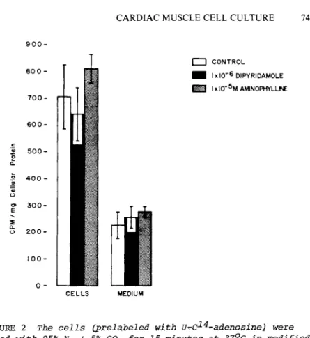
Neither dipyridamole nor aminophylline affected the amount of radio-activity released into the medium during hypoxia (Fig. 2).
Furthermore, dipyridamole had no significant effect on the levels of ATP and ADP (Fig. 3 ) , but AMP was significantly reduced. Dip- yridamole did not alter the levels of hypoxanthine in the medium, but is significantly decreased the levels of inosine (Fig. 3).
Aminophylline caused CFig. 3} no significant differences in the levels of ATP, ADP and AMP in the cells and also did not change the inosine and adenosine concentrations in the medium.
However, there was a significant decrease in the level of hypo- xan thine in the medium CFig. 31.
CARDIAC MUSCLE CELL CULTURE 741
9 0 0 -
CELLS MEDIUM
FIGURE 2 The cells (prelabeled with U^^-4-adenosine) were incubated with 95% N2 + 5% C02 for 15 minutes at 37°C in modified Hanks solution without glucose. The reaction mixture contained 0.1 ml drug (replaced with modified Hanks solution in control) and 0.9 ml modified Hanks solution.
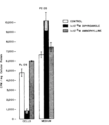
D. Effect of Dipyridamole and Aminophylline on the Uptake of Radio-active Adenosine and on the levels of Adenine Nucleotides and Nucleosides in Cultured Cardiac Muscle Cells
In the presence of dipyridamole, only 7% of the radio-activi- ty was found in the cells; whereas, in the absence of dipyrida- mole, 42% of the radio-activity was present in the cells CFig. 4).
In terms of total cellular radio-activity, the control values were six fold greater than those of cells incubated in the pres- ence of dipyridamole; hence, there was a marked inhibition of U- C^-4-adenosine uptake by the cultured cardiac cells.
ο UJ
u| 8| 0 J D JD|n||90 β ω / β è | è ω u
ι
CM U| 9| 0 J D J 0| n| | 9 Q 6 U J/ 8 8| 0 U I u
Of CJ ϋß ftf
QJ QJ
:^
•Ho •g
4J O QJ t0 ¹ QJ QJ
•S * S ä
U
O M O Q) .
? ci q
^ M o
ftf "H + +J «U
CM O M a? QJ to in to óι 3 Ai
•õ ς
•S * Δ
+J -H tcj
c: eu QJ
•U 4J *H
ftf O
QJ ^ Ü
QJ EH ó»
QJ Ό to to q M O "
(D 3 - \ Ü M M QJ U
-u +J Es 3 CJ
m -õ 5 8
• N q
8
H Pu
q q o o 4J +J
3 3 O O to to
742
CARDIAC MUSCLE CELL CULTURE 743
P 2 . 0 5
C E L L S MEDIUM
FIGURE 4 The cells were incubated with 95% 02 + 5% C02 for 15 minutes at 37°C in modified Hanks solution without glucose.
The reaction mixture contained 0.05 ml U-C^4-adenosine + 0.1 ml of drug (replaced with modified Hanks solution in controls), and the volume made up to 1.0 ml with modified Hanks solution.
Aminophylline produced no significant difference in the pat- tern of distribution of radioactivity from that of the controls
CFig. 4).
The radioactivity in the cells at 1X10~5 M aminophylline was slightly but not significantly higher than that of the controls.
In the presence of oxygen neither dipyridamole nor amino- phylline significantly altered the levels of ATP, ADP, or AMP in the cells CFig, 5). However, adenosine was significantly in-
Lü _l Ο
û. <
cd m
b ¼ ï
ι
Uj9|0Jd J0|Ð||èQ ΤUJ/S8|0UJU
Ç
>
J
I ï
u | 9t0 J d J D | n | | 9 0 6 u u / s e | o w u
&3 •H <H
"H Q) 0
:^
0•M
o •H
q O Ï º- 0 Ï) •
M OJ 0 0 -M
ra eu
CO 3 Q) ro -õ 1 3^ f C M r«
E; O ΙΓ) Q)
M 3
M 0 0 un
<M o
• 0)
( N O . q O O d* CJ q Q) LH •-s
+ +J r i CJ —N CM 0 10 O Ü M
<X° Q) 0 u
LT> •u
3 q
•U o
^ û
E; q
& CJ ••s 0 q Q) 0 4 J 4 J M rff Ü 4 J
*Q rtf 3 3 Q) M
Ü 0
CO 10 Q) O Ü 3 0) Ü
I
3Ë 'S
D q O o
H - H Pu +J
3 O <0
CO q
tJ Q)
*H <M
•H
a .
^ q 4 J o
•H M 3 Q) O CJ co
¹ CO ö q Λ ;
744
CARDIAC MUSCLE CELL CULTURE 745 creased in the medium in the presence of dipyridamole; while,
inosine was significantly reduced CFig. 51, Kypoxanthine levels in the medium were unaffected by dipyridamole, and adenosine, inosine, and hypoxanthine levels in the medium were not altered by aminophylline. Even a concentration of aminophylline as high as 1X10 M was without effect on cell and medium concentrations of the adenine nucleotides, nucleosides, or hypoxanthine.
IV. DISCUSSION
Adenosine triphosphate and other adenine nucleotides have been implicated as the major vehicles for energy coupling between the energy yielding and energy requiring sequences needed for the normal functioning of the cell. It is now becoming evident that many different cells, while they can survive major disturbances in their adenine nucleotide metabolism, show characteristic re- sponse patterns. The consequences to the cell of these fluctua- tion in high energy phosphates can result in disturbed physiolo- gical and metabolic states.
In the present study, efforts have been made to standardize a preparation of cardiac muscle cells, whose ATP values can be com- pared to the control hearts (in vivo). The ATP content of the cardiac muscle cells has been taken as one of the metabolic para- meters Cin addition to their spontaneous beating). Seraydarian and co-workers C35) have shown a definite correlation between intracellular levels of ATP and the maintenance of rhythmic con- tractions. The direct demonstration of ATP in the process of con- traction has been demonstrated unequivocally by Cain and Davis
C71; thus, the relationship between intracellular ATP concentra- tion and the contraction of cardiac cells in culture follows.
The ATP may be required for maintenance of the sodium pump, for proper membrane function, or for active transport of or Na .
Small changes in ATP may alter these functions. The mechanism of ATP involvement in spontaneous beating is not well understood.
The data presented in Table I definitely demonstrate that freshly isolated cardiac muscle cells have significantly lower ATP values (3-4 times] than cultured cardiac muscle cells; where- as, the ATP values of the cultured cardiac muscle cells are very close in ATP levels to those found in intact in situ hearts.
Thus, the use of freshly isolated cardiac muscle cells for vari- ous metabolic and physiological studies should be done with extra caution because of their low ATP content which effects the corre- lation of the normal in vivo condition with the in vitro condi- tion. The interpretation of higher ATP values in cultured cells compared with the freshly isolated cells is that the cells during harsh isolation procedures lose their adenine nucleotide pool (and perhaps other metabolites) due to altered membrane properties.
Culturing the same cells in normal growth medium gives these cells a chance to regain their membrane properties and synthesize vari- ous metabolites to the extent of their original state. The growth medium has all the essential components required for various in- termediary metabolic pathways.
A large number of workers in this area have used the dye ex- clusion method as an assessment of the viability of the prepara- tion. Many of these dyes have acidic or toxic properties (25) that may be injurious to live cells in suspension and, hence, could possibly lead to variable results. The theory of vital dye staining of dead cells is related to the large increases in cell membrane permeability that occur following cell death, resulting into the passage of large molecules such as those of the dye into the cytoplasm. The small changes in membrane permeability which the dye exclusion technique may not show could result in the leakage of macromolecules such as enzymes from the cytoplasm into the extracellular space, changes in electrical resistance or im- pedence, and lack of other cofactors such as coenzymes or adenine nucleotide C21, 27- 37, 39, 401. With these factors in mind, the
CARDIAC MUSCLE CELL CULTURE 747 author devised conditions which would result in a, higher ATP con- tent of the cardiac muscle cells which could only be achieved by culturing the cells under the present situation, This constitutes one of the reasons for employing cell culture techniques. Based on this preparation of cardiac muscle cells the various experi- ments discussed in detail below were carried out.
The present data CFig. 1] show a significant increase in the production of adenosine and its metabolic products, inosine and hypoxanthine when the cultured cardiac muscle cells were subjected to hypoxia. Although there were no significant differences in cellular ATP and ADP, AMP was significantly increased in the cells. The production of nucleosides under hypoxia is obviously by degradation of adenine nucleotides and the sum of adenine nu- cleotides and nucleosides in the control and hypoxic groups are comparable. These finding indicate that the adenosine released by hypoxia in vivo or isolated perfused hearts is primarily from the myocardial cells; however, the observations on isolated per- fused or in vivo hearts to not exclude other sources of cardiac adenosine such as the vasculature and fibrous tissue.
Dipyridamole, a potent coronary vasodilator, enhances the vasodilator activity of exogenous adenosine by blocking its up- take and preventing its degradation by intracellular enzymes C18, 34). In agreement with this observation, the present investiga- tion clearly demonstrates that the uptake of adenosine in cul- tured cardiac muscle cells is blocked by dipyridamole. These re- sults are supported by the finding of higher absolute amounts of adenosine in the medium at the end of the incubation period in the dipyridamole treated cells. The release of adenosine was not influenced by dipyridamole, since the amount of radioactivity re- leased into the medium from the hypoxic cardiac muscle cells Ccell with prelabeled nucleotides! was the same in the absence or presr- ence of the drug. The observation that the amount of adenosine in the medium with, incubation in the absence of oxygen and in the presence of dipyridamole was slightly higher than with hypoxia
alone can be explained on the basis that part of the adenosine re- leased from hypoxic cells is not taken back into the cells because of the inhibition of uptake by dipyridamole. The decrease in the levels of AMP in the cells and inosine in the medium may be at- tributed to a reduction in substrate (adenosine) that is available to the adenosine kinase and adenosine deaminase of the cells re- spectively because of blockade of the uptake of adenosine by dipyridamole. To what extent AMP levels are altered by other in- tracellular enzymatic reactions cannot be determined from the present study.
These observations are not in agreement with the view put for- ward by Kubier and co-workers that dipyridamole blocks the release as well as the uptake of adenosine in myocardial cells (24).
Kubier and co-workers (24) found that the adenosine concentration of the isolated hypoxic dog heart was greater in the presence of dipyridamole than in its absence. They concluded that dipyrida- mole blocked the release of adenosine from myocardial cells and, hence, adenosine could not be responsible for the coronary vaso- dilation observed in hypoxic hearts treated with dipyridamole.
These experiments C24) were conducted on isolated heart prepara- tions that have at least three compartments (vascular, inter- stitial fluid and intracellular) as well as non-myocardial cells and catecholamines. In an attempt to reconcile these observa- tions with those of the present study we suggest that in the in- tact heart the adenosine may be sequestered in interstitial fluid located in the transverse tubules and intercalated discs and may not be completely washed out during coronary perfusion.
The mechanism(s) of action of dipyridamole is not clearly understood. It has been proposed C5, 10) that the drug inhibits adenosine deaminase and prevents endogenous or exogenous adeno- sine from being inactivated; other studies, on the other hand, have failed to detect a significant inhibitory effect on the enr zymes involved in adenosine metabolism, namely adenosine deami^
nase C3, 23, 341 and adenosine kinase (34), m fact an activa^
CARDIAC MUSCLE CELL CULTURE 749 tion of the latter has been demonstrated C91. How dipyridamole
blocks the uptake of adenosine is not known. It is possible that the drug forms a complex with an adenosine carrier or that adeno- sine and dipyridamole compete for the same active site(s) on the carrier.
Aminophylline and theophylline (2, 20} attenuate the vasodi- lator action of exogenous adenosine and as in the case of dipy- ridamole, have been used to test the adenosine hypothesis for the regulation of coronary blood flow by studying their effect on myocardial reactive hyperemia. Our findings do not show a block
in the uptake or release of adenosine in cultured cardiac cells in the presence of these agents. Additional support for these findings was obtained by quantifying the cell and medium fractions for adenosine contents together with other nucleosides and adenine nucleotides. The levels of adenosine released into the medium in the presence or absence of oxygen were not significantly changed by aminophylline. The mechanism whereby aminophylline attenuates the vasodilator action of adenosine is not known. It is possible that aminophylline may cause a direct release of bound C a+ + in the vascular smooth muscle in a manner similar to the effect of caffeine in skeletal muscle (41).
In summary, the use of monolayer cultures of beating cardiac muscle cells provides a useful method for studying many parame- ters of the heart, which can supplement and complement intact animal experiments and which are independent of many variables that plague in vivo studies of the beating heart. Cultured car- diac muscle cells (or other mammalian cells) can also serve as an excellent pharmacological tool for studying the direct effect of drugs on a specific cell type without the influence of neuronal and/or humoral factors.
ACKNOWLEDGMENT S
The technical assistance of Mrs. K ren L, McGrain is greatly appreciated. This study was supported in part from an Intramural Research Grant of the University of South Alabama, College of Medicine, Mobile, Alabama. Part of this work was conducted at the Department of Physiology University of Virginia Medical School, Charlottesville, Virginia and published in Am. J. Physi- ology, 228: 1474-1478, 1975. This portion of the work was car- ried out during the tennure of a postdoctoral training fellowship from Public Health Service Grants NHLI HL-10384 and HL05815.
REFERENCES
Adams, H. (1965). In "Methods of Enzymatic Analysis" (H. V.
Bergmeyer, ed.), pp. 573-577. Academic Press, New York.
Afonso, S. (1970). Circulation, Res. 26, 743-752.
Berne, R. M. (1963)„ Am. J. Physiol. 204, 317-322.
Bretschneider, H. J., Frank, Á., Bernard, U., Kochspeik, Ê., Schier, R. (1959). Arzneimittel. Forsch. 9, 49-59.
Bunag, R. D., Douglas, C. R., Imai, S., Berne, R. M. (1964). Cir- culation. Res. 15, 83-88.
Burns, A. H., Reddy, W. J. (1975). J. Mol. Cell. Cardiol. 7, 553- 561.
Cain, D. F., Davies, R. E. (1962). Biochem. Biophys. Res. Commun.
8, 361-366.
DeHaan, R. L. (1967). In "Factors Influencing Myocardial Con- tractility" (R. D. Tanz, F. Kavaler, J. Roberts, eds.), pp.
217-230. Academic Press, New York.
DeJong, J. W., Kalkman, C. (1973). Biochem. Biophys. Acta. 320, 388-396.
Deuticke, Â., Gerlach, E. (1966). Arch. Exptl. Pathol. Pharmakol.
225, 107-119.
CARDIAC MUSCLE CELL CULTURE 751 Dobson, J, G. Jr., Rubio, R., Berne, R. M. (19711. Circulation.
Res. 29, 375^384.
Glick, M. R., Burns, A. H. f Reddy, W. J. (19741. Anal* Biochem, 61, 32-42.
Greengard, P. (1965). In "Methods of Enzymatic Analysis" (H. V.
Bergmeyer, ed.), pp. 551-555. Academic Press, New York.
Halle, W. (19671, Morphologisches Jahrbuch III, 3.
Halle, W, r Wöllenberger, A. C19681, Zertschrift. Fur. Zellfor^
schung. 87, 292-314.
Harary, I., Farley, B. C19601. Science. 131, 1674-1675.
Harary, I., Farley, B. (19631. Expt. Cell Res. 29, 451-465.
Hashimoto, K., Kumakura, S., Tanemura, I. Cl964), Arzneimittel.
Forschung. 14, 1252-1254,
Imai, S., Riley, A. L., Berne, R. M. 0-964). Circulation. Res. 15, 443-450.
Jurhan, W., Dietmann, K. 0-970). Pflugers. Arch. 315, 105-109.
Kaltenbach, J. P., Kaltenbach, M. H., Lyons, W. B. 0-958). Exp.
Cell. Res. 15, 112-117.
Katori, M., Berne, R. M. 0-966). Circulation. Res. 19, 420-425.
Kubler, W., Bretschneider, H. J. 0-964). Pflugers. Arch. 280, 141- 157.
Kubler, W., Spieckermann, P. G., Bretschneider, H. J. 0-970). J.
Mol. Cell. Cardiol. 1, 23-38.
Laiho, U. Ê., Trump, Â. F. 0-974). Virchows. Arch. Abt. Â. Zell- path. 15, 267-277.
Lehmkuhl, D., Speralakis, N. C1967). In "Factors Influencing Myo- cardial Contractility" (R. D. Tanz, F. Kavaler, J. Roberts, eds.), pp. 245-278. Academic Press, New York.
Lepage, G. Á. 0-950). Cancer. Res. 10, 77-88.
Lowry, O. H., Rosebrough, H. J., Farr, A. L., Randall, R. J.
0-951). J. Biol. Chem. 193, 265-275.
Mark, G. Å., Strasser, F. F. 0-966). Expt. Cell. Res. 44, 217- 233.
Rubio, R., Berne, R. M. (1969). Circulation. Res. 25, 407-415.
Mustafa, S. J., Rubio, R., Berne, R. M. (1975). Am. J. Physiol.
228, 62-67.
Rubio, R., Berne, R, M., Katori, M, (1969). Am, J, Physiol. 216, 56-62.
Rubio, R. , Wiedmeier, V. Ô, , Berne, R, M. (1972). Am. J. Physiol.
222, 550-555.
Schrδder, J., Berne, R. Ì,, Rubio, R. (1972). Am, J. Physiol, 233, 159-166.
Seraydarian, M. W,, Harary, I., Sato, Å. 0-968). Biochem. Bio- phys. Acta. 162, 414^423,
Seraydarian, M. W, , Artaza, L., Abbott, B. C. (1972). J. Mol.
Cell. Cardiol. 4, 477-484.
Slater, T. F. (1969). In "Lysosomes in Biology and Pathology" (J.
T. Dingle and H. B. Amsterdam, eds.), pp. 467-492, North Holland Publishing Co.
Sperelakis, N. (1967). In "Electrophysiology and Ultrastructure of the Heart" (B. Taccardi, G. Marchetti, eds.), pp. 81.
Bunkodo, Tokyo, Japan.
Trump, B. F., Ericsson, J. L. E. (1965). In "The Inflammatory Process" (B. W. Zweifach, L. Grant, and L. McCluskey, eds.), p.
35. Academic Press, New York.
Trump, B. F., Arstila, A. U. (1971). In "Principles of Pathobio- logy" (N. LaVia and R. Hill, eds.), pp. 9-96. Oxford University Press.
Weber, Á., Herz, R. (1968). J. Gen. Physiol. 52, 750-772.
Wildenthal, Ê. (1971). J. Applied. Physiol. 30, 153-157.
A Â 7 C 8 D 9 Å 0 F 1 G 2 H 3 I 4 J 5