Editors’ Suggestion
Near- K -edge single, double, and triple photoionization of C
+ions
A. Müller,1,*A. Borovik, Jr.,2T. Buhr,2J. Hellhund,1K. Holste,2A. L. D. Kilcoyne,3S. Klumpp,4M. Martins,5S. Ricz,6 J. Viefhaus,7,†and S. Schippers2
1Institut für Atom- und Molekülphysik, Justus-Liebig-Universität Gießen, 35392 Giessen, Germany
2I. Physikalisches Institut, Justus-Liebig-Universität Gießen, 35392 Giessen, Germany
3Advanced Light Source, Lawrence Berkeley National Laboratory, Berkeley, 94720-8225 California, USA
4DESY Photon Science, FS-FLASH-D, 22607 Hamburg, Germany
5Institut für Experimentalphysik, Universität Hamburg, 22761 Hamburg, Germany
6Institute for Nuclear Research, Hungarian Academy of Sciences, 4001 Debrecen, Hungary
7DESY Photon Science, FS-PE, 22607 Hamburg, Germany
(Received 11 October 2017; published 16 January 2018)
Single, double, and triple ionization of the C+ion by a single photon have been investigated in the energy range 286 to 326 eV around theK-shell single-ionization threshold at an unprecedented level of detail. At energy resolutions as low as 12 meV, corresponding to a resolving power of 24 000, natural linewidths of the most prominent resonances could be determined. From the measurement of absolute cross sections, oscillator strengths, Einstein coefficients, multielectron Auger decay rates, and other transition parameters of the main K-shell excitation and decay processes are derived. The cross sections are compared to results of previous theoretical calculations. Mixed levels of agreement are found despite the relatively simple atomic structure of the C+ion with only five electrons. This paper is a followup to a previous Letter [A. Mülleret al.,Phys. Rev. Lett.
114,013002(2015)].
DOI:10.1103/PhysRevA.97.013409 I. INTRODUCTION
Carbon is the second most abundant heavy element (with atomic numbers Z >2) in the universe next to oxygen.
It is prominently present in all astrophysical environments.
Moreover, it is the prime constituent of organic chemistry and the building block of life on Earth. In both collisionally ionized and photoionized gases, carbon atoms occur singly or multiply charged and the ions can serve as probes for the state of the plasma in which they are formed, be it of astrophysical or terrestrial origin. Important diagnostic techniques rely on the soft-x-ray spectroscopy of carbon ions, especially near theK edge [1].
Similar spectroscopic techniques based on the production and observation of the decay ofK-vacancy states are employed to characterize the chemical environment of carbon atoms in molecules, clusters, and solids. When aK-shell electron is removed from a neutral carbon atom, the resulting ion is most likely in one of the C+(1s2s22p2 2S,2P ,2D) states which then decay by emitting photons or electrons with characteristic energies. Identical terms can be excited from the ground state of C+ ions by irradiation with photons of the same characteristic energies [2]. Resonance parameters and details of the transitions such as the natural widths, the oscillator strengths, and the exact wavelengths can be determined in photoionization experiments with C+ions.
*alfred.mueller@iamp.physik.uni-giessen.de
†Present address: Helmholtz-Zentrum Berlin, Department Optics and Beamlines, Berlin, Germany.
An important aspect of K-shell excited C+(1s2s22p2 2S,2P ,2D) states is their unique electronic configuration with four electrons in theLshell. Four electrons residing above aK-shell vacancy are the minimum required for observing triple-Auger decay in which one of the four electrons falls into the K vacancy while all three other electrons are ejected into the continuum in a correlated four-electron process. This process has been unambiguously identified in our preceding Letter [3].
Photoprocesses involving C+ ions have been studied in previous experiments. Photoabsorption by boronlike C+ions was investigated by Jannitti et al. [4] with a technique us- ing two laser-produced plasmas. Photoionization of C+ ions has already been addressed in several merged-beam experi- ments. The valence-electron energy range has been studied by Kjeldsenet al.[5,6], while Schlachteret al.[2] explored the 1s→2p excitation resonances. In both cases, absolute single-ionization cross sections were determined. Theoretical calculations for K-shell photoabsorption by C+ ions have produced cross sections usingR-matrix techniques [1,2,7] and, indirectly, by the multiconfiguration Dirac-Fock method [8].
Measurements for the boronlike sequence of ions were recently extended to N2+[9] and O3+[10] by using the photon- ion merged-beam technique. Resonant K-shell excitation of boronlike Fe21+(1s22s22p2P1/2) was observed by detecting fluorescence photons and photoions [11,12] in experiments employing an electron beam ion trap. Deep-core photoexci- tation of ions has recently been reviewed by Müller [13].
Because of the low density of ions in a beam, the pres- ence of high detector backgrounds and limited photon flux available from synchrotron radiation sources, photoioniza-
tion experiments involving K-vacancy production had been limited until recently to the strongest resonance features in the spectrum. Schlachter et al. [2] observed only 1s→2p transitions in C+at resolving powers 2400 to 5800. Gharaibeh et al. [9] measured the strongest 1s→2p and 1s→3p transitions in N2+at resolving powersE/E4500 to 13 500.
McLaughlin et al. [10] investigated the same transitions in O3+ at resolving powers E/E 3200 to 5000. The ion-trap experiments involvingK-vacancy production in B-like Fe21+
ions were restricted to the observation of an unresolved blend of Fe21+(1s2s22p2 2P1/2) and Fe21+(1s2s22p2 2D3/2) levels in the energy range 6583–6589 eV. The associated resonances were not directly measured on an absolute cross-section scale. The energy resolution in the particular measurement on Fe21+was 1 eV corresponding to a resolving power of about 6600.
All those previous experiments were restricted to the single- ionization channel and a few isolated resonances. Since then, significant experimental progress has been achieved in that the energy ranges of experiments have been extended to the region beyond theKedge and multiple ionization up to the removal of three electrons from the initial ion was investigated for the lighter elements up to atomic number Z=10 [3,14–17].L- shell excitation and ionization in Fe+ andM-shell vacancy production in singly and multiply charged xenon ions have been investigated by observing up to sixfold net ionization of the parent ion [18–20].
Here we report absolute cross-section measurements for photoionization of C+ near the K edge. The experi- ments were performed at the photon-ion merged-beam setup PIPE (photon-ion spectrometer at PETRA III) [19] using monochromatized undulator radiation from the PETRA III synchrotron light source in Hamburg. Single and double ionization were investigated covering the whole range of 1s→np(n=2,3,4, . . . ,∞) one-electron and 1s2s→2pn (n=2,3,4, . . . ,∞,=0,2) two-electron excitations occur- ring well beyond the K-shell ionization threshold. For the strongest 1s→2p resonances and the immediate K-edge region, triple ionization was also observed. With the extended capabilities of the PIPE experiment, structures and processes in ionized atoms, molecules, and clusters can be observed which have not been previously accessible to experiments.
The present paper provides details of experiments and additional results from our previous work [3]. Emphasis in the previous publication was on the discovery and unambiguous demonstration of an elusive four-electron Auger process, the triple-Auger decay, in which three electrons are ejected in a single event while a fourth electron falls into a K-shell vacancy. The present paper focuses on the determination of absolute cross sections at very high-energy resolution and the information that can be derived from the combined results of absolute measurements of partial cross sections for different final channels and the natural widths of resonance lines.
This presentation is structured as follows. After this intro- duction, a brief overview of the experiment is provided with details specific to the present measurements. The experimental results are shown in detail. Photoabsorption cross sections for C+ions and decay parameters of core excited states are derived from the measurements and compared with the results of theoretical calculations as well as with measurements available
in the literature. After a summary and acknowledgments, an Appendix describes the formalism and procedure for the ex- traction of transition parameters from absolute photoionization cross sections.
II. EXPERIMENT
The experimental arrangement and procedures have been described in detail previously [17,19]. In short, C+ ions were produced for the present measurements in an electron- cyclotron-resonance (ECR) ion source. The ions were acceler- ated to 6 keV and magnetically analyzed to obtain an isotopi- cally pure beam, which was then transported to the interaction region, collimated, and merged with the photon beam available at beamline P04 [21] of PETRA III. The product ions were separated from the parent ion beam by a dipole magnet inside which the primary beam was collected in a large Faraday cup. The photoionized ions were passed through a spherical 180-deg out-of-plane deflector to suppress background from stray electrons, photons, and ions and then entered a single- particle detector with near-100% detection efficiency. The high brightness and flux of the photon beam (2×10¹¹ s−1 at 288 eV energy and 12 meV bandwidth) permitted tight collimation of the ion beam (1.5 nA) and a significantly improved spatial overlap with the photon beam compared to earlier experiments.
Form factors [17,19,22] in the cross-section measurements were between 3460 and 4800 cm−1; optimized beam tuning resulted even in 9900 cm−1. In the preceding Letter [3], the single-interaction condition (strictly only one photon absorbed and no other interactions) for the measurement of the relatively small triple-ionization cross section has been discussed in detail and unquestionably verified.
The photon flux was measured with a calibrated photodiode.
The photon energy scale was calibrated with an uncertainty of better than±30 meV by remeasuring known photoionization resonances in C3+[23]. Doppler shifts due to the ion velocity directed opposite to the incoming photons were corrected for, the correction factor of the laboratory photon energy being approximately 1.001. The systematic total uncertainty of the measured cross section for single and double ionization is
±15% [19], to which statistical uncertainties have to be added.
In the case of triple ionization, the cross section was derived from ratios between consecutive recordings of the spectra for single, double, and triple ionization with the experimental conditions unchanged and by normalizing to the absolute data for single and double ionization. A conservative estimate of the uncertainty of the extremely small absolute triple-ionization cross section is±50%.
III. RESULTS
Figure 1 provides an overview of the investigated cross sections for single, double, and triple ionization of C+ions by single photons. Theoretical calculations for photoabsorption by ground-term and metastable C+ions [1] are displayed in Fig.1(a). For metastable C+(1s22s2p2 4P) ions with an exci- tation energy of about 5.33 eV [24], the available calculations are restricted to the energy range where 1s→2pexcitations occur. The cross sections are represented by a black solid line with gray shading. For ground-term C+(1s22s22p2P) ions, the
0 50 100 150 200
0 1 2 3 4 5
288 290 306 308 310 312 314 316 318 320 322 324
0.00 0.01 0.02 0 100 200 300
hν+ C1+→ C2++ 1e- resolution 38 meV
Crosssection(Mb)
×10
hν+ C1+→ C3++ 2e- resolution 85 meV
×10
hν+ C1+→ C4++ 3e- resolution 92/130 meV
Photon energy ( eV )
(a)
(b)
(c)
(d)
hν+ C1+photoabsorption R-matrix, natural linewidths
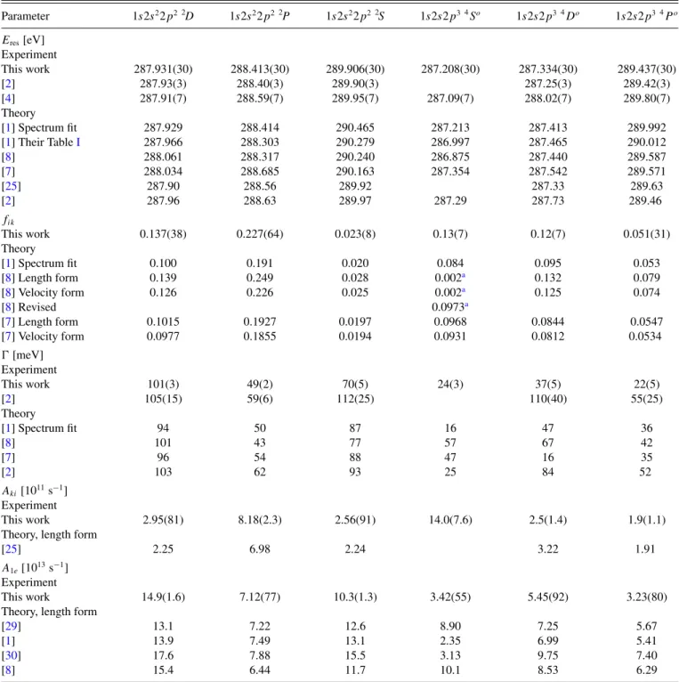
FIG. 1. Overview of the cross sections for photoionization of C+ions addressed in this paper. Panel (a) shows theoretical cross sections [1] for photoabsorption by long-lived excited C+(1s22s2p2 4P) and ground-term C+(1s22s22p2P) parent ions. TheR-matrix results for the metastable ions are restricted to 1s→2ptransitions. They are represented by the black solid line with gray shading. The calculated data for ground-term ions cover the full energy range 287–325 eV investigated in the present experiments. They are represented by the lighter (blue) solid line. Multiplication of the ground-term cross section by a factor of 10 produced the (purple) line, which is shown with a vertical offset for better visibility. The energy axis has a break between 294 and 305 eV, where no resonances are expected. Panel (b) displays experimental results for single photoionization of C+ions measured at an energy resolution of 38 meV. The cross sections obtained beyond 304 eV are shown a second time after multiplication by a factor of 10. They are displayed with an offset. All experimental cross sections are on an absolute scale.
Panel (c) shows experimental results for double photoionization of C+ions measured at an energy resolution of 85 meV. In panel (d), cross sections for triple photoionization of C+ions are displayed. The densely spaced data at around 288 eV were measured at an energy resolution of 92 meV, and the data points with statistical error bars beyond 305 eV were taken at 130-meV resolution.
lighter (blue) solid line was calculated. Since the cross sections at energies beyond 305 eV are relatively small as compared to the 1s→2presonances, they were multiplied by a factor 10 and displayed again in Fig.1(a)with a vertical offset.
Figure1(b) shows the experimental cross section for net single ionization of C+ions by single photons measured at a bandwidth of 38 meV. Like all other cross sections discussed in this paper, these data are on an independently absolute
scale. The spectrum at energies beyond 305 eV was multiplied by a factor of 10 and displayed again in Fig. 1(b) with a vertical offset for better visibility of the small resonance peaks.
In Fig. 1(c), the experimental cross section for net double ionization of the C+ ion by a single photon is displayed. It was measured at a bandwidth of 85 meV. The vertical scale is down from Fig.1(b)by roughly a factor of 40. Figure1(d) shows experimental cross sections for net triple ionization of
287 288 289 290 0
100 200
1s2s2p34 Do1s2s2p34 So 1s2s2p34 Po 1s2s2 2p22 S
1s2s2 2p22 D
this experiment at 38 meV resolution R-matrix model with
90% C1+(1s22s22p2P) 10% C1+(1s22s2p2 4P)
hν+ C1+→C2++ 1e-
Crosssection(Mb)
Photon energy ( eV ) 1s2s2 2p22 P
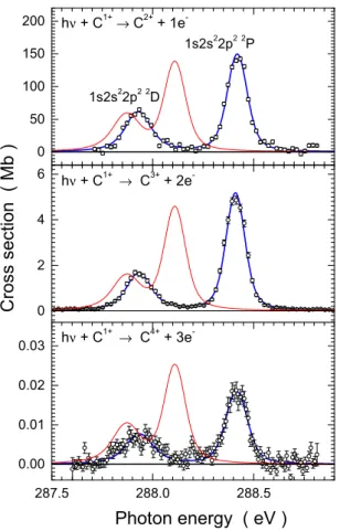
FIG. 2. Experimental cross section for single photoionization of C+ions in the region of 1s→2pexcitations at 38-meV resolution compared with results ofR-matrix photoabsorption calculations [1] convoluted with a 38-meV full-width-at-half-maximum (FWHM) Gaussian distribution function. The terms associated with the individual resonance peaks are indicated. The present measurement is represented by shaded circles with statistical error bars. The area under the experimental cross section is dark shaded (red online). The primary ion beam contained an approximate 10% fraction of metastable C+(1s22s2p2 4P) and 90% ground-term C+(1s22s22p2P) ions. Accordingly, theR-matrix photoabsorption cross sections are a weighted sum of the different parent ion fractions which are individually presented: quartet resonances from metastable parent ions by a solid line with (cyan) shading and doublet resonances from ground-state ions by the dashed black line with (green) shading.
the C+ ion by a single photon. The energy region around 288 eV was scanned in fine steps at a resolution of 92 meV. The isolated cross-section data at higher energies were measured at a bandwidth of 130 meV. Unfortunately, the low signal rates were not sufficient for scanning a wide energy range in small steps, so that resonance structures expected in the energy range 305–325 eV could not be made visible unambiguously. Note that the cross-section scale is down from that of Fig.1(c)by another factor of about 200.
Figure2shows the result of a photon-energy scan at 38-meV resolution of the cross section for single photoionization of C+ ions by single photons in the energy range 286.9–290.9 eV cov- ering all contributions from 1s→2p excitations. Individual resonance terms are identified on the basis of the discussion provided in the work by Haso˘gluet al.[1]. The assignment of the first two peaks is reversed compared to earlier theoretical work [2,7]. Arguments for the new assignment are provided in the theory paper published by Haso˘gluet al.[1]. The quartet terms arise from metastable C+(1s22s2p2 4P) parent ions, and the doublet terms are populated by excitation of ground-level C+(1s22s22p2P) ions. Both ground-state and metastable ions are present in the primary ion beam used in the experiments, as can be seen by comparing the cross sections displayed in Figs.1(a)and1(b)in the range of 1s→2pexcitations.
Comparison of the ratios of experimental resonance strengths for the metastable and the ground-state components with the theoretical results obtained by Haso˘gluet al.[1], Wang and Zhou [7], as well as Shi and Dong [8] results in values
for the fractional abundance of metastable C+(1s22s2p2 4P) parent ions within 10±0.3%.
Figure2includes, as an example, the results of theR-matrix calculations carried out by Haso˘glu et al. [1] for both ion beam components. A weighted sum of the theoretical cross sections for 10% metastable and 90% ground-term components convoluted with a 38-meV FWHM Gaussian is compared with the experimental data. The theory data agree well with the experimental results for the dominant resonances. While peak areas are also predicted quite well for the smaller resonances, there are shifts of up to 0.5 eV in the calculated resonance energies for the 4Po and2S K-vacancy states. Previous R- matrix calculations [2] showed better agreement for the posi- tions of the latter resonances but differed slightly in size and position for the strongest (1s2s22p2 2P) resonance. Further comparisons of the experimental data with the theoretical cross sections mentioned above will be shown and discussed below.
The good agreement of the R-matrix photoabsorption calculations with the experimental cross sections for single ionization by single photons in the energy range 287–291 eV shows that photoabsorption in this energy range is dominated byK-shell excitations, 1s→2p, of C+ions with subsequent single Auger decay populating the final C2+ product ion channel. This situation changes at higher photon energies, as Fig.3illustrates.
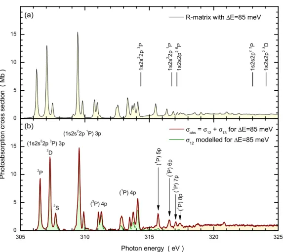
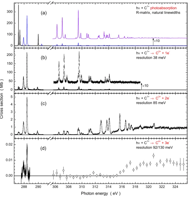
Figure3presents the comparison of theory and experiment at photon energies between 305 and 325 eV. Figure3(a)shows the photoabsorption cross section calculated by Haso˘gluet al.
305 310 315 320 325 0
5 10 15
2D
2P
2S 1 (P)5p 1 (P)6p 1 (P)7p1 (P)8p
(1P) 4p (3P) 4p
(1s2s22p1P) 3p
1s2s2p23 D 1s2s2p23 P 1s2s2p25 P
1s2s2 2p1 P
σabs=σ12+σ13forΔE=85 meV σ12modelled forΔE=85 meV
Photoabsorptioncrosssection(Mb)
Photon energy ( eV ) (a)
(b)
1s2s2 2p3 P
(1s2s22p3P) 3p 0
5 10 15
R-matrix withΔE=85 meV
FIG. 3. Calculated and measured cross sections of C+ ions interacting with photons in the energy range 305–325 eV. Panel (a) shows the theoretical cross section for photoabsorption by ground-state C+ions [1]. For comparison with the experimental results, in panel (b) the R-matrix results were convoluted with a 85-meV FWHM Gaussian. Energies ofK-vacancy levels of C2+ions relative to the ground level of C+were calculated by using theCOWANcode and are indicated by vertical bars. The associatedK-edge terms are also indicated. Panel (b) displays the experimentally derived cross section (see text) for single ionization of C+ions by single photons at 85-meV resolution represented by a thin (green) solid line with (light green) shading. The bold (brown) solid line with light (orange) shading represents the total experimental absorption cross section at 85-meV resolution. It is the sum of the experimental single and double photoionization cross sections at a photon beam bandwidth of 85 meV. Peak features in the spectrum have been identified by calculations using theCOWANcode and, for the 1s2s22p3P3p manifold, by the assignments provided by Sunet al.[25].
[1] convoluted with a 85-meV FWHM Gaussian distribution function [solid line with light (yellow) shading]. The lowest K-shell ionization thresholds relative to the 1s22s22p2P ground-state term of C+ are indicated by vertical bars. They were calculated by using theCOWANcode [26,27]. Computa- tions with different sets of configurations showed substantial configuration interaction in the initial C+andK-shell ionized C2+ions. Therefore, a relatively large set of configurations was used for the calculations of theK-shell ionization thresholds.
For the initial C+ ions, the following configurations were included: 1s22s22p, 1s22p3, 1s22p3s2, 1s22p3p2, 1s22p3d2, 1s22s23p, and 1s2s2p3. For the K-shell ionized C2+ ions, the set of configurations comprised 1s2s22p, 1s2p3,1s2p3s2, 1s2p3p2, 1s2p3d2, 1s2s23p, and 2s2p3. In spite of the relatively large calculation, the resultingKedges may have un- certainties of the order of 1 eV. For example, the 1s2s22p1P1 level which is the series limit of the 1s2s22p(1P)npRydberg resonance series, clearly seen in both the theoretical and experimental spectra, should be near 317.6 eV rather than 316.7 eV as calculated with theCOWANcode.
TheGIPPERcode [27] predicts the cross section for direct C+K-shell ionization to be about 0.8 Mb just above threshold, which is in fairly good agreement with theR-matrix calculation for photoabsorption and the experimental data for double ion- ization at energies beyond 319 eV. TheKedges associated with
3P0(0.20 Mb),3P1 (0.40 Mb), and1P1 (0.20 Mb) constitute almost the fullK-shell photoabsorption cross section, resulting from the R-matrix calculations at energies above the 1P1 threshold.
Figure 3(b) displays the experimental cross section for single ionization of C+ by a single photon as a thin (green) solid line with (light green) shading. There is remarkably good agreement of the experimental single-ionization cross section and the theoretical photoabsorption cross section at energies up to about 312 eV. With increasing photon energy, single ionization appears to die out and there is no sign of theKedge.
Understanding this phenomenon is straightforward. When a K-shell electron is removed from C+, the dominating reaction to be expected is an Auger decay of the resultingK-vacancy state in C2+by which a further electron is removed and a C3+
product ion is formed. Therefore, direct K-shell ionization results predominantly in net double ionization of the initial C+ ion. Indeed, the associated cross-section function shows clear evidence ofKedges at energies above approximately 314.3 eV.
With a much smaller probability, a double-Auger decay may follow the removal of a K-shell electron from C+ producing C4+ and thus contributing to net triple ionization.
This contribution is very small as evidenced by the results shown in Fig.1(d). There is a clear signature of theK edge with a steep rise of the triple-ionization cross section at energies above 316 eV; however, the ionization continuum is about a factor 100 smaller than that in double ionization. As will be demonstrated below, radiative stabilization of a K vacancy in C2+ has a probability that is approximately two to three orders of magnitude smaller than that of Auger decay. Hence, photoabsorption by C+ in the present energy range results almost exclusively in net single or double ionization. The sum of the associated cross sectionsσ12andσ13, respectively, is a very good approximation for the total photoabsorption cross section. Therefore, Fig. 3(b)includes that sum of the experimentally determined σ12 and σ13 at a photon energy bandwidth of 85 meV. It is represented by the bold solid (brown) line with (light orange) shading. Since single ioniza- tion was measured at 38-meV resolution and double ionization at 85-meV resolution, the experimental cross sectionσ12was convoluted with a 76.03-meV FWHM Gaussian to model a resolution of 85 meV=(382+76.032)1/2meV before the two cross sections were added.
The experimentally derived cross sectionσabs =σ12+σ13 is in very satisfying agreement with the theoretical photoab- sorption cross section. Only details in the structure far beyond the K edge appear to be somewhat different with two pro- nounced features in the experimental spectrum at about 318.7 and 320.8 eV, which are not as obvious in the theoretical result.
One has to keep in mind in this context that the theory curve is for ground-state C+ions only, while the ion beam employed in the experiment included a 10% fraction of ions in metastable 1s22s2p2 4Plevels. Apparently, this beam contamination does not have a strong influence on the measured cross sections at energies beyond 305 eV.
The two features at 318.7 and 320.8 eV mentioned above [see also Fig.1, highest-energy resonance peaks in Fig.1(b)]
have not been identified. Most likely, they are associated with double excitations in which aK-shell electron and anL-shell electron are simultaneously promoted by one absorbed photon.
Identification of the most prominent resonance features in the photoabsorption spectra is complicated by the very large number of levels that can potentially contribute. Again the
GIPPERcode [27] was employed to see how much absorption oscillator strength is contributed by which levels at which excitation energies. Given the uncertainty of the calculated energies a level-by-level identification was not attempted.
For the calculations, a large set of configurations was included: 1s22s22p, 1s22p3, 1s22p3s2, 1s22p3p2, 1s22p3d2, 1s22s23p, 1s2s2p2np, 1s2s2p2ns, 1s2s22pnp, 1s2p3np, with n=2,3, . . . ,9 and 1s2s2p2nd, with n=3,4, . . . ,9.
More than 9000 transitions were considered and their dipole oscillator strengthsfafor absorption calculated. Because of the uncertainties of the calculated resonance energies, the relative positions of K-shell excited levels and the magnitude of the
oscillator strengths were used to assign configurations to the most prominent peak features in the spectrum. According to this analysis, the energy range 306–308 eV is dominated by levels in the (1s2s22p3P)3p manifold, the range 309–
310.5 eV by levels in the (1s2s22p1P)3p manifold, and the range 310.5–312 eV appears to be due to (1s2s22p3P)4p levels. The Rydberg series of (1s2s22p3P)np resonances converges to the 1s2s22p3P K-shell ionization edge at about 314.5 eV and is buried under resonances associated with (1s2s22p1P)4p levels. The (1s2s22p1P)np Rydberg sequence withn=5,6,7,8, . . .dominates in the energy range 315.5–317.8 eV.
Figures2and3demonstrate that theK-shell excited levels of C+ in the energy range up to about 312 eV predomi- nantly decay by emission of one electron. These levels are associated with 1s→2p, 1s→3p, and partly with 1s→ 4p excitations. When the principal quantum number of the outermost excited electron increases further, the probability for the emission of two electrons increases and the final product is a C3+ion. At energies beyond the 1s2s22p3P K-shell ionization threshold, even the resonantly excited C+ions decay almost exclusively by two-electron emission. As a result, the cross section for absorption of a photon by a C+ion in that energy range is nearly identical with the cross section for net double ionization.
The present experiment with its low detector background and its high photon flux provided much better conditions than those faced in the experiments of Schlachter et al.[2]
onK-shell excitation followed by single-Auger decay at the Advanced Light Source (ALS) in Berkeley. Hence, it was possible to increase the resolving power by approximately four times that of the previous experiment. The smallest photon energy bandwidth reached by Schlachter et al. was 50 meV. At this bandwidth, only the two strongest resonances in the spectrum could be measured with moderate statistics.
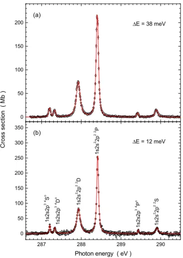
Compared to the previous best resolution, the bandwidth was reduced in the present experiment to about one fourth. The complete spectrum of 1s→2presonances of C+ions in the energy range 286.5–290.5 eV was measured at a resolution of 12 meV. The resulting high-resolution cross section for single ionization of C+ ions is shown in Fig.4(b)together with a 38-meV resolution measurement in Fig.4(a)in which excellent statistical precision was reached.
From the experimental results displayed in Fig.4, resonance parameters for all the six peaks related to 1s→2pexcitations can be extracted using a global fit to both spectra. However, one has to consider that all the K-shell excited levels are multiplets, as are the initial electronic levels of the parent C+ ions. Because of the natural widths of the excited levels, none of these multiplets can be resolved in an experiment, even at infinite resolving power. The electron-cyclotron-resonance ion source used in the present experiments is known to produce a plasma with high electron temperatures. Therefore, it is easily possible to ionize and excite ions in a wide range of charge states. Excited states of ions with sufficiently long lifetimes can survive the time between their population in the plasma and the extraction of the ions from the source. They can further survive the time of flight between the source and the photon-ion interaction region, which was about 30μs in the present case of C+ions. Candidates for such survival are the
0 50 100 150 200
287 288 289 290
0 50 100 150 200 250 300 350 (b)
ΔE = 12 meV
Crosssection(Mb) 1s2s2p34So 1s2s2p34 Do 1s2s2 2p22 D 1s2s2 2p22 P 1s2s2p34 Po 1s2s2 2p22 S
ΔE = 38 meV (a)
Photon energy ( eV )
FIG. 4. Measured cross sections for net single ionization of C+ ions measured at resolution 38 meV (a) and 12 meV (b). The parent ion beam contained 10% metastable4Pand 90%2Pground-term ions.
The solid (red) lines are fits to the two spectra as described in the main text.
first four excited electronic (fine-structure) levels associated with the 1s22s22p2Po ground term and the energetically lowest metastable 1s22s2p2 4P term.
The NIST Atomic Spectra Database [24] provides the fol- lowing energies for the relevant levels populated by the ions in the parent ion beam: 0.00000 eV (2P1/2o ), 0.007863 eV (2P3/2o ), 5.33173 eV (4P1/2), 5.33446 eV (4P3/2), and 5.33797 eV (4P5/2). So the possible initial terms feature fine-structure splitting by up to almost 8 meV. The 1s→2pexcited terms in Fig.4are 1s2s22p2 2D,2P,2Sand 1s2s2p3 4So,4Do,4Po. Ac- cording to Shi and Dong [8], the fine-structure splitting within the2Dterm is 12 meV and it is 13 meV within the2Pterm. Sim- ilar splittings are found for theK-shell excited quartet terms.
Selection rules for electric dipole transitions between the initial and final terms limit the number of possible transitions between the initial and final levels. Nevertheless, the six peaks observed in the experiment in the energy range 287–291 eV comprise 27 significantly contributing level-to-level transitions [8]. The2Dresonance populated from the ground term is made up of three transitions with transition energies differing by up to 19 meV. The 2P resonance comprises four transitions separated by a maximum of 21 meV and the2Speak consists
of two transitions separated by 8 meV. Similarly, the three peaks in the quartet system populated by excitation of ions initially in one of the 1s22s2p2 4P levels are composed of 18 levels in total. The 4So resonance consists of three lines separated by a maximum of 3 meV. The 4Do peak contains eight lines separated by a maximum of 14 meV and the4Po peak comprises seven transitions separated by a maximum of 9 meV. All the fine-structure splittings given above were taken from the theoretical results obtained by Shi and Dong [8].
The multiplet structure complicates the interpretation of linewidths obtained by theory and experiment. In previous work addressingK-shell excitation of B-like ions C+[2], N2+
[9], and O3+[10], the multiplet nature of the resonances was not considered. Not accounting for the fine-structure effects is acceptable as long as the level splitting is much smaller than the natural or lifetime width of the individual transitions.
This has to be investigated for each case. Without further discussion of fine-structure splitting, the Lorentzian width L extracted from resonance fits has to be interpreted as an effective natural width, which is not necessarily identical with the lifetime width.
In order to quantify the possible effects of fine-structure splittings on the widths of the 1s→2p photoexcitation resonances, the information on resonance energies, natural line widths, and oscillator strengths provided by Shi and Dong [8] for each of the 27 possible 1s→2p resonance transition was used to construct a suitable model for fitting the experimental photoionization cross section. By careful inspection of TableIIIof their publication, it became apparent that they had entered wrong numbers for the oscillator strengths for the three transitions 4P5/2o →4S3/2, 4P3/2o →4S3/2, and
4P1/2o →4S3/2. The corrected numbers [28] are 0.577, 0.404, and 0.186, respectively. The gf values calculated in length form were divided by the statistical weights of the4Poinitial levels and then the resonance strengths were calculated for each combination of initial levels|ito final levels |k from the formula (see the Appendix)
S=
Eγ
σtot(Eγ)dEγ =fik×109.761 Mb eV, (1) whereEγ is the photon energy andσtotis the cross section for total photoabsorption via the|i → |ktransition. The quantity fikis the oscillator strength of that transition.
The six apparent peak features were then fitted by 27 Voigt profiles, each of which is characterized by a Lorentzian and a Gaussian width, the resonance energy, and the resonance strength. In addition, a constant background cross section was allowed. Rather than using a 109-parameter fit, a number of constraints were introduced. The Gaussian widths character- izing the experimental energy spread are all the same for each of the 27 profiles within one measured spectrum. Furthermore, the Lorentzian widths associated with each of the fine-structure levels of one term have been assumed to be identical. Only the six energies of the lowest-energy transitions within each of the six apparent resonance features were used as fit parameters while the remaining transition energies were fixed by the fine-structure splittings calculated by Shi and Dong [8]. Also, only six parameters were used to fit the 27 resonance strengths by keeping the calculated relative resonance strengths within
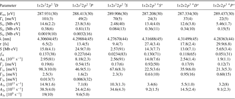
TABLE I. Parameters characterizing the 1s→2pexcitation resonances associated with ground-state C+(1s22s22p2P) (first three columns) and metastable C+(1s22s2p2 4P) parent ions (last three columns). The top entry of each column is theK-vacancy term excited by a single photon. The resonance energiesEresare given with three decimals because their relative uncertainties are of the order of only 5 meV. The absolute calibration uncertainty is 30 meV. Throughout this paper, the numbers in round brackets following a measured quantity denote the uncertainty of the last digits. The following parameters are provided as extracted from resonance fits to the measured spectra:Eres, the resonance energy relative to the initial term energy;L, the Lorentzian width of the excited level;S1e, the partial resonance strength for net single ionization;S2e, the partial resonance strength for net double ionization;S3e, the partial resonance strength for net triple ionization;λ, the transition wavelength;
τ, the lifetime of the excited level;S, the total resonance strength for photoabsorption;fik, the oscillator strength for absorption from level|i to level|k;Aki, the associated Einstein coefficient for radiative decay of the excited level;γ, the radiative (partial) width of the excited level;
1e, the partial width for the emission of one electron;2e, the partial width for the emission of two electrons;3e, the partial width for the emission of three electrons;A1e, the single-Auger decay rate;A2e, the double-Auger decay rate; andA3e, the triple-Auger decay rate. Quantities (S1e,S2e,S3e, andS) that depend on the fraction of ions in the specified initial term are normalized to a fraction of 100% so that the derived parameters (such asfik) retain their intended meaning.
Parameter 1s2s22p2 2D 1s2s22p2 2P 1s2s22p2 2S 1s2s2p3 4So 1s2s2p3 4Do 1s2s2p3 4Po
Eres[eV] 287.931(30) 288.413(30) 289.906(30) 287.208(30) 287.334(30) 289.437(30)
L[meV] 101(3) 49(2) 70(5) 24(3) 37(4) 22(5)
S1e[Mb eV] 14.6(2.2) 23.8(3.6) 2.48(40) 13.4(4.0) 12.6(3.8) 5.46(1.7)
S2e[Mb eV] 0.38(6) 0.81(13) 0.084(13) 0.36(11) 0.34(10) 0.15(5)
S3e[Mb eV] 0.0019(10) 0.0032(16)
λ[nm] 4.30604(45) 4.29884(45) 4.27670(44) 4.31688(45) 4.31499(45) 4.28363(44)
τ[fs] 6.5(2) 13.4(5) 9.4(7) 27.4(3.4) 17.8(2.4) 29.9(6.8)
S[Mb eV] 15.0(4.1) 24.9(7.0) 2.57(91) 14.3(7.7) 13.0(7.1) 5.65(3.4)
fik 0.137(38) 0.227(64) 0.0234(83) 0.130(71) 0.118(65) 0.051(31)
Aki[1011s−1] 2.95(81) 8.18(2.3) 2.56(91) 14.0(7.6) 2.54(1.4) 1.9(1.1)
γ[meV] 0.19(6) 0.54(15) 0.17(6) 0.92(50) 0.17(9) 0.12(7)
1e[meV] 98.3(10.0) 46.9(5.1) 67.6(8.3) 22.5(3.6) 35.9(6.0) 21.3(5.3)
2e[meV] 2.5(3) 1.6(2) 2.3(3) 0.61(10) 0.95(16) 0.60(15)
3e[meV] 0.013(7) 0.0063(32)
A1e[1013s−1] 14.9(1.6) 7.1(8) 10.3(1.3) 3.4(6) 5.5(1.0) 3.2(8)
A2e[1011s−1] 38.5(4.0) 24.4(2.6) 34.6(4.3) 9.2(1.5) 14.5(2.4) 9.1(2.3)
A3e[109s−1] 19(10) 9.6(5.0)
each peak feature identical to those predicted by theory [8].
With all these constraints, the Gaussian width, six Lorentzian widths, six apparent resonance energies, and six factors for correcting the theoretical resonance strengths were extracted from the experiment.
The resulting parameters for the six 1s→2p excitation resonances are provided in Table I. The first row shows the excitation energies found for the six resonances. Their absolute calibration uncertainty is±30 meV. Yet the energies are given with three decimals because the relative uncertainty is less than 5 meV. The second and third rows show the remaining parameters resulting from the fit of the spectra in Fig.4, i.e., the natural line widths =L and the resonance strengths S1efor one-electron removal. The estimated uncertainties are given by the numbers in brackets. In similar fits to experimental spectra (see Figs. 1 and6), the resonance strengths S2e for two-electron removal, andS3efor three-electron removal were also determined. The measured strengths were normalized to 100% considering the relative fractions of ions in the ground term (90%) and the metastable term (10%) in the measurement.
In other words, the resonance strengths obtained from the measurements were divided by 0.9 in case of the resonances populated from the ground-state term and by 0.1 in the case of resonances populated from the metastable parent ion term.
The remaining parameters provided in Table I have all been derived from the observables given in the first five
rows. The relevant formulas are collected in the Appendix, which describes the procedure used for extracting all the additional information about the observed resonances. For convenience, the sixth row of TableIshows the wavelength of the transitions observed in the experiment. These are readily obtained from Eq. (A5). The lifetimes of the core-excited terms are easily calculated from Eq. (A6). The determination of the resonant-absorption strengthS is more involved and requires additional information about radiative transition rates. An iterative procedure which converges after two steps provides Sand the oscillator strengthfikas well as the radiative decay rate Aki and the partial widthγ for radiative decay of the core-excited term. Following from these parameters and the measured resonance strengthsSne(n=1,2,3) for the removal ofnelectrons, the transition ratesAnefor single-, double-, and triple-Auger decay as well as the partial widths of these decays can also be obtained.
The information collected in TableIcan be used for con- structing the natural (unconvoluted) absorption cross section related to 1s→2p excitation of C+. This can be directly compared to the results of theoretical calculations. In principle, for such comparisons the cross sections for exclusively ground- term C+ ions can be separated from the cross sections for exclusively metastable C+ions. Instead, comparisons are made assuming the conditions of the present experiment, i.e., 10%
metastable and 90% ground-level ions. The experimentally
0.1 1 10
100 R-matrix Hasoglu et al.
Crosssection(Mb)
0.1 1 10
100 MCDF Shi and Dong
286 287 288 289 290
0.1 1 10
100 R-matrix Wang and Zhou
Photon energy ( eV ) 0.1
1 10 100
Stot= 36.7 MbeV Stot= 42.9 MbeV Stot= 34.1 MbeV (a)
(b)
(c)
(d)
this experiment
Stot= 39.9 MbeV
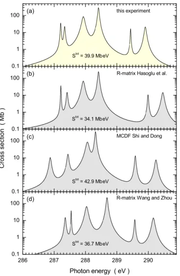
FIG. 5. Natural-line-width absorption cross sections of C+deter- mined by the present experiment with a 90% fraction of2P ground level and 10% of4P metastable ions in the parent ion beam. The experimental spectrum (a) is compared with calculations by Haso˘glu et al.[1] (b), Shi and Dong [8] (c), and Wang and Zhou [7] (d). The theory spectra were modeled assuming the experimental distribution of ground-term and metastable ions. Each panel displays the total resonance strengthSt ot contained in the associated photoabsorption spectrum.
derived natural cross section for such a mixture is shown in Fig.5(a).
The simulated experimental cross section is compared with the results of twoR-matrix calculations [1,7] and the cross section reconstructed from the detailed information provided by Shi and Dong on the basis of their multiconfiguration Dirac-Fock calculations [8]. All cross sections are shown on a logarithmic scale to emphasize the smaller resonances in the comparison. The calculations show spectra which are quite similar to the experimental result. Obvious differences are mainly in the positions of the six resonance features. However, one has to keep in mind that the deviations are typically only within several tens of meV and reach no more than about 550 meV in a few cases. Such deviations at level energies of approximately 290 eV are less than 0.19%.
In Fig.5, the total resonance strength of each spectrum is provided. The average of the theoretical resonance strengths is 37.9 Mb eV, which is only 9.5% from the experimental value.
The maximum deviation is 14.5% which is still within the experimental uncertainty. While the resonance energies and the peak areas in the theory spectra are close to the experiment there are dramatic differences in the peak widths between different theoretical approaches and between theory and ex- periment. Quantitative comparisons are provided in Table II where selected parameters listed in TableIare compared with theoretical and experimental data available in the literature.
The parameters for which comparisons are possible are the resonance energiesEres, the oscillator strengthsfik, the natural widthwhich is equal to the Lorentzian widthLfound in the fits, the radiative decay rateAki, and the single-Auger decay rateA1e.
The resonance energies found in the present investigation agree with most of the other experimental findings within their error bars. Deviations are observed for the positions of the smaller peaks in the spectrum, particularly when comparing with the spectroscopic work of Jannittiet al.[4]. Differences with theoretical calculations have already been discussed and are below a level of 1.90×10⁻³.
The experimentally derived oscillator strengths for ab- sorption have estimated uncertainties between 28% for the strongest ground-term resonances and about 60% for the resonances associated with metastable parent ions. The reason for the relatively large error bars is in the uncertainty of the 10%
fraction of the metastable component in the parent ion beam.
With only few exceptions, the theoretical oscillator strengths are well within the experimental error bars.
With an instrumental width of only 12 meV, the determina- tion of natural line widths for core-excited levels of C+ ions can be expected to be quite accurate. Compared to the previous experiment by Schlachteret al.[2], the uncertainties of some of the values could be substantially reduced by factors up to 8. Nevertheless, the width of the broadest resonance, associated with the core-excited2Dterm, is within 4% in both experiments. In other cases, the uncertainties quoted in the earlier experiment were slightly too optimistic. The theoretical natural widths show substantial scatter by factors up to about 5.
On average, theory predicts broader resonances for metastable parent ions than what the experiment yields.
The radiative decay rates derived from the present ex- periment can be directly compared with the calculations by Sun et al. [25], who used the saddle-point variation and complex-rotation methods including relativistic and mass po- larization corrections in first-order perturbation theory. The calculated radiative transition rates are in agreement with the experiment within the experimental uncertainties. Finally, the total transition rates for single-Auger decay found by the present measurement are compared with the results of four different theoretical calculations. For the resonances associated with ground-term parent ions, theory and experiment are in quite satisfying agreement. However, in a few cases there are deviations exceeding a factor of 2.
The comparisons show that the accurate calculation of atomic transition parameters for a relatively simple atomic ion with only five electrons is still a challenge. With the results obtained in the present experiment for double- and triple-Auger
TABLE II. Selected parameters from TableIin comparison with experimental and theoretical results for the 1s→2pexcitation resonances associated with ground-state C+(1s22s22p2P) (first three columns) and metastable C+(1s22s2p4P) parent ions (last three columns). The peak assignment by Schlachteret al.[2] as well as that by Wang and Zhou [7] have been corrected according to the discussion by Haso˘gluet al.
[1] and the results of Shi and Dong [8]. The calculations by Shi and Dong are differential in the total angular momentum and appropriately weighted sums were entered into this table.
Parameter 1s2s22p2 2D 1s2s22p2 2P 1s2s22p2 2S 1s2s2p3 4So 1s2s2p3 4Do 1s2s2p3 4Po Eres[eV]
Experiment
This work 287.931(30) 288.413(30) 289.906(30) 287.208(30) 287.334(30) 289.437(30)
[2] 287.93(3) 288.40(3) 289.90(3) 287.25(3) 289.42(3)
[4] 287.91(7) 288.59(7) 289.95(7) 287.09(7) 288.02(7) 289.80(7)
Theory
[1] Spectrum fit 287.929 288.414 290.465 287.213 287.413 289.992
[1] Their TableI 287.966 288.303 290.279 286.997 287.465 290.012
[8] 288.061 288.317 290.240 286.875 287.440 289.587
[7] 288.034 288.685 290.163 287.354 287.542 289.571
[25] 287.90 288.56 289.92 287.33 289.63
[2] 287.96 288.63 289.97 287.29 287.73 289.46
fik
This work 0.137(38) 0.227(64) 0.023(8) 0.13(7) 0.12(7) 0.051(31)
Theory
[1] Spectrum fit 0.100 0.191 0.020 0.084 0.095 0.053
[8] Length form 0.139 0.249 0.028 0.002a 0.132 0.079
[8] Velocity form 0.126 0.226 0.025 0.002a 0.125 0.074
[8] Revised 0.0973a
[7] Length form 0.1015 0.1927 0.0197 0.0968 0.0844 0.0547
[7] Velocity form 0.0977 0.1855 0.0194 0.0931 0.0812 0.0534
[meV]
Experiment
This work 101(3) 49(2) 70(5) 24(3) 37(5) 22(5)
[2] 105(15) 59(6) 112(25) 110(40) 55(25)
Theory
[1] Spectrum fit 94 50 87 16 47 36
[8] 101 43 77 57 67 42
[7] 96 54 88 47 16 35
[2] 103 62 93 25 84 52
Aki[1011s−1] Experiment
This work 2.95(81) 8.18(2.3) 2.56(91) 14.0(7.6) 2.5(1.4) 1.9(1.1)
Theory, length form
[25] 2.25 6.98 2.24 3.22 1.91
A1e[1013s−1] Experiment
This work 14.9(1.6) 7.12(77) 10.3(1.3) 3.42(55) 5.45(92) 3.23(80)
Theory, length form
[29] 13.1 7.22 12.6 8.90 7.25 5.67
[1] 13.9 7.49 13.1 2.35 6.99 5.41
[30] 17.6 7.88 15.5 3.13 9.75 7.40
[8] 15.4 6.44 11.7 10.1 8.53 6.29
aEntries in Table III of reference [8] are most likely misprinted; the corrected number, 0.0973, was calculated by Shi and Dong [28] using the FAC code.
decays, the challenge becomes even greater. Theoretical at- tempts are being developed to deal with multielectron ejection subsequent to inner-shell excitation (see, e.g., Ref. [16]).
However, so far only one calculation has been published addressing the multielectron, and particularly the triple-Auger decay processes associated withK-shell excitations of C+ions that have been studied in the present experiment. But very
encouraging theoretical work that includes calculations also of direct triple-Auger decay is under way [31].
The last part of this paper is devoted to the quantitative description of multiple ionization of C+ ions as the result of the absorption of a single photon. For the strongest resonances found in single ionization, additional measurements of single, double, and triple ionization were carried out at a fixed energy
287.5 288.0 288.5 0.00
0.01 0.02
0.03 hν+ C1+ → C4++ 3e-
Photon energy ( eV )
0 2 4
6 hν+ C1+ → C3++ 2e-
C ros s s ec ti on ( M b )
050 100 150
200 hν+ C1+→C2++ 1e-
1s2s22p2 2D
1s2s22p2 2P
FIG. 6. Cross section for single, double, and triple ionization of ground-state C+ions by single photons at 92-meV bandwidth. The resonances seen in all the observed channels are associated with K-shell-excited C+(1s2s22p2 2D,2P) terms. The dark solid (blue) lines represent the fits to the experimental spectra with Voigt profiles from which Auger decay probabilities were obtained. The light (red) solid lines show theory-based absolute cross sections derived from calculations of single-, double-, and triple-Auger decay rates by Zhou et al.[32]. For details, see main text.
resolution of 92 meV [3]. The lowest panel in Fig.6shows the unambiguous observation of C4+ product ions arising from K-shell excited C+(1s2s22p2 2D,2P) states which are popu- lated from the ground-state term of C+via one-photon absorp- tionγ+C+(1s22s22p2P)→C+(1s2s22p2 2D,2P). The re- sultingK-vacancy states can obviously decay by the emission of up to three electrons. Other conceivable explanations for the observation of C4+product ions such as two-photon absorption or one-photon absorption plus one or more interactions with residual-gas particles were discussed in our previous Letter [3]
and shown to be negligible.
Along with the experimental data, Fig. 6 presents theo- retical cross-section curves constructed from calculations of single-, double-, and triple-Auger decay ofK-shell excited C+(1s2s22p2 2D,2P) terms. Decay rates for Auger processes starting from these terms with emission of one, two, or three electrons were computed by Zhouet al.[32] and are compared in Table III with the experimentally derived decay rates.
From the theoretical decay rates as well as the calculated resonance energiesEresand natural widths, also provided in TableIII, relative theoretical cross sections can be constructed
TABLE III. Comparison of the present resonance parameters for C+(1s22s22p2P)→C+(1s2s22p2 2D,2P) excitations with calcula- tions by Zhouet al.[32], including the triple-Auger decay rate. The data obtained by Zhouet al.were taken from their TableIforA1eand derived from the ratios given in their Table IV.
Parameter 1s2s22p2 2D 1s2s22p2 2P
Eres[eV], this work 287.931(30) 288.413(30)
Eres[eV], Zhouet al. 287.87 288.11
[meV], this work 101(3) 49(2)
[meV], Zhouet al. 114 57
A1e[1013s−1], this work 14.9(1.6) 7.1(8) A1e[1013s−1], Zhouet al. 17.4 8.66 A2e[1011s−1], this work 38.5(4.0) 24.4(2.6) A2e[1011s−1], Zhouet al. 53.4 27.1 A3e[109s−1], this work 19(10) 9.6(5.0) A3e[109s−1], Zhouet al. 31.2 15.8
for single, double, and triple ionization of C+(1s22s22p2P) ions by a single photon via excitation of K-shell excited C+(1s2s22p2 2D,2P) terms. For producing absolute cross sections additional information on the absorption oscillator strength is required which has not been provided by Zhouet al.
By using the experimentally derived values offik(see TableII) plus the parameters calculated by Zhou et al. as presented in Table IIIthe light (red) solid curves in Fig. 6 have been computed which represent the absolute cross sections for single (upper panel), double (middle panel), and triple (lower panel) ionization of C+. For direct comparison with the experiment, the theoretical cross sections were convoluted with 92-meV FWHM Gaussians.
The resonance energies obtained by Zhouet al.are slightly off; however, the differences are at most 300 meV corre- sponding to 0.1% of the associated resonance energies. The magnitudes of the cross sections, partly being adjusted to the experimental data by using the experimentally derived oscillator strengths, give remarkable agreement for all three final-charge states of the ionized ions, which requires realistic multielectron-ejection decay rates. The comparison of these rates in Table III shows a level of agreement that is very encouraging for further developments in this field of research.
IV. SUMMARY
In this paper, which is a followup to a previous Letter [3], details of the experiments are presented. In particular, the experimental data are compared to available theoretical results from different computational approaches. From high- resolution absolute cross-section measurements at a resolving power of up to 24 000, exceeding that of previous experiments by a factor of 4, complete sets of transition parameters for 1s→ 2pexcitations of ground-term and metastable C+ions could be derived. These include not only the resonance energies, natural widths, and resonance strengths but also the transition rates for radiative decay as well as single-, double-, and triple-Auger decays. Oscillator strengths, partial decay widths, transition wavelengths, and lifetimes ofK-shell excited terms are also readily computed from the extracted information. Comparison of these quantities with data available in the literature show
![FIG. 2. Experimental cross section for single photoionization of C + ions in the region of 1s → 2p excitations at 38-meV resolution compared with results of R-matrix photoabsorption calculations [1] convoluted with a 38-meV full-width-at-half-maximum (FWHM](https://thumb-eu.123doks.com/thumbv2/9dokorg/1413569.119258/4.911.178.738.102.505/experimental-photoionization-excitations-resolution-compared-photoabsorption-calculations-convoluted.webp)