Surgical anatomy of the extra- and intrahepatic arteries of the human liver
on corrosion casts
PhD thesis
Károly Németh MD.
Semmelweis University
Doctoral School of Pathological Sciences
Tutor: Prof. László Kóbori MD, Ph.D.
Official reviewers: Ákos Szűcs MD, Ph.D.
Zsolt Csapó MD, Ph.D.
Head of the Final Examination Board: Prof. József Sándor MD, Ph.D.
Members of the Final Examination Board: Attila Bursics MD, Ph.D.
Ákos Balázs MD, Ph.D.
Budapest 2016
1. Introduction
Different vascular anatomic variations in the upper abdomen are part of the daily experience of surgeons and radiologists. The various forms of the hepatic arterial structure are of great importance in terms of liver resections, living donor hepatectomies, whole-organ and split liver transplantation. The anatomic variations of both the extra- and intrahepatic arteries are associated with increased perioperative morbidity and mortality due to the ischemic complications implied, such as insufficient remnant liver volume in the donor, reduction of the functional liver mass in the recipient, intrahepatic abscess formation, ischemic cholangiopathy and hepatic artery thrombosis. Moreover, aberrant hepatic arteries can be of major surgical significance during operations of the upper intestinal tract, the gallbladder and pancreas. They can also present technical challenges for infusion therapy and transarterial chemoembolization of neoplasms in the liver.
The first description of the aberrant hepatic arteries was published in 1756 by Haller. Since then the anatomic variations of the extrahepatic arteries have been examined by several authors worldwide. According to the international data available, the incidence of the so called ”normal” anatomical variation varies between 50.7% and 80.9%. Compared to the numerous studies on the extrahepatic arterial supply, the intrahepatic arteries have been less frequently investigated, mostly regarding the arterial branches to the segment IV and the caudate lobe. Few data are available on the segmental branching pattern of the arterial tree in the right lobe.
2. Objectives
The aim of our present study was to investigate the segmental arterial supply of the human liver by using vascular corrosion casting technique, paying particular attention to rare variations not reported in previous studies. To our knowledge, this is the first study in which corrosion procedure is used to
demonstrate the extra- and the intrahepatic arteries together by injecting resin into human cadaveric abdominal organ complexes in relatively large numbers. Our results may contribute to the better understanding of the arterial supply of the liver and may reduce the risk of perioperative complications of surgical and radiological interventions in the upper abdomen.
3. Methods
Vascular corrosion casts were prepared from a total of 50 abdominal organ complexes obtained from fresh human cadavers of Caucasoid race, which were delivered to the Department of Forensic and Insurance Medicine, Semmelweis University, Budapest. Written permission was obtained beforehand from the Ethical Commission of Semmelweis University (TUKEB number: 185/2004). The corpses neither had any history of liver disease, nor presented any signs of abdominal trauma or macroscopic alteration.
Our research group developed several types of resin mixtures to improve the visualization technique of the arterial system on macro- and microvascular scale in humans and other species as well. A special Vinyl Ester resin mixture was prepared for the purpose of injecting the arteries of the human organ complexes through the abdominal aorta. The components of the mixture were: 1. Resin:
Novolac-based Epoxy Vinyl Ester Resin (Derekane 470-300 by Ashland); 2.
Pigments (5%): FP3000 red and FP1021 yellow (by Cytec Surface Specialties Austria GmbH); 3. Accelerator (2%): Cobalt 2-ethylhexanoate, N,N-Dimethyl aniline (Accelerator NL-23 by AkzoNobel); 4. Catalyst (2%): Methyl Ethyl Ketone Peroxid (Butanox M-50 by AkzoNobel). The pigment guaranteed not only different colors of the different vessels, but also provided suitable CT density (approx. 250 HU). The resin polymerized in 3-5 minutes, after which, concentrated
KOH was added to the surrounding water to commence digestion of the soft tissues. The corrosion process lasted one week at 60-65°C temperature. Residual fat and liver parenchyma were removed by rinsing in warm water, leaving only the cast behind. In 16 cases the portal vein system was also visible with a different colored resin.
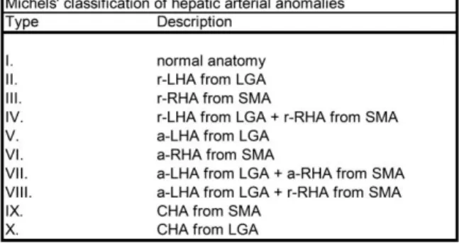
The classification of the extrahepatic arteries was based on Michels’ classic results of 200 autopsies, where the normal extrahepatic arterial structure is defined as (1) the proper hepatic artery (PHA) dividing into the right and left hepatic arteries (RHA, LHA), (2) the right hepatic artery further dividing into the right anterior and posterior hepatic arteries (RAHA, RPHA), (3) the left hepatic artery dividing into branches supplying segment II and segment III, and (4) one or more branches supplying segment IV arising from the right, left, or proper hepatic artery. Hepatic arterial anatomy that differed from the dominant scheme was considered to represent an anatomic variation. According to Michels’ definition, the aberrant vessels may be accessory, occurring in addition to the normal arterial supply, or replaced, representing the primary arterial supply to the lobe (Table 1).
Table 1: Michels’ classification. (a-: accessory, r-: replaced, CHA: common hepatic artery, LGA:
left gastric artery, LHA: left hepatic artery, RHA: right hepatic artery, SMA: superior mesenteric artery)
In addition, since our organ complex casts were suitable for investigation of the complete upper abdominal vascular structure, the right gastric artery (RGA), the gastroduodenal artery (GDA) and the interrelationship between the main portal vein and the hilar hepatic arteries were also taken into account due to their high surgical and radiological significance.
All specimens underwent 3D volumetric CT reconstruction. For this purpose we used a Philips Brilliance 16 unit (parameters: 140 kV, 300mAs, collimation: 16x0.75mm, overlap 50%) with Philips Brilliance 16 Workspace 2007 software.
4. Results
4.1 Anatomical variations of the extrahepatic arterial supply of the human liver
4.1.1 Michels types
In our series of 50 corrosion casts, 41 casts (82%) could be classified according to Michels. Twenty-one cases (42%) showed normal arterial pattern (Michels I), while 29 casts (58%) presented different types of extrahepatic arterial variations. However, 9 casts (18%) displayed variations not described in the Michels’ classification.
Replaced hepatic arteries
Replaced left hepatic artery (r-LHA) arising from the LGA – Michels II – was observed in 3 (6%), while replaced right hepatic artery (r-RHA) originating from the superior mesenteric artery (SMA) – Michels III – was present in 7 (14%) cases. Double replaced system was present in 2 casts (4%). One cast showed a replaced common hepatic artery (r-CHA) arising from the SMA.
Accessory hepatic arteries
Accessory left hepatic artery (a-LHA) – Michels V – was present in 4 (8%) cases, accessory left and right hepatic arteries together (a-LHA and a-RHA) – Michels VII – in 1 (2%) case, while combined a-LHA and r-RHA – Michels VIII – were found in 2 (4%) cases.
It is notable that in 4 cases we observed well-defined Michels types showing additional arteries with extrahepatic destination. In the Michels I group one cast presented RGA and a-LGA originating from LHA, another cast showed an a-LGA arising from the artery of segment II (A2) and segment III (A3). In Michels type V, we found one cast having an a-LGA from the a-LHA. The only case of Michels type VII presented two a-LGA arteries branching from LHA, which represents a triple accessory system, a structure which can be considered as a subtype of Michels VII.
No casts presented a single a-RHA from SMA (Michels VI) or r-CHA originating from the left gastric artery (Michels X).
4.1.2 Arterial variants not mentioned in Michels’ classification (Unclassified variations – UC)
Out of 50 cases, 9 corrosion casts (18%) showed unusual arterial patterns that could not be classified according to Michels.
The UC variations of our series could be divided into 2 groups. The first group consisted of 5 cases presenting arborization abnormalities of the CHA.
Trifurcation of CHA was observed in 4 cases overall, with the CHA giving off the RHA, LHA and the GDA in 3 casts. One cast showed an early, proximal origin of the RHA from the CHA, which results in the CHA trifurcating into LHA, GDA and RGA (Figures 1a-c). In this newly described variation the RHA ran behind the portal vein.
The fifth case within this group is also a new variant, the CHA branching into five arteries: the LHA, RHA, A4, GDA and RGA (Figures 2a-c). It is also remarkable that the caudate artery (artery of segment I - A1) originated dorsally from the RHA only 4 mm away from the point of pentafurcation.
The second group of the UC variations is formed by the 4 cases displaying anomalous origins and courses of the lobar and sectorial arteries, the RHA, LHA and RPHA. In one case the RHA arose from the CA and coursed behind the portal vein. In another cast, displaying a new variant, the RHA originated from the proximal part of the CHA and then passed in front of the portal vein. Thus CHA did not bifurcate into PHA and GDA, but into RHA and LHA-GDA trunk (Figures 3a-c).
In one cast, exhibiting the fourth new variant, the RPHA arose directly from the CHA, in close proximity to the CA-bifurcation. It then passed around the portal vein to reach the right posterior sector of the liver. The celiac axis (CA) bifurcated into the splenic artery (SA) and CHA; and the LGA arose independently from the aorta (Figures 4a-c).
The last, already known UC variation of this group is a proximal branching of the LHA with a considerable distance between the origins of the LHA and RHA. In this case RHA took off of GDA.
Thus, among these 9 UC variations we encountered 4 cases which, to the best of our knowledge, have not been reported before: CHA pentafurcation;
Proximal origin of RHA from CHA with retroportal course, CHA trifurcates into LHA, GDA and RGA; Proximal origin of RPHA from CHA with retroportal course, CHA gives the LHA-RAHA common trunk and GDA (LGA originates separately from aorta); Proximal origin of RHA from CHA with a course in front of the portal vein, CHA later divides into LHA and GDA.
Vessels of approximately 1 mm diameter were visualized during 3D CT evaluation of the specimens and all variations were recognized. Therefore, radiological and anatomical results were identical.
4.2 Branching and coursing patterns of the lobar arteries
We observed numerous abnormalities within almost every Michels type (I, II, III, V, VII, IX) based on the branching and coursing variations of the segmental/subsegmental branches of the RHA and LHA. In Michels types IV and VIII the arborization of the RHA always showed the conventional pattern.
4.2.1 Right hepatic artery
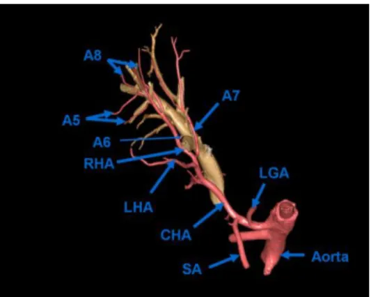
RPHA absence. Proximal deriving of segmental arteries (A6 or A7) were found in 7 cases. Normal branching of the RHA into RAHA and RPHA was not observable due to an early branching of a right-sided segmental artery. (Figure 5).
Figure 5: RPHA absence: Proximal origin and retroportal course of A7. (Yellow: portal vein).
Accessory segmental arteries (Figure 6) deriving from RHA or LHA, proximally to the normal RAHA-RPHA division (i.e. double or triple segmental supply) were seen in 8 cases. In 1 remarkable case the RHA gave 8 separate branches: RAHA, RPHA, 2xA6, 2xCyA (cystic artery), A4, A1.
Figure 6: Accessory segmental arteries (a-A6 + a-A7; a-A5 + a-A8) arising from RHA.
The related segments have double supply.
(On the left picture additionally: RGA-A1 trunk arises from GDA.)
RAHA absence: A4A8 trunk from RHA (2 cases).
Neither RAHA, nor RPHA are present. (3 cases – Figure 7)
RHA branches: A5A6 + A7A8.
RHA branches: A5A6 + A5A8 + A6A7A8.
RHA branches: A4A5, A6A7A8, A6.
Figure 7: Liver No. 18. RAHA and RPHA are absent. RHA branches: A5A6, A5A8, A6A7A8.
Double RAHA. (2 cases – including 1 case of RAHA trifurcation into A5 and two A5A8 trunks)
RHA trifurcation. (2 cases, including 1 case of double RPHA)
4.2.2 Left hepatic artery
Accessory LGA (a-LGA) originating from A2 and A3 (1 case)
Accessory LGA (a-LGA) and RGA take off of LHA (1 case)
Accessory LGA (a-LGA) arises from accessory LHA (a-LHA) (1 case)
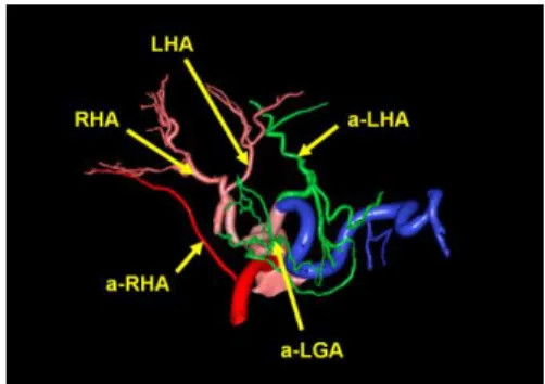
Triple accessory system: a-LHA + a-RHA + a-LGA (Figure 8) (1 case)
Figure 8: Triple accessory system.
In 5 cases we found a combination of extrahepatic and intrahepatic variations as follows:
Proximal branching of LHA + a-A8 from RHA
CHA trifurcation + a-A5 from RHA
CHA trifurcation + retroportal RHA
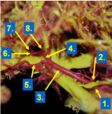
CHA trifurcation + RGA-A1-CyA trunk from LHA (Figure 9)
CHA pentafurcation + double RAHA
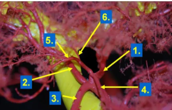
Figure 9: Liver No. 49. RGA-A1-CyA trunk from LHA combined with CHA trifurcation.
1: LHA; 2: RHA; 3: GDA; 4: RGA; 5: CyA; 6: A1. Yellow: portal vein
4.3 Segmental arterial supply of the most important segments
4.3.1 Caudate lobe (Segment I. – S I.)
In the present study we described 49 different types of arterial supply of the caudate lobe (49/50).
In 45 out of 50 cases (90%) communicating arcades with multiple anastomoses (@) were observed between different arteries of the left and the right lobe (Figure 10), often involving extrahepatic vessels, such as GDA in 12 cases, CyA in 5 cases, RGA in 3 cases, SAPD (superior anterior pancreaticoduodenal artery) in 2 cases and IPPD (inferior posterior pancreaticoduodenal artery) in 1 case. These arcades also served as origin for the always fork-like stemming end- arteries of the caudate lobe.
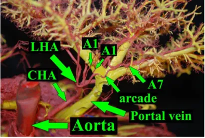
Figure 10: Liver No. 17.
Caudate lobe arteries (A1) originate from the vascular arcade between LHA and A7.
Superior dorsal aspect.
Out of the remaining 5 cases 2 casts showed S I arteries coming from both lobes without forming anastomosis [(RHA @ A6A8 @ IPPD) + A2 and RAHA + A3]; 1 cast showed S I arteries originating from the left lobe only (A2A3 @ A4); 1 cast showed S I arteries stemming from the coeliac axis and the right lobe only (RAHA + PHA); and S I. arteries from the right lobe only were present on 1 cast (RPHA @ CyA).
4.3.2 Segment IV. (S IV.)
We revealed 27 types of arterial supply of S IV, as shown on Table 2.
Table 2: Arterial supply of segment IV in our series.
In 24% (12/50) the A4 was the first branch of the LHA, representing the most frequent variation. In 10% of cases (5/50) the A4 was the second branch from the LHA, deriving from its A3A4 trunk. Segment IV received its arterial inflow from the left side in 62% (31/50), from right-sided origins in 10% (5/50), from both sides in 26% (13/50). In one case A4 took off of CHA as part of the pentafurcation.
One supplying artery irrigated S IV in 48% of cases (24/50), we found two arteries in 30% (15/50), three feeding arteries in 16% (8/50), four arteries in 1 case and five arteries in 2 cases. We observed extrahepatic vessels contributing to the blood supply of S IV in 3 cases. Anastomoses between A4 and GDA were found on 2 casts and a-LHA gave S IV branches on one cast.
5 Discussion
5.1 Corrosion casts and other modalities in the investigation of hepatic arteries
Our results show substantial differences concerning the variations of the extrahepatic arteries, compared to the literary data (Table 3).
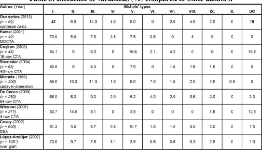
Table 3: Incidence of variations (%) compared to other authors.
While, according to several authors, the incidence of the Michels I type ranges between 50.7% and 80.9%, we found normal anatomy in only 42% of cases. Surprisingly, the second most frequent variation in our study was the UC type with 18%. Our series shares the general findings of the low percentage of Michels’ types VII, VIII and IX, however, discrepancies of other patterns are obvious. These may be explained with the low number of cases in our series, population differences, misinterpretation of radiological findings in other investigations due to respiratory motion artifacts, wrong catheter positioning, narrow diameter or slow flow in the small aberrant vessels.
The limitation of our study is the relatively low number of cases, however, our results and other angiographic investigations in which the patient numbers are ranged between 40 and 63 are comparable to the larger series as well.
5.2 Surgical relevancies of the new unclassified anatomical variations of the extrahepatic arteries
Pentafurcation of the CHA can be beneficial, if the right lobe, segment IV or segments II and III are involved in tumorous transformation. The sufficient length, large diameter and easy identification of the RHA, LHA and A4 – as seen on our preparation – allows the surgeon to safely perform right hepatectomy, left lateral split or Taj Mahal resection, without compromising the arterialization of the liver remnant. This new arterial variation may not necessarily cause problems during superselective chemoembolizations, however, when whole organ chemo- or radioembolization is needed, this anomaly can potentially lead to significant gastrointestinal side effects by shunting the therapeutic agents to the non-hepatic arteries. While this anatomic variant is manageable during whole-organ recovery from a deceased donor, it may be a problem potentially for a full left lobe donation.
Particular attention must be paid when the RHA or the RPHA displays a proximal origin from the CHA (or CA). After passing behind or in front of the portal vein, the distal part of the vessel reaches the right side of the hepatoduodenal ligament. Therefore, one has to be careful during dissection not to inflict accidental damage to the common bile duct, which runs close to it in the hepatic pedicle. In case of proximal branching of the RHA, the point of origin usually lies deep, next to the CA division, consequently – if liver volumetry allows – the left lobe is more preferable for donation in living donor liver transplantation due to its easier accessibility. However, in this case left lateral splitting may endanger the blood supply of segment IV, causing ischemia.
In case of the proximal branching of the RPHA, the right anterior sector of the liver is supplied by the common trunk of the LHA and RAHA and the first branch of this trunk is A3 on our cast. Subsequently, the intrahepatic distribution of the segmental arteries (A3 from LHA-RAHA trunk; A2 from A4) would result in 2 arterial stumps during left lateral splitting and the arterial inflow of S V and S VIII could also be endangered. Furthermore, a separate RPHA arising from CHA may imply a relative contraindication for right lobe living donation and full left – full right split as well, due to the double source of arterial supply of the right lobe (S V and S VIII from LHA; S VI and S VII from CHA). On the other hand, the proximal origin of the RHA or RPHA may have the advantage of an easier selective catheterization and a reduced risk of chemo- or radiotherapeutic agents reaching the wrong liver lobe.
5.3 New arterial structures in the left and right liver lobes
We observed 4 new intrahepatic arborization variations of the LHA: a-LGA from A2 and A3 (1/50); a-LGA from a-LHA (1/50); triple accessory system (1/50); RGA-A1-CyA trunk (1/50).
Radtke et al. proposed the most complex classification for the right-sided segmental variations and set up 7 different types of the peripheral branching patterns. We found 7 arterial structures described neither by Radtke et al., nor by other authors. These variations include RAHA trifurcation (1/50), RHA trifurcation (2/50), absence of RAHA (2/50), absence of RPHA (7/50), nonexistent RAHA and RPHA (3/50), RHA giving A2 (1/50), proximally originating RPHA coursing behind the portal vein (1/50).
6 Conclusions
This method, simulating the MDCT angiography allows us to provide accurate information about the complete upper abdominal vascular anatomy, including detection of new hepatic arterial variations, identifying subvariants of previously described cases. Given the fact that all four new unclassified variations were accurately visualized on our 3D CT reconstructions, these structures should be identifiable during clinical CT examinations as well. Being of great surgical and radiological importance, unusual variations must always be in the focus of surgeons and radiologists during the preoperative evaluations and interventions in the upper abdomen.
In this present study we described the detailed arterial supply of the human liver on 50 corrosion cast preparations and found numerous anatomical variations on both extrahepatic and intrahepatic levels, including newly observed vascular structures which have not been reported before. New extrahepatic variations include combinations of already known variants (CHA trifurcation + accessory A5; proximal branching of LHA from CHA + accessory A8) and entirely new structures (CHA pentafurcation + double RAHA; CHA trifurcation + proximal RHA from CHA with retroportal course; CHA trifurcation + RGA-A1-CyA trunk;
proximal RHA from CHA with anteportal course; proximal RPHA from CHA with retroportal course).
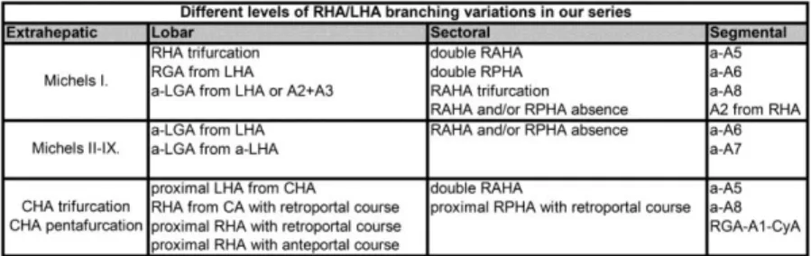
The arborization patterns of the RHA and LHA are summarized on Table 4.
Table 4: Levels of RHA/LHA arborization patterns. The variations can be combined independently of one another.
Concerning the segmental arterial supply, our findings show that the liver segments are often fed by more than one artery. S I receives multiple feeding arteries in 100% of cases; S II in 22%; S III in 30%; S IV in 52%, S V in 74%; S VI in 56%; S VII in 22% and S VIII in 32%.
The different liver segments can be sequenced in the order of arterial variability (Table 5).
Table 5: Distribution of liver segments according to the number of variants and the incidence of the most frequent arterial supply.
The increasing number of living donor and cadaveric liver transplantations, anatomical and non-anatomical hepatic resections, laparoscopic and radiological interventions are the main reason for a renewed interest in the investigation and statistical analysis of arterial variations and reports of new variants. The hepatic arterial anatomy is highly variable and some variations may necessitate the modification of the surgical approach. We believe that our hereby presented data
can contribute to the better understanding of the segmental arterial supply of the human liver and therefore lead to the reduction of complications during surgical and radiological interventions in the upper abdomen.
Main findings
1. Description of 4 new extrahepatic and 11 new intrahepatic arterial variations.
2. First complete documentation of the associated extra- and intrahepatic arterial variations, including previously unknown cases.
3. First highly detailed description of the caudate lobe arteries in healthy livers.
4. To the best of our knowledge, we were the first visualize the intrahepatic arteries up to subsegmental levels.
7 Bibliography of the candidate’s publications Publications related to the subject
Nemeth K, Deshpande R, Mathe Z, Szuak A, Kiss M, Korom C, Nemeskeri A, Kobori L. (2015) Extrahepatic arteries of the human liver - anatomical variants and surgical relevancies. Transpl Int, 28(10): 1216-1226. IF: 2,599
Kiss M, Deshpande RR, Nemeskéri A, Nguyen TT, Kürti Z, Kovács S, Pápai Z, Németh K, Szuák A, Dudás I, Kóbori L.(2015) Optimal line of hepatotomy for left lateral living donor liver transplantation according to the anatomical variations of left hepatic duct system. Pediatr Transplant, 19(5): 510-516. IF: 1,441
Rosero O, Nemeth K, Turoczi Z, Fulop A, Garbaisz D, Gyorffy A, Szuak A, Dorogi B, Kiss M, Nemeskeri A, Harsanyi L, Szijarto A. (2014) Collateral circulation of the rat lower limb and its significance in ischemia-reperfusion studies. Surg Today, 44(12): 2345-2353. IF: 1,526
Rosero O, Németh K, Turóczy Zs, Fülöp A, Garbaisz D, Kiss M, Nemeskéri A, Szijártó A. (2013) Collateral circulation of the rat lower limb and its significance in ischemia-reperfusion studies
European Surgical Research, 50:(Suppl 1) 26. - Abstract
Fülöp András, Rosero Olivér, Turóczi Zsolt, Garbaisz Dávid, Dorogi Bence, Németh Károly, Nemeskéri Ágnes, Harsányi László, Szijártó Attila. (2013) A collateralis hálózat szerepe alsó végtagi ischaemiás modellek kialakítása során.
Magyar Sebészet, 66: 84. - Abstract
Other publications
Tarnoki DL, Tarnoki AD, Nemeth K, Bata P, Berczi V, Karlinger K. (2013) Partial absence of superior vena cava in an adult patient - Case report and literature review. Herz, 38(7): 785-789. IF: 0,912