Chapter 6 Chemical Assay of Adrenocorticosteroids
GRANT G. SLATER
Neurobiochemistry Laboratory T-4b~
Veterans Administration Center Los Angeles, California
I. Introduction 303 A. Bioassay 304 B. Chemical Assay 304 II. Source and Preparation of Test Material 306
A. Biological Source 306 B. Preservation of Samples 308 C. Hydrolysis and Extraction 309 III. Chromatographic Separations 312
A. General 312 B. Thin-Layer Chromatography 314
C. Liquid-Solid and Liquid-Liquid Columns 317
D. Gas-Liquid Columns 321 IV. Fluorescence Assay 323
A. General 323 B. Alkaline 327 C. Acid 329 V. Radioisotope Assay 332
A. General 332 B. Double-Isotope Derivative 332
VI. Other Physical Methods 334
References 334
I. INTRODUCTION
The study of the formation, metabolism, distribution, and excretion of the adrenocortical hormones is one of the most rapidly expanding areas in endocrinology. Since nutrition influences the development of the endocrine system, and hormones are involved at many places in the utilization of foods, it is necessary to know more about the role of adren- ocortical hormones in the chemical control of the vertebrate animal.
For this purpose methods must be available for analysis of their pre- cursors and urinary end products as well as the active steroids.
Since certain corticoids with, as yet, no known specific function occur in the adrenal vein blood, a question arises whether or not the adrenal itself may be a "leaky gland"; that is, during activation small
303
304 GRANT G. SLATER
amounts of steroids which do not influence other tissues may be inad- vertently released. I t is my belief, however, that all such minor compo- nents do have specific uses; it remains only to discover what they are.
It seems most logical to assume, since all vertebrates appear to produce and use physiologically active corticoids, that in the long history of development of the adrenal gland there evolved efficient ways of producing adrenocorticoids without wasting energy on the synthesis of substances to be discarded. It now becomes one of the fascinating possibilities of the near future that we shall be able to measure the effects of nutrition and numerous other factors on the distribution of the minor components of the adrenocortical complex of steroids. The small amount of minor steroids can bear little relationship to their importance if they affect specific tissues or enzyme systems, and also, the minor steroids may show much greater variation with a specific disease or experimental condition, and thus prove to be of great diagnostic or interpretive value. Therefore it is imperative that more attention be paid to the methods which will measure these minor quantities of adrenocortical hormones, and methods to this end have recently become available.
In the past it was possible only to measure the amount of a particular corticoid activity by its effect on some biological system, i.e., by bio- assay; however, as time has passed and more and more physiologically active steroids have been isolated, it has become possible to use more exact chemical methodology. The purpose of this review is to present and evaluate this methodology.
A. Bioassay
Most of the bioassay methods, whether they are in vivo or in vitro determinations, are time consuming and expensive. However, these were important in the isolation of the adrenocorticoids and were essential in determining which chemical structures were important, so that appropriate chemical methods could be originated. Furthermore, the bioassay methods will continue to be important for measuring potentially new synthetic corticoid-like drugs and for determining the importance of adrenocortical hormone action on specific tissues and enzyme systems.
These methods have recently been reviewed by Dorfman (1).
B. Chemical Assay
Considerable latitude has been taken with the word "chemical" in this chapter. It is meant to cover essentially all methods other than bio- assay, whether or not any chemical alterations have been made to the adrenocorticoids during the assay. It should be noted here that no attempt has been made to cover all or even a large part of the rapidly growing
6. CHEMICAL ASSAY OF ADRENOCORTICOSTEROIDS 305 literature in this field. The methods selected are ones with which the author has had some personal experience or which he feels meet the requirements for certain clinical or research needs.
The selection of a method to measure adrenocortical hormones depends in large part on the objectives of the investigation, the equip- ment available and the accuracy demanded. The objectives of a clinical study may be quite different from those of a research study using experi- mental animals. Even the objectives of a clinical study, for instance, measurement of the steroidal synthesis occurring in an adrenal tumor, may be quite remote from those of a study involving dietary effects on the pituitary-adrenocortical system. The cost of equipment in bio- chemical studies is not usually considered very important. However, when the expense of chemicals and labor for doing two different methods is relatively the same, and if an expensive piece of equipment, such as a fluorometer, is required for one method but not for the other, this could become of considerable importance.
The accuracy required for a particular investigation or series of clinical measurements is usually the most important factor in the selection of a method of analysis. The accuracy of measurement is determined by the specificity, the sensitivity, and the reproducibility of the method.
The specificity is controlled by selection of purification steps to isolate the steroid or steroids which most appropriately reflect the change to be measured. For example, if one observed a reduced sodium excretion in a particular disease condition, it would appear desirable to select a method which would lead to the best purification of aldosterone or to use some other specific system of measuring aldosterone, such as an isotope dilution method.
The accuracy demanded can be influenced also by selection of a method with the proper sensitivity; thus, in most cases the fluorometric methods have been found to be more sensitive than the colorimetric methods. However, to some extent this is offset by the requirement of additional purification to remove nonsteroidal fluorescent materials.
The question of reproducibility of a method involves the ability of one group of workers to repeat work of another, but it also involves the ability of the same workers to obtain similar results from more than one determination on the same sample pool. Usually, variability increases with the number of steps in the purification of the substance to be measured.
It is generally believed at the present time that the free corticosteroids in blood are the active form, but it is also recognized that a sizable fraction of the total corticoids may be found conjugated in the form of glucuronides and sulfatides. Although, in the past, acid was used to
306 GRANT G. SLATER
hydrolyze these conjugated forms, it is now recognized that some destruc- tion of sensitive corticoids occurs. Enzymatic splitting of the glucuronide has been used for a number of years and recently the sulfatides have been investigated using hydrolysis with a crude enzyme preparation.
It should be emphasized that biological extracts contain many unknown substances and frequently these unknown substances may be carried through a number of purification steps and cause interference in the final measurements. This becomes increasingly important as the search continues for the minor steroids which are found in very low concentration.
I I . SOURCE AND PREPARATION OF TEST MATERIAL
A. Biological Source
1. Adrenal Gland Tissue
The adrenal gland is known to contain a large number of steroids, but so far only a few of them have been shown to be biologically active.
The extraction and further purification of adrenal gland tissue pose the problem of separation of the physiologically active corticoids from all the steroidal precursors, some of which are closely related in structure and, thus, difficult to separate. On the other hand, the corticoids are found in high concentration in adrenal tissue. As a result of intensive investigation, the major corticosteroids, hydrocortisone and corticos- terone, can now be separated with relative ease from most other lipids in the adrenal tissue, and for many purposes they suffice. Recently, it was shown that the rat has a high level of 18-hydroxydeoxycorticosterone for which new methods must be used; however, measurement of corticoster- one may suffice for many studies. Man produces 18-hydeoxycorticosterone in concentrations greater than aldosterone, but its use is unknown.
Fortunately, relatively little conjugation appears to occur normally in the adrenal gland tissue, and many chromatogens present in blood are found in low concentration. However, recent evidence indicates that steroidal sulfates occur in adrenal tissue (2, 3).
2. Adrenal Vein Blood
The adrencocortical steroids while originating in the adrenal gland can be synthesized and released at a very rapid rate to give high con- centrations in the adrenal vein blood. Holzbauer (4) has shown in the stressed rat that in a little under 3 minutes the content of corticosterone in the adrenal gland is about equivalent to the amount released into the adrenal vein. Most adrenal vein blood corticoids are not conjugated,
6. CHEMICAL ASSAY OF ADRENOCORTICOSTEROIDS 307 and the level of deactivated products, such as the tetrahydro forms, is also relatively lower than the content of free steroids. Recently, very young children were shown to have a large percentage of sulfatides in adrenal vein blood, but little is known as yet about the content of steroid sulfates in disease conditions.
3. Peripheral Blood
Once adrenal corticoids are circulating in the blood they are changed in many ways, thus compounding the problems of their determination.
However, for clinical medicine blood is the only practical place where these corticoids can be measured if it is desired to follow rapid changes in corticoid release. The blood concentration is at all times the result of a balance between the rate of release by the adrenal gland and the rate of storage and deactivation in blood and tissues. The liver has been shown to be the main organ involved in deactivation of the major adreno- corticoids; the minor ones have not yet been extensively studied. The liver is also believed to be implicated in the conjugation process in which the glucuronides are formed. Special steps must be taken to break down the conjugated corticosteroids before assay; while part of the blood corticoids are also loosely attached to protein, no special steps appear necessary to break the attachment to protein other than extraction with fat solvents. More recent studies have attemped to determine the amount of corticoids carried in the blood attached or "bound" to protein and the amount of unattached or "free" corticoids. The active forms are believed to be the "free" steroids and it has been observed that the relative amount of "free" corticosteroids increases in liver disease. While it has been known for some time that there is a diurnal variation in the corticoid level of blood of man, the highest level occurring in the morning and lowest at night, it has only recently been shown that the ratio of "free" to
"bound" corticoids also goes through a diurnal pattern. Because of the very low concentration of the minor steroids in peripheral blood and the practical limit frequently imposed on the amount of blood which can be used, most of the minor corticoids cannot be assayed without using the most sophisticated of methods.
4. Urine
The steps through which corticosteroids pass from adrenal gland to site of excretion are apparently not reversible to any extent at any place in the chain; however, only part of the quantity of corticoids that appear in the adrenal vein blood can be accounted for by the urinary corticoid products. Regardless, both blood and urinary corticoids show relatively good agreement in relation to the known physiological state
308 GRANT G. SLATER
of the animal. In the urine is found the greatest number of derivatives and the highest concentration of conjugated forms of adrenocorticos- teroids. In addition are found many chromatogens which interfere in all methods unless first separated.
5. Other Tissues and Body Fluids
Under some experimental conditions it may be desirable to measure the adrenocorticosteroids in tissues other than the adrenal gland.
Especially difficult is the separation of corticosteroids from brain tissue since it contains very high concentrations of steroids (not corticosteroids) and other lipids. All methods for tissues are faced with the special problem of separating out very small quantities of corticosteroids from a large mass of other substances. The adrenal corticoids have been measured in a number of body fluids: ascitic fluid (5), cerebrospinal fluid (6), amniotic fluid (7), bile (8), and synovial fluid (9).
B. Preservation of Samples
1. Tissue
The adrenal glands of larger animals can be quickly frozen by pressing them between two blocks of "dry ice" and then storing at — 20°C until used. However, if it is desired to determine the adrenal corticoids per milligram of tissue, weights should be taken before storing, or instead the tissue should be protected from drying after freezing by sealing it in aluminum foil or plastic film. Small glands of animals can be wrapped and placed at low temperature immediately since they will freeze quickly.
A system used in our laboratory for rat adrenals, first proposed by Silber et al. (10), is to weigh, then homogenize immediately in ethanol, dilute to 13% ethanol with water, and store frozen in sealed screw-top test tubes.
Tissues other than adrenal have usually been protected from drying and have been stored without further treatment at low temperature about
— 20°C. Some proteins are denatured by freezing, but apparently corti- costeroid-binding globulin is not and it may be stored for long periods—
as long as two years (11).
2. Blood
Blood should not be stored as whole blood but should be centrifuged immediately to separate plasma (12). We have confirmed this in our laboratory; that is, after about hour, corticoids begin to be lost presumably by absorption into or onto red blood cells. On the other hand, Mattingly (13) claims that, if refrigerated immediately, plasma may be kept 72 hours at 4°C. Once separated the plasma may be frozen and kept as long as two years at low temperature without change.
6. CHEMICAL ASSAY OF ADRENOCORTICOSTEROIDS 309 3. Urine
Urine samples have been stored in a number of ways. Perhaps it would be best to point out some conditions that should be avoided.
Since adrenocorticosteroids are destroyed under alkaline conditions it is desirable to keep them in acid media. However, acid also can be destruc
tive during long storage periods if the pH is too low. It has been suggested usually that urine be adjusted to about pH 6.5 even when it is to be frozen.
Too acid a condition, furthermore, is to be avoided when it is planned to measure both free and conjugated steroids since some hydrolysis could occur, and thus give increased ratios of free to conjugated corticoids.
In the past, acetic acid has been used to acidify urine, but because of its solubility in most organic solvents it is difficult to remove at later stages and should be avoided. Silber and Porter (14) observed that phenol
(1 mg/ml), toluene (3 μΐ/ml), or boric acid (10 mg/ml) in urine at pH 6.0 gave lower values for conjugated corticosteroids after storage for 6 months at 5°C than did storage as frozen samples at pH 6.0. Using toluene to pre
serve urine samples which will later be used for the determination of 17- ketogenic steroids has been shown by Birke et al. (15) to be adequate.
C. Hydrolysis and Extraction
The ratio of "free" to conjugated corticoids decreases as we go from adrenal gland to adrenal vein blood, to peripheral blood, to urine. How
ever, in each compartment some conjugated forms appear and for certain studies it may be desirable to measure the "free," conjugated and total corticoids or even the type of conjugated steroid. The water-soluble conjugates are generally insoluble in organic solvents so that in order to utilize the many methods available for free steroids it is necessary to hydrolyze the conjugates and release the organic solvent-soluble form.
While some methods have been proposed especially for the hydrolysis of steroid conjugates in blood (16) it has been assumed generally that the conditions for hydrolysis of urine would apply also to blood. Almost all the corticosteroids in urine appear as the water-soluble conjugated metab
olites, whereas in blood, only part are found to be conjugated. It is generally thought that the major corticoids are reduced in the liver before they are conjugated and that the glucuronides are the most common conjugated product. Baulieu (2) demonstrated that the human adrenal gland can produce dehydroisoandrosterone sulfate, and recently Adams (17) revealed the presence of sulfokinases which could esterify a number of steroid substrates. In addition to the above conjugates there are phosphates (18), and an acid-labile conjugate of aldosterone, possibly a glucuronide, which, however, is not readily hydrolyzed by the usually utilized ^-glucuronidases (19, 20).
310 GRANT G. SLATER
1. Acid Hydrolysis
The classical method of hydrolyzing an ester is to use a strong mineral acid, but most of the adrenal steroids, "free" or conjugated, are extremely labile to attack by mineral acids (21-23). An exception is the use of acid at pH 1.0, to release aldosterone. Kliman and Peterson (24) showed that above and below pH 1.0 less aldosterone was recovered after a 24-hour extraction. About 10% destruction occurs, but this is not considered excessive since all the aldosterone cannot be released by enzymatic hydrolysis.
2. Enzyme Hydrolysis
The enzymatic hydrolysis of steroid conjugates has recently been surveyed by Voigt (25), who discussed glucuronides, sulfates, and phos
phates. Future use may dictate a mixed enzyme preparation for maximal steroid release but up to the present most studies on urine have examined the glucuronides as the principal conjugated form. The discovery of β-glucuronides by Cohen and Marrian (26) led to a number of commercial preparations from various animal sources for the enzymatic hydrolysis of steroidal glucuronides. However, Silber (27) recommended the use of the bacterial preparation and stated that when substitution of mammalian enzyme in Fishman units is made for bacterial enzyme in Sigma units then urines require about 10 times as many Fishman units and a lower pH than when mammalian enzyme is used. If a large number of deter
minations is planned it is better to order one large batch of enzyme than a number of smaller batches. A test run should be made on a typical urine sample using various concentrations of enzyme to make certain that an optimal amount is used. The enzyme, even in solution, if stoppered and kept at 4°C, will keep 3 to 4 weeks. Obviously, it should not be used if the enzyme solution contains bacteria or molds.
The hydrolysis method of Silber (27) is as follows: Urine samples at pH 6.5 are washed 3 times with 3 volumes of methylene chloride to remove any free steroids, and 1.5 to 6 ml samples are incubated overnight at 37°C after the addition of bacterial ^-glucuronidase (about 250 Sigma units per milliliter). The enzyme and buffer may be added in dry form or in an equal volume of water.
3. Extraction
Free or conjugated adrenocorticoids must be separated and concen
trated in order to use the relatively nonspecific chemical methods. The extraction attempts to retain all the corticoids to be measured while excluding fats, acids, phospholipids, and pigments which interfere in
6. CHEMICAL ASSAY OF ADRENOCORTICOSTEROIDS
various methods and occur in different amounts depending on whether tissue, blood, or urine is being analyzed.
Many solvents, such as petroleum ether, isooctane, 2,2,4-trimethyl- pentane, and cyclohexane, have been used to remove fats, waxes, and other nonpolar compounds. This preliminary step is particularly valuable when the corticoids are to be chromatographed on paper or thin-layer systems, since it is desirable to reduce the bulk of noncorticosteroid material. However, the most recently used systems (13, 28) for rapid determination of corticoids in blood have eliminated the fat-removal step unless the plasma sample is heavily loaded with fat, since the slight added error is insignificant for clinical determination. The fat-removal step has also been omitted when extraction of certain tissue is followed by silica gel column chromatography since the nonpolar compounds can be separated by the column.
A number of solvents have been used to extract the free corticoids.
Romanoff et al. (29) showed that the efficiency in extracting Porter-Silber reacting material increased progressively as follows: ether-chloroform, 1:3, methylene dichloride, isopropyl acetate, ethyl acetate. They found that ethyl acetate and the isopropyl acetate also extract less urinary pigments than ether-chloroform and, thus, produced lower blank values when colorimetric methods of assay were used. The use of isopropyl acetate is recommended when large volumes are involved and emulsions are to be avoided; recently, isopropyl acetate was used by Levy et al. (30) for extracting corticoids from blood.
Most of the more recent methods use extraction of adrenocorticoids from an aqueous phase with methylene dichloride or chloroform. From the studies of Burstein (31) on partition coefficient of C2i 05 and C2i 06
steroids in aqueous chloroform and ethyl acetate solvent systems, it might seem that ethyl acetate would be a superior solvent. However, this is not always so. In the case of aldosterone not only is the partition coefficient in chloroform/water (27.2) somewhat better than expected, since C o r t i s o l is 9.5, but the ethyl acetate/water coefficient is considerably worse (aldosterone 3.8, Cortiso l 15.1) (32). Usually extractions follow approximately the system used by Kliman and Peterson (24), in which an extract is dissolved in 1.5 ml of ethanol, to which 5 ml of water is added, and the solution is then washed with 3 volumes of a fat solvent such as cyclohexane. The fat solvent is removed and the aqueous phase is extracted with 7 volumes of dichloromethane. Until the last few years most extraction methods also included a wash with dilute NaOH, but in the rapid blood steroid methods of recent times at least 2 authors (13, 28) feel that the NaOH wash can be excluded safely except when the plasma samples are hemolyzed. When studies are concerned more with the minor
312 GRANT G. SLATER
components in blood and tissue, special attention should be directed to using solvents of high purity. Bush (33) and Neher (34) give many details in their books on the purification of solvents.
The "free" and conjugated corticosteroids have been measured for a number of years by extracting the free steroids, hydrolyzing the con
jugated steroids, and then extracting the liberated free steroids by the same procedure. Conjugated has meant generally the glucuronides which could be split by treatment with β-glucuronidase. While preparations of enzyme have been made from mammalian liver and spleen, as well as from bacteria, and vary in their pH optimum and substrate specificity, all preparations are used in about the same way (25). However, in addition to the glucuronides, the adrenocorticoids may be found as sulfates, phosphates, and glucuronides not split by bacterial or mammalian liver
^-glucuronidase (20). When it is desirable to determine the conjugates, it is essential that the material be worked up quickly since blood and tissue contain enzymes that may split the conjugates, and urine samples may be contaminated with bacteria that can hydrolyze the steroidal com
pounds. A number of researchers have used butanol to extract the free and conjugated steroids. Butanol extractions of small samples of plasma following the precipitation of proteins with zinc sulfate produced recov
eries of 94 to 110% for the free adrenal steroids and 92 to 106% for added conjugated derivatives (35). Before the butanol extraction an initial extraction of urine with ether-ethanol (3:1, v/v) has been used to avoid the emulsion formation with butanol. The ether-ethanol is removed with a flash evaporator, the most recent modification being that of Siiteri et al (36).
Recently Arcos and Lieberman (37) published an extraction of steroid conjugates from urine using charcoal as an absorbent. This method needs further testing but it may prove to be easier to use than butanol. They claim that the results are comparable to extraction by the method of Edwards et al (38).
III. CHROMATOGRAPHIC SEPARATIONS
A. General
The separation of steroids from the mass of biological material and the primary extraction to produce a mixture which is principally steroidal has been a slowly developing field much in contrast to the rapidly expand
ing field of separation of the individual adrenocortical steroids. From the point of view of understanding the mechanisms for control of corticos
teroid release and their action on tissue, such separation methods are extremely important. Furthermore, in the past the clinician was concerned
6. CHEMICAL ASSAY OF ADRENOCORTICOSTEROIDS 313 with gross changes, but as more specific pathological and nutritional changes are related to specific corticoid changes more demands will be placed on the laboratory to separate the individual adrenocorticosteroids.
Filter paper chromatography has been the vehicle which has made possible the present understanding of the complexity of the mixtures of steroids in nature. However, two other techniques which may replace filter paper chromatography are rapidly growing, i.e., thin-layer chroma- tography (TLC) and gas chromatography (GC). The use of filter paper to separate steroids has been discussed fully by Bush (33) and more recently by Neher (34), who also made comparisons with TLC. While new solvent systems continue to be developed for paper chromatography, the newly developing methods for thin-layer chromatography are faster and many of the solvent systems developed for paper can be used on TLC, especially the cellulose layering material.
In addition to the three systems discussed above, partition and absorption column chromatography continues to develop and will find considerable use in the separation of larger amounts of steroids. Two other technical advances have been made recently, the use of fiber glass in thin sheets, both coated and uncoated, and the thin-layer coated plastic sheet. Both of these show promise for the future. The fiber glass sheets have the advantage that they can be handled like paper but can be treated with reagents that would destroy the filter paper. Solvents, also, run fast on glass paper. Thin thin-layer silica gel-coated plastic- backed sheets have been evaluated recently by Quesenberry, Donaldson, and Ungar (39), who compared them to thin-layer silica gel on glass.
They claim that TLC sheets are superior to conventional TLC plates in ease of handling and facility of recovery of chromatographed steroids.
However, some ultraviolet material migrates near the solvent front in a broad band and is only partly removed by washing with methanol and dichloromethane. These investigators also found that the blue tetrazolium color test on cortisone failed.
Steroids may be detected on the TLC sheets by spraying the sheets with concentrated sulfuric acid and heating for 5 minutes at 80°C, but the charring method with H2S 04 cannot be used. The Rf values for 19 steroids were found to be similar to those found on glass.
A large number of people are now familiar with paper chromatography, and many techniques have been developed for its use. Any new advance which will keep it competitive with TCL is likely to find many users.
Bush and Crowshaw (40) recently showed that by pretreating paper with alcohol, water, and diethyl ether and drying for 1 to 5 minutes sufficient water is retained in the paper to make it possible largely to eliminate the long equilibration period normally used. When they com-
314 GRANT G. SLATER
bine this with the use of small spots, shorter length of paper, and fast- running solvent systems, they claim that chromatograms comparable in size to TLC can "be run with a rapidity and final quality close to, or identical with, that achieved by the published TLC methods for com
parable mixtures and quantities of comparable substances."
B. Thin-Layer Chromatography
In 1956 and in later papers, Stahl (41-43) standardized the methods for making thin layers of absorbents on glass plates. Reviews on thin- layer chromatography of steroids appear at least annually, see, e.g., Heftmann, 1965 (44), Neher, 1964 (45), Bobbitt, 1963 (46), Randerath, 1962 (47), and Stahl, 1961 (48).
A number of different absorbents have been used to coat the plates;
for steroids, silica has been the most useful. In general the method is quite simple. Silica gel plus binder is spread in a layer 200-1000 μ thick, using a spreading device, on a number of glass plates, usually 8 X 8 or 2 X 8 inches in size, of the same thickness. The steroids to be separated are dried in spots 2 mm in diameter, 1-2 cm apart, and 2 cm from one side. The spotted side is placed in a shallow trough containing the solvent which moves up through the adsorbing material by capillary action. By adsorption and partitioning processes the steroids are retarded to various degrees behind the advancing front and are, thus, separated in 20 to 60 minutes. The chromatography is terminated by simply removing the plate. After drying, the steroids may be visualized by any of a number of techniques.
The technical details necessary to obtain reproducible results have been adequately presented by Neher (45). However, Brenner et al. (49) pointed out that the running rate and thus the Rf in TLC is dependent on the following factors as in paper chromatography: (a) quality and activity of adsorbent; (b) layer thickness; (c) quantity of solvent (storage, aging); (d) solvent saturation; (e) running technique, immersion dis
tance, and running distance; (f) quantity of spotted substance; (g) temper
ature. Although it may be difficult to maintain strictly reproducible conditions day after day, it is possible to carry out parallel runs with very small quantities of several substances on the same plate and obtain reproducible results for both qualitative and quantitative evaluations.
For the detection and evaluation of separated substances most of the methods used in paper chromatography are applicable, with two notable exceptions. The alkali fluorescence will not work on silica gel layers, but sulfuric acid fluorescence followed by charring will work and
6. CHEMICAL ASSAY OF ADRENOCORTICOSTEROIDS 315 can be quite sensitive (detection limit of corticosterone, 0.01 μg). Von Arx and Neher (50) have discovered that when H2S04-ethyl alcohol (1:1) is used the same fluorescence occurs but does not fade as with concentra
ted H2S 04 alone. The adrenocorticoids all absorb ultraviolet light at 254 ταμ and thus will quench the fluorescence of certain dyes, e.g., morin, fluorescein, quinine sulfate, and sulfosalicylic acid. This property has been used to observe separated corticoids after incorporation of a dye (51). The A4-3-oxosteroids can be observed down to 0.1 μg on a silica gel plate with a fluorescent dye incorporated. Furthermore, the great advantage of this detection method is that the corticosteroids are not destroyed unless exposed too long to ultraviolet light.
Lisboa (52) describes the separation and characterization of 37 Δ4-3- ketosteroids of the pregnene series using Silica Gel G and seven color reactions. However, all his systems use alcohol, which may cause destruc
tion of 18-hydroxysteroids (53, 54). Quesenberry and Ungar (53), con
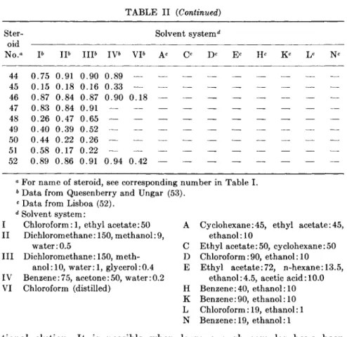
cerned primarily with aldosterone and the 18-hydroxylated compounds, devised six systems for separating 26 steroids, some of which do not use alcohols. Two-dimensional separations can be used, but considerable spot spreading will occur unless conditions of temperature and humidity are well controlled. In Table I are listed the names and trivial names of 52 steroids likely to be found in tissue and blood; and in Table II, the Rf values for 37 A4-3-ketosteroids (and some other steroids likely to be found with adrenocortical steroids) using the chromatographic solvent systems of Lisboa (52), Lisboa and Diczfalusy (55), and Quesenberry and Ungar (53).
P£ron (54) has described some of the difficulties to be encountered with the unstable 18-hydroxy pregnenes. More recently McCarthy et al.
(51) showed that when 18-hydroxy deoxycorticosterone, but not 18- hydroxy corticosterone, was chromatographed in either chloroform- ethanol-water (92:8:0.5) or ethyl acetate-chloroform (90:10), it migrated with a greater Rf when spotted from a mixture than when spotted alone.
However, an irreversible molecular change does not appear to have occurred, since rechromatography of the fast moving 18-hydroxy DOC isolated from a mixture, and of the slower migrating pure sample, showed both to have the same Rf. Although Lisboa (52) does not show R/s for 18-hydroxy pregnenes, he does show a scheme for separating 32 Δ4-3- ketosteroids using a 5-solvent system. Some other workers who have made TLC separations of corticoids are cited in the references (56-69).
In order to obtain many quantitative data on corticosteroids, it is impor
tant to obtain high recovery. Quantitative recovery has been shown for a number of the more clinically important corticoids (61, 62, 65-69).
3 1 6 GRANT G. SLATER TABLE I
SYSTEMATIC AND TRIVIAL NAMES OF VARIOUS STEROIDS
No. Systematic name Trivial name
1 Pr egn-4-en-3-on e —
2 Pregn-4,17(20)-dien-3-one —
3 20j8-Hydroxy-pregn-4-en-3-one —
4 20a-Hydroxy-pregn-4-en-3-one —
5 20j8-Hydroxy-pregn-4-6-dien-3-one —
6 17a,20j8,21-Trihydroxy-pregn-4-en-3-one 20-Dihydro-Sa 7 11 β, 17α, 20/3,21-Tetr ahydroxy-pr egn- E«
4-en-3-one
8 1 la, 17a,20/3-21-Tetrahydroxy-pregn- epi-Ea 4-en-3-one
9 17a,20j3,21-Trihy droxy-pr egn-4-ene- U«
3,11-dione
10 17a,20a,21 -Trihy droxy-pr egn-4-en e- epi-Ua 3,11-dione
11 Pregn-4-ene-3,20-dione Progesterone
12 Pregn-4,6-diene-3,20-dione 4-Dehydroprogesterone 13 Pregn-4,16-diene-3,20-dione 16-Dehydroprogesterone 14 6/3-Hydroxy-pregn-4-ene-3,20-dione 6/3-Hydroxyprogesterone 15 llj8-Hydroxy-pregn-4-ene-3,20-dione 11/3-Hydroxyprogesterone 16 lla-Hydroxy-pregn-4-ene-3,20-dione 1 la-Hydroxyprogesterone 17 16a-Hydroxy-pregn-4-ene-3,20-dione 16a-Hydroxyprogesterone 18 17a-Hydroxy-pregn-4-ene-3,20-dione 17a-Hy droxyprogesteron e 19 21-Hy droxy-pr egn-4-ene-3,20-dione Cortexone, 11-deoxy-
Corticosterone, DOC 2 0 11/3,17a,Dihydroxy-pregn-4-ene- 21-Deoxy Cortisol
3,20-dione
21 ll/3,21-Dihydroxy-pregn-4-ene-3,20-dione Corticosterone, B6 22 lla,21-Dihydroxy-pregn-4-ene-3,20-dione Epicorticosterone 23 16a,21-Dihydroxy-pregn-4-ene-3,20-dione 16a-Hydroxy cortexone 24 17a,21-Dihydroxy-pregn-4-ene-3,20-dione S*
25 19,2 l-Dihydroxy-pregn-4-en e-3,20-dione 19-Hy droxy cortexone 26 11 j8,21-Dihydroxy-18-al-pregn-4-ene- Aldosterone
3,20-dione
27 6/3,11 β, 21 -Trihy droxy-pr egn-4-ene- 6|3-Hydroxy corticosterone 3,20-dione
28 ll/3,17a,21-Trihydroxy-pregn-4-ene- Cortisol, F6 3,20-dione
29 1 la, 17a,21-Trihydroxy-pregn-4-ene- Epicortisol 3,20-dione
30 16a, 17a, 21 -Trihy droxy-pregn-4-ene- 16a-Hydroxy-Sa 3,20-dione
31 17a, 19,21-Trihydroxy-pregn-4-ene- 17a, 19-Dihy droxy cortexone 3,20-dione
32 11 β, 16a, 17a, 21-Tetr a h y d r o x y - p r egn- 1 6 a- H y d r o x y C o r t i s o l 4 - e n e - 3 , 2 0- d i o n e
6. CHEMICAL ASSAY OF ADRENOCORTICOSTEROIDS 317 TABLE I (Continued)
No. Systematic name Trivial name
3 3 Pregn-4-ene-3,11,20-trione 11-Ketoprogesterone
3 4 21-Hydroxy-pregn-4-ene-3,11,20-trione 11-Dehydrocorticosterone, A&
3 5 17a,21-Dihydroxy-pregn-4-ene- Cortisone, E6 3,11,20-trione
36 6/3,17a,2 l-Trihydroxy-pregn-4-ene- 6/3-Hydroxycortisone 3,11,20-trione
37 16α, 17a,21-Trihydroxy-pr egn-4-ene- 16a-Hydroxy cortisone 3,11-20-trione
3 8 170-Hy droxy-androst-4-en-3-one Testosterone
39 11/3,17£-Dihydroxy-androst-4-en-3-one 11/3-Hydroxy testosterone 4 0 11 j8-Hydroxy-androst-4-en e-3,17-dione 11 /3-Hydroxyandrostenedione 41 18,21-Dihydroxy-pregn-4-ene-3,ll,20- 18-Hydroxy-l 1-dehydrocorticos-
trione terone
42 11/3,18,2 l-Trihydroxy-pregn-4-ene-3,20- 18-Hydroxy corticosterone dione
4 3 18,21-Dihydroxy-pregn-4-ene-3,20-dione 18-Hydroxy-l 1-deoxy corticosterone 4 4 18-Hydroxy-pregn-4-ene-3,20-dione 18-Hydroxyprogesterone
4 5 ll/3,20|9,21-Trihydroxy-pregn-4-ene-3,20- 20/3-Hydroxy corticosterone dione
4 6 3/3-Hydroxy-pregn-5-en-20-one Pregnenolone
47 20jS-Hydroxy-pregn-4-ene-3,20-dione 20/3-Hydroxyprogesterone 4 8 3a,21-Dihydroxy-pregnane-ll,20-dione Tetrahydro-A&
49 3α, 11 j3,2 l-Trihydroxy-pregnan-20-one Tetrahydro-Bb 5 0 3a, 17a,21-Trihydroxy-pregnane-l 1,20- Tetrahydro-E6
dione
51 3a, 11/3,17a,21-Tetrahydropregnan-20-one Tetrahydro-F&
52 Cholest-5-en-3jS-ol Cholesterol
α Reichstein.
6 Kendall.
C. Liquid-Solid and Liquid-Liquid Columns
1. Liquid-Solid
Chromatography in the classical sense meant the separation of sub
stances on a column of adsorbing material by developing with the proper solvents, then extruding the adsorbent and extracting the separated substances from the column material. Today, almost all substances separated on columns are allowed to run in the solvent, from the bottom of the column, and are collected in fractions. This method is superior to the older methods because it separates the substances from each other and from the column support. The advent of reliable fraction collection apparatus and the use of gradient changes (70) in solvent composition have also increased the ease of use and the resolution possible by frac-
3 1 8 G R A N T G . S L A T E R
T A B L E II
Rf OF ADRENOCORTICAL AND RELATED STEROIDS USING 1 3 SOLVENT SYSTEMS
Ster- Solvent system4
oid
No." Ρ I P I I P IV& VP Ac Ο Dc Ec Hc Kc Lc Nc 1 0. . 7 0 0 . 5 9 0, .75 0, .83 0 .72 0, .64 0 .68 0 . 5 2 2 0. .69 0 . 5 6 0. .75 0. ,83 0. .70 0, ,62 0, ,68 0 . 5 3 3 0. .55 0 . 2 6 0. .63 0. ,70 0. .56 0, ,38 0. ,50 0 . 2 0 4 0. .50 0 . 2 4 0. .60 0. ,67 0. .54 0. ,36 0. ,47 0 . 1 8
5 0. .53 — 0. .61 0. ,69 0, .57 —
6 0. .15 — 0. .21 0. ,28 0. .30 0. ,13 —
7 0. .07 — 0. .18 0. ,17 0. .21 -
8 0. .03 — 0 .04 0, .09 0, .12 —
9 0. .09 — 0 .16 0, . 2 0 0, .25 —
10 0. .09 — 0, .17 0, .21 0, .25 —
11 0. .93 0 . 9 3 0 . 9 3 0 . 9 6 0 . 3 4 0. .58 0 . 3 6 0, .73 0, .74 0 .65 0 .57 0 .65 0 . 4 1
12 0. .57 — 0 .71 0. .73 0, .65 0, .55 —
13 0. ,59 0 . 3 8 0. .72 0. .74 0, ,65 0 . 4 0
14 0. ,78 0 . 7 6 0 . 8 3 — — 0. ,49 0 . 1 5 0. 55 0. 61 0. 54 0. .32 0. .34 0 . 1 4 15 0. .77 0 . 8 2 0 . 9 0 — — 0. .46 0 . 1 4 0. .57 0. . 6 0 0, .54 0, .32 0, ,39 0 . 1 4 16 0. .31 0 . 0 6 0. .43 0. .43 0, .45 0, . 2 0 0, .23 0 . 0 7 17 0. 50 0 . 7 2 0 . 7 3 — — 0. ,29 — 0. ,47 0. .44 0. .51 0, .23 0 . 0 8 18 0 . 8 9 0 . 9 2 0 . 9 5 — — 0. .56 0 . 2 7 0, . 6 4 0. .72 0, .58 0. . 4 0 0. .49 0 . 2 2 19 0 . 7 2 0 . 9 2 0 . 9 0 0 . 9 2 — 0. .37 0 . 1 3 0. .62 0, .55 0, .54 0, .35 0, .51 0 . 2 0 20 0, .43 0 . 1 1 0. . 4 0 0. .62 0. .50 0. 22 0, .19 0 . 0 7 21 0. .47 0 . 6 0 0 . 7 5 0 . 7 2 — 0. .22 — 0. .31 0. .37 0. .38 0, .16 0, .14 0 . 0 5
2 2 0. 13 — 0. ,19 0. ,22 0. ,26 0, .08 0. .07 —
2 3 0. ,14 — 0. 22 0. 26 0. 31 0. 11 0, .09 —
2 4 0, .70 0 . 6 0 0 . 7 3 — — 0, .38 0 . 0 9 0, .41 0, .57 0, ,49 0. .23 0, .22 0 . 0 8
25 0, .16 — 0, .28 0. .30 0, ,31 0. .16 —
26 0, .20 0 . 4 0 0 . 5 0 0 . 4 9 — 0, .13 — 0. .24 0, .26 0. ,31 0. .16 0 . 0 3
27 0. .12 — 0, .14 0, .26 0. ,24 —
28 0 . 4 5 0 . 2 6 0 . 3 8 0 . 6 5 — 0. .27 — 0, .17 0, .44 0, .34 0. .11 0. .06 —
29 0 . 1 6 — 0 . 1 0 0 .28 0 .25 0, .06 0, .03 —
3 0
31 0 .18 — 0, .16 0 . 3 3 0. .26 0. ,10 —
3 2
33 0, .44 0 . 1 6 0. .70 0 . 6 2 0. 60 0. ,44 0. 60 0 . 2 7 3 4 0, .21 0 . 0 4 0. 46 0. 36 0. 4 0 0. 22 0. ,27 0 . 0 8 35 0 .56 0 . 4 8 0 . 6 4 0 . 7 1 — 0. ,31 0 . 0 5 0 . 3 2 0. .47 0. 4 0 0. 17 0. 13 0 . 0 5
36 0. .17 — 0. .16 0. 31 0. ,28 —
37
3 8 0 . 8 7 0 . 7 9 0 . 8 7 — — —
39 0 .52 0 . 3 8 0 . 4 0 4 0 0 .86 0 . 7 7 0 . 8 8
41 0 . 3 9 0 . 4 4 0 . 5 7 0 . 5 3 — —
42 0 .17 0 . 3 0 0 . 2 3 0 . 3 7 — —
43 0 .85 0 . 8 2 0 . 8 5 0 . 6 1 — —
6. CHEMICAL ASSAY OF ADRENOCORTICOSTEROIDS 319
Ster- Solvent system4
oid
No.° Ρ I P I I P IV6 V P Ac Ο Dc Ec Hc Kc Lc Nc
44 0.75 0.91 0.90 0.89 — 45 0.15 0.18 0.16 0.33 — 46 0.87 0.84 0.87 0.90 0.18 47 0.83 0.84 0.91 — — 48 0.26 0.47 0.65 — — 49 0.40 0.39 0.52 — — 50 0.44 0.22 0.26 — — 51 0.58 0.17 0.22 — — 52 0.89 0.86 0.91 0.94 0.42
a For name of steroid, see corresponding number in Table I.
6 Data from Quesenberry and Ungar (53).
c Data from Lisboa (52).
d Solvent system:
I Chloroform: 1, ethyl acetate:50 A Cyclohexane:45, ethyl acetate:45, II Dichlorom ethane: 150, methanol: 9, ethanol: 10
water: 0.5 C Ethyl acetate: 50, cy clohexane: 50 III Dichlorom ethane: 150, meth- D Chloroform: 90, ethanol: 10
anol: 10, water: 1, glycerol:0.4 Ε Ethyl acetate:72, n-hexane: 13.5, IV Benzene:75, acetone:50, water:0.2 ethanol:4.5, acetic acid: 10.0 VI Chloroform (distilled) Η Benzene:40, ethanol: 10
Κ Benzene:90, ethanol: 10 L Chloroform: 19, ethanol: 1 Ν Benzene: 19, ethanol:1
tional elution. It is possible when large enough samples have been separated, to measure the amount of substance in each fraction by (a) weighing the sample after removal of the solvent; (b) color reactions;
(c) spectrophotometric methods; (d) radioactivity measurements; and (e) thin-layer chromatography. Liquid-solid chromatography is most frequently used when large amounts are to be separated, since the convenience of the thin-layer method has brought TLC into common use when small quantities are to be separated. The solids used for adsorbents with steroids are alumina, magnesium silicate, silica gel, magnesium carbonate, aluminum silicate, and charcoal. Of these, silica gel is the most useful by far for adrenocorticosteroid separations.
Many of the older separations of the adrenocortical steroids were done using adsorption columns, since they have high capacity. However, although the use of alumina may continue for the less polar steroids, for the more polar adrenocortical steroids its use will decline since structural alterations can occur to the corticosteroids on such columns. Even silica gel has been reported to cause changes when used with a very low water
TABLE II (Continued)
320 GRANT G. SLATER
content (71). Silica gel, silicic acid, and silica are terms used interchange
ably for a powdered solid of the general formula S i 02 X H20 . The water content can vary considerably depending on the mode of preparation.
It may have a mono- or multimolecular layer of absorbed water, "free water," which can be reversibly removed by heating at about 100°C. In order to use the silica gel with a known amount of water, it is completely dried, usually for 3 hours or longer at 120°C, then the desired amount of water is added and the powder is mixed and sealed in a container over
night so that it is uniform in water content before use.
2. Liquids-Liquid
Silica gel with a "free water" content above 17% is thought to separate substances using liquid-liquid partition (70) and to separate corticosteroids with less tailing than when it is used in an adsorbent system with water below 17% (71, 72). However, silica gel is most likely producing its effects by both partition and adsorption. In addition to silica gel, a number of other substances are used to hold the liquid on the column, such as Hyflow Supercel, Celite 535 (kieselguhr), cellulose, and aluminum silicate. While the adsorption column works best with less polar compounds, such as the acetate derivatives of corticosteroids, the partition systems work better on the unesterified, more polar, corti
costeroids. A number of later partition-chromatographic procedures (73-76) are derived from that of Butt et al. (77) who used kieselguhr as a supporting medium for aqueous alcohol as the stationary phase and hexane-benzene-chloroform-ethyl acetate as the mobile phase.
A completely automated system has been designed by Haines and Karnemaat (78) using ethylene glycol on silica gel columns and cyclo- hexane-CH2Cl2 for the solvent. Gradient elution has been used by Heftmann et al. (79, 80) with petroleum ether-CH2Cl2 on a silica gel column containing added water.
Reversed-phase partition systems have also been used (81), but they suffer from the disadvantage that the eluent is largely aqueous and difficult to evaporate. Liquid-solid and liquid-liquid chromatography were used for years before the advent of gas chromatography, which has replaced them for numerous separations of volatile organic compounds.
However, there are new developments which may bring back the use of liquid-liquid columns for the separation of the adrenal corticoids. Recently a detector has been developed (82) for liquid-liquid columns which is a modification of the system used on gas chromatographs. With this detector, as little as 0.01 μξ can be measured as rapidly as the substance is eluted from the column. The eluent is evaporated as it comes from the column onto a platinum chain; the solid remaining is passed through the
6. CHEMICAL ASSAY OF ADRENOCORTICOSTEROIDS 321 flame of a hydrogen flame ionization detector, where the change in electric current through the flame is amplified and then recorded on a chart recorder. The chain can handle solvent flows from about 0.24 to 4.8 ml/hour.
D. Gas-Liquid Columns
Gas chromatography (GC) can be gas-solid or gas-liquid but essen
tially all studies with steroids have been with the gas-liquid system, and that is implied in the further use of this term. GC is a type of partition chromatography where the stationary phase is a liquid film spread on a solid support in a column, and the moving phase is a carrier gas passing over the surface of the liquid film under controlled conditions. The chromatographic column is controlled at a temperature where steroids in a mixture are partitioned between the liquid and gas phases. The order in which components elute from the column depends on their individual partition coefficients as expected from a consideration of the basic principles of partition chromatography. Bush (33) has discussed these principles as they concern steroid separations on paper chromatography, and these apply also to GC.
With thin-film columns (which now appear to be necessary for corti
coids) only small samples can be used, and thus very sensitive detectors must be employed. Ionization detectors and more recently hydrogen flame detectors are most usually used. The columns are generally operated isothermally, but temperature programming has been used where mix
tures contain substances of widely different vapor pressure.
The following parameters must be known about the column: kind and size of column (usually glass 3-5 mm diameter and 3-12 feet long), type and size of support, and amount and kind of liquid phase. Furthermore, certain conditions relative to operation of the column must be known;
these include operational temperature (usually 200-225°C), type of gas and amount of pressure (argon or nitrogen usually at 10-30 psi), gas flow rate, sensitivity settings of the electronic detector system, sample size (on the order of 0.1-10 μΐ or 1-20 μg), and chart speed (usually }4 to }4 inch per minute).
Gas chromatography has certain advantages that make it a desirable analytical method. Its greatest value is that steroids can be quantitated as well as separated; furthermore, it has great resolving power and with the proper equipment, steroids can be isolated and collected. Also steroids can be resolved in microgram and submicrogram quantities in a short period of time. The system is especially valuable in separating complex mixtures of biological origin and may be eventually quite valuable in nutritional research work. The combination of small sample size, speed,
322 GRANT G. SLATER
and ability to do multiple determinations on one run allows easy changes in experimental conditions and could prove to be one of its greatest assets. The general methods used in gas chromatography and biochemical applications are well known (83, 84), and reviews of the application of GC to the separation and determination of steroids have been published (85, 34). Since the practical demonstration of the separation of steroids using gas chromatography by VandenHeuvel et al. in 1960, it has become evident that separation of the adrenocortical hormones would be difficult because of their liability to rearrangement and decomposition at high temperature (86, 87). Only at rather high temperature do the steroids have sufficient vapor pressure to be chromatographed. In order to reduce decomposition at high temperature, VandenHeuvel introduced the use of relatively thin-film coating liquids with high thermostability (86).
Since metal appears to catalyze decomposition of steroids, all-glass columns are generally used. To the present time improved liquid phases and support materials continue to be developed although the basic approach remains the same (88, 89). But new techniques continue to be developed and used on steroid separations; these include (a) the use of electron-capture detectors for selective analysis of acetates and other derivatives (90-92); (b) the application of isotopic labeling to quantitative analysis (93); and (c) mating of mass spectrometry with GC (94).
The adrenocorticosteroids cannot be separated as satisfactorily as the free steroids yet, and considerable effort has gone into an examination of derivatives. Ideally such a derivative must be (a) volatile at reasonable temperatures; (b) preparable in near quantitative yield; (c) stable to GC; and (d) less polar than parent corticoid. Some derivatives which have use in the separation of steroid mixtures are the trimethylsilyl ethers, acetyl derivatives, oxidized derivatives with H I 04, and methyl esters.
Brooks (95) has recently made a study of acetylated corticosteroids and related 20 oxopregnane derivatives and has suggested that consider
able improvement can still be made to reduce the loss of corticosteroids during gas chromatography. He stated that "the deficiency appears to be in the chromatographic conditions rather than in the derivatives, as we have found that cortisone acetate and 3£,17a,21-triacetoxy-5a- pregnane-ll,20-dione are both recoverable in excellent yield by vacuum sublimation."
The ketosteroids of urine are partly derived from adrenocortical steroids, and in this area considerable progress has been made using GC.
As an example, a recent (88) separation on a new liquid phase, a modified Carbowax (96), was made of the trimethylsilyl derivatives of 9 urinary 17-ketosteroids and progesterone derivatives. Cholesterol was used as an internal standard, and it was found that there was a linear response for androsterone from about 5 to as little as 0.01 μg. The study used 6.8 foot
6. CHEMICAL ASSAY OF ADRENOCORTICOSTEROIDS 323 by }£ inch o.d. glass column packed with 3 % liquid phase on 1 0% 2 0 mesh support. Temperatures were: column 233°C, detector 235°C, injector 275°C. Helium was used as the gas at 19 ml/min with a hydrogen flame detector. Sample size was 1 μΐ.
Although the free corticosteroids cannot be chromatographed without considerable loss, their oxidized and methylated derivatives can be measured. The "Quantitative GC of Rat Adrenal Gland Steroids" by Kittinger (97) is a good example of the use of these derivatives.
Kittinger has shown that where experimental studies are made on rats and mice which produce little or no 17-hydroxylated steroids it is possible to determine the following corticosteroids in the adrenal gland: proges
terone, deoxycorticosterone, Ιΐβ-hydroxyandrostenedione, dehydrocorti- costerone, 110-hydroxyprogesterone, corticosterone, 18-hy droxy deoxy
corticosterone, and aldosterone. This separation and quantitation would seem to have considerable application in experimental nutrition studies.
The method used by Kittinger treats the steroids obtained from an extraction of adrenal glands with HIO4, in aqueous dioxane, to quantita
tively convert α-ketolic steroids to the corresponding etiocholenic acids.
Aldosterone and 18-hydroxy deoxycorticosterone (180HDOC) form internal esters. Gas chromatographic analysis of the product prior to esterification determines progesterone, Ιΐβ-hydroxyprogesterone, 18β- hydroxyandrostenedione, 180HDOC, and aldosterone. Esterification of the same product with diazomethane yields volatile esters of the free etiocholenic acids in the mixture. Rechromatography then determines the previously mentioned compounds plus deoxycorticosterone, corticoste
rone, and dehydrocorticosterone as methyl esters of the corresponding etiocholenic acids.
Kittinger determined the amount of steroid which reacted with blue tetrazolium and used an amount equal to 10 to 100 μg of blue tetrazolium- positive material, equivalent to corticosterone as a standard, for the determination. About 5μg of mixture is needed for each column, so that, allowing for dilutions, a determination can be made easily on a 20-M g sample. Furthermore Kittinger has worked out the program so that the data can be calculated using a computer.
A new book on the "Gas Chromatography of Steroids in Biological Fluids" by M. Lipsett, has recently been published (98).
I V . FLUORESCENCE ASSAY
A. General
The determination of adrenocortical steroids by fluorescent methods has shown great progress in recent years, due largely to improvements in instrumentation. Lamps which produce excitation, which give out a
324 GRANT G. SLATER
broad-spectrum high-intensity ultraviolet light, have contributed greatly to the advances of the better instruments. It is now possible to scan, with a high degree of resolution, both the wavelengths of excitation as well as the wavelengths of fluorescence, and these spectra can serve for identifica- tion purposes. A considerable range of instruments is available to the biochemist from the simple filter type, which can be quite sensitive when equipped with a photomultiplier detection tube, to the automatically corrected spectrophotometer for the most accurate spectra analysis. The instrument selected depends on a number of factors, such as the sensi- tivity, which is related to the kind and amount of corticoids to be deter- mined, and the type of use, where the investigator may be doing clinical measurements or a more definitive research study. For a discussion of instruments and also the fundamentals of fluorescence assays in biological systems see Udenfriend (99). The book is quite complete as of 1961, and most of the instruments described therein are still being used and sold;
in addition, for some of the instruments accessories have been made that have improved their performance.
One of the great advantages of the fluorescent assay is the large number of substances which can be determined; on the other hand, it increases the difficulty of purification of test materials, especially when determining microgram quantities of steroids. The reagents must have a special type of purity, and extreme care must be used to eliminate contaminating substances, which may be of two general types: those which produce excess fluorescence, and those which produce decreased fluorescence in the test solution. Contamination may arise from the solvents but can be reduced greatly by known methods (100). It may arise from many sources, e.g., rubber particles, dust, stopcock grease, cleaning agents, and filter paper (101).
In addition to contamination, losses may occur because of special problems involved with dilute solutions, e.g., adsorption onto surfaces.
Brodie et al. (102) showed that small quantities of quinine were adsorbed on glass from organic solvents; thus, in a concentration of 1 Mg/ml, quinine could be completely removed from a benzene solution by glass surfaces. That quinine could be removed from the glass by dilute H2S 04
showed that it was, indeed, adsorbed. This loss from dilute solutions is a common phenomenon; however, this effect decreases with an increase in polarity of the solvents. Bird (103) states that new glass surfaces may be very adsorptive and that such glassware should be put through cleaning solution or heated in strong acid or alkali before use. This also suggests that special care should be taken to ensure that clean cuvettes are used, since any adsorbed substances on the inside or outside of the cuvettes might cause error. Since the quantities of steroids adsorbed on glass
6. CHEMICAL ASSAY OF ADRENOCORTICOSTEROIDS 325 surfaces may be very small, a determination of recovery in an analytical method may give appreciably different results when a large amount is run through this procedure as compared to a quantity near the amount that may be adsorbed. Thus, if 0.5 μg was adsorbed in a test with only 1 μg of total steroid, a 50% loss would occur, whereas had 50 μg been used for the test, a 0.5 μg loss would only be 1%. Therefore, if any adsorp
tion loss is anticipated in a recovery experiment, quantities approxi
mating those expected in test solutions should be used.
In addition to glass surfaces, there is the problem of separating steroids from proteins after precipitation. In all methods for the deter
mination of steroids in plasma and tissue, the adrenocorticosteroids must be separated from proteinaceous substances. The use of alcohol with chloroform or methylene chloride helps to prevent adsorption of steroids onto these proteins and is commonly used in extraction procedures.
In addition to adsorption, losses may also occur by oxidation. In extremely dilute solutions the loss of only a very small quantity can be quite significant and occurs so readily that solutions should be prepared fresh frequently. While peroxides in ether may easily cause oxidation and lowered recoveries, they can be removed with a ferrous sulfate wash.
Another method by which steroids may be lost is through photo- decomposition. It is known that the steroids are decomposed by ultra
violet light and when they are exposed in very dilute concentrations, photodecomposition may be rapid. Precautions can be taken to prevent the decomposition, e.g., working in darkened rooms, wrapping the sample tubes with aluminum foil, or using low actinic glass containers which protect corticoids from destruction by ultraviolet light. Since photo- decomposition may occur even while a sample is being read in the fluorom- eter, and since the decomposition rates of individual steroid derivatives have not been carefully studied, readings should be made rapidly unless it is determined that the samples being measured do not decompose.
Samples can be tested easily by determining whether the fluorometric readings decrease with time. With samples that decompose rapidly, a series of readings can be obtained and a plot of readings versus time made; extrapolation to zero time will then give the corrected reading.
A further source of error in fluorescent measurement is the lack of temperature control. It is fairly well known that the fluorescence decreases with an increase of temperature. This is due to a number of factors;
one of the most important is that the viscosity of the solvent is decreased.
With a new fluorometer it is desirable to determine whether the temper
ature of the cuvette compartment shows an increase with time. Normally, the cuvette compartment temperature is a little higher than room temper
ature, a few degrees is generally acceptable. Actually, the best results