III./3.3.2. Neurodegeneration with brain iron accumulation
Introduction
Neurodegeneration with brain iron accumulation (NBIA) refers to a group of neurogenetic diseases that are characterized by movement disorder, intellectual decline, and a typical MRI appearance. NBIA disorders exhibit low signal in the globus pallidus and often the substantia nigra on T2-weighted images (together with an isointense appearance on T1-weighted images), consistent with iron deposition. Pantothenate kinase-associated neurodegeneration (PKAN) accounts for 50-70% of cases of NBIA, but neuroferritinopathy (NFT), aceruloplasminemia (AC), fatty acid hydroxylase-associated
neurodegeneration (FAHN), Kufor-Rakeb syndrome, and Woodhouse-Sakati syndrome also belong to this group.
Keywords: NBIA, iron deposition, basal ganglia, PKAN, PANK2, Hallervorden-Spatz
Hallervorden-Spatz syndrome was originally described in 1922 by two German neuropathologists, Julius Hallervorden and Hugo Spatz. For decades, most of the patients with radiographic or pathologic evidence of iron
deposition in the basal ganglia received this diagnosis. Later, the first subtype of Hallervorden-Spatz syndrome, PANK2 mutation was identified, and this subgroup was designated as pantothenate kinase-associated neurodegeneration (PKAN). In 2002, a new nomenclature was proposed and the name NBIA was introduced to replace Hallervorden-Spatz syndrome (Hallervorden and Spatz were involved in the active euthanasia of “mental defectives” during World War II).
Pantothenate kinase-associated neurodegeneration (PKAN)
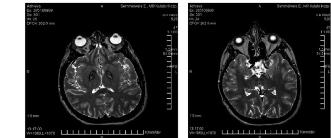
PKAN is caused by the mutation of the PANK2 gene. Its main symptoms are severe generalized dystonia and intellectual decline. There are two forms of PKAN, classic PKAN is characterized by early onset (< 6 years) and rapid progression, and patients may have retitinis pigmentosa. Atypical PKAN comprises a more heterogeneous group, which has a later onset and slower progression. Its typical symptoms are severe dystonia, speech difficulty (palilalia, dysarthria), but parkinsonian and psychiatric symptoms are also common. In PKAN, the typical MRI abnormality on T2-weighted images is the “eye of the tiger sign” (Fig. 1) bilaterally in the pallidum, which is a central signal hyperintensity with a surrounding hypointense region. This sign may be associated with PANK2 mutation. Pharmacologic and surgical
interventions are aimed at symptomatic relief. Clinical trials of the chelating agent deferiprone are currently underway for PKAN. According to clinical experience, deep brain stimulation of bilateral GPi can decrease generalized dystonia with 30% in PKAN (Chapter N_III_3_4).
Fig. 1:Typical T2-weighted MRI changes of PKAN bilaterally in the pallidum, the “eye of the tiger sign”, central signal hyperintensity with a surrounding hypointense region.
Neuroferritinopathy (NFT)
The only autosomal dominant form of NBIA, NFT is caused by mutations in the ferritin light chain gene, FTL. NFT leads to a complex neurological
phenotype characterized by parkinsonism, choreoathetosis, dystonia, spasticity, ataxia, dementia, and autonomic features, and typically occurring in adults. In NFT, iron deposition may be observed in the caudate, putamen, globus pallidus, thalamus, and the dentate nucleus.
Aceruloplasminemia (ACP)
ACP is caused by mutations in the CP gene. It has an early onset.
Ceruloplasmin has a very low level or it is undetectable in the blood, depending on whether a heterozygous or a homozygous mutation of the CP gene is present. Ceruloplasmin acts as a catalyst in iron oxidation, which is necessary for the binding of iron to transferrin. Copper metabolism is often normal. Clinical symptoms may include ataxia, chorea, parkinsonism, dysarthria, and progressive dementia, as well as retinopathy, and anemia.
Neuroimaging typically demonstrates widespread iron deposition in the basal ganglia, and also in the liver, pancreas, and other visceral organs, with concurrent diabetes mellitus observed in some patients.
Reference
Gregory A, Hayflick SJ. Neurodegeneration with brain iron accumulation.
Folia Neuropathol. 2005;43(4):286-296.