Role of Lysophosphatidic Acid in the Regulation of Vascular Tone
PhD thesis
Éva Ruisanchez MD
Doctoral School of Basic Medicine Semmelweis University
Supervisor: Zoltán Benyó MD, D.Sc.
Official rewievers: Violetta Kékesi, Ph.D.
Tóth Attila MD, D.Sc.
Head of the Final Examination Committee: Viktor Bérczi MD, D.Sc.
Members of the Final Examination Committee:
Tamás Radovits MD. Ph.D.
Attila Mohácsi MD, Ph.D.
Budapest 2017
1 1. INTRODUCTION
Lysophosphatidic acid (LPA) is a family of lipid mediators composed of a glycerol backbone, a phosphate headgroup and a fatty acid with various chain length and saturation. LPA is ubiquitously present in living organisms, and its effects are mainly mediated by G-protein-coupled receptors called LPA1-6, but it can also activate the nuclear receptor PPARγ. LPA plays important regulatory roles in many physiological and pathophysiological processes including neural development, inflammation, reproductive functions, embryo implantation, bone metabolism and tumor progression. LPA is synthesized by an ectoenzyme called lysophospholipase D or autotaxin (ATX) and is metabolized by lipid phophate phosphatases.
LPA is produced and degraded in the vascular system and has diverse effects on almost all types of vascular and blood cells. The plasma concentration of LPA is in the low to medium nanomolar range. However, upon platelet activation it can rise to several micromolar concentrations. Its role is well known in the vascular system for its vasculo-, angio-, athero- and thrombogenic effects, although its involvement in the regulation of vascular tone is obscure.
Early research on the effects of LPA implicated its potential role in the regulation of the vascular tone. In 1978 Tokumura et al.
described that intravenous application of LPA induces a transient hypertension in rats, independently of sympathetic activation. Later the same group found that LPA can also decrease blood pressure, depending on the animal species investigated. It is still unknown whether this controversial blood pressure response is due to a direct effect of LPA on the vascular tone or not. Later on, Tigyi and
2
colleagues published that LPA evokes a dose dependent constriction of the pial arteries of newborn piglets. A similar response was described by Caner et al. in rabbit basilar arteries. According to literature data, LPA may have a role in the regulation of mechano- transduction of the vessel wall. In the presence of shear stress LPA stimulated endothelial thromboxane release and subsequent changes in the vascular reactivity. In another study LPA was found to modulate myogenic tone of small arteries by the production of free- radicals.
There are several studies indicating the role of LPA in the modulation of endothelial nitric oxide (NO) production, but these data are controversial. According to Chen et al. LPA causes endothelial dysfunction via the inhibition of the endothelial nitric oxide synthase (eNOS) by the production of free radicals.
Interestingly, others found that LPA activates eNOS in different endothelial cell cultures and eNOS phosphorylation by phosphoinositol 3 kinase (PI3K) was implicated in this process. By the time our research was started, it was unknown whether LPA can affect the vascular endothelial NO production in situ, or in vivo.
3 2. OBJECTIVES
Although LPA has diverse roles in the cardiovascular regulation both in physiological and pathophysiological conditions, our knowledge about the effects of LPA on the vascular tone are obscure. Literary data suggest that LPA may serve as a direct vasoactive agent and/or modulate the effects of different biomechanical and pharmacological stimuli. While the potential vasoconstrictive action of LPA was partly described by the groups of Tigyi and Caner by the time we started our research, we suspected that LPA could also evoke or modulate vasodilation as it was able to affect NO production.
Therefore we wanted to answer the following questions:
Does LPA have any endothelium-dependent vasoactive effect? In particular,
is LPA able to affect the vascular tone through the modulation of NO production? If so,
which LPA receptors and signal transduction pathways are involved in the effect?
Are LPA receptors and autotaxin expressed in vascular endothelial cells?
4
3. METHODS
3.1. Animals
The experiments have been performed in vessels of wild type (WT) and genetically modified adult male mice with C57Bl/6J background. The genotypes of the tested mice were the following:
cyclooxygenase 1 knockout (COX1 KO), phospholipase Cε knockout (PLCε KO), eNOS KO, LPA1 and LPA2 receptor KO.
C57Bl/6J mice served as control and referred as WT. All experiments have been carried out according to the guidelines of the Institutional Animal Care and Use Committees of the Semmelweis University and the University of Tennessee Health Science Center.
3.2. Preparation of vessels
Mice were transcardially perfused with 10 mL heparinized (10 IU/mL) Krebs solution under deep ether or isoflurane anesthesia.
Thoracic and abdominal aorta (TA and AA) were removed and cleaned of fat and connective tissue under a dissection microscope, and immersed in a Krebs solution of the following composition (mmol/L): 119 NaCl, 4.7 KCl, 1.2 KH2PO4, 2.5 CaCl2·2H2O, 1.2 MgSO4·7H2O, 20 NaHCO3, 0.03 EDTA, and 10 glucose at room temperature and pH 7.4. Vessels were cut into ~3 mm-long segments and mounted on stainless steel vessel holders (200 µm in diameter) in a conventional myograph setup. Special care was taken to preserve the endothelium. However in some experiments, the endothelium was mechanically removed by gently moving a surgical thread through the lumen. The successful removal of the endothelium was verified by the lack of acetylcholine-induced vasorelaxation.
5 3.3. Myography
Wells of the myographs were filled with 8 mL Krebs solution aerated with carbogen. The vessels were allowed a 30-min resting period, during which the bath solution was warmed up to 37 °C and the passive tension was adjusted (TA=15 mN, AA=10 mN).
Subsequently, the tissues were exposed to 124 mmol/L K+
Krebs solution (K+-Krebs) for 1 min, followed by several washes with normal Krebs solution. According to a contraction evoked by 10 μmol/L phenylephrine (PE) followed by administration of 0.1 μmol/L acetylcholine (ACh), nonreacting vessels were excluded from further investigation.
After repeated washing, during which the vascular tension returned to the resting level, the segments were exposed to K+-Krebs solution for 3 min in order to elicit a reference maximal contraction.
Subsequently, after a 30-min washout, increasing concentrations of PE (0.1 nmol/L to 10 μmol/L) and ACh (1 nmol/L to 10 μmol/L) were administered to determine the reactivity of the smooth muscle and the endothelium, respectively.
The effect of LPA species with different fatty acid chain length and saturation on vascular tone was tested either with or without precontraction. The level of precontraction was set to 70- 90% of the reference contraction by an appropriate concentration of PE. Oleoil-LPA was used to test the signal transduction pathways of LPA-induced vasorelaxation, as it appeared to have the highest vasorelaxant activity.
In search of the signal transduction pathways of LPA-induced vasorelaxation, different enzyme inhibitors and receptor antagonists were administered. In case of wortmannin, MK-2206, Ki16425 and AM095, inhibitors were added to the organ bath 30 min prior to LPA exposition. In order to inhibit the PLC in an endothelium-specific
6
manner, the inhibitors were used in the perfusion solution in the appropriate concentration. Perfusion was slowed down to ensure a long contact with the endothelial cells. Control experiments were performed similarly: U73343 – the inactive analogue of U73122 – and their vehicle dimethyl sulfoxide (DMSO), as well as ethanol – the vehicle of edelfosine – were also added to the perfusion fluid.
The time between perfusion with inhibitors and the testing of LPA was reduced by skipping the first exposition with PE and ACh. The endothelium-selectivity of the inhibition was confirmed by a normal PE and diminished ACh dose-response. Vasorelaxation to NO-donor sodium nitroprusside (SNP) was tested to confirm that the vascular smooth muscle had remained reactive to NO.
3.4. Expression analysis of endothelial LPA receptors and autotaxin
Endothelial cells were freshly isolated from murine aorta.
After mice were anesthetized and perfused, the lumen of the aorta was flushed with DMEM and filled with DMEM containing 2 mg/ml collagenase type II. The aorta was ligated with silk threads on both ends, dissected out, immersed in DMEM and incubated at 37 °C for 45 min. Endothelial cells were removed from the lumen of the aorta by flushing with 3 ml of DMEM. The cell suspension was divided into two tubes and centrifuged at 1200 rpm for 5 min. The pellet of the first tube was suspended in 300 μl RNAlater and stored at 4 °C until RNA isolation. The pellet of the other tube was resuspended in DMEM containing 20% FBS, 100 U/ml penicillin and 100 μg/ml streptomycin, plated on coverslips, and incubated overnight at 37 °C until cells were attached enabling immunocytochemical analysis for evaluation of the relative number of endothelial cells obtained (i.e.
“purity” of the endothelial cell culture).
7
Claudin-5-immunocytochemistry has been performed for the identification of endothelial cells and DAPI staining for counting of the total number of cells. Coverslips were washed four times with PBS and the cells were fixed in 4% paraformaldehyde for 20 min at room temperature. After washing with 100 mM glycine-PBS solution for 10 min at room temperature, and permeabilizing with 0.1%
Triton X-100 for 20 min, cells were blocked with 3% BSA in PBS for 1 hr. Next, cells were incubated with the mouse anti-mouse claudin-5 monoclonal antibody (1:200 dilution) in 1% BSA-PBS solution at room temperature for 1 hr. After washing with PBS, cells were incubated for 1 hr at room temperature with Alexa 568- conjugated anti-mouse IgG antibody (1:500 dilution). Nucleus staining was performed with Alexa 488-conjugated DAPI (1:1000 dilution) for 10 minutes at room temperature. Endothelial vs. total cell numbers were counted with a fluorescence microscope.
Expression analysis was performed only if the immunocytochemistry-verified endothelial cell content of the sample was >98%. RNA was isolated from the cells with RNeasy micro kit, and RNA concentration and quality was assessed with Nanodrop. 50 ng total RNA was converted to SPIA amplified cDNA. The amplified SPIA cDNA was purified with QIAquick PCR.
Assessment of mRNA expression was performed by quantitative real-time PCR using 4 ng of SPIA amplified cDNA. PCR reactions were carried out in triplicate using RT2 SYBR Green/ROX PCR master mix in 25 μl final volume, in an Applied Biosystems 7300 Real-Time PCR System. Cycle parameters were: one initial incubation step at 50 °C for 2 min, one denaturation step at 95 °C for 10 min and 40 cycles of denaturation at 95 °C for 10 s, annealing and elongation at 60 °C. Relative gene expression of each mRNA to GAPDH was determined using the dCt method.
8 3.5. Statistics
Changes of the vascular tension were recorded with the MP100 System and analyzed with the AcqKnowledge 3.7.3 software of BIOPAC System Inc. Vasoconstrictions were normalized to the reference contraction induced by 124 mM K+, whereas vasorelaxations were expressed as percent of the precontraction produced by PE. All data are presented as mean ± SE and n indicates the number of vessels tested. Statistical analysis was performed using Student’s unpaired t-test when comparing two variables. All other comparisons were made between the different experimental groups by ANOVA followed by the appropriate post-hoc test. A p value of less than 0.05 was considered to be statistically significant.
3.6. Reagents
LPA (18:1) and VPC31143 were purchased from Avanti Polar Lipids and dissolved in saline immediately prior to administration.
Stock solutions of LPA and VPC31143 were 100-times more concentrated as compared to their final concentrations in the organ baths. AM095 was synthesized as described previously. Ki16425, edelfosine and U73122 were purchased from the Cayman Chemical Company, whereas insulin, U73343 and wortmannin were from Tocris Bioscience, and MK-2206 was from Selleckchem. All other drugs and chemicals used in the present study were purchased from Sigma-Aldrich. If drugs were dissolved in organic solvents (U73122, U73343, Ki16425, wortmannin and MK-2206 in DMSO, whereas AM095 and edelfosine in ethanol, respectively) the final concentration of the solvent was ≤0.1% in vitro and 1% during transcardial perfusion. In these experiments vehicle treatment was used as control. In case of myograph experiments all concentrations are expressed as the final concentration of each drug in the organ bath.
9 4. RESULTS
4.1. Lysophosphatidic acid induces endothelium-dependent vasorelaxation
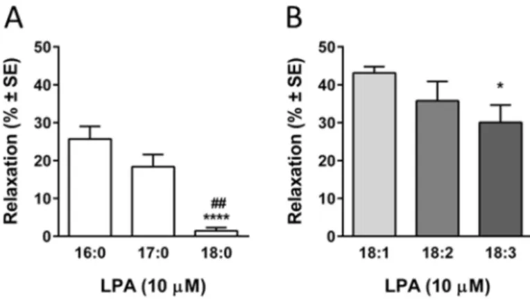
Thoracic aorta segments with intact endothelium were used to test the vasodilator activity of LPA. LPA species with fatty acyl chain of different length and saturation were applied in 10 μM concentration after a stable precontraction evoked by phenylephrine.
Saturated palmitoyl (16:0) and heptadekanoyl (17:0) LPA evoked a fast, transient relaxation but stearoyl LPA (18:0) had no vasorelaxant effect.
Evaluation of the maximal vasorelaxant effect showed that vasorelaxant activity is inversely proportional with the acyl chain length in case of saturated LPA species (Figure 1 Panel A:
26,3±3,6%, 18,6±3,8% and 1,4±0,8%, ##p<0.01 vs. 17:0 LPA,
****p<0.0001 vs. 16:0 LPA, n=16, 10, 10; one way ANOVA, followed by Tukey’s post-hoc test).
Figure 1 Vasorelaxing effect of LPA is acyl-chain dependent
10
Since LPAs with 18 carbon long acyl chain is very common in saturated as well as in unsaturated forms, oleoyl (18:1), linoleoyl (18:2) and linolenoyl LPA (18:3) were also investigated. Mono unsaturated oleoil- as well as polyunsaturated linoleoyl- and linolenoyl LPA evoked fast, transient vasorelaxation. Evaluation of the maximal vasorelaxant effect showed that relaxation was parallel to the number of unsaturated bonds in the acyl chain (Figure 1 Panel B: 43.1±1.6%, 37±6.7% and 30.2±6.4%, *p<0.05 vs. 18:1 LPA, n=59, 10, 11; one way ANOVA, followed by Tukey’s post-hoc test).
Interestingly, only polyunsaturated linoleoyl- and linolenoyl LPA evoked biphasic effect on vascular tone with a transient relaxation and a subsequent constriction.
Oleoil LPA was chosen to further investigate the vasorelaxation, as it appeared to have the most potent vasorelaxing effect. The maximum of the vasorelaxation (Emax) was 53.9%
(compared to precontraction), while half-maximal effective concentration (EC50) was 400 nM. This EC50 value is in the range of physiological plasma and serum concentration of LPA both in human and experimental animals.
4.2. Vasorelaxant effects of LPA in aortic rings depend on presence of intact endothelium and genetic deletion of eNOS
Next, we sought to identify the mediator(s) of the LPA- induced vasorelaxation. We have hypothesized a potential role of endothelium-derived vasoactive compounds, specifically NO and prostanoids, in mediating the LPA-induced vasorelaxation. We tested this hypothesis in two ways by using de-endothelialized WT vessels and vessels isolated from eNOS or cyclooxygenase-1 (COX1) KO mice.
11 Figure 2 Vasorelaxing effect of LPA is endothelium dependent
Removal of the endothelium abolished the vasorelaxation elicited by LPA (Figure 2
****p<0.0001 vs. „WT E+”, n=59, 21, 27, 8; one way ANOVA, followed by Dunnett’s post-hoc test).
Interestingly, in the absence of the endothelium LPA increased vascular tone sugges ting that endothelium- derived signals and also smooth muscle-dependent vasoconstrictor signals were involved in the vasoactive effect. In addition, absence of eNOS but not that of COX1 abolished LPA-induced vasorelaxation.
4.3. Identification of the LPA receptor(s) mediating LPA- induced vasorelaxation
Since the endothelium was found to mediate LPA-induced vasorelaxation we first aimed to analyze the expression of LPA receptors and ATX in mouse aortic endothelial cells (MAEC) using quantitative real-time PCR. From the EDG-like subtypes of LPA receptors, both LPA1 and
LPA2, but no LPA3
transcripts were detectable.
In addition, expression of the non-EDG LPA receptor subtypes LPA4 and LPA5 as well as that of ATX were also found in MAEC (Figure
3). Figure 3 Endothelial expression of LPA
receptor mRNA and ATX in the mouse aorta
12
Our next aim was to identify the LPA receptor subtype(s) that mediate(s) eNOS-dependent vasorelaxation. We used a combination of pharmacological and genetic manipulation of the LPA receptors.
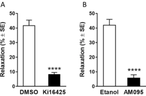
The LPA1/3-receptor antagonist, Ki16425, significantly reduced LPA-induced vasorelaxation (Figure 3 Panel A, n=7, 18). The selective LPA1-receptor blocker AM095 also reduced LPA-induced vasorelaxation by approximately 90% at 10 µM as compared to vehicle control (Figure 3 Panel B, n=7, 8; on both panels
****p<0.0001 vs. vehicle treated vessels, Student’s unpaired t-test) without influencing the eNOS-mediated vasorelaxation elicited by acetylcholine.
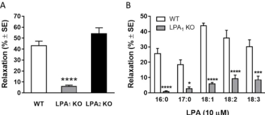
Figure 4 LPA induced vasorelaxation is mediated by LPA1 receptor We also tested vessels isolated from LPA1- and LPA2-KO mice. Aortic rings isolated from LPA1-KO failed to show relaxation in response to LPA, whereas their reactivity to acetylcholine was not altered. On the other hand, vessels isolated from LPA2-KO animals showed unaltered relaxation to both LPA and acetylcholine (Figure 5 Panel A, n=19, 15, 4; ****p<0.0001 vs. „WT”, one way ANOVA, followed by Dunnett’s post hoc test).
13
Figure 5 LPA induced vasorelaxation is mediated by LPA1 receptor
Thereafter, the vasorelaxant effects of the other LPA species have been tested on LPA1 KO TA segments. The relaxing responses to the different LPA species were markedly reduced in vessels of LPA1 deficient mice (Figure 5 Panel B, n=19, 13, 52, 10, 11 in WT and n=6, 6, 20, 7, 9 in LPA1 KO; *p<0.05, ***p<0.001,
****p<0.0001 vs. „WT”, two way ANOVA, followed by Sidak’s post hoc test, stearoyl LPA was not tested due to the lack of its vasorelaxant activity).
4.4. Identification of the intracellular signaling pathway(s) of LPA-induced vasorelaxation
What signals couple LPA1 to eNOS activation in the endothelium? To address this question, we examined first the potential role of phosphoinositide 3 kinase (PI3K) and protein kinase B/Akt signaling pathway, since in a previous study using cultured BAEC it has been shown to mediate LPA induced Ser1179 phosphorylation and consequent activation of eNOS. However, the inhibitors of this signaling cascade, 100 nM wortmannin and 5 µM MK-2206, failed to alter the LPA-induced vasorelaxation (Figure 6 Panel A, n=27, 16, 17; ****p<0.0001 vs. “DMSO”, one way ANOVA, followed by Dunnett’s post hoc test), although they
14
significantly reduced the vasorelaxing effect of insulin, which is mediated reportedly by the PI3K-Akt pathway. Thus, in murine vessels LPA1 coupling to eNOS does not appear to involve the PI3K- Akt pathway.
Figure 6 Vasorleaxing effect of LPA is mediated by the PLC pathway LPA1 is known to activate inositol trisphosphate (IP3) production and the ensuing elevation of intracellular Ca2+ can activate eNOS. To further delineate the mechanism of eNOS activation, we focused on phospholipase C (PLC) enzymes that could stimulate NO production via IP3-mediated intracellular Ca2+-release.
Because several effects of LPA are mediated reportedly by PLCε, the involvement of this PLC isoenzyme was examined first by testing vessels isolated from PLCε-KO mice. However, in our experiments, LPA-induced vasorelaxation did not differ between vessels from PLCε-KO and WT mice (Figure 6 Panel B, „Vehicle” bars; n=18 WT; 20 PLCε-KO), ruling out the involvement of PLCε in mediating the effect.
Subsequently we focused on other PLC enzymes. To achieve a selective pharmacological inhibition of PLC in the endothelium without abolishing PE-induced precontraction, the vessels were pretreated intraluminally with 10 mM U73122. Interestingly, LPA- induced vasorelaxations were abolished in aortae from both WT and PLCε-KO mice following treatment with U73122, but not with its inactive analogue, U73343 (Figure 6 Panel B; n=18, 7, 18 WT; 20,
15
11, 20 PLCε-KO; ****p<0.0001 vs. “Vehicle”, two way ANOVA followed by Tukey’s post hoc test). Furthermore, the structurally independent PLC inhibitor, edelfosine, also abolished the vasorelaxation by LPA (Figure 6 Panel C, n=10, 11; ****p<0.0001 vs. “Vehicle”, Student’s unpaired t-test) without altering the ability of the smooth muscle to relax in response to exogenous NO. Taken together, these results indicate the pivotal role of PLC enzyme(s) other than PLCε in mediating the LPA-induced vasorelaxation response.
4.5. Lysophosphatidic acid induces vasoconstriction if the endothelium is damaged
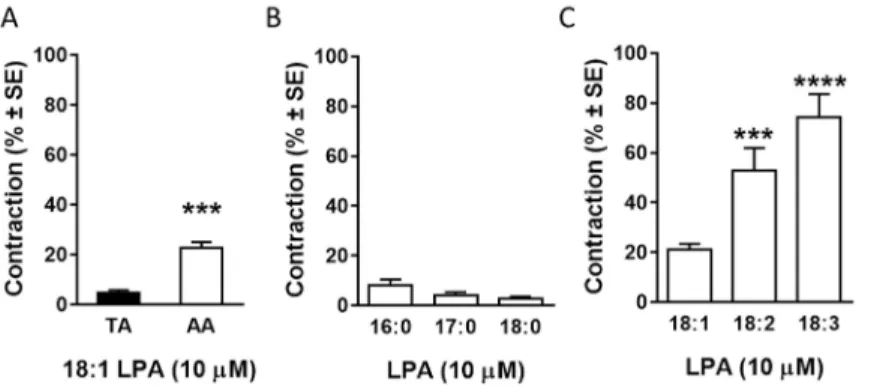
In some cases, application of LPA resulted in the increase of the vascular tone. For instance, vessels with intentionally damaged endothelium and vessels prepared from eNOS KO mice showed vasoconstriction to LPA. These observations suggested that the vasoactive effect of LPA was the superimposition of a constrictor and a relaxant component, the relaxation being dominant if intact endothelium is present. To investigate the vasoconstrictor activity of LPA, the endothelium was removed and oleoil-LPA was administered to the resting tone. From these experiments, we concluded the following:
removal of the endothelium resulted in a stronger vasoconstriction (1.2±0.3%, n=17 vs. 4.8±1.0%, n=14, compared to the 124 mM K+ reference contraction, p<0.001, Student’s unpaired t- test),
the amplitude of the contraction is higher in the abdominal aorta, than in thoracic aorta (Figure 7 Panel A, n=14, 42; ***p<0.001 vs.
“TA”, Student’s unpaired t-test) and
in the abdominal aorta, the contraction is dose-dependent (Emax=30.3 % of the reference contraction, EC50=3.15 μM, n=4-42, nonlinear regression).
16
In conclusion, vasoconstrictor effect of oleoyl-LPA is weak, considering the vasoconstrictive effect of edg-receptor agonist VPC31143 which evokes a 3-times stronger contraction in deendothelialized AAs (results not presented in this thesis).
Therefore, vasoconstrictor activity of LPA species with different acyl chain was investigated.
Figure 7 Vasoconstrictor effect of LPA
The poly-unsaturated linoleoyl- and linolenoyl-LPA have significantly higher vasoconstrictive activity compared to mono- unsaturated oleoyl-LPA (Figure 7 Panel C, n=48, 12, 13; ***p<0.001,
****p<0.0001 vs. 18:1 LPA, one-way ANOVA, followed by Tukey’s post-hoc test). These LPA types also evoked a transient contraction but the amplitude of the contraction was comparable to that of VPC31143. However, saturated LPA types did not evoke any vasoconstriction (Figure 7 Panel B, n=9, 9, 6).
17 5. CONCLUSIONS
Our aim was to investigate the vasoactive effects and the underlying signal transduction mechanisms of lysophosphatidic acid (LPA). We have confirmed that in mouse aorta:
• LPA evokes dose-dependent vasodilation in the presence of intact endothelium. The amplitude of the vasodilation depends on the acyl- chain of the LPA: in case of saturated LPA, length of the chain, in case of unsaturated 18 carbon-long LPA the number of double bonds decreases the vasorelaxing potency.
• LPA evokes vasoconstriction when the endothelium is damaged.
This effect is more pronounced in the abdominal aorta and its amplitude increases with the level of unsaturation.
• Endothelial cells express mRNA of LPA1, LPA2, LPA4 and LPA5 receptors and mRNA of the LPA producing enzyme autotaxin. LPA1 mediates the LPA-induced vasorelaxation.
• LPA-induced vasorelaxation is mediated by endothelial nitric oxide synthase but is independent of cyclooxigenase metabolites.
• Activation of eNOS in mediated by phospholipase C (PLC) – but not PLCε – and is independent of the phosphoinositol-3-kinase – Akt/PKB pathway.
18
6. BIBLIOGRAPHY OF THE CANDIDATE’S PUBLICATIONS
Publications related to the thesis
Ruisanchez, E., Dancs, P., Kerek, M., Nemeth, T., Farago, B., Balogh, A., Patil, R., Jennings, B. L., Liliom, K., Malik, K. U., Smrcka, A. V., Tigyi, G., Benyo, Z. (2014) Lysophosphatidic acid induces vasodilation mediated by LPA1 receptors, phospholipase C, and endothelial nitric oxide synthase. FASEB J 28: 880-890
Dancs, P. T., Ruisanchez, E., Balogh, A., Panta, C. R., Miklos, Z., Nusing, R. M., Aoki, J., Chun, J., Offermanns, S., Tigyi, G., Benyo, Z. (2017) LPA1 receptor-mediated thromboxane A2 release is responsible for lysophosphatidic acid-induced vascular smooth muscle contraction. FASEB J 31: 1547-1555
Publications not related to the thesis
Donko, A., Ruisanchez, E., Orient, A., Enyedi, B., Kapui, R., Peterfi, Z., de Deken, X., Benyo, Z., Geiszt, M. (2010) Urothelial cells produce hydrogen peroxide through the activation of Duox1.
Free Radic Biol Med 49: 2040-2048
Ruisanchez, E., Cselenyak, A., Papp, R. S., Nemeth, T., Kaldi, K., Sandor, P., Benyo, Z. (2012) Perivascular expression and potent vasoconstrictor effect of dynorphin A in cerebral arteries. PLoS One 7: e37798
Iring, A.*, Ruisanchez, E.*, Leszl-Ishiguro, M.*, Horvath, B., Benko, R., Lacza, Z., Jarai, Z., Sandor, P., Di Marzo, V., Pacher, P., Benyo, Z. (2013) Role of endocannabinoids and cannabinoid-1
19
receptors in cerebrocortical blood flow regulation. PLoS One 8:
e53390
Masszi, G., Novak, A., Tarszabo, R., Horvath, E. M., Buday, A., Ruisanchez, E., Tokes, A. M., Sara, L., Benko, R., Nadasy, G. L., Revesz, C., Hamar, P., Benyo, Z., Varbiro, S. (2013) Effects of vitamin D3 derivative--calcitriol on pharmacological reactivity of aortic rings in a rodent PCOS model. Pharmacol Rep 65: 476-483 Szekeres, M., Nadasy, G. L., Turu, G., Soltesz-Katona, E., Benyo, Z., Offermanns, S., Ruisanchez, E., Szabo, E., Takats, Z., Batkai, S., Toth, Z. E., Hunyady, L. (2015) Endocannabinoid-mediated modulation of Gq/11 protein-coupled receptor signaling-induced vasoconstriction and hypertension. Mol Cell Endocrinol 403: 46-56 Benyo, Z., Ruisanchez, E., Leszl-Ishiguro, M., Sandor, P., Pacher, P. (2016) Endocannabinoids in cerebrovascular regulation. Am J Physiol Heart Circ Physiol 310: H785-801
Polycarpou, A., Hricisak, L., Iring, A., Safar, D., Ruisanchez, E., Horvath, B., Sandor, P., Benyo, Z. (2016) Adaptation of the cerebrocortical circulation to carotid artery occlusion involves blood flow redistribution between cortical regions and is independent of eNOS. Am J Physiol Heart Circ Physiol 311: H972-H980
* equal contribution