Increasing safety of vascular access with ultrasound guidance
Ph.D thesis
Dr. Géza Reusz
Doctoral School of Clinical Medicine Semmelweis University
Consultant: Ákos Csomós, MD, Ph.D
Official reviewers: Dr .Attila Szijártó, MD, Ph.D Dr. Róbert Almási, MD, Ph.D Dr. Zoltán Máthé, MD, Ph.D Head of the Final Examination Committee:
Dr. László Harsányi MD, Ph.D
Members of the Final Examination Committee:
Dr. Attila Doros, MD, Ph.D Dr. Tamás Végh, MD, Ph.D
Budapest
2014
1
INTRODUCTIONCannulation of various vessels (such as central and peripheral veins, arteries) is a common and important part of daily anaesthetic practice. Although traditional (landmark-based) techniques are routine procedures, occasionally cannulations prove to be difficult or impossible, and can lead to serious complication. With the help of ultrasound, deep, impalpable and invisible vessels can be visualized.
Besides being of help to localize vessels, pathological conditions, e.g.
vessel wall abnormalities, thrombosis can be identified. Furthermore, real-time ultrasound guidance allows for controlled needle manipulation towards the target.
Ultrasound guidance has been used for more than 25 years for vascular access, and basic techniques (in-plane and out-of-plane guidance) were described and utilized already long ago. Ultrasound- based techniques have become exponentially widespread in the last 10-15 years, thanks to technological development and the availability of laptop-sized mobile ultrasound machines.
Several studies proved that ultrasound guidance generally increases success rates for vascular access procedures, and complication rates can be largely reduced. However, because this is a relatively new technique, indications are not precisely described yet, and technical details of procedures performed by various operators are
2
very different. It is now clear that technical details and the experience of the operator have a significant influence on results, and procedures performed under inadequate conditions, by inexperienced operators, can jeopardize patient safety.
Ultrasound-guided interventions are only safe if the operator is able to orientate himself confidently on the ultrasound image, and track needle advancement during the whole time of the intervention.
Most vascular access - related complications are consequences of unnoticed puncture of vital organs, so it is essentially important to correctly identify the needle tip on the monitor, and continuously track its position throughout the procedure. This is not easy because the operator has to orientate himself in a three-dimensional space but at present common ultrasound machines only provide two-dimensional images. Correct interpretation of the needle is also commonly interfered by ultrasound artefacts.
Thanks to technological development: better image quality and needle visualization (especially under steep needle angle), ultrasound guidance has become easier even in traditionally “difficult”
applications, but skills, experience and technique of the operator are still important factors. At present it is not clear that among several ultrasound-based techniques of various operators which is (are) the safest, and, similarly to any other new methods, early phases of the learning curve are critical. Furthermore, we do not know whether
3
proper ultrasound guidance makes some traditional safety precautions (e.g. correction of coagulopathy before central venous cannulation), that have been used for decades with the traditional techniques to decrease mechanical complications, unnecessary.
4
OBJECTIVESTo prove that correct utilization of ultrasound guidance improves safety of vascular access procedures. In order to achieve this, to execute the following:
1. Detailed description of ultrasound image of needles used for vascular access. Correct identification of the needle tip on the image under different conditions. Description of artefacts that interfere with identification and tracking of the needle tip position.
2. Modification of the in-plane cannulation technique based on precise description of needle tip visualization. Utilization of this modified technique in clinical practise. Evaluation of the technique in an observational clinical study.
3. Introduction of ultrasound-guided vascular access procedures into clinical practise in the Department of Anaesthesia and Intensive Care in a county teaching hospital, documenting all important data related to the procedures with special regards to success rates and
5
complications. Evaluation of safety of ultrasound-guided interventions in the early phases of the learning curve.
4. Study of safety of ultrasound-guided central venous access in patients with uncorrected coagulopathy.
6
METHODSOur thesis consists of two parts: a theoretical and a clinical section.
1. Theoretical section: analysis of the ultrasound visualization of needles
Analysis of a large number of ultrasound images, saved both in vivo and in vitro. Explanations for aberrations on the ground of physical concepts and known, in other context described, ultrasound artefacts. Proof of assumptions with in vitro experiments if needed.
Looking for connections with clinical practise.
2. Clinical studies
2.1. Observational study of a new cannulation technique
Thanks to our theoretical studies, we managed to precisely identify needle tip position on the ultrasound image during interventions. This lead to the development of a new cannulation technique that can potentially increase success rate of small vessel (e.g. radial artery, peripheral vein) cannulation where precise manipulation with the needle is mandatory. We continuously track the
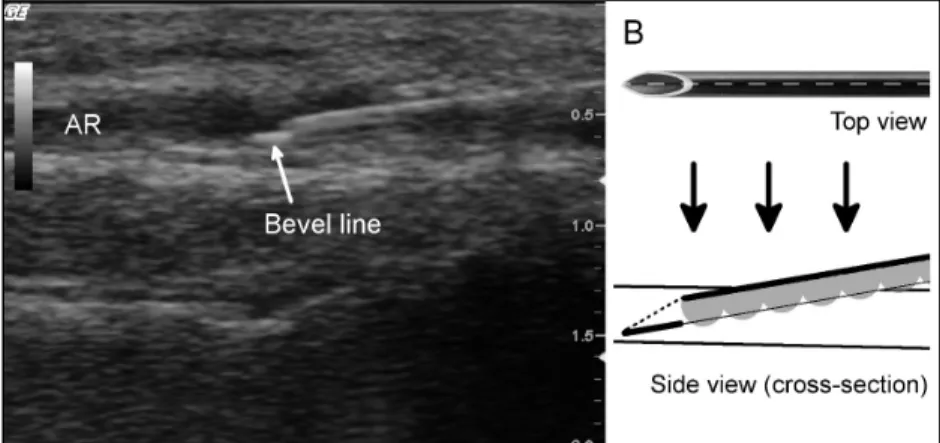
“bevel line”, that corresponds to the needle tip as we showed (See:
7
Results: 1. Visualization of the needle by ultrasound), and keep it in the plane of ultrasound, so needle entry into the vessel is under direct visual control. We evaluated this technique in an observational study (ethical permission: IKEB/UDARC/2012/2). Cannula placements were performed by five clinicians through 6 months in the Intensive Care Unit in Markhot Ferenc Hospital, Eger, Hungary. Cannulation site and outcome of the procedure (successful / unsuccessful) were recorded for all cannulations. When time allowed, detailed data were also recorded including factors potentially influencing cannulation success (blood pressure, vasopressors, coagulopathy, body mass index, diameter and depth of the artery, local vessel-abnormalities) and recorded outcomes of single attempts and the procedure (cannulation time: from skin puncture to successful cannula placement, number of attempts, type and number of cannulas used).
2.2. Introduction of ultrasound-guided vascular access procedures into clinical practise in the Department of Anaesthesia and Intensive Care in a county teaching hospital
Ultrasound was introduced into practise in February 2011 in the Department of Anaesthesia and Intensive Care in the Markhot Ferenc Hospital; only one doctor had experience with the technique at the time. So the first one-two years can be considered as a learning phase not only for a given doctor but for the department as well.
8
Colleagues were trained systematically from the first moment on. We organised in vitro teaching sessions to practice needle manipulation under ultrasound, and the first, supervised, procedures at the bedside were only performed thereafter. Doctors used ultrasound without supervision only when they already had experience.
We recorded most important data (intervention, date, operator, difficulties, complications) for all interventions. We regularly audited our practise during the first 1000 interventions, mainly from the point of view of success rates and complications.
2.3. Ultrasound-guided central venous access in patients with uncorrected coagulopathy
In a retrospective study we involved all ultrasound-guided central venous cannula placements for patients with coagulopathy in the intensive care unit. Besides our ultrasound register we retrieved data from the electronic (MEDWORKS) and paper-based patient records of the department; we were looking for the presence of coagulopathy and any recorded procedure-related complications within 24 hours after the intervention (ethical permission:
IKEB/UVCV/2012/2). Patients with an INR or APTT ratio above 1.5, platelet count below 100 G/l, or recent clopidogrel, heparin/LMWH administration were considered as having coagulopathy, whilst
9
INR/APTT ratio above 2.0, platelet count below 50 G/l, or therapeutic anticoagulation were considered as severe coagulopathy. Any potentially procedure-related mechanical complication that was either documented in the patient records, or reported on check chest X-rays were considered a complication, including bleeding to any degree (e.g. leakage from entry site, haematoma, haemothorax), pneumothorax, and injuries of any organ, nerve, vessel nearby.
We used Wilson test for confidence interval (CI)
calculations and chi-squared test for comparisons of success
rates. Intercooled STATA 8.2 software (StataCorp, Texas, USA)
was used for calculations.
10
RESULTS1. Visualization of needles by ultrasound
When using in-plane technique, the needle typically appears as one long line on the ultrasound image despite its tubular structure.
When the needle is exactly in the plane of ultrasound and the bevel faces upwards, another short echo-dense line can be observed at the distal end of the main needle line. Although this line had been described before, it was considered as an artefact with no meaning.
We showed that this line (i.e., “bevel line”) is not an artefact but part of the needle and carries significance. We proved that the resolution of modern ultrasound machines is sufficient to visualize the structure of the needle, i.e., to show both the anterior and posterior needle walls with a lumen in between; however, because of strong echo-reflections from the echogenic needle surface, the lumen and the posterior wall of the needle shaft are in the acoustic shadow of the anterior wall and obscured. However, the distal end of the posterior needle wall - uncovered by the anterior wall - gets out of the acoustic shadow at the bevel and is recognizable. The tip of the needle is at the distal end of this short “bevel line” (Figure 1). The bevel line can be used as a direct visual confirmation of the needle tip; furthermore, as it is best seen when the tip is exactly in the plane of the ultrasound, it can be a useful guide to keep the needle central throughout the procedure.
11
Many ultrasound artefacts can impair needle visibility, e.g.
reverberation, comet tail, beam width, side lobe artefacts. Although these artefacts are well known, their relations to needles had not been extensively described yet before our studies. All of them can interfere with needle, and especially needle tip, identification. We published the results of our work in a review article in the British Journal of Anaesthesia
Figure 1. Bevel line and explanation. AR= Arteria radialis.
2.1. Cannulation of the radial artery with a new technique
During the 6-months study period in the intensive care unit, a total number of 66 ultrasound-guided arterial cannulations were attempted with the new technique, which is based on the identification and continuous tracking of the bevel line during the procedure.
12
At least one factor leading to potentially difficult cannula placement (e.g. shock, obesity, weak or absent arterial pulsation, oedema, agitated patient, diameter of radial arteries less than 2 mm, local vessel-abnormalities) was identified in 70.3% of cases. Success rate was 100% (95% CI: 94.5-100%), placement was successful in 75.7%, 21.6% and 2.7% at first, second and third attempt, respectively. Over- the-needle type cannula (Flow switch, Beckton Dickinson) was used in 65 cases and Seldinger technique once. Median cannulation time was 92 sec. The smallest, successfully cannulated artery had a diameter of 1.4 mm. The bevel line was visible in 83% of all attempts, and the success rate was significantly higher when this was visible (p=0.002).
2.2. Introduction of ultrasound-guided procedures into clinical practise
The first 1000 ultrasound guided procedures, performed by anaesthetists, were done between February 2011 and January 2013 (central venous access: 364, arterial cannulation: 307, peripheral venous cannulation: 76, all vascular access procedures: 746). The interventions were performed by 19 doctors; all but one used ultrasound the first time in the study period. Success rates were between 96-98.7% in different applications. Serious complications did not occur, minor complications were observed at rates between 0-
13
1.3% (central venous access: artery puncture in 2 cases – 0.5%, peripheral venous access: artery puncture on one occasion – 1.3%).
By February 2013 already 5 doctors gained extensive experience (>100 ultrasound-guided interventions per person) with the technique.
2.3. Ultrasound-guided central venous access in patients with uncorrected coagulopathy
We placed a total number of 310 central venous cannulas in the ICU between February 2011 and January 2013, 43% of patients had coagulopathy on the day of the procedure. Coagulopathy was moderate in 80, severe in 54 cases. Moderate coagulopathy was corrected in one (1.25%), severe coagulopathy in 9 (16.7%) patients.
So at the time of cannulation 124 patients had uncorrected coagulopathy (moderate: 79, severe: 45). Despite the large number of patients with haemorrhagic diathesis, there were no bleeding-related or other mechanical complications (complication rate: 0%, 95%CI: 0- 3,0%, for patients with severe coagulopathy 95%CI: 0-7,7%).
14
CONCLUSIONS1. During ultrasound-guided procedures the real structure of the needle is obscured due to acoustic shadowing of the strong echo- reflector metal surface. Visualization of the needle is impaired by other ultrasound artefacts, too. However, if we pay attention to these factors, we will be able to accurately identify and continuously track the needle tip on the ultrasound image during the intervention. This can be particularly important when cannulating small vessels, as here precise needle manipulation is mandatory.
2. Despite the large number of difficult-to-cannulate patients, we achieved a high success rate for radial artery cannulation with our modified in-plane cannulation technique that is based on identification and continuous tracking of the bevel line. This technique can be used efficiently for all ultrasound-guided procedures, where, because of the small size of the target, precise needle manipulation is important, e.g., for small vessel cannulation.
3. Although complications are generally most frequent at the early phase of the learning curve for a given intervention, after proper training, with initial supervision, utilizing the principle of
15
progressivity, excellent success and very low complication rates can be achieved during the learning stages of ultrasound-guided interventions, even if performed in large numbers and by many operators. When taking these precautions, ultrasound guidance increases patient safety in the anaesthetic department from the very beginning.
4. Before ultrasound-guided central venous access, as long as it is performed by an experienced operator with a good technique, routine correction of mild / moderate coagulopathy is unnecessary, and the procedure may be safe even when the coagulopathy is severe.
16
BIBLIOGRAPHY OF THE CANDIDATE’S PUBLICATIONS
Publications related to the Ph.D thesis:
1. Reusz G, Jakab L, Langer Cs, Varga R, Kovács Sz, Sárkány P.
(2011) Műtermékek az ultrahangos képalkotásban. Aneszt Int Terápia, 41: 175-181.
2. Langer Cs, Reusz G, Varga R, Kovács Sz, Egervári G, Sárkány P.
(2011) Ultrahang-vezérelt atípusos vénakanülálások trombotizált vénákkal rendelkező hemodializált beteg intenzíves kezelése során Aneszt Int Terápia, 41: 182-186.
3. Reusz G, Langer C, Jakab L, Morvay Z. (2012) Ultrasound-guided vascular access: the importance of the needle bevel. Can J Anesth, 59:
499-500.
Impact factor: 2,127 4. Reusz G, Csomós Á. (2013) Ultrahangvezérelt perifériás vénabiztosítás. Orv Hetil, 154: 187-190.
5. Reusz G, Langer Cs, Müller M, Kovács Sz, Varga R, Sárkány P, Csomós Á. (2013) Az első 1000 ultrahang-vezérelt beavatkozás tapasztalatai osztályunkon. Aneszt Int Terápia, 43: 66-71.
6. Reusz G, Sarkany P, Gal J, Csomos A. (2014) Needle-related ultrasound artifacts and their importance in anaesthetic practice. Brit J Anaesth, 112: 794-802.
Impact factor: 4,237 Impact factor: 6,364
17
Other publications:7. Reusz G, Lámer Z, Székely I, Bodnár J, Vincze K, Ökrös I. (2007) Korai percutan tracheostomia sebészeti intenzív osztályon. Aneszt Int Terápia, 36: 16-22.
8. Erdős G, Reusz G. (2008) Nehézségek Cook tubuscserélő katéter használata során Aneszt Int Terápia, 38: 90-93.
9. Reusz G, Ökrös I. (2010) Vesepótló kezelések hatékonysága akut veseelégtelenségben. Aneszt Int Terápia, 40: 39-47.
10. Horváth A, Reusz G, Gál J, Csomós Á. (2012) A betegbiztonság javításának lehetőségei nagy sebészeti műtétek perioperatív szakában.
Orv Hetil, 153: 1447–1455.
Abstracts related to the Ph.D thesis:
1. Reusz G, Langer Cs, Müller M, Kovács Sz, Varga R, Sárkány P.
(2012) Az ultrahang bevezetésében szerzett tapasztalataink az első 13 hónapban végzett 464 ultrahang-vezérelt beavatkozás tükrében.
Aneszt Int Terápia, 42 (Suppl. 1): 27
2. Müller M. Reusz G, Langer Cs, Kiss N, Horváth N, Sárkány P.
(2012) Kiserek (artéria, perifériás véna) kanülálása ultrahanggal.
Aneszt Int Terápia, 42 (Suppl. 1): 27
3. Reusz G, Langer Cs, Egervári G, Sárkány P, Csomós Á. (2013) Safety of ultrasound-guided central venous access in critically ill patients with uncorrected coagulopathy. Crit Care, 17 (Suppl 2): P173 4. Reusz G, Langer Cs, Hevessy T, Egervári G, Sárkány P, Csomós Á. (2013) Ultrahang-vezérelt centrális vénabiztosítás korrigálatlan koagulopátiában. Aneszt Int Terápia, 43 (Suppl. 1): 19
18
5. Reusz G, Langer Cs, Müller M, Varga R, Csomós Á. (2013) Cannulation of the radial artery with ultrasound guidance – evaluation of a new technique. Infection, 41 (Suppl 1): S52-53.
Other abstracts:
6. Reusz G, Szabó K, Magyar I, Csomós Á, Debreceni K, Rácz K.
(2001) Tracheostomia a költséghatékonyság tükrében. Aneszt Int Terápia, 31 (1. Suppl.): 39
7. Reusz G, Tóth J, Szabó K, Csomós Á. (2003) A „PERCUTWIST”- szettel végzett percutan tracheostomia ismertetése. Aneszt Int Terápia, 33 (1. Suppl): 60-61
8. Reusz G, Bodnár J, Székely I, Lámer Z, Szedlák B, Ökrös I. (2007) Lélegeztetési idő előrejelzése intenzív osztályon. Aneszt Int Terápia, 37 (Suppl. 1):
9. Reusz G,Szedlák B,Székely I, Bodnár J,Lámer Z, Debreceni K, Bakos L, Ormay C, Ökrös I. (2007) Prediction of prolonged mechanical ventilation in critically ill patients Intensive Care Med, 33 (Suppl 2): 85
10. Reusz G, Székely I, Szedlák B, Bodnár J, Ökrös I. (2008) Percutan tracheostomia bevezetése osztályunkon: tapasztalataink 125 beteg alapján. Aneszt Int Terápia, 38 (Suppl. 1): 48-49
11. Szedlák B, Székely I, Reusz G, Ökrös I. (2008) Szepszis osztályunkon 2005-2007. Aneszt Int Terápia, 38 (Suppl. 1): 46-47 12. Székely I, Szedlák B, Reusz G, Ökrös I. (2008) Magas kardiális rizikójú beteg perioperatív ellátása. Aneszt Int Terápia, 38 (Suppl. 1):
26
13. Reusz G, Kvaszta Sz, Székely I, Bodnár J, Kánya Z, Ökrös I.
(2009) Magas kockázatú betegek vesepótló kezelése osztályunkon.
Aneszt Int Terápia, 39 (Suppl. 1): 37
19
14. Langer Cs, Reusz G, Kovács Sz, Varga R, Müller M, Egervári G.
(2012) Ultrahang-vezérelt felső végtagi idegblokádok osztályunkon – eredményeink az első 13 hónapban végzett 94 beavatkozás alapján.
Aneszt Int Terápia, 42 (Suppl. 1): 9