Pathogens from the Genus Armillaria
Liqiong Chen1, Bettina Bóka1, Orsolya Kedves1, Viktor Dávid Nagy1, Attila Sz ˝ucs1 , Simang Champramary2, Róbert Roszik2, Zoltán Patocskai3, Martin Münsterkötter2, Thu Huynh1, Boris Indic2, Csaba Vágvölgyi1 , György Sipos2,4 and LászlóKredics1,*
1 Department of Microbiology, Faculty of Science and Informatics, University of Szeged, Szeged,
Közép fasor 52, H-6726 Szeged, Hungary; liqiongchen2016@163.com (L.C.); boka.tina@gmail.com (B.B.);
varga_orsi91@yahoo.com (O.K.); viktor.david.nagy@gmail.com (V.D.N.); a.szunyi@gmail.com (A.S.);
huynh_thu@hcmut.edu.vn (T.H.); mucor1959@gmail.com (C.V.)
2 Functional Genomics and Bioinformatics Group, Research Center for Forestry and Wood Industry, University of Sopron, Bajcsy-Zsilinszky str. 4., H-9400 Sopron, Hungary; simang5c@uni-sopron.hu (S.C.);
roszikster@gmail.com (R.R.); M.Muensterkoetter@web.de (M.M.); boris.indjic@phd.uni-sopron.hu (B.I.);
gyoergy.sipos@wsl.ch (G.S.)
3 Bakonyerd˝o Ltd., Jókai Mór u. 46., H-8500 Pápa, Hungary; patocskai@bakonyerdo.hu
4 Swiss Federal Research Institute WSL, Zürcherstrasse 111, CH-8903 Birmensdorf, Switzerland
* Correspondence: kredics@bio.u-szeged.hu; Tel.:+36-62-544516
Received: 18 October 2019; Accepted: 9 November 2019; Published: 13 November 2019
Abstract:Research Highlights: A large scale effort to screen, characterize, and select Trichoderma strains with the potential to antagonize Armillaria species revealed promising candidates for field applications. Background and Objectives: Armillaria species are among the economically most relevant soilborne tree pathogens causing devastating root diseases worldwide. Biocontrol agents are environment-friendly alternatives to chemicals in restraining the spread of Armillaria in forest soils. Trichoderma species may efficiently employ diverse antagonistic mechanisms against fungal plant pathogens. The aim of this paper is to isolate indigenous Trichoderma strains from healthy and Armillaria-damaged forests, characterize them, screen their biocontrol properties, and test selected strains under field conditions. Materials and Methods: Armillaria and Trichoderma isolates were collected from soil samples of a damaged Hungarian oak and healthy Austrian spruce forests and identified to the species level. In vitro antagonism experiments were performed to determine the potential of the Trichoderma isolates to control Armillaria species. Selected biocontrol candidates were screened for extracellular enzyme production and plant growth-promoting traits. A field experiment was carried out by applying two selected Trichoderma strains on two-year-old European Turkey oak seedlings planted in a forest area heavily overtaken by the rhizomorphs of numerous Armillaria colonies. Results: AlthoughA. cepistipesandA. ostoyaewere found in the Austrian spruce forests,A. melleaandA. gallicaclones dominated the Hungarian oak stand. A total of 64 Trichoderma isolates belonging to 14 species were recovered. Several Trichoderma strains exhibited in vitro antagonistic abilities towards Armillaria species and produced siderophores and indole-3-acetic acid.
Oak seedlings treated withT. virensandT. atrobrunneumdisplayed better survival under harsh soil conditions than the untreated controls. Conclusions: Selected native Trichoderma strains, associated with Armillaria rhizomorphs, which may also have plant growth promoting properties, are potential antagonists of Armillaria spp., and such abilities can be exploited in the biological control of Armillaria root rot.
Keywords: Armillaria; Trichoderma; root rot; biocontrol; antagonism; siderophore;
indole-3-acetic acid
Forests2019,10, 1013; doi:10.3390/f10111013 www.mdpi.com/journal/forests
1. Introduction
Armillaria and Desarmillaria species (Physalacriaceae and Basidiomycota) are globally distributed fungal plant pathogens varying in host range and pathogenicity [1,2]. They cause white rot, a severe destructive disease (also known as Armillaria root rot) on a wide range of woody hosts growing in managed plantations, natural forests, orchards, and amenity plantings in urban areas, and their impact often leads to devastating forest damages and immense economic losses [3]. Armillaria colonies are spread in the soil by root-like rhizomorphs, which can attack host trees through root contacts, and then the penetrating hyphae colonize heartwood and invade the cambium as mycelial fans [4]. In general, Armillaria root disease results in reduced forest productivity due to direct mortality or permanent non-lethal infections affecting the health and growth of the trees [5].
It is well known that most Armillaria species exhibit specialization towards either coniferous or broadleaf hosts. Although native coniferous forests in the Northern hemisphere are predominantly inhabited byA. ostoyaeandA. cepistipes, various oak and other broadleaf species are most exposed to A. mellea,A. gallica, andDesarmillaria tabescens[6–8].
The use of naturally occurring antagonistic fungi (e.g., certain Trichoderma species) and bacteria (e.g., Bacillus and Pseudomonas species) has uncovered great potential to successfully reduce the pathogenic activities of Armillaria. Particularly, native microorganisms isolated from soil, rhizosphere, or directly from plant roots usually have a better adaptation to that specific soil and plant environment, and thus can display more efficient control of diseases, than introduced exotic microorganisms [9].
Species of the soilborne genus Trichoderma have been widely used as biocontrol agents. Results of field trials showed that mycelia and conidia of five combined isolates of Trichoderma species significantly reduced the root colonization ofA. luteobubalinaand may have had additional inhibitory effects on fruiting body development as well [10]. Application ofT. harzianumto the soil surrounding the wood-borne inoculum of Armillaria caused a significant reduction in the viability of the pathogen [11].
Armillaria failed to invade the stem sections colonized byT. harzianumand had low viability in the plant materials inoculated with Trichoderma [12]. The use of air-spading combined withT. harzianum inoculation also proved to be a potential joint cultural/biocontrol strategy against A. melleain a forest [13].
The biocontrol abilities of Trichoderma strains are based on a wide arsenal of various antagonistic mechanisms [14]. Trichoderma species are excellent competitors for space and nutrients. Their extracellular enzyme systems, including cellulases (e.g., endocellulases, cellobiohydrolases, and β-glucosidases) and xylanases (e.g., endoxylanases andβ-xylosidases), enable the efficient utilization of plant polysaccharides, while they can successfully compete for iron by the production of siderophores [14]. Antifungal secondary metabolites including pyrones, polyketides and non-ribosomal peptides play important roles in their antibiotic effects against fungal plant pathogens [14]. Direct mycoparasitism is closely associated with the production of extracellular cell wall-degrading enzymes (CWDE)—such as glucanases, chitinases, and proteases—playing an important role in the degradation of the cell wall and the penetration into the host hyphae [14,15]. Besides the above-mentioned direct mechanisms of antagonism, the ability of Trichoderma to promote plant growth via mechanisms including phosphorous mobilization, by extracellular phosphatases and the production of indole-3-acetic acid derivatives [14], and induce systemic resistance in the host plant can also be considered when screening for biocontrol agents. The antagonistic behavior of some Trichoderma strains may result from the interaction with plant roots, promotion of plant growth, and improving tolerance to abiotic stresses as well as plant resistance to diseases [13,16].
The presence of Armillaria root rot disease in the forests of the Northern hemisphere, and its economic consequences have consumed a lot of environmentally harmful and polluting fungicides.
Woody plants, beyond their commercial values, provide essential components of wildlife habitats worldwide. However, Armillaria species often seem to dominate in the forests and may cause serious diseases leading to compromised seedlings. Commercial products based on Trichoderma used to protect plants have been available on the market. However, isolating and screening for antagonistic
Trichoderma strains from diverse populations distributed at different geographic regions may be more helpful for developing efficient biocontrol agents against a broad range of pathogens from the genus Armillaria. The aim of this study is to select and characterize Trichoderma strains with the potential to control Armillaria and examine their performance during application in the field.
2. Materials and Methods
2.1. Isolation of Armillaria and Trichoderma
Samples of bulk soil (soil outside the rhizosphere), upper rhizospheric soil, Armillaria rhizomorphs and their surrounding soil, as well as Armillaria fruiting bodies were collected from a heavily Armillaria-damaged oak stand (Keszthely Hills, Hungary) and healthy native spruce forests (Rosalia, Austria). The rhizomorph samples were taken as aliquots of the soil pools associated with the collected rhizomorphs. The Roth and Shaw medium [17] supplemented with 15 mg/L benomyl and 250 mg/L streptomycin was applied for Armillaria isolation from the field samples. For Trichoderma isolation, 1 g of fresh soil per sample was suspended in sterile 0.9% NaCl solution, diluted serially (the 10−1, 10−2, and 10−3dilution) and spread on Trichoderma selective media. The composition of the media for selectively isolating Trichoderma strains was 10 g/L glucose, 5 g/L peptone, 1 g/L KH2PO4, 0.5 g/L MgSO4×7H2O, 20 g/L agar, amended with 0.25 mL/L 5% Rose-Bengal in water, 0.5 mL/L 0.2% dichloran in ethanol, 0.01% streptomycin, 0.01% oxytetracycline, and 0.01% chloramphenicol [18]. After 3 days of incubation at 25±0.5◦C, fungal colonies including Trichoderma were detected and transferred onto potato dextrose agar (PDA).
2.2. Identification of Armillaria and Trichoderma Isolates
One-hundred mg of fresh mycelia from each fungal isolate was collected for DNA extraction following the manufacturer’s instructions of the E.Z.N.A.® Fungal DNA Mini Kit (Omega Bio-tek, USA). The Internal Transcribed Spacer (ITS) region of the nuclear ribosomal RNA gene cluster was amplified using the ITS4 (50-TCCTCCGCTTATTGATATGC-30) and ITS5 (50-GGAAGTAAAAGTCGTAACAAGG-30) universal primers for fungi [19]. The PCR reactions were carried out in a final volume of 25.1µL, consisting of 2.5µL 10×DreamTaq Buffer with 20 mM MgCl2, 2.5µL of 2 mM dNTP mix, 0.1µL of 5 U/µL DreamTaq DNA Polymerase (Thermo Scientific), 0.5µL of each primer (10µM), 18µL bidistilled water, and 1µL template DNA. Amplifications were performed in a Doppio Thermal Cycler (VWR, Hungary). Thermal cycling parameters were as follows; initial denaturation at 94◦C for 5 min, 35 cycles of DNA denaturation at 94◦C for 30 s, primer annealing at 50◦C for 30 s, elongation at 72◦C for 50 s, and a final elongation at 72◦C for 7 min. For amplification of the translation elongation factor 1-alpha (tef1α) gene fragment, reaction mixtures were the same as described above, but with universal primers TEF-LLErev (50-AACTTGCAGGCAATGTGG-30) and EF1-728F: (50-CATCGAGAAGTTCGAGAAGG-30) [20] and the thermal cycling program, with an initial denaturation at 94◦C for 5 min, 40 cycles of DNA denaturation at 94◦C for 45 s, primer annealing at 57◦C for 30 s, elongation at 72◦C for 90 s, and a final elongation at 72◦C for 7 min. The amplicon quality was detected by 2% agarose gel electrophoresis of 4µL samples from the reaction mixtures.
Direct sequencing of the unpurified PCR products was performed by the sequencing platform of the Biological Research Centre, Szeged. The resulting sequences were analyzed byTrichOkey 2.0 [21], TrichoBLAST [22] and NCBI Nucleotide BLAST. The isolated and identified Armillaria and Trichoderma strains were deposited in the Szeged Microbiology Collection (SZMC,www.szmc.hu), Szeged, Hungary, whereas the sequences were submitted to the GenBank Nucleotide database (ncbi.nlm.nih.gov) under the accession numbers listed in Table1.
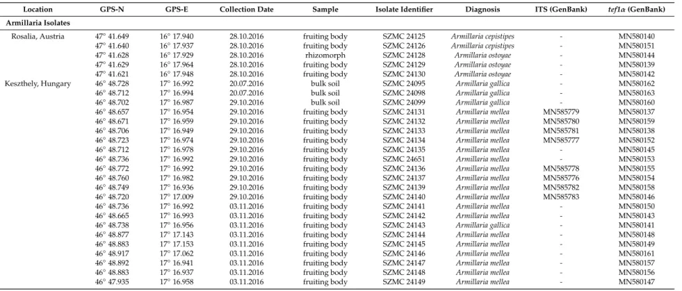
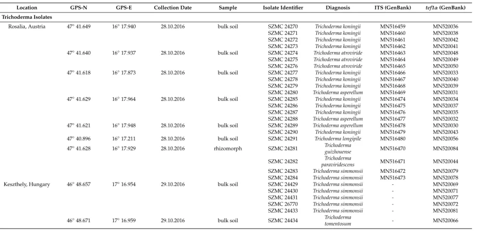
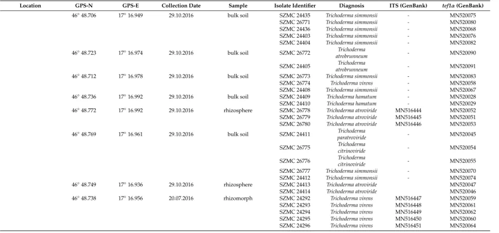
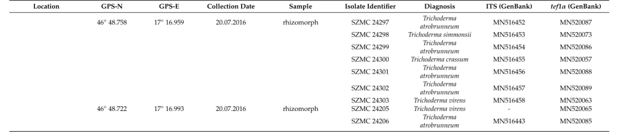
Table 1.Armillaria and Trichoderma isolates collected during the study.
Location GPS-N GPS-E Collection Date Sample Isolate Identifier Diagnosis ITS (GenBank) tef1α(GenBank)
Armillaria Isolates
Rosalia, Austria 47◦41.649 16◦17.940 28.10.2016 fruiting body SZMC 24125 Armillaria cepistipes - MN580140
47◦41.640 16◦17.937 28.10.2016 fruiting body SZMC 24126 Armillaria cepistipes - MN580151
47◦41.628 16◦17.929 28.10.2016 rhizomorph SZMC 24128 Armillaria ostoyae - MN580144
47◦41.629 16◦17.964 28.10.2016 fruiting body SZMC 24129 Armillaria ostoyae - MN580139
47◦41.621 16◦17.948 28.10.2016 fruiting body SZMC 24130 Armillaria ostoyae - MN580142
Keszthely, Hungary 46◦48.728 17◦16.992 20.07.2016 bulk soil SZMC 24095 Armillaria gallica - MN580162
46◦48.712 17◦16.994 20.07.2016 bulk soil SZMC 24098 Armillaria gallica - MN580163
46◦48.702 17◦16.987 29.10.2016 bulk soil SZMC 24099 Armillaria gallica - MN580160
46◦48.657 17◦16.954 29.10.2016 fruiting body SZMC 24131 Armillaria mellea MN585779 MN580137 46◦48.671 17◦16.959 29.10.2016 fruiting body SZMC 24132 Armillaria mellea MN585780 MN580159 46◦48.706 17◦16.949 29.10.2016 fruiting body SZMC 24133 Armillaria mellea MN585781 MN580138 46◦48.723 17◦16.974 29.10.2016 fruiting body SZMC 24134 Armillaria mellea MN585777 MN580152
46◦48.712 17◦16.978 29.10.2016 fruiting body SZMC 24135 Armillaria mellea - MN580145
46◦48.736 17◦16.992 29.10.2016 fruiting body SZMC 24651 Armillaria mellea - MN580153
46◦48.772 17◦16.992 29.10.2016 fruiting body SZMC 24136 Armillaria mellea MN585778 MN580155 46◦48.760 17◦16.982 29.10.2016 fruiting body SZMC 24137 Armillaria mellea MN585776 MN580154 46◦48.749 17◦16.936 29.10.2016 fruiting body SZMC 24139 Armillaria mellea MN585782 MN580158 46◦48.720 17◦17.009 29.10.2016 fruiting body SZMC 24140 Armillaria mellea MN585783 MN580146
46◦48.736 17◦16.992 03.11.2016 fruiting body SZMC 24141 Armillaria mellea - MN580150
46◦48.665 17◦16.993 03.11.2016 fruiting body SZMC 24142 Armillaria mellea - MN580143
46◦48.738 17◦16.956 03.11.2016 fruiting body SZMC 24143 Armillaria gallica - MN580141
46◦48.877 17◦17.143 03.11.2016 fruiting body SZMC 24144 Armillaria mellea - MN580148
46◦48.883 17◦17.153 03.11.2016 fruiting body SZMC 24145 Armillaria mellea - MN580149
46◦48.917 17◦17.062 03.11.2016 fruiting body SZMC 24146 Armillaria mellea - MN580161
46◦48.892 17◦16.941 03.11.2016 fruiting body SZMC 24147 Armillaria mellea - MN580157
46◦48.883 17◦16.937 03.11.2016 fruiting body SZMC 24148 Armillaria mellea - MN580156
46◦47.935 17◦16.958 03.11.2016 fruiting body SZMC 24149 Armillaria mellea - MN580147
Table 1.Cont.
Location GPS-N GPS-E Collection Date Sample Isolate Identifier Diagnosis ITS (GenBank) tef1α(GenBank)
Trichoderma Isolates
Rosalia, Austria 47◦41.649 16◦17.940 28.10.2016 bulk soil SZMC 24270 Trichoderma koningii MN516459 MN520036 SZMC 24271 Trichoderma koningii MN516460 MN520038 SZMC 24272 Trichoderma koningii MN516461 MN520042 SZMC 24273 Trichoderma koningii MN516462 MN520041 47◦41.640 16◦17.937 28.10.2016 bulk soil SZMC 24274 Trichoderma atroviride MN516463 MN520048 SZMC 24275 Trichoderma atroviride MN516464 MN520049 SZMC 24276 Trichoderma atroviride MN516465 MN520050 47◦41.618 16◦17.873 28.10.2016 bulk soil SZMC 24277 Trichoderma koningii MN516466 MN520033 SZMC 24278 Trichoderma koningii MN516467 MN520040 SZMC 24279 Trichoderma koningii MN516468 MN520039 SZMC 24280 Trichoderma asperellum MN516469 MN520031 47◦41.629 16◦17.964 28.10.2016 bulk soil SZMC 24285 Trichoderma koningii MN516474 MN520034 SZMC 24286 Trichoderma koningii MN516475 MN520037 SZMC 24287 Trichoderma koningii MN516476 MN520035 SZMC 24288 Trichoderma asperellum MN516477 MN520032 47◦41.621 16◦17.948 28.10.2016 bulk soil SZMC 24289 Trichoderma asperellum MN516478 MN520030 SZMC 24290 Trichoderma koningii MN516479 MN520043 47◦40.896 16◦17.211 28.10.2016 bulk soil SZMC 24291 Trichoderma longipile MN516480 MN520056 47◦41.628 16◦17.929 28.10.2016 rhizomorph SZMC 24281 Trichoderma
guizhouense MN516470 MN520084
SZMC 24282 Trichoderma
paraviridescens MN516471 MN520044
SZMC 24283 Trichoderma simmonsii MN516472 MN520079 SZMC 24284 Trichoderma simmonsii MN516473 MN520078
Keszthely, Hungary 46◦48.657 17◦16.954 29.10.2016 bulk soil SZMC 24429 Trichoderma simmonsii - MN520069
SZMC 24430 Trichoderma simmonsii - MN520071
SZMC 24431 Trichoderma simmonsii - MN520077
SZMC 26770 Trichoderma simmonsii - MN520072
SZMC 24433 Trichoderma simmonsii - MN520081
46◦48.671 17◦16.959 29.10.2016 bulk soil SZMC 24434 Trichoderma
tomentosum - MN520066
Table 1.Cont.
Location GPS-N GPS-E Collection Date Sample Isolate Identifier Diagnosis ITS (GenBank) tef1α(GenBank)
46◦48.706 17◦16.949 29.10.2016 bulk soil SZMC 24435 Trichoderma simmonsii - MN520075
SZMC 26771 Trichoderma simmonsii - MN520080
SZMC 24436 Trichoderma simmonsii - MN520068
SZMC 24403 Trichoderma simmonsii - MN520076
SZMC 24404 Trichoderma simmonsii - MN520082
46◦48.723 17◦16.974 29.10.2016 bulk soil SZMC 26772 Trichoderma
atrobrunneum - MN520090
SZMC 24405 Trichoderma
atrobrunneum - MN520091
46◦48.712 17◦16.978 29.10.2016 bulk soil SZMC 26773 Trichoderma simmonsii - MN520083
SZMC 26774 Trichoderma virens - MN520058
SZMC 24408 Trichoderma simmonsii - MN520067
46◦48.736 17◦16.992 29.10.2016 bulk soil SZMC 24409 Trichoderma hamatum - MN520028
SZMC 24410 Trichoderma hamatum - MN520029
46◦48.772 17◦16.992 29.10.2016 rhizosphere SZMC 26778 Trichoderma atroviride MN516444 MN520052 SZMC 26779 Trichoderma atroviride MN516445 MN520051 SZMC 26780 Trichoderma atroviride MN516446 MN520053 46◦48.769 17◦16.961 29.10.2016 bulk soil SZMC 24411 Trichoderma
paratroviride - MN520045
SZMC 26775 Trichoderma
citrinoviride - MN520054
SZMC 26776 Trichoderma
citrinoviride - MN520055
SZMC 26777 Trichoderma simmonsii - MN520070
SZMC 24412 Trichoderma simmonsii - MN520074
46◦48.749 17◦16.936 29.10.2016 rhizosphere SZMC 24413 Trichoderma atroviride MN520047
SZMC 24414 Trichoderma atroviride MN520046
46◦48.738 17◦16.956 20.07.2016 rhizomorph SZMC 24292 Trichoderma virens MN516447 MN520059 SZMC 24293 Trichoderma virens MN516448 MN520061 SZMC 24294 Trichoderma virens MN516449 MN520062 SZMC 24295 Trichoderma virens MN516450 MN520060 SZMC 24296 Trichoderma virens MN516451 MN520064
Table 1.Cont.
Location GPS-N GPS-E Collection Date Sample Isolate Identifier Diagnosis ITS (GenBank) tef1α(GenBank)
46◦48.758 17◦16.959 20.07.2016 rhizomorph SZMC 24297 Trichoderma
atrobrunneum MN516452 MN520087
SZMC 24298 Trichoderma simmonsii MN516453 MN520073 SZMC 24299 Trichoderma
atrobrunneum MN516454 MN520086
SZMC 24300 Trichoderma crassum MN516455 MN520057 SZMC 24301 Trichoderma
atrobrunneum MN516456 MN520088
SZMC 24302 Trichoderma
atrobrunneum MN516457 MN520089
SZMC 24303 Trichoderma virens MN516458 MN520063
46◦48.722 17◦16.993 20.07.2016 rhizomorph SZMC 24205 Trichoderma virens - MN520065
SZMC 24206 Trichoderma
atrobrunneum MN516443 MN520085
2.3. Antagonistic Activity Assessment In Vitro by Dual Culture Assay
Trichoderma isolates were screened for their antagonistic abilities against Armillaria isolates in vitro using dual-culture confrontation test. During the experiments, 25 Armillaria isolates were confronted with 62 Trichoderma isolates on PDA plates. Armillaria strains were inoculated with agar plugs (5 mm in diameter, cut from the edge of 14-day-old colonies) 1.5 cm from the center of PDA plates. After 14 days, the Trichoderma isolates were inoculated in a similar way, 1.5 cm from the center of PDA plates in the opposite direction, resulting in a distance of 3 cm between the two inoculation positions. After a further 5 days of incubation, image analysis of plate photographs was performed by ImageJ. Biocontrol Index (BCI) values were calculated with Microsoft Excel 2010 according to the formula BCI=(area of Trichoderma colony/total area occupied by the colonies of both Trichoderma and the plant pathogenic fungus)×100 [23]. All confrontation tests were repeated three times under the same experimental conditions. Values were recorded as the means with standard deviations for triplicate experiments.
2.4. Extracellular Enzyme Activity Measurements
Conidiospores (2×10⁵/plate) of Trichoderma strains were transferred into Petri plates (9 cm in diameter), each containing 3 g spelt bran and 10 mL distilled water. After 9 days of incubation at room temperature, the enzyme extraction was carried out in 25 mL distilled water at 5◦C for 3 h, followed by filtering through gauze, to remove fungal hyphae and spelt bran, and centrifugation of the crude extract in a Heraeus Multifuge 3SR (Thermo Fisher Scientific, Hungary) at 4300gfor 10 min. One mg/mL stock solutions were prepared from chromogenic substrates in distilled water.
β-Glucosidase, cellobiohydrolase,β-xylosidase, and phosphatase enzyme activities were measured with p-nitrophenyl-β-d-glucopyranoside, p-nitrophenyl-β-d-cellobioside, p-nitrophenyl phosphate, p-nitrophenyl-β-d-xylopyranoside, and disodium salt hexahydrate (all from Sigma-Aldrich, Hungary), respectively. One-hundred microliters of substrate solution, 25µL 10-fold diluted culture supernatant, and 75µL distilled water were mixed in the wells of a microtiter plate. After 1 h of incubation at room temperature, 50µL 10% Na2CO3was added to stop the reaction. The optical densities were measured with a Spectrostar Nano microplate reader (BMG Biotech) at 405 nm. Background values of the crude extract and the value resulting from the self-degradation of the substrate were subtracted from the optical density of the enzymatic reactions. The U/mL values were calculated according to the formula ((A/ε×l)×106)/60, where “A” is the absorbance of the solution at 405 nm, “ε” is the molar extinction coefficient (for p-nitrophenol: 1.75×10⁴M−1cm−1) and “l” is the pathlength of the light in the solution.
All measurements were carried out in 3 biological replicates.
2.5. Quantitative Analysis of Indole-3-Acetic Acid Production
The indole-3-acetic acid (IAA) production of Trichoderma isolates was analyzed by colorimetric analysis using Salkowsky’s reagent [24] with some modification. The isolates were inoculated into 20 mL tryptone soy broth (TSB) (15 g/L tryptone, 5 g/L peptone from soy, 5 g/L NaCl, 1 mg/mL tryptophan) and incubated for 7 days at 25◦C with shaking at 150 rpm. After the incubation period, 2 mL of each culture was centrifuged in a Heraeus Fresco 17 Microcentrifuge (Thermo Fisher Scientific, Hungary) at 8000 rpm for 15 min. The supernatant was preserved and 100µL was mixed with 200µL of Salkowski’s reagent (300 mL H2SO4(98%), 15 mL FeCl3(0.5 M), and 500 mL distilled water) and incubated at room temperature in the dark for 1 h. The optical density (OD) was measured at 530 nm with a Spectrostar Nano microplate reader (BMG Labtech, Germany) after 30 min. The IAA concentration was determined using a calibration curve of standard IAA solutions. All measurements were carried out in 3 biological replicates.
2.6. Siderophore Production
Siderophore production of Trichoderma isolates was determined by using a modified chrome azurol S (CAS) agar test [25]. One half was CAS blue agar and the other half was an iron-free medium in 9 cm diameter Petri plates. The CAS agar was prepared according to Schwyn and Neilands [26].
The iron-free medium was MEA agar medium (10 g/L glucose, 12.5 g/L yeast extract, 5 g/L malt extract, and 20 g/L agar). A fungal mycelial disc (4 mm) of active culture was transferred to the plates with iron-free medium. Orange and purple halos around the colonies on the blue medium were indicative of siderophore production. All measurements were carried out in 3 biological replicates.
2.7. Field Study in the Keszthely Hills
A field study was set up on the 13 April 2017 in the Keszthely Hills, in a forest clearing surrounded by a 2-meter-high fence, located in the central part of a heavily Armillaria-damaged Turkey oak (Quercus cerris) stand. A total of 235 two-year-old, bare rooted seedlings ofQ. cerrisfrom the nursery of the Bakonyerd˝o Ltd. forestry company, with a stem length of 10–52 cm, a main root length of minimum 25 cm, and a stem base diameter of minimum 6 mm, were planted. Before planting, 10 L plastic buckets were used to soak the roots of 115 seedlings for at least 2 h in tap water (control group), whereas the roots of the other 120 seedlings were soaked in tap water containing conidia ofT. virensSZMC 24205 andT. atrobrunneumSZMC 24206, both at a concentration of 106conidia per mL (treated group) for at least 2 h. The seedlings were planted in groups of 40 into parcels of 6.4×6.3 m resulting in a density of 1 seedling per m2. The allocation of the parcels was random in a block design of 3×4 parcels, with 3 parcels treated, 3 parcels untreated (control), and 6 parcels left empty, to cover a larger area for balancing the eventual differences in soil quality and distribution of potential Armillaria inoculum in the soil.
Seedlings were planted into 20 cm deep holes made with 10 cm wide drain spades. Seedling stem height (in mm) and stem base diameter (in mm with one decimal precision) were recorded individually for each tree with measuring tape and slide calipers, respectively. From the recorded values a biomass index (BMI) was calculated for each seedling according to the following formula: (BD/2)2×π×L, where BD is the stem base diameter and L is the stem length. The area received no further treatment. Half a year later, on the 17 October 2017, the seedlings were evaluated for survival, their L and BD values recorded again, and the BMI values calculated. A seedling was recorded as “dead” if it was degraded or showed a dry brown appearance without any leaves, and it was not possible to excoriate the surface around the stem base with the orifice of a 1 mL plastic pipette tip. Stem height extensions (dL), stem base diameter extensions (dBD), and BMI changes (dBMI) were calculated. Seedlings with green leaves and an increase in biomass production were taken as “growing” live plants. All other seedlings without a significant biomass extension but with stems still green under the bark and slightly damp to the touch were considered “surviving” ones. In the end, after the second round of the measurements, size values lower than the ones measured directly after planting were considered as the result of measurement error and were removed from the total pools. The percentage of dead and surviving seedlings was calculated for each parcel, and their total numbers were compared between the control and treated groups by testing “independence” with the aid of theχ2test with Yates’s correction.
3. Results
3.1. Diversity of the Genera Armillaria and Trichoderma in Healthy and Armillaria-Damaged Forests
Armillaria and Trichoderma strains were isolated from different locations of healthy and Armillaria-infested forests. The sampling sites were two different regions, one in Northwest Hungary (Keszthely-hills) and one in Northeast Austria (Rosalia Mountains) (Table1). Four Armillaria species could be identified by the sequence analysis of a fragment of thetef1αgene: the conifer-specific species A. cepistipes(2 isolates) andA. ostoyae(3 isolates) were abundant in the neighboring Rosalia spruce forest stands, whereas the presence ofA. mellea(18 isolates) andA. gallica(4 isolates) was revealed in the Keszthely oak stand (Table1).
A total of 64 strains showing typical morphology of Trichoderma were also isolated from soil, rhizosphere or Armillaria rhizomorph-associated samples collected in the two examined forest areas (Table1). Forty-two and 22 isolates were collected from the oak stand near Keszthely (Hungary) and the spruce forest at Rosalia (Austria), respectively. As the ITS sequences did not enable an exact, species-level identification in the case of many Trichoderma isolates, the species identification was set up based on the sequence of a tef1αgene fragment. The isolates proved to represent 14 Trichoderma species:T. simmonsii (17),T. koningii(11),T. virens(8),T. atroviride(8),T. atrobrunneum(7),T. asperellum(3),T. hamatum(2),T.
citrinoviride(2),T. tomentosum(1),T. paratroviride(1),T. crassum(1),T. guizhouense(1),T. paraviridescens(1), andT. longipile(1) (Table1). The diversity of Trichoderma species showed a difference between the two forests. Only two species—T. atrovirideandT. simmonsii—were isolated from both locations. The speciesT.
virens,T. atrobrunneum,T. citrinoviride,T. hamatum,T. tomentosum,T. paratroviride, andT. crassumwere only isolated from the oak stand near Keszthely (Hungary), whereasT. koningii,T. asperellum,T. guizhouense,T.
paraviridesens, andT. longipilewere only found in the spruce forest at Rosalia (Austria) (Table1). Eleven samples revealed isolates from a single species. A frequent species pair detected in Rosalia samples was T. koningii–T. asperellum, whereas in the Keszthely samples, the co-occurrence ofT. simmonsii–T. virensand T. virens–T. atrobrunneum. Communities consisting of more than 2 species in the same sample wereT.
guizhouense–T. paraviridescens–T. simmonsii(Rosalia),T. paratroviride–T. citrinovirie–T. simmonsii(Keszthely), andT. atrobrunneum–T. simmonsii–T. crissum–T. virens(Keszthely).
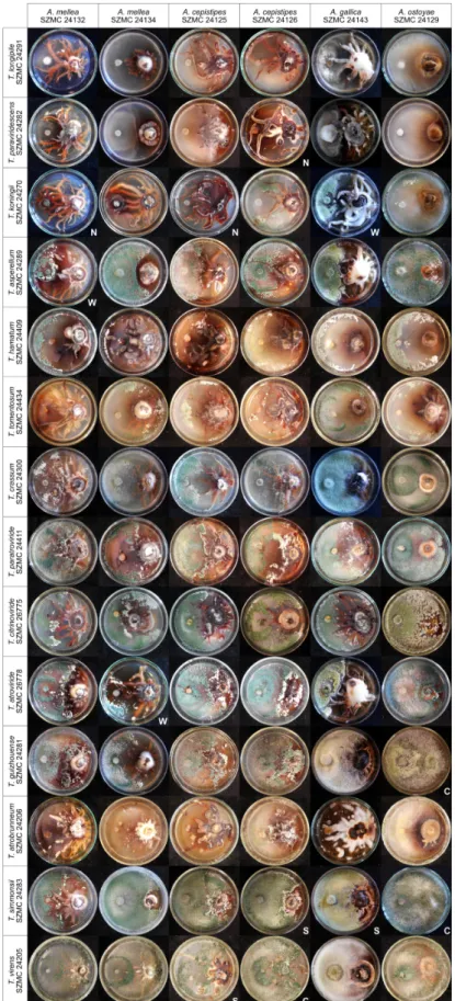
3.2. In Vitro Antagonism of the Isolated Trichoderma Strains Towards Armillaria Species
All 8 isolates ofT. virens, and some isolates ofT. simmonsii, T. atrobrunneum, T. guizhouense, T. atroviride, T. citrinoviride, T. paratroviride, T. hamatum, and T. tomentosum, proved to be highly effective against the 25 examined Armillaria isolates. Figure1shows a representative set of plate photographs taken during the in vitro antagonism experiments and reflecting all species combinations of Trichoderma and Armillaria. In many cases, antagonistic Trichoderma isolates were able to overgrow Armillaria colonies and intensely produce conidia on their surface, thereby potentially restricting Armillaria growth. Isolates ofT. virens, such as SZMC 24205, SZMC 24294, SZMC 24303, SZMC 24293, and SZMC 26774, proved to be the best in vitro antagonists with BCI values above 80 for more than 17 out of 25 Armillaria strains (Table2). Isolates ofT. simmonsiishowed high in vitro antagonistic abilities with BCI values above 80 for more than 15 out of the 25 tested Armillaria isolates, whereas theT.
koningii,T. asperellum,T. paraviridescens, andT. longipileisolates had the lowest BCI values against almost all of the tested Armillaria isolates. The distribution of antagonistic Trichoderma species with higher BCI values showed a geographical pattern. Except for the two species—T. simmonsiiandT. atroviride, isolated from both the oak stand in Keszthely—and the spruce forest in Rosalia, having relatively high antagonistic activities, species that were only isolated from the Keszthely Hills, includingT.
virens,T. atrobrunneum,T. hamatum,T. citrinoviride,T. paratroviride, andT. tomentosum, exhibited good in vitro antagonistic abilities against most of the tested Armillaria isolates. All isolates ofT. koningii,T.
asperellum,T. paraviridesens, andT. longipile, which seem to dominate in the soil of the Rosalia forest, showed lower BCI values against most Armillaria isolates.
Figure 1.In vitro antagonism of Trichoderma strains from different species againstArmillaria mellea, A. cepistipes,A. gallica, andA. ostoyae. Example plates are marked with N: no inhibition; W: weak inhibition; S: strong inhibition; C: complete overgrowth of Armillaria by Trichoderma.
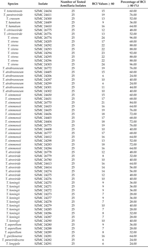
Table 2.In vitro antagonistic abilities of Trichoderma isolates towards Armillaria isolates.
Species Isolate Number of Tested
Armillaria Isolates BCI Values≥80 Percentage of BCI
≥80 (%)
T. tomentosum SZMC 24434 25 15 60.00
T. paratroviride SZMC 24411 25 19 76.00
T. crassum SZMC 24300 25 13 52.00
T. hamatum SZMC 24409 25 9 36.00
T. hamatum SZMC 24410 25 16 64.00
T. citrinoviride SZMC 26775 25 12 48.00
T. citrinoviride SZMC 26776 25 13 52.00
T. virens SZMC 26774 25 24 96.00
T. virens SZMC 24205 25 17 68.00
T. virens SZMC 24292 25 22 88.00
T. virens SZMC 24293 25 22 88.00
T. virens SZMC 24294 25 23 92.00
T. virens SZMC 24295 25 22 88.00
T. virens SZMC 24296 25 22 88.00
T. virens SZMC 24303 25 24 96.00
T. atrobrunneum SZMC 26772 25 19 76.00
T. atrobrunneum SZMC 24405 25 15 60.00
T. atrobrunneum SZMC 24206 25 6 24.00
T. atrobrunneum SZMC 24297 25 10 40.00
T. atrobrunneum SZMC 24299 25 6 24.00
T. atrobrunneum SZMC 24301 25 11 44.00
T. atrobrunneum SZMC 24302 25 10 40.00
T. simmonsii SZMC 24430 25 15 60.00
T. simmonsii SZMC 24431 25 17 68.00
T. simmonsii SZMC 26770 25 21 84.00
T. simmonsii SZMC 24433 25 16 64.00
T. simmonsii SZMC 24435 25 19 76.00
T. simmonsii SZMC 24436 25 18 72.00
T. simmonsii SZMC 24403 25 17 68.00
T. simmonsii SZMC 24404 25 18 72.00
T. simmonsii SZMC 26773 25 19 76.00
T. simmonsii SZMC 24408 25 10 40.00
T. simmonsii SZMC 26777 25 17 68.00
T. simmonsii SZMC 24412 25 16 64.00
T. simmonsii SZMC 24298 25 13 52.00
T. simmonsii SZMC 24283 25 18 72.00
T. simmonsii SZMC 24284 25 16 64.00
T. atroviride SZMC 26778 25 12 48.00
T. atroviride SZMC 26779 25 8 32.00
T. atroviride SZMC 26780 25 10 40.00
T. atroviride SZMC 24413 25 16 64.00
T. atroviride SZMC 24414 25 14 56.00
T. atroviride SZMC 24274 25 14 56.00
T. atroviride SZMC 24275 25 12 48.00
T. atroviride SZMC 24276 25 10 40.00
T. koningii SZMC 24270 25 9 36.00
T. koningii SZMC 24271 25 9 36.00
T. koningii SZMC 24272 25 9 36.00
T. koningii SZMC 24273 25 7 28.00
T. koningii SZMC 24277 25 9 36.00
T. koningii SZMC 24278 25 7 28.00
T. koningii SZMC 24279 25 10 40.00
T. koningii SZMC 24285 25 7 28.00
T. koningii SZMC 24286 25 8 32.00
T. koningii SZMC 24287 25 7 28.00
T. koningii SZMC 24290 25 10 40.00
T. asperellum SZMC 24280 25 9 36.00
T. asperellum SZMC 24288 25 7 28.00
T. asperellum SZMC 24289 25 8 32.00
T. guizhouense SZMC 24281 25 16 64.00
T. paraviridescens SZMC 24282 25 6 24.00
T. longipile SZMC 24291 25 6 24.00
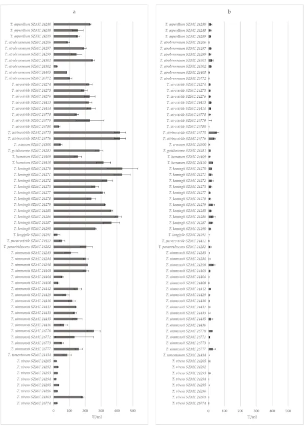
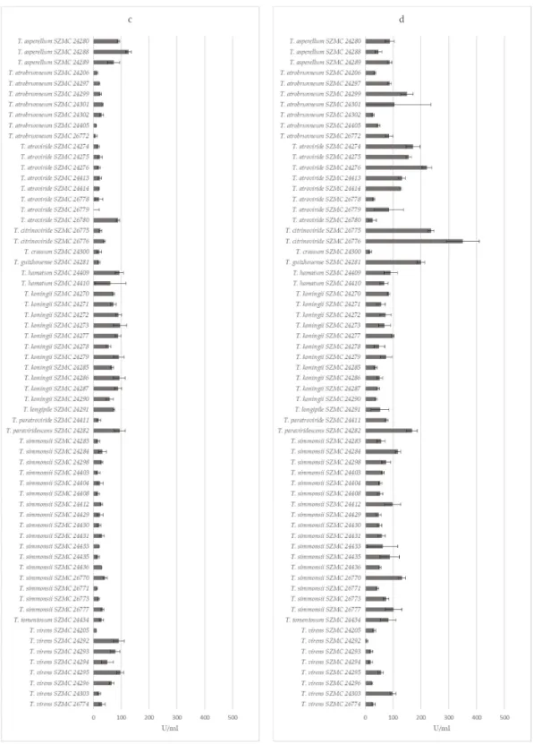
3.3. Extracellular Enzyme Production of the Trichoderma Isolates
The extracellular enzyme measurements revealed that isolates of the same Trichoderma species have similar enzyme activity values (Figure2). Altogether, most of the isolates could be characterized by highβ-glucosidase (Figure2a) and phosphatase (Figure2d), but lower cellobiohydrolase (Figure2b) andβ-xylosidase (Figure2c) activities. The 11T. koningiiisolates, along withT. asperellum SZMC
24288, SZMC 24280, andT. paraviridescensSZMC 24282, showed goodβ-glucosidase andβ-xylosidase activities. Among them,T. paraviridescensSZMC 24282 had good phosphatase, whereasT. koningii SZMC 24286 and SZMC 24270 good cellobiohydrolase activities as well. The examinedT. virens,T.
atrobrunneum,T. simmonsii, andT. atrovirideisolates showed lower activity levels for all enzymes tested, except forForests 2019, 10, x FOR PEER REVIEW T. atrovirideSZMC 26780, which had a very highβ-xylosidase activity. 13 of 23
Figure 2.Cont.
Figure 2. Extracellular enzyme activities of Trichoderma isolates derived from forest soil samples: (a) β-glucosidase, (b) cellobiohydrolase, (c) β-xylosidase, and (d) phosphatase.
3.4. Potential Plant Growth-Promoting Traits of the Isolated Trichoderma Strains
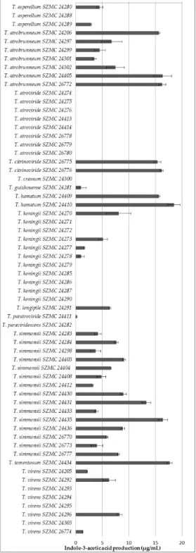
All of the examined isolates from T. atrobrunneum, T. simmonsii, T. hamatum, and T. citrinoviride, along with the single isolates of T. tomentosum, T. longipile, T. paratroviride, and T. guizhouense, proved to be IAA producers, whereas the T. atroviride isolates, as well as the examined single isolates of T.
paraviridescens and T. crissum, were unable to produce this metabolite (Figure 3). Both producers and Figure 2. Extracellular enzyme activities of Trichoderma isolates derived from forest soil samples:
(a)β-glucosidase, (b) cellobiohydrolase, (c)β-xylosidase, and (d) phosphatase.
3.4. Potential Plant Growth-Promoting Traits of the Isolated Trichoderma Strains
All of the examined isolates fromT. atrobrunneum,T. simmonsii,T. hamatum, andT. citrinoviride, along with the single isolates ofT. tomentosum,T. longipile,T. paratroviride, andT. guizhouense, proved to be IAA producers, whereas theT. atrovirideisolates, as well as the examined single isolates ofT.
paraviridescensandT. crissum, were unable to produce this metabolite (Figure3). Both producers and non-producers were found among the examined isolates ofT. virens,T. koningii, andT. asperellum.The
highest amounts of IAA were detected in the case of the isolates ofT. tomentosum,T. citrinoviride,T.
hamatum, as well as certain isolates ofThe highest amounts of IAA were detected in the case of the isolates of T. tomentosum, T. citrinoviride, T. atrobrunneum,T. simmonsii, andT. koningii.
T. hamatum, as well as certain isolates of T. atrobrunneum, T. simmonsii, and T. koningii
Figure 3.Indole-3-acetic acid production of Trichoderma isolates derived from forest soil samples.
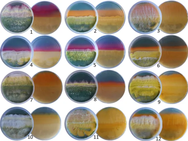
All theTrichodermaisolates tested were able to produce siderophores, which was indicated by the change of the color of blue medium to orange or purple (Figure4). The different colors of the medium suggested that the produced siderophores were structurally different. There are two major groups of siderophores, known as catechol-type and hydroxamate-type [25]. In the case of catechol-type siderophores the medium turns to purple, which was detected in the case of theT.
atroviride,T. paraviridescens, andT. koningiiisolates, whereas the hydroxamate-type siderophores result
Forests2019,10, 1013 16 of 22
in an orange color, as it was the case for all other examined species (Figure4). The isolates of the speciesT. asperellumseemed to produce both types of siderophores (see plate 8 on Figure4).
Figure 3. Indole-3-acetic acid production of Trichoderma isolates derived from forest soil samples.
All the Trichoderma isolates tested were able to produce siderophores, which was indicated by the change of the color of blue medium to orange or purple (Figure 4). The different colors of the medium suggested that the produced siderophores were structurally different. There are two major groups of siderophores, known as catechol-type and hydroxamate-type [25]. In the case of catechol- type siderophores the medium turns to purple, which was detected in the case of the T. atroviride, T.
paraviridescens, and T. koningii isolates, whereas the hydroxamate-type siderophores result in an orange color, as it was the case for all other examined species (Figure 4). The isolates of the species T.
asperellum seemed to produce both types of siderophores (see plate 8 on Figure 4).
Figure 4. Production of siderophores on modified CAS agar medium by forest-derived Trichoderma isolates belonging to different species. (1) T. atroviride SZMC 24275, (2) T. virens SZMC 24205, (3) T.
hamatum SZMC 24410, (4) T. paraviridescens SZMC 24282, (5) T. koningii SZMC 24287, (6) T. citrinoviride SZMC 26776, (7) T. simmonsii SZMC 24431, (8) T. asperellum SZMC 24280, (9) T. atrobrunneum SZMC 24206, (10) T. guizhouense SZMC 24281, (11) T. tomentosum SZMC 24434, and (12) T. crassum SZMC 24300.
3.5. Field Experiment in a Heavily Armillaria-Damaged Forest in the Keszthely Hills
Two Trichoderma isolates—T. virens SZMC 24205 and T. atrobrunneum SZMC 24206—were selected for a field experiment. Both strains were isolated from a Keszthely soil sample associated with decaying Armillaria rhizomorphs, which have not revealed any Armillaria growth upon isolation attempts; furthermore, both exerted very good in vitro antagonistic abilities towards the tested Armillaria isolates and were able to produce hydroxamate-type siderophores. The isolates were applied to Turkey oak seedlings as a root treatment before planting in the form of a conidial suspension (106 conidia per mL for both). The total survival rates calculated after 6 months for 120
Figure 4.Production of siderophores on modified CAS agar medium by forest-derived Trichoderma isolates belonging to different species. (1) T. atrovirideSZMC 24275, (2)T. virens SZMC 24205, (3)T. hamatumSZMC 24410, (4)T. paraviridescens SZMC 24282, (5)T. koningiiSZMC 24287, (6)T.
citrinovirideSZMC 26776, (7)T. simmonsiiSZMC 24431, (8)T. asperellumSZMC 24280, (9)T. atrobrunneum SZMC 24206, (10)T. guizhouenseSZMC 24281, (11)T. tomentosumSZMC 24434, and (12)T. crassum SZMC 24300.
3.5. Field Experiment in a Heavily Armillaria-Damaged Forest in the Keszthely Hills
Two Trichoderma isolates—T. virensSZMC 24205 andT. atrobrunneumSZMC 24206—were selected for a field experiment. Both strains were isolated from a Keszthely soil sample associated with decaying Armillaria rhizomorphs, which have not revealed any Armillaria growth upon isolation attempts;
furthermore, both exerted very good in vitro antagonistic abilities towards the tested Armillaria isolates and were able to produce hydroxamate-type siderophores. The isolates were applied to Turkey oak seedlings as a root treatment before planting in the form of a conidial suspension (106conidia per mL for both). The total survival rates calculated after 6 months for 120 treated and 115 control trees were 84.3% and 54.7%, respectively (Table3), indicating that the applied treatment had a beneficial effect on the survival of oak seedlings planted into the soil of an Armillaria-infested forest area.
Table 3. Survival of Trichoderma-treated and control trees 6 months after planting into heavily Armillaria-infested soil.
Parcel Total No.
1Dead No.
2Growing No.
3Survivor No.
4FM No.
5Corrected Total No.
Dead
%
Growing
%
Growing+ Survivor %
1 (treated) 40 5 24 3 8 32 15.6 75.0 84.4
2 (treated) 40 5 27 5 3 37 13.5 73.0 86.5
3 (treated) 40 7 28 4 1 39 17.9 71.8 82.1
Total 120 17 79 12 12 108 15.7 73.1 84.3
4 (untreated) 40 23 5 2 10 30 76.7 16.7 23.3
5 (untreated) 39 13 19 2 5 34 38.2 55.9 61.8
6 (untreated) 36 7 24 0 5 31 22.6 77.4 77.4
Total 115 43 48 4 20 95 45.3 50.5 54.7
1already degraded/disappeared and dry seedlings;2positive biomass production;3still alive but no biomass production;4failed measurement (size values after 6 month lower than the ones measured directly after planting);
5Total—FM.
4. Discussion
Armillaria fruiting bodies, rhizomorphs, and soil samples were collected at previously established study sites from both spruce and oak stands; all of them with abundant rhizomorph and mushroom production. The conifer sampling sites selected in Rosalia represented a native environment for Norway spruce with single clones ofA. ostoyae andA. cepistipescolonies appearing only around relatively freshly cut trunks. All identified genets appeared non-damaging and tolerable by the surrounding live trees. In contrast, the Turkey oak stand from Keszthely, Hungary was a heavily infested area with multiple A. melleaandA. gallicaclones merged to form a continuous coverage of the whole stand. All remaining standing trees were showing symptoms of Armillaria infections. The same bulk, rhizospheric and rhizomorph-associated soil samples were also subjected to Trichoderma isolation. The reported diversity of Trichoderma had expanded to ~75 species in temperate Europe [27,28]. All the Trichoderma species collected in this study from Keszthely and Rosalia (T. tomentosum,T. paratroviride, T. crassum,T. hamatum,T. citrinoviride,T. atrobrunneum,T. virens,T. simmonsii,T. atroviride,T. koningii,T.
asperellum,T. guizhouense,T. paraviridescens, andT. longipile) had already been reported from Southern Europe [29]. Among them, the speciesT. citrinoviride,T. atroviride,T. koningii,T. paraviridescens, and T. longipilewere also identified from Central Europe [30]. TheT. harzianumspecies complex (also known asT. harzianum sensu lato) from the Harzianum clade of the genus Trichoderma was supposed to comprise at least 14 species [31], including the more recently described, biocontrol-relevant species ofT. atrobrunneum,T. guizhouense, andT. simmonsiithat were also found at both locations of our current investigation.
The application of biocontrol agents as alternatives to chemical fungicides reduces the impacts and risks on human health as well as on the environment [32]. Trichoderma species as effective biocontrol agents against diverse genera of pathogenic fungi can be used for plant disease management, especially in the case of soilborne diseases. Strains of theT. koningii,T. asperellum,T. atroviride,T. hamatum,T.
virens, andT. harzianumspecies complex and other Trichoderma taxa have been officially registered and commercialized as crop protection products and microbial fungicides throughout the world including the European countries [33].
Antagonistic activity assessment in vitro by dual culture assay has demonstrated in this study that, besidesT. virens,T. atroviride, andT. hamatumand the members of theT. harzianumspecies complex (T. simmonsii,T. atrobrunneum, andT. guizhouense), strains ofT. citrinoviride,T. paratroviride, andT.
tomentosumalso proved to be effective in vitro antagonists of Armillaria species with the potential to be used as biocontrol agents against Armillaria root rot. On the other hand, certain species and strains of Trichoderma showed weak antagonistic abilities against Armillaria strains reflected by low BCI values. For example, all the tested isolates ofT. koningiiandT. asperellumhad lower BCI values than the isolates ofT. virens,T. simmonsii,T. atrobrunneum,T. atroviride, andT. hamatum. Previously,T.
koningiiandT. asperellumshowed excellent antagonistic activities during the application against other plant pathogens. For instance,T. koningiishowed the highest growth inhibition ofRhizoctonia solani
causing root rot in cotton, followed byT. viride,T. harzianum, andT. virens[34]. Similarly,T. asperellum showed effective antagonistic activity against the white-rot fungusPhellinus noxius, the causal agent of an epidemic brown root rot disease of various coniferous and broad-leaved tree species [35].
The Trichoderma isolates collected during this study were characterized for their abilities to produce polysaccharide-degrading enzymes of the cellulolytic (β-glucosidase and cellobiohydrolase) and xylanolytic (β-xylosidase) enzyme systems that are important for efficient competition in habitats rich of plant-derived polysaccharides, as well as acidic phosphatase playing a role in phosphorus mobilization. Interestingly, the isolates of species with the best in vitro antagonistic abilities against Armillaria (T. virens,T. atrobrunneum,T. simmonsii, andT. atroviride) were among the worst producers of these extracellular enzymes and vice versa, suggesting that the main antagonistic mechanism of these Trichoderma species against Armillaria may be mycoparasitism of hyphae and rhizomorphs rather than competition for polysaccharides or increasing phosphorous availability to the tree roots. Certain Trichoderma species (e.g.,T. reesei) can be characterized with a predominantly saprophytic behavior, while others (e.g.,T. virensandT. atrovirideand members of the Harzianum clade) are described as successful mycoparasitic species [14]. Extracellular hydrolytic enzymes are known as key players of both the saprophytic and the mycoparasitic behavior: the former is relying on the production of plant polysaccharide-degrading enzyme systems like cellulases or xylanases, whereas the latter is based on CWDEs targeting the cell wall of the fungal host (glucanases, chitinases, and proteases).
The competition for iron may also contribute to the anti-Armillaria activity of the examined Trichoderma iolates, as the production of siderophores proved to be a general feature among them.
Previous studies reported that certain strains ofT. asperellum,T. atrobrunneum,T. atroviride,T. gamsii,T.
hamatum,T. harzianum,T. polysporum,T. reesei,T. virens,T. paratroviride,T. pyramidale,T. rufobrunneum, T. thermophilum,T. viridulum,T. guizhouense, andT. simmonsiiwere mainly used as biocontrol agents due to their siderophore producing abilities [36–40]. Wang and Zhuang [40] firstly reported the siderophore-producing ability ofT. guizhouenseandT. simmonsii. To the best of our knowledge, the production of siderophores byT. citrinoviride,T. koningii,T. crassum,T. longipile, andT. paraviridescens strains is firstly demonstrated in the present study.
From the forest-derived Trichoderma isolates of our study, 40 were able to produce IAA with T. hamatumSZMC 24410,T. citrinovirideSZMC 26776, andT. atrobrunneumSZMC 24206 producing the highest quantities (18.49, 16.198, and 15.64µg/mL, respectively). Data in the literature about the IAA-producing ability of Trichoderma strains is limited. Chagas et al. [41] investigated the IAA production ofT. harzianum,T. pinnatum,T. longibrachiatum, andT. asperelloides, as well as two strains of T. virens, and recorded production values of 2.9–3.2µg/mL. In the present study, the detected values were in a wider concentration range (1.349–8.248µg/mL). A previous study used a similar method to show that strains ofT. atrobrunneum,T. guizhouense,T. paratroviride, andT. simmonsiiproduce IAA at concentrations of 6.6, 10.3–21.8, 4.1–8.5, and 6.0–7.2µg/mL [40]. In our study, the examinedT.
guizhouenseandT. paratrovirideisolates produced lower amounts of IAA. We also present the first data about the IAA production ofT. koningii,T. longipile,T. tomentosum,T. hamatum, andT. citrinoviride.
A comparison of the data about the in vitro antagonism and the production of indole-3-acetic acid, siderophores as well as extracellularβ-glucosidase, cellobiohydrolase,β-xylosidase, and phosphatase enzymes among the Trichoderma isolates mostly revealed very similar values for isolates deriving from the same sample and belonging to the same species, suggesting that the respective isolates are clonal and represent the same strain, which is, in many cases, also supported by identical sequences of thetef1αfragment used for species-level identification (Table1). Examples for probable clonality are the isolate groupsT. koningiiSZMC 24277/24278/24279, T. atrovirideSZMC 24274/24275/24276 and SZMC 24413/24414,T. simmonsiiSZMC 24435/26771/24436/24403/24404 and SZMC 26777/24412, orT. atrobrunneumSZMC 26772/24405. On the other hand, in certain cases, the differences in the physiological parameters ortef1αsequences clearly revealed the presence of multiple strains from the same species in the same sample, e.g.,T. koningiiSZMC 2470/2471 vs.T. koningiiSZMC 2472/2473,T.
simmonsiiSZMC 26770 vs. SZMC 24429/24430/24431/24433,T. hamatumSZMC 24409 vs. SZMC 24410,