Secondary Metabolites and Bioactivities of Aspergillus ochraceopetaliformis Isolated from Anthurium brownii
Hao-Chun Hu,
+Chi-Ying Li,
+Yi-Hong Tsai, Dai-Yun Yang, Yang-Chang Wu, Tsong-Long Hwang, Shu-Li Chen, Ferenc Fülöp, Attila Hunyadi, Chia-Hung Yen, Yuan-Bin Cheng, and Fang-Rong Chang*
Cite This:ACS Omega2020, 5, 20991−20999 Read Online
ACCESS
Metrics & More Article Recommendations*
sı Supporting InformationABSTRACT: Five new polyketides, asperochrapyran (1) and asperochralactones A−D (2−5), along with 12 known polyketides (6−17), were obtained from the fungal strain Aspergillus ochraceopetaliformis. Structures of all isolates were elucidated by their spectroscopic parameters. The relative configurations of the new compounds were deduced by the data of coupling constants and NOESY spectra. The absolute configurations were determined by the comparison of experimental and calculated ECD spectra.
Moreover, the plausible biosynthesis pathway of major isolates was proposed as well. Anti-inflammatory activity of compounds5and 7−17 were evaluated with human neutrophils in response to the stimulation of formyl-methionyl-leucyl phenylalanine (fMLP).
Asperlactone (9), aspyrone (13), and (−)-(3R)-mellein (14)
exerted superoxide anion inhibition at 30±9%, 29±9%, and 26±12%, respectively, at 10μM. The capacities of asperlactone (9), aspilactonol B (10), penicillic acid (12), and (−)-(3R)-mellein (14) in elastase release inhibition were revealed as 25±4%, 38± 8%, 25±5%, and 34±9%, respectively, at 10μM.
■
INTRODUCTIONFungi generate diverse groups of secondary metabolites with intriguing activities.1 Several bioactive secondary metabolites have been applied in agriculture or pharmaceutical industries.1 Following the cooperation of a high-throughput screening project with the Dr. Cecilia Koo Botanic Conservation Center (KBCC) in Taiwan providing extraordinary plant materials, we obtained a fungal strain, isolated from leaves of Anthurium brownii(A. brownii) Mast, which was identified asAspergillus ochraceopetaliformis (A. ochraceopetaliformis) Bat. and Maia.
TheAspergillusgenus contains a large number of species and is widely distributed in natural environments. This genus was reported as a prolific source in producing bioactive secondary metabolites, such as alkaloids,2 glycosides,3 peptides,4 poly- ketides,5steroids,6and terpenoids.7These molecules exhibited diverse biological activities, including antibacterial,8,9 anti- fungal,10 cytotoxic,11 nematicidal,12 radical scavenging,13 ovicidal, and insect growth-regulating14 activities. The chemical diversity and broad spectrum of bioactivities make this genus to be a potential resource in the discovery of new drug development. For instance, the renowned case of cholesterol-lowing drug, lovastatin, was discovered from Aspergillus terreus (A. terreus). It was proved as a trigger of HMG-CoA reductase inhibition and clinically used as a hypercholesterolemia and cardiovascular disease ameliorator.15 Moreover, simvastatin, a derivative synthesized and modified
substance generated from A. terreus, is another clinical drug used for a similar purpose of lovastatin.15These cases attracted our attention and encouraged us on exploring new potential bioactive molecules from theAspergillusgenus.
Although A. ochraceopetaliformis was considered as not a common pathogen to humans, it had once been found in human skin lesions and reported as an invader to cause onychomycosis.16,17Notably, this fungal species was also found in ocean sponges and even discovered from an Antarctic soil sample.5,18,19 Furthermore, Wang et al. reported that this fungal species would produce various types of bioactive sesquiterpenoids with fascinating activities such as those against of influenza viruses or lipopolysaccharide-induced NO release in RAW 264.7 cell lines.5,18
In a current study, we fermented A. ochraceopetaliformis through a liquid fermentation methodology and the ethyl acetate extract ofA. ochraceopetaliformiswas found to possess an anti-inflammatory effect on inhibiting superoxide anion
Received: May 27, 2020 Accepted: July 28, 2020 Published: August 14, 2020
Article http://pubs.acs.org/journal/acsodf
copying and redistribution of the article or any adaptations for non-commercial purposes.
Downloaded via 213.197.95.141 on September 9, 2020 at 07:18:58 (UTC). See https://pubs.acs.org/sharingguidelines for options on how to legitimately share published articles.
generation and elastase release at 10 μg/mL (103 ± 1% and 107±6%, respectively).
Continuing our efforts on discovery of the chemical diversity and biological activities of natural products, we performed further chemical and biological investigation on this fungus and identified 17 polyketide secondary metabolites, including 5 new polyketides (1−5) and 12 known polyketides (6−17).
Herein, we described the structural elucidation of new secondary metabolites (1−5) and the extensive determination of the absolute configurations by computational approaches.
We evaluated the isolated compounds for several bioactivity assays, including those for cytotoxicity and anti-inflammatory properties. Moreover, we proposed the plausible biosynthesis pathway of the isolated polyketide secondary metabolites.
■
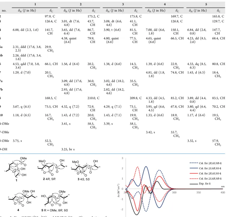
RESULTS AND DISCUSSIONFive new polyketides, asperochrapyran (1) and asperochra- lactones A−D (2−5), together with 12 known polyketides, (5S,6R,8S,9R)-8,9-dihydroxy-8,9-deoxyaspyrone (6), aspyro- nol (7), dihydroaspyrone (8), asperlactone (9), aspilactonol B (10), asperochrin B (11), penicillic acid (12), aspyrone (13), (−)-(3R)-mellein (14), aspinonediol (15), aspinotriols A (16), and aspinotriols B (17) were isolated from the ethyl acetate extract of A. ochraceopetaliformis (Figure 1). New
compounds were elucidated and identified by their spectro- scopic data as well as by analyzing their stereochemistries with experimental and electronic circular dichroism (ECD) calculations and conformational searches to establish their absolute configurations.
Asperochrapyran (1) was obtained as a colorless oil with a specific rotation of [α]D24 = −83 (c 0.08, MeOH). The molecular formula (C10H16O5) was confirmed by the analysis of its13C NMR and HR-ESI-MS data (m/z239.08887 [M + Na]+), implying 3 degrees of unsaturation. The infrared (IR) spectrum presented prominent absorption bands for hydroxyl (3415 cm−1), conjugated ester carbonyl (1708 cm−1), and C− O functional groups (1087 cm−1). The UV spectrum showed an absorption at 218 nm. The1H NMR data of1 (Table 1) indicated two methyls atδH1.18 (d,J= 6.5 Hz) and 1.29 (d,J
= 7.0 Hz), one olefinic methane at δH6.88 (dd,J = 2.3, 1.6 Hz), two oxymethines at δH 3.67 (q, J = 6.5 Hz) and 4.53
(qdd, J= 7.0, 3.8, 3.4 Hz), one methoxy atδH3.75 (s), and one methylene atδH2.26 (ddd,J= 17.6, 3.4, 1.6 Hz) and 2.31 (ddd, J = 17.6, 3.8, 2.3 Hz). Moreover, the 13C NMR and DEPT data (Table 1) revealed 10 carbon signals. These signals resulted from one ester carbonyl (δC 168.5), one olefinic methine (δC141.7), one nonprotonated sp2carbon (δC126.4), one hemiacetal carbon (δC 97.9), two oxygenated methines (δC66.1 and 73.5), one methylene (δC29.9), one methoxy (δC
52.3), and two methyls (δC16.7 and 20.1). According to its MS and NMR data, 2 degrees of unsaturation, CC double bond and carbonyl functionalities, were disclosed and reduced to one degree of unsaturation. By comparing these data with a previous study,1would be inferred as a polyketide framework secondary metabolite.20
In interpretation of the COSY spectrum, two fragments, H-4 (δH6.88)/H2-5 (δH2.26 and 2.31)/H-6 (δH4.53)/H-7 (δH
1.29) and H-9 (δH 3.67)/H-10 (δH 1.18), were observed (Figure 2). The HMBC correlations were found from H2-5 to C-3 (δC126.4). These correlations established a 3,6-dihydro- 2H-pyran ring system. The side-chain moiety of C-9 (δC73.5) and C-10 (δC16.7) was linked to the pyran ring by HMBC cross peaks from both H-9 and H-10 to C-2 (δC97.9). Further, the HMBC correlations from 8-OMe (δH3.75), and H-4 (δH
6.88) to C-8 (δC 168.5) were used to predict the linkage of ester carbonyl to C-3 (δC 126.4) (Figure 2). Based on the above data, the gross structure of1was established.
In terms of the stereochemistry of1, the C-6 can be scripted by the coupling constants (J5,6= 3.8 and 3.4 Hz). As a result, the H-6 and CH3-7 were sited in pseudo-equatorial and pseudo-axial positions, respectively. All the possible absolute results of 1, 2S,6S,9S-1, 2R,6R,9R-1, 2S,6R,9S-1, 2R,6S,9R-1, 2S,6S,9R-1, 2R,6R,9S-1, 2S,6R,9R-1, and 2R,6S,9S-1, were computed by the conformational search algorithms. The experimental J5,6 coupling constants of these conformers are composed with an anti and a gauche orientation. However, the calculated results showed the data with both gauche orientations. Hence, the prediction of calculated conformers, 2S,6S,9S-1, 2R,6R,9R-1, 2S,6S,9R-1, and 2R,6R,9S-1, cannot match to theJ5,6coupling constants. On the other hand, all the other conformers, such as 2S,6R,9S-1, 2R,6S,9R-1, 2S,6R,9R-1, and 2R,6S,9S-1, not only conform with the J5,6 coupling constants but also meet the data of the absence in NOESY correlation between H3-7 and H-10 (Figure S6). Thus, the absolute configuration of 1 was further determined by comparing the calculated and experimental ECD spectra with Gaussian 09 software. The ECD spectra of possible con- formers, 2S,6R,9S-1, 2R,6S,9R-1, 2S,6R,9R-1, and 2R,6S,9S-1, are shown inFigure 3. The experimental ECD spectrum of1 was approximate to 2R,6S,9S-1, which exhibited a calculated ECD spectrum with positive Cotton effects at 240 and 270 nm and negative Cotton effects at 223 and 246 nm. Therefore, the structure and absolute stereochemistry of 1 were completely elucidated as descripted, and it was named asperochrapyran (1).
Asperochralactone A (2) was isolated as a colorless oil, and the high-resolution ESI-MS data showed a molecule peak at m/z 239.08889 [M + Na]+, which indicated the molecular formula as C10H16O5 and 3 as the index of hydrogen deficiency. The γ-lactone, ketone, and C−O signals were found in the IR spectrum at 1766, 1720, and 1078 cm−1, respectively. In the 1H NMR spectrum of 2 (Table 1), the proton resonances suggested two methyls atδH1.43 (d,J= 7.2 Hz) and 1.56 (d, J= 6.4 Hz), three oxymethines atδH3.65 Figure 1.Structures of all isolates (1−17).
(dd,J= 7.6, 6.4 Hz), 4.32 (q,J= 7.2 Hz), and 4.38 (quint,J= 6.4 Hz), one methylene atδH2.93 (dd,J= 17.6, 4.8 Hz) and 3.09 (J= 17.6, 4.8 Hz), one methine atδH3.01 (dt,J= 7.6, 4.8 Hz), and one methoxy atδH 3.41 (s). Furthermore, the13C and DEPT NMR data of2 (Table 1) denoted one carbonyl (δC210.0), one ester carbonyl (δC175.2), three oxygenated methines (δC 72.9, 79.9, and 86.7), one methoxy (δC 58.5), one methylene (δC 36.0), one methine (δC 43.7), and two methyls (δC 20.0 and 20.2). According to the measured IR, MS, NMR, and UV (212 nm) spectral data analysis and referring to a previous report,20compound2was identified as a polyketide secondary metabolite with 3 unsaturated degrees counted inγ-lactone and ketone moieties.
The COSY correlations between H2-7 (δH2.93 and 3.09)/
H-3 (δH3.01)/H-4 (δH3.65)/H-5 (δH4.38)/H3-6 (δH1.56) and H-9 (δH 4.32)/H3-10 (δH 1.43) indicated two partial fragments (Figure 2). The HMBC correlations of H2-7/C-2 (δC 175.2) and 4-OMe (δH 3.41)/C-4 (δC 86.7) and aforementioned COSY fragments were used to elaborate the γ-lactone moiety (Figure 2). Moreover, the HMBC corre- spondence from H2-7 and H3-10 to C-8 (δC210.0) and the remaining COSY fragment suggested the connection with the γ-lactone moiety (Figure 2). Thus, the planar structure of2 was established. Additionally, the NOESY correlation of H-3/
H-4/H-5 was observed (Figure S12), which indicated that those protons were at the same orientation.
Table 1.1H (400 MHz) and13C (100 MHz) NMR Spectroscopic Data of Compound 1 in CD3OD and Compounds 2, 4, and 5 in CDCl3; 1H (600 MHz) and13C (150 MHz) NMR Spectroscopic Data of Compound 3 in CDCl3
1 2 3 4 5
no. δH(Jin Hz) δC δH(Jin Hz) δC δH(Jin Hz) δC δH(Jin Hz) δC δH(Jin Hz) δC
2 97.9, C 175.2, C 175.8, C 169.7, C 165.8, C
3 126.4, C 3.01, dt (7.6,
4.8)
43.7, CH
3.08, dt (6.6, 4.6)
41.1, CH
126.8, C 129.7, C
4 6.88, dd (2.3, 1.6) 141.7, CH
3.65, dd (7.6, 6.4)
86.7, CH
3.90, t (6.6) 81.5, CH
7.00, dd (6.6, 1.8)
150.1, CH
6.84, dd (2.6, 0.8)
147.7, CH
5 4.38, quint
(6.4)
79.9, CH
4.80, quint (6.6)
77.5, CH
4.65, quint (6.6)
66.5, CH 4.23, dd (8.5, 2.6)
68.4, CH
5a 2.31, ddd (17.6, 3.8, 2.3)
29.9, CH2 5b 2.26, ddd (17.6, 3.4,
1.6)
6 4.53, qdd (7.0, 3.8, 3.4)
66.1, CH 1.56, d (6.4) 20.2, CH3
1.38, d (6.6) 14.3, CH3
1.39, d (6.6) 22.9, CH3
4.33, dq (8.5, 6.3)
80.8, CH
7 1.29, d (7.0) 20.1,
CH3
4.81, dd (1.8, 1.8)
74.8, CH 1.43, d (6.3) 18.4, CH3
7a 3.09, dd (17.6,
4.8)
36.0, CH2
3.02, dd (18.2, 4.6)
35.5, CH2
7b 2.93, dd (17.6,
4.8)
2.82, dd (18.2, 4.6)
8 168.5, C 210.0, C 209.8, C 4.33, dd (4.5,
1.8)
85.2, CH 3.89, dd (4.4, 0.8)
83.5, CH
9 3.67, q (6.5) 73.5, CH 4.32, q (7.2) 72.9, CH
4.29, q (7.1) 73.1, CH
3.95, qd (6.6, 4.5)
67.8, CH 3.80, qd (6.4, 4.4)
70.2, CH
10 1.18, d (6.5) 16.7,
CH3
1.43, d (7.2) 20.0, CH3
1.43, d (7.1) 19.9, CH3
1.33, d (6.6) 18.9, CH3
1.17, d (6.4) 19.5, CH3
4-OMe 3.41, s 58.5,
CH3
3.39, s 58.1,
CH3
7-OMe 3.42, s 55.7,
CH3
8-OMe 3.75, s 52.3,
CH3
3.32, s 57.9,
CH3
9-OH 3.25, br s
Figure 2.Key COSY (1H−1H) and HMBC (H→C) correlations of 1−5.
Figure 3.Calculated and experimental ECD spectra of1.
The absolute configuration was elucidated with ECD spectroscopies. Four possible candidates, 3S,4R,5R,9S-2, 3R,4S,5S,9S-2, 3R,4S,5S,9R-2, and 3S,4R,5R,9R-2, were computed by molecular modeling software, Spartan 16, to conduct the results of the conformation search. ECD spectra of each conformer were further calculated by Gaussian 09 as well.
The results showed that the experimental ECD spectrum exhibited positive Cotton effects at 221 and 278 nm and negative ones at 243, 316, and 377 nm. The data displayed high similarity to the calculated ECD pattern of 3S,4R,5R,9S-2 (Figure S34). Therefore, the absolute stereochemistry of compound 2 was deduced, and the name of the new compound, asperochralactone A, was given.
Asperochralactone B (3) was purified as a colorless oil with a specific optical rotation of [α]D24=−74 (c 0.03, MeOH) and possessed a molecular formula of C10H16O5and 3 degrees of unsaturation deduced from HR-ESI-MS data (m/z 239.08889 [M + Na]+). Two methyl groups [δH1.38 (d,J= 6.6 Hz) and 1.43 (d,J= 7.2 Hz)], three oxymethines [δH3.90 (t,J= 6.6 Hz), 4.29 (q,J= 7.1 Hz), and 4.80 (quint,J= 6.6 Hz)], one methylene [δH2.82 (dd,J= 18.2, 4.6 Hz) and 3.02 (dd, J= 18.2, 4.6 Hz)], one methine [δH3.08 (dt,J= 6.6, 4.6 Hz)], and one methoxy [δH3.39 (s)] were revealed from the1H NMR spectrum of3(Table 1). The13C and DEPT NMR spectra of 3(Table 1) indicated that 10 carbons can be categorized into one carbonyl (δC209.8), one ester carbonyl (δC175.8), three oxygenated methines (δC73.1, 77.5, and 81.5), one methoxy (δC58.1), one methylene (δC 35.5), one methine (δC41.1), and two methyls (δC14.3 and 19.9). The IR, MS, NMR, and UV data of3shared high similarity to compound2, implying that these two molecules possess the same framework.
The relative configuration of3was assigned on the basis of NOESY correlations. The NOESY cross peaks of H3-6/H-3/4- OMe and H-4/H-5 suggested that those two groups showed different orientations (Figure S18). The absolute configuration of 3 was further determined by comparing the ECD spectra between computational and experimental results to 2. Two possible conformers, 3S,4S,5S,9S-3 and 3R,4R,5R,9S-3, were simulated by Gaussian 09, which provided the calculated ECD spectra (Figure S35) for conformation search. The pattern of ECD curves of 3S,4S,5S,9S-3 demonstrated high similarity to the experimental ECD spectrum of3. Therefore, on the basis of the above data, the structure of3was identified and named asperochralactone B.
Asperochralactone C (4) was obtained as a colorless oil.
Three degrees of unsaturation was estimated on the basis of its molecular formula (C10H16O5), which was determined by the analysis of its HR-ESI-MS data (m/z239.08889 [M + Na]+).
The hydroxyl (3420 cm−1), α,β-unsaturated-γ-lactone (1748 and 1666 cm−1),20 and C−O (1087 cm−1) signals were observed in the IR spectrum. The 1H NMR of 4 (Table 1) displayed two methyl groups atδH1.33 (d,J= 6.6 Hz) and 1.39 (δH,J= 6.6 Hz), one olefinic methane atδH7.00 (dd,J= 6.6,1.8 Hz), four oxymethines atδH3.95 (qd,J= 6.6, 4.5 Hz), 4.33 (dd,J= 4.5, 1.8 Hz), 4.65 (quint,J= 6.6 Hz), and 4.81 (dd,J= 1.8, 1.8 Hz), and one methoxy atδH3.42 (s). The13C and DEPT NMR spectra (Table 1) revealed one ester carbonyl (δC 169.7), one olefinic methine (δC 150.1), one non- protonated sp2 carbon (δC 126.8), four oxygenated methines (δC66.5, 67.8, 74.8, and 85.2), one methoxy (δC55.7), and two methyls (δC18.9 and 22.9).
The1H−1H COSY spectrum (Figure 2) showed correlations between H-4 (δH7.00)/H-5 (δH4.65)/H3-6 (δH1.39) and H-
7 (δH 4.81)/H-8 (δH4.33)/H-9 (δH3.95)/H3-10 (δH1.33).
The α,β-unsaturated-γ-lactone moiety was established by the COSY fragment (H4/H5/H3-6) and the HMBC correlations of H-4/C-2 (δC169.7) and H-5/C-3 (δC126.8) (As shown in Figure 2). Furthermore, the side-chain fragment was attached to the α,β-unsaturated-γ-lactone by HMBC cross-peak correlations (H-4/C-7 and H-7/C-2 and C-3). The methoxy group was substituted to C-7, which was confirmed by an HMBC correlation of 7-OMe/C-7. According to aforemen- tioned data, the planar structure of4was determined.
The relative configuration of 4 was assigned by coupling constant analyses and experimental ECD. According to a previous report for xylogibloactone,20the coupling constant of erythrowas more than 4.0 Hz and that of thethreo was less than 2.5 Hz.20,21The coupling constants of H-7/H-8 (1.8 Hz) and H-8/H-9 (4.5 Hz) exhibited threo and erythro relative configurations, respectively. Notably, the absolute configura- tion at theγsite with a methyl or methoxy substitution onα,β- unsaturated-γ-lactone can be determined by the Cotton effect between 200−235 and 235−270 nm. The positive Cotton effect at 200−235 nm and a negative one at 235−270 nm represent aβconfiguration, and the negative Cotton effect at 200−235 nm and a positive one at 235−270 nm show anα configuration.22−24 According to the experimental ECD spectra (Figure S35), the H-5 of4displayed anαorientation and the configuration was in the R form. Additionally, the calculated ECD spectra of 5R,7R,8R,9R-4 and 5R,7S,8S,9S-4 were conducted by Gaussian 09. The absolute stereochemistry of4was assigned to be 5R,7S,8S,9Sby ECD data (Figure S36).
Finally, the structure of compound 4 was elucidated and named asperochralactone C.
Asperochralactone D (5) was isolated as a colorless oil. The molecular formula of C10H16O5was deduced for5based on a pseudo ion peak atm/z239.08881 [M + Na]+in the HR-ESI- MS and indicated 3 degrees of unsaturation. The IR spectrum illustrated hydroxyl (3409 cm−1), α,β-unsaturated-δ-lactone (1721 and 1650 cm−1),25 and C−O (1084 cm−1) function- alities. Furthermore, in analyzing its UV (208 nm) and 1D and 2D NMR spectral data, this compound exhibited high similarity to compound 7.20 However, the opposite optical rotation values of compounds 5 (+64) and 7 (−41.6)20 implied that these two compounds have different stereo- chemistries. The relative configurations of C-5, C-6, C-8, and C-9 were further determined by coupling-constant analyses.
Considering the conformation between H-5 and H-6, the coupling constant of the trans form was usually detected around 7.0 Hz, and thecis form would be observed near 3.0 Hz.2,25−27The coupling constant of H-5/H-6, calculated as 8.5 Hz, suggested the existence of atrans conformation. On the other hand, according to previous studies, the conformation between H-8/H-9 should be regarded as threo, while the coupling constant was more than 6.3 Hz. Furthermore, a coupling constant less than 5.0 Hz would be erythro.20,28−30 TheJvalue between H-8/H-9 was found to be 4.4 Hz, and the conformation would be considered aserythro. In addition, the absolute configuration of 5 was elucidated by comparing the experimental ECD spectra with the calculated results. The experimental ECD spectrum of5 showed a negative Cotton effect near 270 nm (n to π*), which suggested an S configuration at C-5,31 and C-6 was assigned to an R configuration accordingly. Due to the relative conformation between C-8 and C-9 assigned aserythro, the possible absolute stereochemistry of 5 would be 5S,6R,8S,9R or 5S,6R,8R,9S.
Compound 7 was reported to possess a 5S,6R,8S,9R configuration,20which implied that the structure of5 should be 5S,6R,8R,9Saccording to the aforementioned information.
In order to deduce the absolute configuration, the computa- tional procedures were carried out. The pattern of the ECD curve of the experimental result was approximate to the ECD curve simulated for 5S,6R,8R,9S-5 (Figure S37) and con- formation search result was calculated by carbon chemical shifts (Figure S42). Consequently, the structure and absolute configuration of5 were elucidated, and the name asperochra- lactone D was given.
Because the absolute configuration of compound6 has not been established yet,32 a series of computational experiments were carried out for determining this issue. The experimental ECD displayed a negative Cotton effect at 260 nm. It suggested an S configuration at C-5. Moreover, the experimental ECD spectrum of 6 was compared with 5 (8R,9S) and7(8S,9R).25The pattern of the ECD curve of6 exhibited high similarity to that of7 (Figure S37). Thus, this suggested that the absolute stereochemistry of 6 can be assigned as 5S,6R,8S,9R and named (5S,6R,8R,9R)-8,9- dihydroxy-8,9-deoxyaspyrone.
In this study, the conformational search was used to simulate the dynamic balance of compounds in the solvent phase. The conformational search conformers including structures with a flexible chain were also considered and calculated. Due to insufficient samples for chemical modification to help in determination of stereochemistry, absolute configurations of isolated compounds were speculated by spectral data.
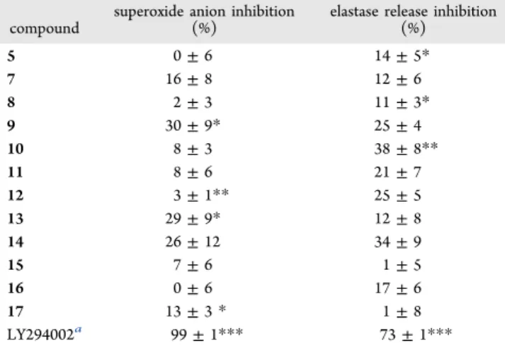
Fungi produce many secondary metabolites that exhibit a wide range of biological activities.33Polyketides are a class of fungal secondary metabolites, and many of them demonstrate fascinating biological activities.34 Therefore, the isolated compounds were tested for several bioactivities such as cytotoxicity and anti-inflammation. Due to limited sample amounts, compounds1−4and6were not able to be evaluated in their anti-inflammation assay. Compounds5and7−17were tested for anti-inflammation activity against the response of human neutrophils stimulated by formyl-methionyl-leucyl phenylalanine (fMLP). Compounds9, 13, and 14exerted an anti-inflammatory effect on inhibiting superoxide anion generation with 30 ± 9%, 29 ± 9%, and 26 ± 12%, respectively, at a concentration of 10 μM (Table 2).
Furthermore, the capacities of elastase release inhibition after administrations of compounds9,10,12, and14were revealed as 25±4%, 38±8%, 25±5%, and 34±9%, respectively, at a concentration of 10μM (Table 2). In these assays, LY294002 was taken as the positive control and showed 99 ± 1%
superoxide anion inhibition and 73 ± 1% elastase release inhibition at a concentration of 10μM.35
In addition, the cytotoxicity of compounds 1−17 was evaluated against three human cancer cell lines, including HepG2 (hepatoma), MDA-MB-231 (human breast carcino- ma), and A549 (human lung adenocarcinoma). Compounds9 and 12were active in the cytotoxicity evaluation against the HepG2 cancer cell line (IC50= 42.9±0.5 and 32.9±0.0μM, respectively). Moreover, compound 12 displayed cytotoxic activity against MDA-MB-231 and A549 cancer cell lines with the IC50values of 39.4±0.0 and 25.9±1.8μM, respectively (Table 3).
The plausible biosynthesis pathway (Figure 4) was proposed in that compounds 1−7, 11, and 13 were composed of 3- oxobutanoic acid with 3,5-dioxohexanoic acid or 3-oxopenta-
noic acid. All new compounds were derived from the polyketide synthase (PKS) pathway with a series of condensation, cyclization, decarbonation (acetyl-CoA carbox- ylase), dehydration (dehydratase), methylation (methyl trans- ferase), reduction (enoyl reductase), and oxidation (oxi- dase).36
■
CONCLUSIONSIn the current study,five new polyketides, asperochrapyran (1) and asperochralactones A−D (2−5), together with 12 known secondary metabolites (6−17) were isolated from the fungal strain A. ochraceopetaliformis. The structure of each new compound possesses at least three chiral centers, which causes difficulty to determine their absolute stereochemistry. Previous research had never reported the determination of the absolute configurations from this type of polyketide secondary metabolites. Thus, this unusual issue is an imperative task that needs to be clarified. Additionally, the new compounds exhibited an oil-like appearance, whose configurations are difficult to elucidate by a single crystal X-ray analysis method.
Therefore, coupling constant, NOESY, experimental, and calculated ECD spectroscopic analyses were used extensively to assign the absolute configuration. Our findings suggested the stereochemistry in a series of special fungal polyketide secondary metabolites, and the plausible biosynthesis pathway of key isolates was proposed.
■
MATERIALS AND METHODSGeneral Experimental Procedures. Polymerase chain reaction (PCR) amplifications were reacted by FlexCycler PCR. Optical rotations were measured with a JASCO P-2000 Table 2. Anti-Inflammatory Results of Compounds 5 and 7−17
compound
superoxide anion inhibition (%)
elastase release inhibition (%)
5 0±6 14±5*
7 16±8 12±6
8 2±3 11±3*
9 30±9* 25±4
10 8±3 38±8**
11 8±6 21±7
12 3±1** 25±5
13 29±9* 12±8
14 26±12 34±9
15 7±6 1±5
16 0±6 17±6
17 13±3* 1±8
LY294002a 99±1*** 73±1***
aLY294002 was used as the positive control.35 Percentage of inhibition (Inh %) at 10 μM concentration. Results are presented as mean±SEM (n= 3−4).*P< 0.05,**P< 0.01 compared with the control (DMSO).
Table 3. Cytotoxicity Results (IC50,μM)a
compoundb HepG2 MDA-MB-231 A549
9 42.9±0.5 >100 >100
12 32.9±0.0 39.4±0.0 25.9±1.8
doxorubicinc 0.4±0.0 0.8±0.2 0.2±0.0
aIC50values are taken as mean±SD (n= 3).bCompounds1−8,10−
11, and13−17were inactive with IC50values of >100μM.cPositive control.
polarimeter. UV spectra were recorded on a JASCO V-530 UV−vis spectrophotometer, and experimental ECD spectra were measured on a JASCO J-815 spectropolarimeter. IR spectra were obtained on a JASCO FT/IR-4600 Fourier transform infrared spectrometer. 1D and 2D NMR spectra were performed on a JEOL JNM-ECS 400 MHz NMR spectrometer (1H, 400 MHz;13C, 100 MHz), Varian Mercury Plus 400 MHz FT-NMR (1H, 400 MHz;13C, 100 MHz), and Varian VNMRS 600 MHz FT-NMR (1H, 600 MHz;13C, 150 MHz) in CDCl3, CD3OD, and CD3COCD3, respectively. Mass spectra were obtained from a Waters 2695 separation module (ESI-MS) and Bruker FT-MS SolariX (HR-ESI-MS). Column chromatography was carried out on silica gel 60 (0.063−0.200 mm and 0.040−0.063 mm, Merck) and Sephadex LH-20 (Fine Chemicals AB, Uppsala, Pharmacia). Thin-layer chromatog- raphy (TLC) analyses were performed using silica gel 60, F254, and RP-18, F254S (0.20 nm, Merck, Germany). Semi- preparative HPLC was performed on Shimadzu LC-10 AD, Shimadzu LC-20AT, or Jasco PU-980 pumps, an SPD-M10A diode array, SPD-10A UV−vis or UV-970 UV−vis detectors, and SCL-10A or CBM-20A controllers with Luna phenyl- hexyl, 100 Å, 250×10 mm, Phenomenex or Luna CN, 100 Å, 250×10 mm, Phenomenex columns.
Fungal Material. The fungus,A. ochraceopetaliformis, was isolated from A. brownii collected from the Dr. Cecilia Koo Botanic Conservation Center (KBCC), Pingtung, Taiwan.
KBCC is the biggest botanical garden and deposits over 30,000 living plant materials. The leaves of A. brownii were washed and air-dried. The dry leaves were soaked in 0.01% Tween 20 (aq), ddH2O, and 0.01% bleach (aq) to clean the surface. The washed leaves were moved into a laminar flow after the treatment of 75% alcohol (aq), and we used sterilized scissors and tweezers to cut the central part of leaf (5 mm×5 mm).
The mesophyll of the leaf was bisected and seeded on the potato dextrose agar (PDA) plate. Then, the plates were incubated in a 25°C incubator. After repeated purification, the pure strains were stored in 2 mL cryogenic vials (Nalgene,
Thermo) with 1.5 mL of potato dextrose broth (PDB) and 0.2 mL of sterilized glycerol and stored in a−80°C refrigerator.
The fungal strain was identified by D.-Y.Y. and C.-Y.L. A voucher specimen (code no. K004643) was deposited in the Graduate Institute of Natural Products, College of Pharmacy, Kaohsiung Medical University, Kaohsiung, Taiwan.
Species Identification.The fungusA. ochraceopetaliformis was identified on the basis of its morphology and a pair of internal transcribed spacers (ITS1-5.8S-ITS2) rRNA gene analysis using universal fungal primers. DNA was extracted by using the AxyPrep Multisource Genomic DNA miniprep kit (AxyPrep,#02815KC1) following the manufacturer’s protocol.
PCR amplifications were accomplished by using FlexCycler2 (Analytik Jena, Germany) with the following conditions: 95°C (5 min), 30 cycles of 95°C (30 s), 55°C (30 s), and 72°C (40 s), with the last extension at 72 °C (7 min). The PCR products were sent to the Mission Biotech Co., Ltd. for sequencing services after purification. The results of 18S rRNA gene sequences were blasted with the National Center for Biotechnology Information (NCBI) database for species identification. The reversal and forwarding of the 18S rRNA gene sequence displayed 99% sequence identity with A.
ochraceopetaliformis(GenBank accession no. FJ7976981).
Fermentation, Extraction, and Isolation.The fungusA.
ochraceopetaliformis was cultivated by using 120 Erlenmeyer flasks (500 mL) with each flask containing 300 mL of PDB medium. Theseflasks were incubated on the rotator shaker at 150 rpm at 25°C for 7 days. The whole broth wasfiltered to give the filtrate from mycelia. The filtrate was extracted by ethyl acetate (EtOAc), and the EtOAc layer was concentrated by rotary evaporators to obtain the crude extract (10.9 g). The crude extract was loaded to the Sephadex LH-20 column and eluted with methanol (MeOH) to yieldfive fractions (Fr. 1− 5). Compound1425(3.8 mg) was precipitated from Fr. 5. Fr. 3 was isolated by a silica gel column stepwise eluted with dichloromethane (CH2Cl2) and MeOH from 29:1 to 0:1 to give 10 fractions (Fr. 3.1−3.10). Fr. 3.3 was further separated Figure 4.Plausible biosynthesis pathway of new compounds and their analogues.
by a silica gel column eluted with CH2Cl2 and MeOH from 24:1 to 0:1 to furnish nine fractions (Fr. 3.3.1−3.3.9). Fr. 3.3.5 (100.4 mg) was purified by reversed-phase (RP) HPLC (Luna phenyl-hexyl, 100 Å, 250×10 mm, Phenomenex,flow rate of 2.0 mL/min, 35% MeOH (aq)) to afford compounds925(20.4 mg) and 1312 (4.5 mg). Fr. 3.5 was submitted to silica gel column chromatography eluted with CH2Cl2and MeOH from 39:1 to 0:1 to get 10 fractions (Fr. 3.5.1−3.5.10). Fr. 3.5.5 (16.8 mg) was subjected to RP-HPLC (Luna phenyl-hexyl, 100 Å, 250×10 mm, Phenomenex,flow rate of 2.0 mL/min, 32% MeOH (aq)) to give compound 2 (1.0 mg). Fr. 3.5.6 (80.3 mg) was isolated by RP-HPLC (Luna phenyl-hexyl, 100 Å, 250×10 mm, Phenomenex,flow rate of 2.0 mL/min, 33%
MeOH (aq)) to yield compound 118 (3.2 mg) and three fractions (Fr. 3.5.6.2−3.5.6.4). Compound 3 (1.1 mg) was purified by normal-phase (NP) HPLC (Luna CN, 100 Å, 250
×10 mm, Phenomenex,flow rate of 2.0 mL/min, hexanes and EtOAc = 1:1). Fr. 3.5.7 (85.3 mg) was isolated by RP-HPLC (Luna phenyl-hexyl, 100 Å, 250×10 mm, Phenomenex,flow rate of 2.0 mL/min, 40% MeOH (aq)) to obtain compounds1 (1.6 mg) and1212(56.2 mg). Fr. 3.5.9 was separated by a silica gel column (CH2Cl2 and MeOH from 24:1 to completely MeOH) to give eight fractions (Fr. 3.5.9.1−3.5.9.8).
Compounds4 (2.5 mg),5 (4.8 mg), and 722(3.0 mg) were isolated by RP-HPLC (Luna phenyl-hexyl, 100 Å, 250 ×10 mm, Phenomenex,flow rate of 2.0 mL/min, 25% MeOH (aq)) from Fr. 3.5.9.5 (36.1 mg). Furthermore, Fr. 3.7 (107.7 mg) was subjected to RP-HPLC (Luna phenyl-hexyl, 100 Å, 250× 10 mm, Phenomenex, flow rate of 2.0 mL/min, 25%
MeOH(aq)) to afford compounds 837 (30.4 mg) and 1511 (1.6 mg). Compounds632(3.0 mg),1035(1.6 mg),1611(6.4 mg), and 1711 (6.5 mg) were purified by RP-HPLC (Luna phenyl-hexyl, 100 Å, 250×10 mm, Phenomenex,flow rate of 2.0 mL/min, 15% MeOH (aq)) from Fr. 3.9 (98.9 mg).
Asperochrapyran (1): colorless oil; [α]D24 = −83 (c 0.08, MeOH); UV (MeOH)λmax(logε) 218 (4.00) nm; ECD (6.5× 10⁻⁵M, MeOH)λmax(Δε): 289 (−0.25), 265.5 (+0.19), 251.0 (−0.95), 223.5 (−2.71) nm; IR (ATR)vmax: 3415, 2979, 2918, 1708, 1659, 1557, 1438, 1263, 1235, 1087, 885 cm−1;1H and
13C NMR spectroscopic data, see Table 1; HR-ESI-MSm/z 239.08887 [M + Na]+(calcd for C10H16O5Na, 239.08899).
Asperochralactone A (2): colorless oil; [α]D24=−53 (c0.06, MeOH); UV (MeOH)λmax(logε) 212 (3.92) nm; ECD (6.5× 10⁻⁵ M, MeOH)λmax (Δε): 279.5 (+0.57), 242.5 (−1.03), 221.0 (+1.01) nm; IR (ATR)vmax: 3381, 2928, 2898, 1766, 1720, 1449, 1282, 1189, 1140, 1078, 883 cm−1; 1H and 13C NMR spectroscopic data, see Table 1; HR-ESI-MS m/z 239.08905 [M + Na]+(calcd for C10H16O5Na, 239.08899).
Asperochralactone B (3): colorless oil; [α]D24=−74 (c0.03, MeOH); UV (MeOH)λmax(logε) 212 (3.91) nm; ECD (6.5× 10⁻⁵M, MeOH)λmax(Δε): 310 (−0.50), 274 (+1.23), 237.0 (−0.09), 220.5 (+0.72), 207 (−0.23) nm; IR (ATR) vmax: 3414, 2979, 2920, 1762, 1716, 1650, 1455, 1373, 1234, 1082, 889 cm−1;1H and13C NMR spectroscopic data, seeTable 1;
HR-ESI-MS m/z 239.08889 [M + Na]+ (calcd for C10H16O5Na, 239.08899).
Asperochralactone C (4): colorless oil; [α]D24=−26 (c0.05, MeOH); UV (MeOH)λmax(logε) 215 (3.64) nm; ECD (6.5× 10⁻⁵M, MeOH)λmax(Δε): 263.5 (+0.30), 228.0 (−1.05) nm;
IR (ATR) vmax: 3420, 2978, 2916, 1748, 1666, 1584, 1401, 1382, 1234, 1087, 883 cm−1;1H and13C NMR spectroscopic data, see Table 1; HR-ESI-MS m/z 239.08882 [M + Na]+ (calcd for C10H16O5Na, 239.08899).
Asperochralactone D (5): colorless oil; [α]D24= +64 (c0.05, MeOH); UV (MeOH)λmax(logε) 208 (4.24) nm; ECD (6.5× 10⁻⁵M, MeOH)λmax(Δε): 270.0 (−8.55), 236.0 (+7.53), 212 (−1.55) nm; IR (ATR) vmax: 3409, 2987, 2912, 1721, 1650, 1446, 1378, 1232, 1084, 1042, 888 cm−1; 1H and13C NMR spectroscopic data, seeTable 1; HR-ESI-MS m/z 239.08881 [M + Na]+ (calcd for C10H16O5Na, 239.08899).
In Silico Calculations. Structures were built, and we optimized the minimized energy conformers in the MM2 level.
After the gross optimization, the data were used to outputxyz typefiles and inputfiles for calculating conformational results at MMFF94 by Spartan 16 software (Wavefunction Inc.;
Irvine, CA, U.S.A.). These data were submitted into Gaussian 09 software (Gaussian Inc.; Wallingford, CT, U.S.A.) and optimized using the time-dependent density functional theory (TDDFT) methodology at the B3LYP/6-311++G(d,p) level for ECD and the GIAO-DFT at the mpw1pw91/6-311+g- (2d,p) level for NMR in the solvent phase. During the computation of Gaussian 09 software, the calculated ECD and NMR spectra were generated by GaussSum 2.2.5 and GaussView 5.0.8, respectively. TMS was calculated at the same level of theory (δref = 183.143 ppm) as the reference compound for the calculated NMR spectra. For conforma- tional searches, the calculated ECD and NMR spectra of compounds were averaged by the proportion of each conformer.38
Anti-inflammatory Activity Assay. The assay on super- oxide anion generation and elastase release in response to fMLP stimulation of neutrophils was evaluated by the methods published by a co-author of this study, T.-L.H.39
Cytotoxicity Assay.The method for cytotoxicity assay was performed as previously described.40,41 Briefly, three human cancer cell lines, HepG2 (1×10⁴cells), A549 (5×10³cells), and MDA-MB-231 (1×10⁴cells), were inoculated onto 96- well plates and treated with the samples (20 μg/mL). The medium was removed after 72 h of incubation then the 3-(4,5- dimethylthiazol-2-yl)-2,5-diphenyltetrazolium bromide (MTT) solution (100μL, 0.5 mg/mL) was added into each well. Then, the plates were incubated at 37°C for 1 h. The MTT dye was detected by the addition of 100μL of dimethyl sulfoxide. The absorbance was estimated at 550 nm. The positive control was doxorubicin.
■
ASSOCIATED CONTENT*sı Supporting Information
The Supporting Information is available free of charge at https://pubs.acs.org/doi/10.1021/acsomega.0c02489.
1D and 2D NMR for compounds1−5, conformational search results of1−5, calculated and experimental ECD spectra of 1−7, and calculated 13C chemical shifts against the experimental data of1−5(PDF)
■
AUTHOR INFORMATION Corresponding AuthorFang-Rong Chang− Graduate Institute of Natural Products, College of Pharmacy and Drug Development and Value Creation Research Center and Department of Medical Research, Kaohsiung Medical University Hospital, Kaohsiung Medical University, Kaohsiung 807, Taiwan; Department of Marine Biotechnology and Resources, National Sun Yat-Sen University, Kaohsiung 804, Taiwan; orcid.org/0000-0003-2549-4193;
Phone: +886-7-312-1101 (ext. 2162); Email:aaronfrc@
kmu.edu.tw
Authors
Hao-Chun Hu−Graduate Institute of Natural Products, College of Pharmacy, Kaohsiung Medical University, Kaohsiung 807, Taiwan
Chi-Ying Li−Department of Pharmacology and Pharmaceutical Sciences, School of Pharmacy, University of Southern California, Los Angeles, California 90089, United States
Yi-Hong Tsai−Graduate Institute of Natural Products, College of Pharmacy, Kaohsiung Medical University, Kaohsiung 807, Taiwan
Dai-Yun Yang−Graduate Institute of Natural Products, College of Pharmacy, Kaohsiung Medical University, Kaohsiung 807, Taiwan
Yang-Chang Wu−Graduate Institute of Integrated Medicine and Chinese Medicine Research and Development Center, China Medical University, Taichung 404, Taiwan
Tsong-Long Hwang−Graduate Institute of Natural Products, College of Medicine, Chang Gung University, Taoyuan 333, Taiwan; Research Center for Chinese Herbal Medicine, Research Center for Food and Cosmetic Safety, and Graduate Institute of Health Industry Technology, College of Human Ecology, Chang Gung University of Science and Technology, Taoyuan 333, Taiwan; Department of Anesthesiology, Chang Gung Memorial Hospital, Taoyuan 333, Taiwan
Shu-Li Chen−Graduate Institute of Natural Products, College of Pharmacy, Kaohsiung Medical University, Kaohsiung 807, Taiwan
Ferenc Fülöp−Institute of Pharmaceutical Chemistry, University of Szeged, Szeged 6720, Hungary; MTASZTE Stereochemistry Research Group, Hungarian Academy of Sciences, Szeged 6720, Hungary; orcid.org/0000-0003- 1066-5287
Attila Hunyadi− Institute of Pharmacognosy, Interdisciplinary Excellence Center, and Interdisciplinary Centre for Natural Products, University of Szeged, Szeged 6720, Hungary Chia-Hung Yen− Graduate Institute of Natural Products,
College of Pharmacy, Kaohsiung Medical University, Kaohsiung 807, Taiwan
Yuan-Bin Cheng−Graduate Institute of Natural Products, College of Pharmacy, Kaohsiung Medical University, Kaohsiung 807, Taiwan; Department of Marine Biotechnology and Resources, National Sun Yat-Sen University, Kaohsiung 804, Taiwan; orcid.org/0000-0001-6581-1320
Complete contact information is available at:
https://pubs.acs.org/10.1021/acsomega.0c02489
Author Contributions
+H.-C.H. and C.-Y.L. contributed equally to this work. H.-C.H.
and Y.-H.T. contributed to writing the manuscript, in silico calculations, and design of the Table of Contents image. C.- Y.L. and D.-Y.Y. contributed to collecting the physical data, optimizing fungal cultivation, and natural product purification and identification. C.-Y.L., D.-Y.Y., Y.-C.W., and A.H.
contributed to the data analysis. T.-L.H., S.-L.C., and C.-H.Y.
contributed to the bioassays. The experiment was designed, and the manuscript was revised by Y.-B.C., F.F., and F.-R.C.
Notes
The authors declare no competingfinancial interest.
■
ACKNOWLEDGMENTSThis research was funded by the Ministry of Science and Technology, Taiwan, awarded to F.-R.C., grant nos. MOST 105-2628-B-037-001-MY3, MOST 106-2320-B-037-008-MY2, MOST 106-2911-I-037-504, MOST 108-2320-B-037-022- MY3, 108-2811-B-037-511, and 109-2927-I-037-502. In addition, this research was partially funded by the Drug Development and Value Creation Research Center of Kaohsiung Medical University & Department of Medical Research of Kaohsiung Medical University Hospital awarded to F.-R.C. (grant no. KMU-TC108A03-11). We appreciate the Dr. Cecilia Koo Botanic Conservation Center (KBCC) for providing plant materialA. brownii.We also thank Prof. Chia- Wei Li who is the CEO of KBCC. The plant material and fungal strain were obtained from a project supported by the Ministry of Science and Technology, Taiwan, awarded to Prof.
Yi-Ming Arthur Chen, aimed to build an herbal medicine database with KBCC.
■
(1) Newman, D. J.; Cragg, G. M. Natural products as sources of newREFERENCES drugs over the nearly four decades from 01/1981 to 09/2019.J. Nat.Prod.2020,83, 770−803.
(2) Mikkola, R.; Andersson, M. A.; Hautaniemi, M.; Salkinoja- Salonen, M. S. Toxic indole alkaloids avrainvillamide and stephacidin B produced by a biocide tolerant indoor moldAspergillus westerdijkiae.
Toxicon2015,99, 58−67.
(3) Liu, T.; Yu, H.; Liu, C.; Wang, Y.; Tang, M.; Yuan, X.; Luo, N.;
Wang, Q.; Xu, X.; Jin, F. Protodioscin-glycosidase-1 hydrolyzing 26- O-β-D-glucoside and 3-O-(1→4)-α-L-rhamnoside of steroidal sap- onins fromAspergillus oryzae. Appl. Microbiol. Biotechnol. 2013,97, 10035−10043.
(4) You, M.; Liao, L.; Hong, S. H.; Park, W.; Kwon, D. I.; Lee, J.;
Noh, M.; Oh, D. C.; Oh, K. B.; Shin, J. Lumazine peptides from the marine-derived fungusAspergillus terreus.Mar. Drugs2015,13, 1290−
1303.
(5) Wang, J.; He, W.; Kong, F.; Tian, X.; Wang, P.; Zhou, X.; Liu, Y.
J. Ochracenes A−I, humulane-derived sesquiterpenoids from the Antarctic fungusAspergillus ochraceopetaliformis.J. Nat. Prod. 2017, 80, 1725−1733.
(6) Qiao, M. F.; Ji, N. Y.; Miao, F. P.; Yin, X. L. Steroids and an oxylipin from an algicolous isolate ofAspergillus f lavus.Magn. Reson.
Chem.2011,49, 366−369.
(7) Ishikawa, K.; Sato, F.; Itabashi, T.; Wachi, H.; Takeda, H.;
Wakana, D.; Yaguchi, T.; Kawai, K.; Hosoe, T. Asnovolins A−G, spiromeroterpenoids isolated from the fungusAspergillus novof umiga- tus, and suppression of fibronectin expression by Asnovolin E.J. Nat.
Prod.2016,79, 2167−2174.
(8) Liu, Y.; Li, X. M.; Meng, L. H.; Wang, B. G. Polyketides from the marine mangrove-derived fungus Aspergillus ochraceus MA-15 and their activity against aquatic pathogenic bacteria. Phytochem. Lett.
2015,12, 232−236.
(9) Phainuphong, P.; Rukachaisirikul, V.; Tadpetch, K.; Sukpondma, Y.; Saithong, S.; Phongpaichit, S.; Preedanon, S.; Sakayaroj, J. γ- Butenolide and furanone derivatives from the soil-derived fungus Aspergillus sclerotiorum PSU-RSPG178. Phytochemistry 2017, 137, 165−173.
(10) Choudhary, M. I.; Musharraf, S. G.; Mukhmoor, T.; Shaheen, F.; Ali, S.; Rahman, Atta-ur. Isolation of bioactive compounds from Aspergillus terreus.Z. Naturforsch. B2004,59, 324−328.
(11) Kito, K.; Ookura, R.; Yoshida, S.; Namikoshi, M.; Ooi, T.;
Kusumi, T. Pentaketides relating to aspinonene and dihydroaspyrone from a marine-derived fungus,Aspergillus ostianus.J. Nat. Prod.2007, 70, 2022−2025.
(12) Kimura, Y.; Nakahara, S.; Fujioka, S. Aspyrone, a nematicidal compound isolated from the fungus, Aspergillus melleus. Biosci., Biotechnol., Biochem.1996,60, 1375−1376.
(13) Yun, K.; Feng, Z.; Choi, H. D.; Kang, J. S.; Son, B. W. New production of (R)-(−)-5-bromomellein, a dihydroisocoumarin derivative from the marine-derived fungus Aspergillus ochraceus.
Chem. Nat. Compd.2013,49, 24−26.
(14) Balcells, M.; Canela, R.; Coll, J.; Sanchis, V.; Torres, M. Effect of fungal metabolites and some derivatives against Tribolium castaneum (Herbst) and Nezara viridula (L.). Pestic. Sci. 1995, 45, 319−323.
(15) Alberts, A. W. Lovastatin and simvastatin-inhibitors of HMG CoA reductase and cholesterol biosynthesis.Cardiology1990,77, 14−
21.
(16) Visagie, C. M.; Varga, J.; Houbraken, J.; Meijer, M.; Kocsube, S.; Yilmaz, N.; Fotedar, R.; Seifert, K. A.; Frisvad, J. C.; Samson, R. A.
Ochratoxin production and taxonomy of the yellow aspergilli (Aspergillussectioncircumdati).Stud. Mycol.2014,78, 1−61.
(17) Brasch, J.; Varga, J.; Jensen, J. M.; Egberts, F.; Tintelnot, K. Nail infection by Aspergillus ochraceopetaliformis. Med. Mycol. 2009, 47, 658−662.
(18) Wang, J.; Wei, X.; Qin, X.; Tian, X.; Liao, L.; Li, K.; Zhou, X.;
Yang, X.; Wang, F.; Zhang, T.; Tu, Z.; Chen, B.; Liu, Y. Antiviral merosesquiterpenoids produced by the antarctic fungus Aspergillus ochraceopetaliformisSCSIO 05702.J. Nat. Prod.2016,79, 59−65.
(19) Liu, J. T.; Wu, W.; Cao, M. J.; Yang, F.; Lin, H. W. Trienicα- pyrone and ochratoxin derivatives from a sponge-derived fungus Aspergillus ochraceopetaliformis.Nat. Prod. Res.2018,32, 1791−1797.
(20) Haroon, M. H.; Premaratne, S. R.; Choudhry, M. I.;
Dharmaratne, H. R. W. A new β-glucuronidase inhibiting butyrolactone from the marine endophytic fungusAspergillus terreus.
Nat. Prod. Res.2013,27, 1060−1066.
(21) Chang, Y. C.; Lu, C. K.; Chiang, Y. R.; Wang, G. J.; Ju, Y. M.;
Kuo, Y. H.; Lee, T. H. Diterpene glycosides and polyketides from Xylotumulus gibbisporus.J. Nat. Prod.2014,77, 751−757.
(22) Chen, X. W.; Li, C. W.; Cui, C. B.; Hua, W.; Zhu, T. J.; Gu, Q.
Q. Nine new and five known polyketides derived from a deep sea- sourcedAspergillussp. 16-02-1.Mar. Drugs2014,12, 3116−3137.
(23) Gawronski, J. K.; van Oeveren, A.; van der Deen, H.; Leung, C.
W.; Feringa, B. L. Simple circular dichroic method for the determination of absolute configuration of 5-substituted 2(5H)- furanones.J. Org. Chem.1996,61, 1513−1515.
(24) Uchida, I.; Kuriyama, K. The π-π circular dichroism of δβ- unsaturatedγ-lactones.Tetrahedron Lett.1974,15, 3761−3764.
(25) Garson, M. J.; Staunton, J.; Jones, P. G. New polyketide metabolites from Aspergillus melleus: structural and stereochemical studies.J. Chem. Soc. Perkin Trans. 11984, 1021−1026.
(26) Zhang, D.; Yang, X.; Kang, J. S.; Choi, H. D.; Son, B. W.
Chlorohydroaspyrones A and B, antibacterial aspyrone derivatives from the marine-derived fungusExophialasp.J. Nat. Prod.2008,71, 1458−1460.
(27) Jarvis, B. B.; Comezoglu, S. N.; Rao, M. M.; Pena, N. B.;
Boettner, F. E.; Williams, T. M.; Forsyth, G.; Epling, B. Isolation of macrocyclic trichothecenes from a large-scale extract of Baccharis megapotamica.J. Org. Chem.1987,52, 45−56.
(28) Takeshita, M.; Sato, T. Synthesis of optically active 1-phenyl- 1,2-propanediol by use of baker’s yeast.Chem. Pharm. Bull.1989,37, 1085−1086.
(29) Ayer, W. A.; Trifonov, L. S. Metabolites of Peniopbora polygonia, Part 2. some aromatic compounds.J. Nat. Prod.1993,56, 85−89.
(30) Jarvis, B. B.; Wang, S.; Ammon, H. L. Trichoverroid stereoisomers.J. Nat. Prod.1996,59, 254−261.
(31) Beecham, A. F. The CD of αβ-unsaturated lactones.
Tetrahedron1972,28, 5543−5554.
(32) Dong, Z.; Zheng, Z. H.; Lu, C. H.; Shen, Y. M. Two new compounds isolated from a seaweed-associated fungus,Aspergillussp.
AF044.Chin. J. Nat. Med.2010,8, 370−372.
(33) Li, C. Y.; Chang, C. C.; Tsai, Y. H.; El-Shazly, M.; Wu, C. C.;
Wang, S. W.; Hwang, T. L.; Wei, C. K.; Hohmann, J.; Yang, Z. J.;
Cheng, Y.; Wu, Y. C.; Chang, F. R. Anti-inflammatory, antiplatelet aggregation, and antiangiogenesis polyketides from Epicoccum
sorghinum: toward an understating of its biological activities and potential applications.ACS Omega2020,5, 11092−11099.
(34) Rustamova, N.; Bozorov, K.; Efferth, T.; Yili, A. Novel secondary metabolites from endophytic fungi: synthesis and biological properties.Phytochem. Rev.2020,19, 425−448.
(35) Hsu, Y. M.; Chang, F. R.; Lo, I. W.; Lai, K. H.; El-Shazly, M.;
Wu, T. Y.; Du, Y. C.; Hwang, T. L.; Cheng, Y. B.; Wu, Y. C.
Zoanthamine-type alkaloids from the zoanthid Zoanthus kuroshio collected in Taiwan and their effects on inflammation.J. Nat. Prod.
2016,79, 2674−2680.
(36) Dewick, P. M. Medicinal Natural Products: A Biosynthetic Approach; 3rd ed., John Wiley & Sons: 2011; 39−131.
(37) Fuchser, J.; Zeeck, A. Secondary metabolites by chemical screening, 34. − Aspinolides and aspinonene/aspyrone co-metabo- lites, new pentaketides produced byAspergillus ochraceus.Liebigs Ann./
Recl.1997,1997, 87−95.
(38) Cheng, Y. B.; Hu, H. C.; Tsai, Y. C.; Chen, S. L.; El-Shazly, M.;
Nonato, M. G.; Wu, Y. C.; Chang, F. R. Isolation and absolute configuration determination of alkaloids fromPandanus amaryllifolius.
Tetrahedron2017,73, 3423−3429.
(39) Yang, S. C.; Chung, P. J.; Ho, C. M.; Kuo, C. Y.; Hung, M. F.;
Huang, Y. T.; Chang, W. Y.; Chang, Y. W.; Chan, K. H.; Hwang, T. L.
Propofol inhibits superoxide production, elastase release, and chemotaxis in formyl peptide-activated human neutrophils by blocking formyl peptide receptor 1.J. Immunol. 2013, 190, 6511−
6519.
(40) Li, C. Y.; Lo, I. W.; Wang, S. W.; Hwang, T. L.; Chung, Y. M.;
Cheng, Y. B.; Tseng, S. P.; Liu, Y. H.; Hsu, Y. M.; Chen, S. R.; Hu, H.
C.; Chang, F. R.; Wu, Y. C. Novel 11-norbetaenone isolated from an entomopathogenic fungus Lecanicillium antillanum. Bioorg. Med.
Chem. Lett.2017,27, 1978−1982.
(41) Li, C. Y.; Lo, I. W.; Hsueh, Y. P.; Chung, Y. M.; Wang, S. W.;
Korinek, M.; Tsai, Y. H.; Cheng, Y. B.; Hwang, T. L.; Wang, C. C. C.;
Chang, F. R.; Wu, Y. C. Epigenetic manipulation induces the production of coumarin-type secondary metabolite fromArthrobotrys foliicola.Isr. J. Chem.2019,59, 432−438.