92
DETERMINATION OF MACROPHAGE-MEDIATED ANTIBACTERIAL RESISTANCE
R. J. North G. L. Spitalny
INTRODUCTION
Unless they possess an antiphagocytic capsule, most bac- teria inoculated intravascularly are rapidly cleared from the blood almost exclusively by fixed macrophages of the reticulo- endothelial system. Because by far the largest number of bac- teria are ingested by fixed macrophages (Kuppfer cells) that line the sinusoids of the liver, it is obvious that the liver is the organ of choice for measuring antibacterial resistance
in vivo. Provided the bacterial species chosen is clearedfrom the blood in several minutes, it is possible to determine the total number of bacteria initially ingested by macrophages of the liver, and the number of bacteria inactivated during any one period of time thereafter. This is achieved by sam- pling a population of mice at progressive times after inocula- ting them with Listeria, and determining changes in the number of bacteria in liver homogenates capable of forming colonies on nutrient agar.
METHODS FOR STUDYING Copyright © 1981 by Academic Press, Inc.
MONONUCLEAR PHAGOCYTES 1011 All rights of reproduction in any form reserved.
ISBN 0-12-044220-5
The facultative, intracellular, bacterial parasite, Lis- teria monocytogenes, is a popular test organism for measuring both innate and acquired, nonspecific, antibacterial resistance expressed by liver macrophages in vivo. It should be realized that Listeria is far more susceptible to inactivation by macro- phages than certain other facultative intracellular pathogens, such as mycobacteria. Consequently, the liver macrophages of normal mice are capable of inactivating 50% or more of the Listeria load in the liver over the first several hours of in- fection, depending on the state of "cleanliness" of the mice and the virulence of the pathogen. Nevertheless, the rate and extent of bacterial inactivation is greatly increased in ani- mals generating an activated macrophage system in response to bacterial infection. This increased bactericidal activity can be measured in vivo by the procedure described here.
II. REAGENTS
Animals: For obvious reasons mice are the most popular choice, although the same procedures can be used for rats and guinea pigs.
Listeria monocytogenes with at least moderate virulence
for the host species chosen.
Disposable 1-cm
3tuberculin syringes fitted with 26-gauge needles (Becton-Dickinson, Oxnard, California, No. 5625).
Dissecting materials: Cork board and pins; blunt-end
5 1/2 in. stainless scissors; Mayo 5 1/2 in. dissection scissors; rat tooth forceps (4 1/2 in.) (Clay Adams, Parsip- pany, New Jersey); ethanol, 95%.
Petroff-Hauser bacterial counting chamber (Scientific Products, McGraw Park, Illinois, No. C8400-1).
Glass homogenizing tubes with matching Teflon pestles (Tri-R Instruments, Rockville Center, New York, Nos. S32 and S22).
A large number of 5-ml plastic tubes (Falcon Plastics, Oxnard, California, No. 2058) containing 0.9 ml of sterile 0.9% sodium chloride solution for performing serial dilutions.
A large number of sterile 1-ml disposable serological pipettes (Scientific Products, McGraw Park, Illinois, No. P643-1) for performing serial dilutions.
Bacterial culture dishes (Falcon Space Saver No. 1506)
containing sterile trypticase-soy agar (TSA). The nutrient
agar is made by dissolving 30 gm trypticase-soy broth (Balti-
more Biological Labs, Cockeysville, Maryland, No. 11768) and
20 gm of granulated agar (Baltimore Biological Labs, Cockeys-
ville, Maryland, No. 11849) in 1 liter of distilled water in
an autoclave at 15 psi for 15 min. The dissolved nutrient
XI. MONONUCLEAR PHAGOCYTES IN VIVO 1013 agar is immediately poured while hot into culture plates and allowed to solidify at room temperature under sterile condi- tions. Water that has condensed on the agar and on the inside of plates and their covers is evaporated by removing the covers and placing the plates and covers upside down in a hot air oven.
Tubes with 5 m l of trypticase-soy broth (TSB) prepared as above, but without agar.
A vertically mounted, low-speed (100 r p m ) , electric drill with footpedal switch and a chuck for holding pestles.
III. PROCEDURES
A. Listeria Passage
If need b e , the virulence of Listeria can be increased by repeated passage in animals. Starting with a log phase culture with a known number of organisms per milliliter, as determined by counting in a Petroff-Häuser chamber, a sample of the cul- ture is diluted in physiological saline to 5 x 10^/ml and 0.2 ml inoculated intravenously into one or more mice via a lateral tail vein using a tuberculin syringe fitted with a 26-gauge needle. Three days later a mouse is sacrificed, its spleen re- moved aseptically and homogenized in 8 m l of physiological sa-
line by hand. The homogenate is used to seed 5 m l of TSB in plastic tubes. It is advisable to seed three tubes with 0.1,
0.5, and 0.01 m l of homogenate, respectively, and to use the culture that is still in log phase after overnight incubation at 37°C. The larger the volume of spleen homogenate added, the sooner the culture is ready for u s e . The bacteria are counted and diluted appropriately in physiological saline for another passage in mice, and the procedure repeated as necessary. For worthwhile experiments, the L D ^ Q dose of Listeria for mice should be 1 0 ^ or less. Therefore, after several passages, it will be necessary to determine the intravenous L D C Q dose by following procedures adequately described in the literature ( 1 ) .
B. Standard Frozen Cultures for Experiments
Having demonstrated that the intravenous L D ^ Q dose of Lis- ter la is 105 or less, a spleen homogenate prepared as described above is used to seed 100 m l of TSB to obtain a log phase
(1-5 x 10 ml) culture for distribution in 1 m l ampoules for freezing at -70°C. The culture is dispensed directly into am- poules that are then placed in a suitable -70°C freezer. The viability of the culture before freezing and after thawing is determined in each case by plating ten-fold serial dilutions of it on nutrient agar. It has been found consistently that prac-
tically no loss of viability is caused by freezing at -70°C.
Moreover, because the viability and virulence remain stable for at least 6 months, the frozen ampoules serve as a standard culture for a large number of experiments. For each experiment an ampoule is thawed and diluted appropriately in physiological saline for intravenous inoculation.
C. Measurement of Macrophage-Medlated Antibacterial Re-
sistance
In essence, the method involves inoculating a population of mice intravascularly with Listeria, and sampling the popu- lation over time for changes in the number of bacteria in the liver capable of forming colonies on nutrient agar. Mice are inoculated intravenously via a lateral tail vein with 1 05 Listeria in 0.2 ml physiological saline. This size of the inoculum is determined by the expected bactericidal capacity of the liver. At 30 m i n , and at 3, 6, 1 2 , 2 4 , and 48 hr after inoculation, five mice are killed by cervical dislocation, pinned out on a cork board, and their abdomens swabbed with 95% ethanol. The abdominal skin is reflected, the peritoneal lining held with rat-tooth forceps and cut along the midline with scissors to expose the peritoneal contents. The liver is removed whole by gently grasping the porta with forceps and, while gently pulling, cutting all ligamentous connections close to the liver from the exit of the hepatic veins down to the porta. Care should be taken not to cut through the esophagus.
Each liver is placed in an homogenizing tube containing 8 ml of cold (4°C) saline. This volume is chosen because an adult mouse liver occupies a volume of about 2 m l , thereby making the
final volume in the tubes approximately 10 m l . Homogenization is performed with cold (-20°C) Teflon pestles driven at 100 rpm by a foot-pedal-controlled electric drill fitted with an appro- priate chuck. To achieve complete homogenization, each tube is held by hand and moved firmly up and down several times against the rotating pestle for about 6 sec. A thick leather glove is worn to protect the hand from possible glass breakage, and the procedure is performed inside a hood with an air flow powerful enough to draw away the aerosol that inevitably forms during homogenization.
Serial tenfold dilutions of 0.1 ml homogenate are performed with plastic tubes containing 0.9 ml physiological saline. A fresh pipette is used for each dilution. Serial dilution is performed according to procedures adequately described in nu- merous bacteriological texts, and selected dilutions are plated on the quadrants of TSA plates. Four dilutions are adequate for calculating the number of Listeria per total organ homo- genate. Knowing the number of dilutions to make and which ones to plate comes from a familiarity with the experimental system.
XI. MONONUCLEAR PHAGOCYTES IN VIVO 1015 Since an intravascular inoculum of Listeria is cleared from the circulation in less than 5 m i n , and since 9 5 % is ingested by liver macrophages, the liver will contain almost all of a
10 inoculum. At the end of 30 m i n , over 5 x 1 04 viable bac- teria will be present in the liver. Previous experiments have shown (2) that maximum bacterial kill is achieved between 6 and 12 hr after inoculation, and it is useful to know that while the livers of normal mice can inactivate about 0.5 log of bacteria during this period, the livers of mice with a BCG- activated macrophage system, for example, can inactivate 1.5- 2.0 logs.
IV. CALCULATION OF DATA
The number of bacterial colonies that emerge on TSA plates from known tenfold dilutions of the homogenate allows a calcu- lation of the total number of bacteria per whole organ homo- genate. This involves multiplying the number of bacterial colonies on a given quadrant by 1 0n, where n equals the dilu- tion that was used to plate the quadrant counted. It should be realized that since it is 0.1 m l of the 10 m l of homogenate that is subjected to serial dilution, the zero dilution (0.1 ml of undiluted homogenate) itself represents 1 0 ~2 of the whole homogenate and the first dilution, 10""^. Thus, if mice are inoculated with 10 Listeria, and if one expects 0.5 log of inactivation at 6 h r , the 0, 1, 2, and 3 dilutions are plated. This would result theoretically, in 500 colonies on the zero dilution, 50 on the 1 dilution, and 5 on the 2 dilu- tion. Since 500 colonies is too high a number to count, and since 5 colonies is too small a number to be accurate, the 50 colonies on the 1 dilution would be recorded and used for cal- culation. If, in fact, 50 colonies appeared on this dilution, then, the total number of viable bacteria in the liver at the time of homogenization was 5 x 10 , which is converted to the log-,Q number. It is a good idea to count periodically the numbers of colonies on different quadrants of a plate to d e - termine whether approximate tenfold differences exist. If this is not the case, then it is obvious that the dilutions are not being accurately performed, probably because bacteria are not being adequately suspended in the dilution tubes.
V. CRITICAL COMMENTS
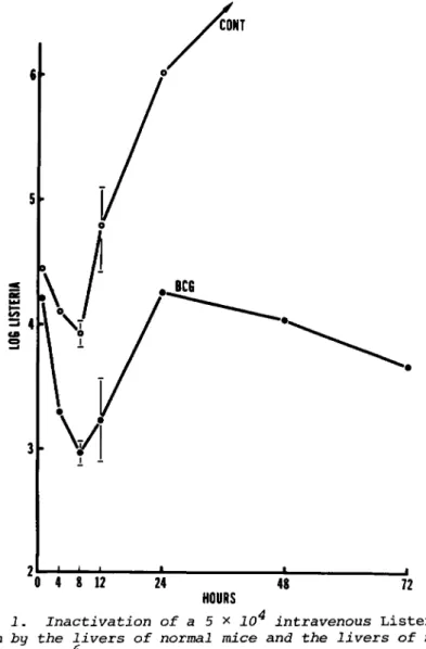
The fate over a 48-hr period of 5 x 1 04 Listeria ingested by liver macrophages can be appreciated from an examination
of Fig. 1, which compares the growth of the organism in t h e livers of mice with macrophage systems activated by a BCG in- fection initiated 12 days earlier. It can be seen in this particular experiment, that ingestion of the organism by liver macrophages w a s followed by progressive bacterial inactivation in the liver for a period of approximately 8 h r , and that this
CO = 4
3h
0 4 8 12 24
HOURS 48 72
Fig. 1. Inactivation of a 5 x 10 intravenous Listeria inoculum by the livers of normal mice and the livers of mice infected with 10° BCG 12 days previously. The liver macro- phages of BCG-infected mice were capable of destroying a much larger proportion of the bacterial load during the first 8 hr.
Means of five mice per time point ±2 x SE.
XI. MONONUCLEAR PHAGOCYTES IN VIVO 1017 was followed in turn by substantial multiplication of those bacteria that survived. In fact, the major difference between normal mice and mice with an activated macrophage system is seen in the events that take place over the first 8 h r , in that the latter mice are capable of inactivating Listeria at a faster rate and to a much larger extent. This results in BCG- infected mice, for example, being able to reduce rapidly p o - tentially lethal numbers of Listeria in their livers to sub- lethal numbers. Indeed, the bacterial activity expressed dur- ing the first 8-12 hr of infection is the only legitimate mea- sure of macrophage-mediated nonspecific resistance, because the bacterial inactivation that occurs after this time is d e - pendent on the generation of specific immunity to the test organism itself. It is significant, in differentiating between these two antibacterial events, that, whereas the mechanism r e - sponsible for the initial destruction of Listeria is radio- resistant, the acquired antibacterial mechanism expressed later is highly radiosensitive (2,3). This supports the interpreta- tion that the initial 8-hr period of destruction is achieved by macrophages that are already resident in the liver at the time of intravascular bacterial inoculation. Even so, this 8-hr period of bacterial inactivation is much longer than the 120- min period of destruction expressed by macrophages in vitro.
The possibility exists, therefore, that the 8-hr period of bacterial destruction in vivo is not achieved solely by those liver macrophages that initially clear Listeria from the circu- lation. It seems more likely that those liver macrophages that support the growth of the bacterium are joined during the 8-hr period by more resistant macrophages in close proximity.
It should be realized that Listeria monocytogenes is classi- fied as a class II agent by the Center for Disease Control, which means that it is an agent that is contained by "ordinary laboratory techniques." In fact, Listeria is an opportunistic pathogen (4,5) that infects the aged, the very young, and per- sons with underlying diseases. However, in spite of the fact that it is ubiquitous in nature and that the incidence of lis- teriosis in humans is low, the organism should be handled with caution, particularly during homogenization of heavily infected livers and other organs. It is important, therefore, to per- form organ homogenization in a fume hood with an adequate air flow and preferably with a filtered exhaust. All tubes, pi- pettes, culture plates, and instruments should be sterilized after being used, and the working space wiped down with 75%
ethanol.
Acknowledgment
Work supported by NIH Grants AI-10351, CA-21360, and RR05705 from the Division of Research Resources.
REFERENCES
1. L. J. Reed and H. Munch. Am. J. Hyg. 27:493, 1938.
2. R. J. North. T-Cell dependence of macrophage activation and mobilization during infection with Mycobacterium tuberculosis. Infect. Immun. 10:66-71, 1974.
3. M. F. Newborg and R. J. North. On the mechanism of the T-cell independent anti-Listeria resistance in nude mice.
J. Immunol. 124:511-516, 1980.
4. M. L. Gray and A. H. Killinger. Listeria monocytogenes and listeric infection. Bacteriol. Rev. 30:309-382, 1966.
5. L. A. Busch. Human listeriosis in the United States.
J. Infect. Dis. 123:328-331, 1971.