Neurochemistry International 142 (2021) 104920
Available online 22 November 2020
0197-0186/© 2020 The Authors. Published by Elsevier Ltd. This is an open access article under the CC BY license (http://creativecommons.org/licenses/by/4.0/).
Species-specific neuronal localization of kynurenine aminotransferase-2 in the mouse cerebellum
Emma Balog
a,1, Gyula Jenei
a,1, Levente Gell ´ ert
a, Etsuro Ono
b,c, L ´ aszl ´ o V ´ ecsei
d,e, J ozsef Toldi ´
a, Zsolt Kis
a,*aDepartment of Physiology, Anatomy and Neuroscience, Faculty of Science and Informatics, University of Szeged, Szeged, Hungary
bDepartment of Biomedicine, Graduate School of Medical Sciences, Kyushu University Fukuoka, Japan
cCenter of Biomedical Research, Research Center for Human Disease Modeling, Graduate School of Medical Sciences, Kyushu University Fukuoka, Japan
dDepartment of Neurology, Faculty of Medicine, University of Szeged, Szeged, Hungary
eMTA-SZTE Neuroscience Research Group, Hungary
A R T I C L E I N F O Keywords:
Kynurenine aminotransferase Kynurenic acid
Cerebellum Purkinje cells Astrocytes
Immunohistochemistry
A B S T R A C T
The immunohistochemical pattern of kynurenine aminotransferase-2 (KAT-2) - the key role enzyme in the production of neuroactive and neuroprotective kynurenic acid (KYNA) - was studied in the cerebellum of mice. It is known from literature that KAT-2 is localized mainly in astrocytes in different parts of the cerebrum.
Kynurenine aminotransferase (KAT) activity in the cerebellum is relatively low and alternative production routes for KYNA have been described there. Therefore we examined the immunohistochemical pattern of KAT-2 in this part of the brain. Surprisingly, the cellular localization of KAT-2 in mice was proven to be unique; it localized characteristically in Purkinje cells and in some other types of neurons (not identified) but was not found in astrocytes nor microglia. The exclusive neuronal, but not glial localization of KAT-2 in the cerebellum is novel and may be related to its low activity and to the alternative pathways for KYNA production that have been described.
1. Introduction
Kynurenic acid (KYNA) is one of the neuroactive end-products of the kynurenine pathway (KP) of tryptophan degradation, which has multi- ple effects in the mammalian brain (Fujigaki et al., 2017). Alone or jointly, these effects play an important role in the mechanism of neu- roprotective and neuromodulatory effects of endogenous KYNA in the central nervous system. Consequently, the pathological change of KP balance may result in hypo- or hyperfunction of neuroactive metabo- lites, which is associated with neurological, as well as psychiatric dis- eases (V´ecsei et al., 2013). One of the major questions is the biosynthesis of KYNA within the brain, which has been examined in considerable detail. Up to now, four aminotransferases have been shown to catalyze the transamination of the pivotal KP metabolite L-kynurenine to KYNA (Han et al., 2010). Out of the four isoforms of KAT enzymes, KAT-2 is of greatest importance both in the murine and the human brain (Nem- atollahi et al., 2016). Due to the importance of KATs in regulating the level of KYNA, studies aimed to describe the spatiotemporal expression
pattern of the different KATs at the cellular level, and it was found in the rat brain that KAT-2 is localized mainly in astrocytes (Guidetti et al., 2007), which glial cells release in situ “de novo” synthesized KYNA.
Kynurenic acid production and release from the astrocytes and brain slices were studied in detail (Turski et al., 1989), and turned out that KAT-2 is expressed in other cells too, including neurons (Her´edi et al., 2017).
The role of KP in neurodegenerative and neuropsychiatric diseases is evident (V´ecsei et al., 2013), consequently the investigation of that topic is essential. However, most studies were carried out on rat models and human samples. In addition to it, these studies were focused on different parts of the forebrain but not on the cerebellum. In recent years we started to study the KAT activity in mice (Her´edi et al., 2017, 2019). The impor- tance of these experimental animals in these studies is also evident, considering that mice strains are the main subjects of gene manipula- tion. To our best knowledge, there is no description of KAT-2 immu- nohistochemistry in the cerebellum of mice.
This study aimed to investigate the possible localization of KAT-2
* Corresponding author.
E-mail address: zskis@bio.u-szeged.hu (Z. Kis).
1 Authors (EB and GyJ) contribute equally to this paper.
Contents lists available at ScienceDirect
Neurochemistry International
journal homepage: www.elsevier.com/locate/neuint
https://doi.org/10.1016/j.neuint.2020.104920
Received 1 May 2020; Received in revised form 16 November 2020; Accepted 18 November 2020
2 enzyme in the cerebellum of mice and to answer these questions: i) is there any sign of KAT-2 enzyme in the cerebellum, ii) if yes, is it local- ized in glial cells and/or in neurons?
2. Materials and methods 2.1. Animals
12-14-week-old male C57Bl/6 mice obtained from The National Fig. 1.Investigating the distribution of KAT-2+cells in the mouse cerebellum with fluorescent immunohistochemistry using neuronal markers. A: Widely distributed KAT-2+ neurons (red) were found in the whole mouse cerebellum, among them the most expressive were the Purkinje cells (insert: Purkinje cells in larger magnifica- tion). Scale bars: 400 μm (A) and 40 μm (insert). The photomicrographs (C, E, G) in the right column correspond to the rectan- gles in photomicrographs B, D, F, with higher magnifications. B–C: Double labeling of KAT-2 (red) with the neuronal marker NeuN (green). The NeuN protein is not present in Purkinje cells, while KAT-2 shows strong expression (arrows). D-E: Double la- beling of KAT-2 (red) with GABAergic neuronal marker GAD67 (green). KAT-2 and GAD67 showed complete overlap (or- ange). Not only Purkinje cells (arrows), but other GABAergic neurons express KAT-2 as well (asterisk). F-G: Double labeling of KAT- 2 (red) with calcium-binding protein neuronal marker Calbindin (green).
Several types of neurons express Calbindin, including Purkinje cells. In the Purkinje cell layer, Calbindin and KAT-2 showed signifi- cant co-localization (arrows). Scale bars: 40 μm (B, D, F) and 20 μm (C, E, G).
Institute of Oncology (Budapest, Hungary) were used (n =12) for his- tological studies. Animals were kept under controlled laboratory con- ditions and had free access to food and water. All experiments complied with the guidelines of the European Communities Council Directives (2010/63/EU) and the Hungarian Act for the Protection of Animals in Research (XXVIII.tv. 32. §) and were approved by the ethical license: I- 74-16/2015.
2.2. Tissue preparation
For the immunohistochemical experiments animals were deeply anesthetized with an intraperitoneal injection of urethane (1.6 g/bwkg) and were perfused transcardially with ice-cold 0.1 M phosphate buffer (PB, pH 7.4) following 4% paraformaldehyde (PFA, dissolved in 0.1 M PB, pH 7.4). The brains were removed and post-fixed overnight in 4%
PFA at 4 ◦C. 20 μm coronal sections were obtained with a vibratome
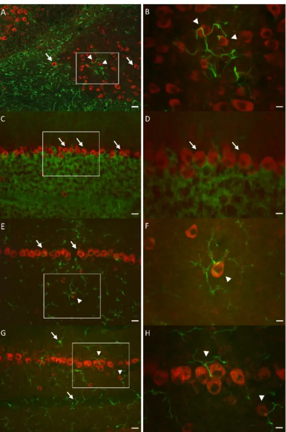
Fig. 2. Investigating the distribution of KAT-2+cells in the mouse cerebellum with fluorescent immunohistochemistry using glial markers. Photomicrographs (B, D, F, H) in the right column corre- spond to the rectangles in photomicro- graphs (A, C, E, G) with higher magnifications. A-B: Double labeling of KAT-2 (red) with astrocyte marker GFAP (green). The processes of GFAP+ glial cells surround the soma of KAT-2+ neurons (arrowheads), but double la- beling of GFAP and KAT-2 cannot be observed (arrows). C-D: Double labeling of KAT-2 (red) with astrocytic end-feet marker AQP4 (green). KAT-2 expres- sion was detected in neurons (arrows) but not in the astrocytic end-feet. E-F:
Double labeling of KAT-2 (red) with microglia marker CD11b (green). The CD11b+ microglial cells surround the KAT-2+neurons (arrowhead). However, double labeling of CD11b and KAT-2 cannot be detected (arrows). G-H:
Double labeling of KAT-2 (red) with microglia marker Iba1 (green). Similar to the results of CD11b labeling, we observed microglial cells’ processes surrounding Purkinje cells (arrow- heads), but there was no KAT-2 posi- tivity in the microglia (arrows). Scale bars: 40 μm (A, C, E, G) and 20 μm (B, D, F, H).
4 (Leica VT1000S).
2.3. Immunohistochemistry
Free-floating sections were washed in PB containing 0,4% Triton X- 100 (PBT) and incubated in 2% normal donkey serum (NDS) at room temperature. For the detection of KAT-2 and identification of cells containing the enzyme, the sections were exposed to primary antibodies (rabbit anti-KAT-2, 1:1000, Proteintech; rat anti-GFAP, 1:4000, Sigma;
mouse anti-NeuN, 1:4000, Millipore; mouse anti-GAD67, 1:1000, Mil- lipore and mouse anti-Calbindin, 1:1500, Swant; rat anti-CD11b, 1:200, Bio-Rad; goat anti-Iba1, 1:400, Abcam and mouse anti-AQP4, 1:200, Sigma) overnight at 4 ◦C.
The next day, samples were incubated in the appropriate secondary antibodies (1:500; Jackson Immuno Research) for 2 h at room temper- ature. Primary antibodies were diluted in 0.1 M PBT containing 1% NDS, while secondary antibodies were diluted in 0.1 M PB. Negative control was prepared from sections incubated without the primary antibodies.
After the incubations, the sections were coverslipped with antifade mounting medium (ProLong® Gold, Life Technologies). Fluorescent photomicrographs were obtained with an Olympus BX51 microscope fitted with a DP70 digital imaging system and a Zeiss Axio Imager 2 microscope.
3. Results
The presence of KAT-2 was studied with single and double fluores- cent immunolabeling using neuronal and glial markers. We observed broad KAT-2 distribution in the whole mouse cerebellum (Fig. 1A).
Using double fluorescent immunolabeling we found neurons in different cerebellar layers, with distinct size and appearance, which were positive both for NeuN and KAT-2, except for the Purkinje cell layer (Fig. 1B). In Purkinje cells, only KAT-2 labeling was found, and this KAT-2 positivity was prominent (Fig. 1B and C). To further verify the Purkinje cell identity, and to detect other GABAergic neurons in the cerebellum, GAD67 (Fig. 1D) and Calbindin (Fig. 1F) markers were used. Both GAD67 and Calbindin are expressed in several types of neurons, including Purkinje cells. These markers showed complete overlap with KAT-2+Purkinje cells (Fig. 1 E, G arrows). These results undoubtedly show that the most prominent KAT-2 positive cells in the cerebellum are Purkinje cells. That was indicated by i) labeling with GAD67 and by ii) labeling with Calbindin, iii) cell diameter and iiii) the lack of labeling with NeuN as an indirect proof.
GFAP astrocyte marker (Fig. 2A) and AQP4 astrocytic end-feet marker (Fig. 2C) were used to identify astrocytes and to examine the glial expression of KAT-2. Although KAT-2 has been described in the cerebrum partly as a glial enzyme (mainly astrocytic), it is not expressed either in astrocytes (Fig. 2B) or in astrocytic end-feet surrounding Pur- kinje cells in the investigated regions of the cerebellum (Fig. 2D).
Microglial cells were labeled with CD11b (Fig. 2E) and Iba1 microglia markers (Fig. 2G). Both with CD11b and Iba1 markers we observed microglial cells’ processes surrounding Purkinje cells (Fig. 2F, H ar- rowheads), but there was no KAT-2 positivity in the microglia.
4. Discussion
Kynurenine aminotransferase has four isoforms (KAT-1, -2, -3, -4) which are capable of catalyzing the transamination of L-kynurenine to KYNA. KAT-2 has the highest importance in the mammalian brain (Nematollahi et al., 2016). The present study demonstrated for the first time that KAT-2 enzyme did not localize in astrocytes, but notable expression was observed in the Purkinje cells of mice cerebellum. The finding that the prominently labeled cells are Purkinje cells is strongly supported by the position of their layer, the size of the cell, the fact that they can be labeled with GAD67 and Calbindin, and mostly by the fact that they are not labeled by NeuN. It is a well-known fact that the
sympathetic chain ganglia, the cells of the internal nuclear layer of the retina, and the Purkinje cells are immunonegative for NeuN (Wolf et al., 1996). Therefore, GAD67 and Calbindin markers were used for the identification of Purkinje cells. The finding that in the cerebellum of mice, KAT-2 is expressed in neurons (mainly in Purkinje cells) but not in astrocytes is not only new but also surprising, since KAT-2 enzyme in rat localizes mostly in astrocytes in different parts of the cerebrum (Guidetti et al., 2007).
One of our recent studies in mice showed that KAT-2 is expressed not only in astrocytes but also in interneurons in different structures of the cerebrum (Her´edi et al., 2017).
Based on these results, one can say that KAT-2 in the cerebrum of different species is expressed in astrocytes and also in other cell types, including neurons. On the other hand, in the cerebellum of mice KAT-2 is expressed in neurons (mainly in Purkinje cells, and also in other types of interneurons) but not in astrocytes. Besides this finding also draws our attention to species-specific differences in KAT-2 immunoreactivity in the central nervous system: in the rat cerebellum, KAT-2 has been re- ported to be localized in astrocytes but not in Purkinje cells (Guidetti et al., 2007). According to the literature, there is another major differ- ence between mice and rats. In contrast to rats, KAT-2 plays only a minor role in kynurenic acid production in the brain of adult mice (Yu et al., 2004).
Even if KAT-2 plays only a minor role in kynurenic acid production in the brain of adult mice, the prominent KAT-2 expression in Purkinje cells raises an interesting question. If we accept that the Purkinje cell itself (which has a key role in the cerebellar network) produces and releases in situ “de novo” synthesized KYNA, it may have widespread modulatory effects. For example, it may also suggest the existence of an autor- egulatory activity on these neurons. It is because KYNA-sensitive post- synaptic responses were recorded at the proximal pole of the Purkinje cell dendrites evoked by climbing fibers (Lopez et al., 1991).
The ontogenetic pattern of KAT activity in different parts of the brain was studied in detail. The KAT activity increased in all regions of the rat brain between 3rd postnatal day and 3rd months. However, the KAT activity was always the lowest in the cerebellum (Baran and Schwarcz, 1993). At the same time, alternative KYNA synthesis routes were described in the cerebellum. Generally, KYNA is attributed to the enzymatic conversion of L-kynurenine by KATs. In the cerebellum, it was found that KYNA may be produced from D-kynurenine by D-amino acid oxidase and by direct transformation of kynurenine to KYNA by reactive oxygen species (Blanco Ayala et al., 2015). These alternative ways are highly efficient in the cerebellum (Wang et al., 2012; Blanco Ayala et al., 2015).
According to the literature cited above and to our studies, KAT-2 is expressed in the brain in a region and cell type-specific manner, as Song and coworkers suggested (Song et al., 2018).
Because of the increasing attention that is paid to the role of endogenous kynurenic acid in brain physiology and pathology, the demonstration of fundamental differences in the cellular localization of KAT-2 in the forebrain and the cerebellum has far-reaching implications.
Therefore, we tried to pay special attention to the specificity of our antibody.
The validation of our antibody has happened earlier with several molecular biological methods. We performed immunohistochemistry on mouse brain sections and immunocytochemistry on the HeLa cell culture transfected with mouse kat-2 cDNA. We observed no KAT-2 positivity in the negative controls.
In addition to the immunohistochemical studies, we tested the pri- mary antibody specificity with Western blot analysis both on transfected HeLa cell culture and on mouse brain tissue homogenates from cere- brum and cerebellum. We detected a single immunoreactive band in the transfected HeLa cell culture preparation and the cerebral and cerebellar tissue homogenates at ~47 kDa, which is the estimated size of KAT-2 (not shown here, Her´edi et al., 2017).
Considering the antibody validation results mentioned above we did
not carry out an experiment in which parallel tissue sections were incubated with the antibody that had been preadsorbed with pure an- tigen and we did not verify the inhibition of KAT-2 enzyme activity by our antibody.
We are convinced that KAT-2 labeling shows a unique pattern in the cerebellum, which may be related to the multiple ways of KYNA pro- duction (low level of the classical way of KYNA production, low KAT activity) and the existence of alternative KYNA synthesis routes.
In summary, the present study is the first to report the prominent expression of KAT-2 in Purkinje cells but the lack of glial protein expression in mice cerebellum. Further studies are needed to clarify i) the functional role of KAT-2 expression in cerebellar Purkinje cells and ii) the role of KAT-2 expression in other types of neurons in mice cerebellum.
Conflicts of interest
The authors declare that they have no conflict of interest.
CRediT authorship contribution statement
Emma Balog: Conceptualization, Methodology, Investigation, Visualization. Gyula Jenei: Conceptualization, Methodology, Investi- gation, Visualization. Levente Gellert: Conceptualization, Supervision. ´ Etsuro Ono: Resources, Supervision, Funding acquisition. Laszl´ o ´ V´ecsei: Resources, Funding acquisition, Supervision. J´ozsef Toldi: Su- pervision, Writing - original draft. Zsolt Kis: Supervision, Project administration, Writing - review & editing.
Declaration of competing interest None.
Acknowledgments
This work was supported by grants GINOP-2.3.2-15-2016-00034, JSPS-HAS (NKM-25/2019), TUDFO/47138-1/2019-ITM, and the MTA- SZTE Neuroscience Research Group of the Hungarian Academy of Sci- ences and the University of Szeged. The authors are grateful to P´eter S´antha for expert histological technical assistance, University of Szeged, Department of Physiology. This work was supported by the University of Szeged Open Access Fund, grant number: 5041.
References
Baran, H., Schwarcz, R., 1993. Regional differences in the ontogenetic pattern of kynurenine aminotransferase in the rat brain. Brain Res. Dev. Brain Res. 74, 283–286. https://doi.org/10.1016/0165-3806(93)90014-2.
Blanco Ayala, T., Lugo Huitr´on, R., Carmona Aparicio, L., Ramírez Ortega, D., Gonz´alez Esquivel, D., Pedraza Chaverrí, J., P´erez de la Cruz, G., Ríos, C., Schwarcz, R., P´erez de la Cruz, V., 2015. Alternative kynurenic acid synthesis routes studied in the rat cerebellum. Front. Cell. Neurosci. 9, 178. https://doi.org/10.3389/
fncel.2015.00178.
Fujigaki, H., Yamamoto, Y., Saito, K., 2017. L-Tryptophan-kynurenine pathway enzymes are therapeutic target for neuropsychiatric diseases: focus on cell type differences.
Neuropharmacology 112, 264–274. https://doi.org/10.1016/j.
neuropharm.2016.01.011.
Guidetti, P., Hoffman, G.E., Melendez-Ferro, M., Albuquerque, E.X., Schwarcz, R., 2007.
Astrocytic localization of kynurenine aminotransferase II in the rat brain visualized by immunocytochemistry. Glia 55, 78–92. https://doi.org/10.1002/glia.20432.
Han, Q., Cai, T., Tagle, D.A., Li, J., 2010. Structure, expression, and function of kynurenine aminotransferases in human and rodent brains. Cell. Mol. Life Sci. 67, 353–368. https://doi.org/10.1007/s00018-009-0166-4.
Her´edi, J., Berk´o, A.M., Jankovics, F., Iwamori, T., Iwamori, N., Ono, E., Horv´ath, S., Kis, Z., Toldi, J., V´ecsei, L., Gell´ert, L., 2017. Astrocytic and neuronal localization of kynurenine aminotransferase-2 in the adult mouse brain. Brain Struct. Funct. 222, 1663–1672. https://doi.org/10.1007/s00429-016-1299-5.
Her´edi, J., Cseh, E.K., Berk´o, A.M., Veres, G., Zadori, D., Toldi, J., Kis, Z., V´ ´ecsei, L., Ono, E., Gell´ert, L., 2019. Investigating KYNA production and kynurenergic manipulation on acute mouse brain slice preparations. Brain Res. Bull. 146, 185–191. https://doi.org/10.1016/j.brainresbull.2018.12.014.
Lopez, L., Chan, C.Y., Okada, Y.C., Nicholson, C., 1991. Multimodal characterization of population responses evoked by applied electric field in vitro: extracellular potential, magnetic evoked field, transmembrane potential, and current-source density analysis. J. Neurosci. 11, 1998–2010. https://doi.org/10.1523/JNEUROSCI.11-07- 01998.1991.
Nematollahi, A., Sun, G., Jayawickrama, G.S., Church, W.B., 2016. Kynurenine aminotransferase isozyme inhibitors: a review. Int. J. Mol. Sci. 17 https://doi.org/
10.3390/ijms17060946.
Song, C., Clark, S.M., Vaughn, C.N., Nicholson, J.D., Murphy, K.J., Mou, T.-C.M., Schwarcz, R., Hoffman, G.E., Tonelli, L.H., 2018. Quantitative analysis of kynurenine aminotransferase II in the adult rat brain reveals high expression in proliferative zones and corpus callosum. Neuroscience 369, 1–14. https://doi.org/
10.1016/j.neuroscience.2017.11.001.
Turski, W.A., Gramsbergen, J.B., Traitler, H., Schwarcz, R., 1989. Rat brain slices produce and liberate kynurenic acid upon exposure to L-kynurenine. J. Neurochem.
52, 1629–1636. https://doi.org/10.1111/j.1471-4159.1989.tb09218.x.
V´ecsei, L., Szal´ardy, L., Fülop, F., Toldi, J., 2013. Kynurenines in the CNS: recent ¨ advances and new questions. Nat. Rev. Drug Discov. 12, 64–82. https://doi.org/
10.1038/nrd3793.
Wang, X.-D., Notarangelo, F.M., Wang, J.-Z., Schwarcz, R., 2012. Kynurenic acid and 3- hydroxykynurenine production from D-kynurenine in mice. Brain Res. 1455, 1–9.
https://doi.org/10.1016/j.brainres.2012.03.026.
Wolf, H.K., Buslei, R., Schmidt-Kastner, R., Schmidt-Kastner, P.K., Pietsch, T., Wiestler, O.D., Blümcke, I., 1996. NeuN: a useful neuronal marker for diagnostic histopathology. J. Histochem. Cytochem. 44, 1167–1171. https://doi.org/10.1177/
44.10.8813082.
Yu, P., Di Prospero, N.A., Sapko, M.T., Cai, T., Chen, A., Melendez-Ferro, M., Du, F., Whetsell, W.O., Guidetti, P., Schwarcz, R., Tagle, D.A., 2004. Biochemical and phenotypic abnormalities in kynurenine aminotransferase II-deficient mice. Mol.
Cell Biol. 24, 6919–6930. https://doi.org/10.1128/MCB.24.16.6919-6930.2004.