UNCORRECTED
PROOF
Inorganica Chimica Acta xxx (2017) xxx-xxx
Contents lists available at ScienceDirect
Inorganica Chimica Acta
journal homepage: www.elsevier.com
Research paper
Oligopeptide models of the metal binding loop of the bacterial copper efflux regulator protein CueR as potential Cu(I) chelators
Edit Mesterházy
a, b, Bastien Boff
b, Colette Lebrun
b, Pascale Delangle
b,⁎, Attila Jancsó
a,⁎aDepartment of Inorganic and Analytical Chemistry, University of Szeged, Dóm tér 7, Szeged H-6720, Hungary
bINAC/SyMMES UMR 5819 CEA, CNRS, Université Grenoble Alpes, 17 rue des Martyrs, 38 054 Grenoble, France
A R T I C L E I N F O
Article history:
Received 20 April 2017
Received in revised form 20 June 2017 Accepted 27 June 2017
Available online xxx
Keywords:
Copper(I) Competition Peptide complexes Thiolate coordination Chelators
A B S T R A C T
Copper(I) binding features of oligopeptides, mimicking the metal binding loops (MBL) of the bacterial copper efflux regulator protein CueR, were investigated with an aim of exploring potential candidates for Cu(I)-se- questration. Two of the studied ligands comprise the MBL ofE. coliCueR (Ac-ACPGDDSADCPI-NH2;EC) andV. choleraeCueR (Ac-SCPGDQGSDCPI-NH2;VC) while the third peptide is a His-containing variant ofVC(Ac-SCHGDQGSDCSI-NH2;HS). UV-titrations of the ligands by Cu(I) at pH = 7.4 and monitoring the characteristic SCys−→ Cu(I) charge transfer bands, together with CD-experiments, indicated the exclusive formation of monomeric Cu(I)-complexes (CuL) up to a 1:1 Cu(I):L ratio and the presence of oligometallic/
cluster type complexes at Cu(I)-excess. Such a change in speciation and the domination of the latter species was also supported by comparing the ESI-MS spectra obtained at 0.9:1 and 2:1 Cu(I):peptide ratios. pH-de- pendence of the UV spectra of the Cu(I):peptide 1:1 complexes reflected the formation of the two thio- late-Cu(I) bonds well below neutral pH, as two successive deprotonation processes could be fitted by apparent pKavalues falling in the pH range of 1.8–3.0 and 4.3–5.5. Cu(I)-binding affinity of the ligands was deter- mined by competition experiments applying the bicinchoninate ion for the displacement of the Cu(I)-ligated peptides. The obtained conditional stability constants ( = 15.3, 15.8 and 16.3 for Cu(EC), Cu(VC) and Cu(HS), respectively) indicate a strong affinity of Cu(I) to each ligand and suggest a slight destabilizing effect of charge repulsion between sidechain groups inECwhile a possible Cu(I)-coordination of the histi- dine residue inHS.
© 2017.
1. Introduction
Copper is an essential element for life but in excess becomes toxic, therefore several proteins, with a wide range of metal binding mo- tifs, participate in copper homeostasis [1]. Copper is found both in Cu(I) and Cu(II) oxidation states in living organisms. Cu(I) has a soft Lewis-acid character [2], and thus it is usually coordinated by 2, 3 or 4 sulphur donors in linear, trigonal planar or tetrahedral geometry [3].
The borderline Lewis-acid Cu(II) is generally coordinated by 4–6 ni- trogen and oxygen donors in a square planar, (distorted) pentacoordi- nate or (distorted) octahedral geometries [3,4]. In copper binding pro- teins these donor atoms are provided by the thiolate moieties of cys- teines, the thioether groups of methionines, the carboxylate groups of glutamates and aspartates, the imidazole rings of histidines, and be- sides, the N- and O-atoms of the polypeptide backbone can also play a role in the coordination of Cu(II).
⁎Corresponding authors.
Email addresses:pascale.delangle@cea.fr (P. Delangle); jancso@chem.u-szeged.
hu (A. Jancsó)
The metal binding motifs of the various proteins participating in the sensing, transport or storage of copper in cellular compartments with a reducing environment perfectly accommodate this metal in its Cu(I) state. Metallothioneins, encompassing cysteine rich sequence patterns, e.g. CXC or CXXC, are known to participate in the detox- ification of harmful metal overload [5]. These low molecular weight proteins are able to bind different number of Cu(I) ions in clusters [6,7]. A number of transport proteins utilize a variation of methio- nine and cysteine residues for the binding of Cu(I). Representative examples are the plasma membrane transporter Ctr1 with an 11-re- peat consensus MxxM motif [8] or Cu-transporter P-type ATPases possessing a MxCxxC metal binding domain [9]. There are several other fascinating Cu(I)-binding sequences, such as the CxCxxxxCxC found in the CopY repressor protein of thecopoperon [10], the me- thionine-rich LVMTAMPGMEHSPMGV Cu(I)-binding sequence of the periplasmic copper trafficking CopC [11], the unusual His36,Tr- p44,Met47,Met49 motif of the copper trafficking CusF [12,13] or the short CCxC fragment, which – together with three additional sepa- rated Cys residues – forms Cu(I)-clusters in the Cox17 chaperon [14].
The Cu(I)-responsive Cu Efflux Regulator CueR protein [15,16], a representative member of the large MerR family [17], regulates the synthesis of the copper transporter CopA [18] and the multicopper
http://dx.doi.org/10.1016/j.ica.2017.06.062 0020-1693/© 2017.
UNCORRECTED
PROOF
oxidase CueO [19,20] at the transcription level. Each monomer of the dimeric CueR protein binds a Cu(I) ion by two conserved cysteine residues located at the two ends of a short loop, restricting the metal ion into a linear coordination fashion [21]. CueR displays an outstand- ing sensitivity for Cu(I) as it is able to sense Cu(I) in a zeptomolar (10−21M) concentration. Besides, the protein can effectively differen- tiate between mono and divalent metal ions by providing a transcrip- tional response for Cu(I), Ag(I) or Au(I), but remaining inactive in the presence of Zn(II) or Hg(II) [21].
It has been previously demonstrated by Delangle et al. that short linear or cyclic decapeptides, inspired by the Cys-rich metal ion bind- ing domains of proteins participating in the homeostasis of soft metal ions could be efficiently used for Cu(I)-chelation. The studied ligands, a linear and a cyclic peptide mimicking the metal binding MxCxxC motif of the Atx1 chaperon and two cyclodecapeptides containing two cysteine residues, displayed high affinity for Cu(I) and a high selectiv- ity with regard to the potential cellular competitor Zn(II) [22–24].
With an aim of constructing potential peptide-based Cu(I)-seques- tering ligands benefitting from the large sensitivity and selectivity of CueR, three 12-mer peptides were designed (Scheme 1). Two of the ligands comprise the metal binding loop of CueR found in E.
coli (Ac-ACPGDDSADCPI-NH2; EC) and V. cholerae (Ac-SCPGDQGSDCPI-NH2;VC). The third studied peptide is a vari- ant of the ligandVC, where the two proline residues were replaced by a histidine and a serine (Ac-SCHGDQGSDCSI-NH2;HS) with a pur- pose of influencing the flexibility and thus the metal binding proper- ties of the molecule. His and Ser residues are, indeed, found in these positions in some of the metalloregulatory members of the MerR fam- ily [21]. Previously, we have reported on the Zn(II), Cd(II), Hg(II) and Ag(I) coordination features of these ligands [25–27]. In this work we present a detailed characterization of the Cu(I)-binding properties of the three 12-mer peptides, with a special attention devoted to the sta- bility of species formed under neutral conditions with regard of their potential use in Cu(I)-chelation in medical applications [28].
2. Experimental 2.1. Materials
The investigated peptides were synthesized by solid phase syn- thesis following the standard Fmoc strategy and purified by reversed phase semi preparative HPLC, as described earlier [25]. Metal ion so- lutions were made of tetrakis(acetonitrile)copper(I) hexafluorophos- phate (Cu(CH3CN)4PF6, Aldrich) dissolved in acetonitrile (Sigma) and mercury(II) chloride (HgCl2, Aldrich) dissolved in water.
5,5′-dithiobis-(2-nitrobenzoic acid) (DTNB) used for determining the concentration of the peptides, bathocuproinedisulfonic acid disodium salt (BCS) and bicinchoninic acid disodium salt hydrate (BCA) ap
Scheme 1.Schematic structures of the studied peptides (double column).
plied for determining the concentration of the copper(I) stock solu- tions and the conditional stability constants of the Cu(I) complexes, respectively, were purchased from Sigma-Aldrich. For the calibration of the mass spectrometry instrument a Pierce™ LTQ ESI Positive Ion Calibration Solution, containing Caffeine, MRFA and Ultramark 1621 was obtained from Thermo Fischer Scientific. Potassium phosphate monobasic (Sigma) and ammonium acetate (Acros) salts were used for buffer preparations. All the chemicals and solvents were used without further purification.
2.2. Sample preparations
Since both the peptides and Cu(I) are sensitive to oxidation, all of the sample manipulations were performed in a glovebox under argon atmosphere. Fresh solutions were prepared before each ex- periment. The Cu(I) stock solution was prepared in acetonitrile to avoid Cu(I) disproportionation into Cu(0) and Cu(II). Indeed acetoni- trile forms coordination complexes with Cu(I), which stabilizes the low oxidation state of copper in solution [29]. The peptides were dissolved in buffered media or in water containing 10 V/V% ace- tonitrile. The final concentration of the solutions was determined by Ellman’s procedure [30,31]. The protocol uses DTNB; the DTNB2−
anion reacts with free thiols resulting in the formation of the yel- low colored TNB− ion (2-nitro-5-thiobenzoate) per each thiol group (λmax= 412 nm;ε= 14,150 M−1cm−1). The Cu(I) concentration of the stock solutions was determined by addition of excess BCS and mea- suring the absorbance of the [Cu(BCS)2]3−complex (λmax= 483 nm;
ε= 13,300 M−1cm−1) [32].
2.3. UV spectroscopy
UV–Vis spectroscopic measurements were carried out on a Varian Cary50 spectrophotometer equipped with optical fibers connected to an external cell holder in the glove box. A quartz UV cell from Hellma Analytics with a 1.0 cm optical path length, equipped with a Teflon stopper, was used. For each titration 2.5 mL of a freshly prepared peptide solution (cpeptide∼30 µM, in phosphate buffer, pH = 7.4, cbuffer= 20 mM) were transferred to the cell. Aliquots of a Cu(I) stock solution (c∼3 mM), corresponding to 0.1 equivalent of Cu(I) per lig- and, were added by a 10 µL capacity Hamilton syringe. For pH titra- tions 2.0 mL of the peptide stock solution (c∼30 µM) was transferred into the quartz UV cell and then 0.9 equivalent of Cu(I) was added.
The desired pH values were adjusted with KOH solutions of appropri- ate concentrations (e.g. 0.1 M, 0.01 M). The pH of the solution was measured by a Metrohm 702 SM Titrino system equipped with a Met- tler Toledo InLab® Microelectrode.
2.4. CD spectroscopy
Circular dichroism spectra were acquired on an Applied Photo- physics Chirascan spectrometer. (1S)-(+)-10-camphorsulfonic acid served as calibration material for the instrument. 2.5 mL of freshly prepared peptide solution (cpeptide∼80 µM, in phosphate buffer, pH = 7.4,cbuffer= 20 mM) were transferred into a 1.0 cm optical path length sealable quartz cell (Hellma Analytics) with a silicone rubber seal. Aliquots of a Cu(I) stock solution (c∼3 mM), corresponding to 0.25 equivalents of Cu(I) per ligand, were added by a 10 µL capac- ity Hamilton syringe in the glove box. The spectra were recorded in the 190–400 nm wavelength range with 1 nm steps and a dwell time of 2 s per data points. The range of wavelength reported (typically 220–400 nm) corresponds to non-saturating CD signals, which was checked by looking at the HT voltage. Data were acquired and treated
UNCORRECTED
PROOF
by Pro-Data and Pro-Data Viewer programs (Applied Photophysics).
From each sample 3 parallel spectra were recorded and the average of these spectra was smoothed by the Savitzky-Golay method with a
“window size” 3.
2.5. ESI-MS
Mass spectra were acquired on a LXQ-linear ion trap (THERMO Scientific, San Jose, USA) instrument equipped with an electrospray source. Electrospray full scan spectra in the range ofm/z= 50–2000 amu were obtained by infusion through a fused silica tubing at 2–10 µL/min. The samples were analyzed in the negative mode. The LXQ calibration (m/z= 50–2000) was achieved according to the stan- dard calibration procedure from the manufacturer (using a mixture of caffeine, MRFA and Ultramark 1621). The temperature of the heated capillary for the LXQ was set in a range of 200–250 °C, the ion-spray voltage was adjusted to 3–6 kV and the injection time varied between 5–200 ms. The ligand concentration wasc∼100 µM in ammonium acetate buffer (pH = 6.9,cbuffer= 20 mM) / acetonitrile, 90/10, V/V.
Three samples were prepared for each peptide with 0.9 and 2.0 equiv- alents of copper(I).
2.6. Determination of apparent stability constants
The affinity constants for the Cu(I)-binding of the different pep- tides were determined by competition reactions using BCA as a com- petitor [33]. Three samples were prepared from each peptide (cpeptide∼65 µM, in phosphate buffer, pH = 7.4,cbuffer= 20 mM) con- taining 0.9 equivalents of Cu(I) relative to the concentration of the peptides. As the next step, different equivalents of the competitor, rel- ative to the Cu(I)-content, were added. The progression of the dis- placement of Cu(I) from the peptide complexes was followed by the measurement of the absorbance of the [Cu(BCA)2]3− complex (λmax= 562 nm;ε= 7900 M−1cm−1; logβ2= 17.2 [33]). The samples were equilibrated for several days in order to ensure that the recorded spectra represented the state of equilibrium.
3. Results and discussion
3.1. The mononuclear Cu(I) complexes evidenced by UV/Vis and CD spectroscopies
UV/Vis spectroscopic measurements were performed by adding aliquots of a Cu(I) stock solution in acetonitrile to the samples of pep- tides in phosphate buffer at pH 7.4. The observed changes for the Cu(I)-VCsystem are presented in Fig. 1, while the UV/Vis spectra obtained for Cu(I)-ECand Cu(I)-HSare shown in the Supplementary data (Fig. S1). The increase of the Cu(I) concentration is accompanied by the appearance of two main absorption bands atλ∼260 nm and λ∼300 nm (Fig. 1 and Fig. S1). The first one refers to a characteris- tic charge transfer transition from a thiolate group to Cu(I) (LMCT) [34] indicating the coordination of the ligands to Cu(I) via the thiolate moieties of the cysteine residues. The lower energy band appearing aroundλ∼300 nm was previously attributed to copper-thiolate clus- ters in metallothioneins [7]. However, such a band was also present in the spectra of other short peptides containing two cysteines where the exclusive formation of mononuclear complexes was proved by diffu- sion NMR [23]. In excess of Cu(I) a third transition emerges in the UV spectra atλ∼340 nm representing a formally spin-forbidden 3d → 4s metal to metal transition [7,35] indicating the formation of copper-thi- olate clusters, as often observed in the interaction of Cu(I) with thiol compounds [6,7,35,36]. The evolution of the LMCT band at 260 nm in all three Cu(I)-peptide systems, relative to the concentration of Cu(I), reflect molar absorbances in the range of∼7000 M−1cm−1per bound Cu(I) ions, which is compatible with values reported for other thiol compounds and for metallothioneins [7,37].
The recorded absorbance at 264 nm increases linearly until a breakpoint at ca.1.4 equivalents of Cu(I) in case ofVC(Fig. 1) and HS (Fig. S1), while in the sample ofEC (Fig. S1) the increasing absorbance follows a non-linear trend, which might suggest that the complex formation occurs in separated stages. A similar phenomenon was observed by Le Brun and co-workers in the Cu(I)-titration of the copper chaperone CopZ, possessing an MXCXXC metal binding frag- ment [38]. It was proposed that two major forms of the Cu(I)-CopZ complex existed, depending on the applied Cu(I)-to-protein ra
Fig. 1.UV titration ofVCwith Cu(I) at pH = 7.4. The insert shows the absorbances at 264, 300 and 340 nm as a function of thecCu(I)/cVCratio. (cVC= 30.0 μM) (single column).
UNCORRECTED
PROOF
tio. Cu(I)(CopZ)2 dominates at low Cu(I)-protein stoichiometries, where only one Cys residue of each monomer is coordinated to the metal ion. The latter species transforms into the dimeric structure Cu(I)2(CopZ)2above 0.5:1 Cu(I)-protein ratio, where all the four Cys take part in the coordination, two of them bridging the two Cu(I) ions.
The formation of these two Cu(I)-CopZ complexes was more evident on the CD titrations, which showed two clear endpoints at 0.5 and 1 Cu(I) equivalents on the evolution of LMCT bands.
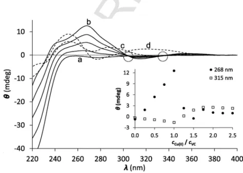
CD spectroscopy is also a useful tool for studying metal ion com- plexes of peptides since it provides information about the chiral back- bone and also about the charge transfer transitions. Thus we attempted to obtain complementary information on the Cu(I)-coordination by EC,VCandHS. The intense negative CD band below 220 nm is at- tributed to π → π∗transitions possibly overlapping with n → π∗tran- sitions, both belonging to the amide bonds of the peptide backbone [39,40] with an essentially disordered structure [25,26,41]. The addi- tion of aliquots of a Cu(I) solution to the ligands results in a gradual change of the intensities atλ= 220 nm, referring to changes in the av- erage conformation of the ligands induced by metal ion ligation (Fig.
2 and Fig. S2). However, similarly to the UV–Vis, the main infor- mation in the CD spectroscopy can be obtained from the S−→ Cu(I) LMCT bands. When the peptides were titrated by Cu(I), CD-bands ap- peared in the 240–380 nm wavelength range in parallel with the in- creasing Cu(I) concentration. With all the three peptides, up to 1.0 equivalent of Cu(I), the ellipticity at ca. 260 nm follows a linearly in- creasing trend in parallel with the increasing Cu(I):peptide concentra- tion ratio (see the inserts of Fig. 2 and Fig. S2) and isodichroic points are systematically observed. These results unambiguously show that the freeVC,ECandHSpeptides are transformed into one single com- plex species up to reaching the 1:1 Cu(I):peptide ratio. Only the CuL mononuclear complexes are formed with all the peptides, even with EC, where presence of more than one species was suspected based on the UV-data.
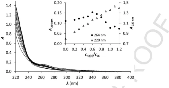
A further proof that the Cu(I)EC complex is the only formed species for Cu(I):ECratios below 1 and that a CuS4type Cu(EC)2 complex does not exist was obtained by using mercury(II) as a metal ion competitor. A sample with a Cu(I):ECratio of 0.5:1 was titrated with Hg(II). The S−-Cu(I) LMCT (∼264 nm) does not change signifi
cantly until 0.5 equivalents of Hg(II) as can be seen in Fig. 3. This sug- gests that the highly thiophilic Hg(II) can find free thiolates to coordi- nate to and does not disturb the Cu(I) coordination center. The slight increase can be attributed to the overlapping effect of the S−→ Hg(II) LMCT below λ= 220 nm [23,26,42–45]. For further addition of Hg(II) the S−-Cu(I) LMCT starts to collapse, while the S−-Hg(II) LMCT continues to increase, evidencing the transformation of Cu(EC) into the more stable Hg(EC) complex. These observations confirm that the CuS4type Cu(EC)2complex does not exist.
Comparison of the CD spectra obtained at 1:1cCu(I):cpeptideratios for the three ligands reflect a notable difference betweenHSand the other two peptides (Fig. 2, Fig. S2, see spectra denoted by letter “b”).
Considering that the CD-features observed for the free peptides are rather similar to each other, the different shapes of the CD spectra of the Cu(I) complexes must be correlated to different structures around the Cu(I) ion. Indeed, the absence of prolines inHSprovides a larger conformational flexibility of the peptide backbone, which may adopt different conformations compared toVCandEC, affecting the Cu(I) center. Nevertheless, the most plausible explanation for the differ- ent CD-features of Cu(I)-HSmay be the participation of the histidine residue in Cu(I) coordination, as it was demonstrated before for a few Cu(I)-binding proteins and model peptides [46–48]. An additional co- ordinating donor group besides the two thiolates should modify the chiral environment of the metal ion and therefore change the shape of CD-spectra.
3.2. Polynuclear Cu(I) complexes in excess of metal ions evidenced by UV/Vis and CD spectroscopies
The speciation in any of the Cu(I)-peptide systems above one equivalent of Cu(I) per ligand is rather complex, as reflected by the changes in the UV/Vis. The formation of clusters is indicated by the emerging UV-band atλ∼340 nm. Pronounced changes are also ob- served in the shapes of the CD spectra, when Cu(I) is added in excess to any of the ligands, which indicates a rather complex speciation, as a possible result of the formation of different clusters or polymetallic structures.
Fig. 2.CD spectra of theVCpeptide titrated with Cu(I) at pH = 7.4 from 0.0 to 1.0 equivalents of Cu(I) (solid lines). Spectra denoted by the letters “a”, “b”, “c” and “d” represent samples of 0.0, 1.0, 1.25 and 2.0 equivalents of Cu(I), respectively. Dotted circles highlight isodichroic points. The insert shows the evolution of ellipticities at two selected wave- length values. (cVC= 80 μM) (single column).
UNCORRECTED
PROOF
Fig. 3.UV spectra for Cu(I):EC0.5:1 titrated with Hg(II) at pH = 7.4. The insert shows the absorbances at 264 nm (left axis) and 220 nm (right axis) as a function of thecHg(II)/cEC
ratio. (single column).
The recorded absorbance values at 260 and 300 nm continue to follow a close to linear increasing trend even up to∼1.3–1.5 equiva- lents of Cu(I) per peptide (Fig. 1 and Fig. S1), even though CD titra- tions show clear endpoints at 1 Cu(I) equivalent. This shows that the LMCT absorbance band at ca. 260 nm is not sensitive to the nature of the different complexes (monometallic or polymetallic) but only to the number of thiolate to Cu(I) bonds. A similar behaviour was observed previously for tripodal pseudopeptide ligands bearing three cysteines orD-penicillamines [37,49,50], where the LMCT at 260 nm increased linearly up to 2.0 equiv. of Cu(I), even though it was demon- strated that two different types of complexes formed, the mononuclear CuL from 0.0 to 1.0 equivalent Cu(I) and the Cu4L2or Cu6L3clus- ters in excess of Cu(I). This indicates that the average number of thi- olate-Cu(I) bonds per metal ion does not change significantly in the species formed at low Cu(I) excess over the ligands and accordingly the molar absorbance per bound Cu(I) ion may also be similar to that of the monomeric complex. The nature and formation of these poly- metallic species could be inferred by ESI-MS that is sensitive to the stoichiometry of the complexes.
3.3. ESI-MS experiments for studying the nuclearity of the complexes Samples containing the peptides and 0.9 and 2 equivalents of Cu(I) in ammonium acetate buffer were analyzed by (−)ESI-MS. 10% ace- tonitrile in volume was added to minimize Cu(I) oxidation during the injection. The spectra acquired for theECpeptide are shown in Fig. 4 as an example. For 0.9 equivalent of Cu(I), the major signals arise from the free ligand as the negatively charged ions [L-H]−and [L−2H]2−:m/z= 1202.3 and 600.8, respectively (Fig. 4 upper panel).
The CuL complex is detected as a significant species as [L+Cu−2H]− and [L+Cu−3H]2−:m/z= 1264.2 and 631.7, respectively (Fig. 4 upper panel). Similar observations can be seen in Fig. S3 for peptidesVC andHS.
Many different polymetallic species were detected with 2 Cu(I) equivalents per ligands (see Fig. 4 lower panel forECas an example):
Cu2(EC), Cu3(EC), Cu4(EC)2. Similar mixtures of polymetallic com- plexes are detected for the three peptides as seen in Fig. S3 forVC andHS. The isotopic patterns of the Cu(I) complexes formed with the three peptides are shown in Fig. S4.
Fig. 4.(−)ESI-MS spectra recorded for Cu(I)-EC0.9:1 (upper panel) and 2:1 (lower panel) systems. * sodium adduct of the free peptide, ● oxidized free peptide (single col- umn).
The qualitative results obtained by ESI-MS provide a further sup- port for the Cu(I):L ratio dependent speciation suggested in each of the studied systems based on the UV/Vis and CD-data. They also confirm the complicated speciation in excess of Cu(I).
UNCORRECTED
PROOF
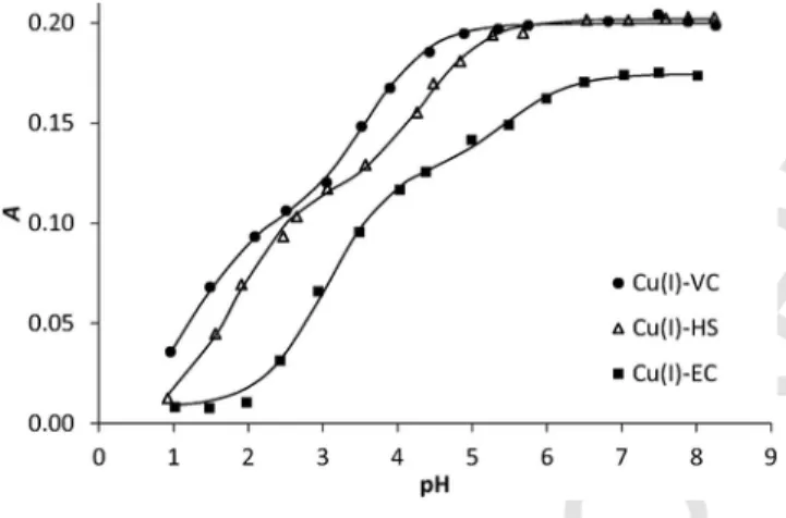
3.4. Effect of pH on thiolate-binding to Cu(I)
The evolution of the LMCT bands in the Cu(I)L complexes was studied with pH as seen in Fig. 5 reporting the absorbance at 264 nm as a function of pH. The absorbance recorded in the three systems is stable at pH larger than 7 showing that the complexes are fully formed, with absorption coefficients in the range 6300–7800 M−1cm−1. When the pH decreases, two successive processes are detected for each Cu(I) complex. The data were satisfactorily fitted with SPECFIT [51–54], with two successive (de)protonation steps according to equilibrium (1) and (2); the corresponding apparent pKavalues are given in Table 1.
The first protonation takes place with logKH1values between 3.6 and 5.5 and corresponds to the loss of about half the intensity of the S− to Cu(I) LMCT absorbance of the Cu(I) complex at pH 7. This is consistent with the protonation of one of the cysteines in equilib- rium (1) to give a Cu(I) complex with only one thiolate coordinated to the metal center. The apparent logKH1values found for the three complexes are significantly lower than the logKH(=pKa) previously reported for the Ag(I) complexes formed withECandVC(pKa= 6.5) [27]. The protonation processes reported here for the three Cu(I) pep- tide complexes are irrelevant with regard to the ways of potential
Fig. 5.Absorbances recorded atλ= 264 nm in the Cu(I):peptide 0.9:1 systems as a function of pH. The continuous lines represent the fitted values for the same wave- length. Data fitting was performed by SPECFIT in the wavelength range of λ= 250–400 nm based on a simple model involving two Cu(I)-promoted thiol deproto- nation processes (see details in the text) (single column).
Table 1
Apparent stability constants of the Cu(I)-peptide complexes at pH = 7.4, apparent pKa
( = logKH) values and absorption coefficients of the Cu(I)-peptide complexes. Experi- mental errors on the last characters are indicated in parentheses.
Ligand pKa1 pKa2 ε(M−1cm−1) (264 nm)
EC 15.3(1) 5.5(3) 3.0(1) 6370
VC 15.8(1) 3.6(2) 1.3(2) 7825
HS 16.3(1) 4.3(2) 1.8(1) 7740
Cu(I)-sequestration in physiological conditions since they are all tak- ing place at pH values significantly lower than pH 7.4. The second protonation steps are detected for lower pH values, with apparent logKH2between 1.3 and 3.0, and result in a total loss of the LMCT absorption to form the CuLH2complexes with no thiolate bound to Cu(I), according to equilibrium (2). The latter species in acidic condi- tions may be low stability Cu(I) complexes in which the metal center is coordinated to other functions than thiolates, or may represent the total release of free Cu(I) from the protonated ligand.
3.5. Stability of the Cu(I)-peptide complexes at pH 7.4
The various spectroscopic studies used for the characterization of the Cu(I)-binding ofVC, HSandECdemonstrate that mononu- clear complexes dominate in solutions where the Cu(I):peptide ra- tio is close to 1:1. The apparent stability constant of these species at pH = 7.4 were determined by a competition reaction. Previous studies showed that the Cu(I)-binding affinity of two cysteine containing lin- ear or cyclic peptides should fall in the range of logβ11pH 7.4∼15–17, β11pH 7.4 being the conditional stability constant of the Cu(I) com- plex at pH 7.4 [23,24]. According to Wedd’s recommendation [33,55]
bathocuproine disulfonate (BCS) is a perfect choice for a competitor ligand to conveniently determine stabilities with a logβ11pH 7.4value near 17 (logβ2([Cu(BCS)2]3−) = 19.8 [32]). Nevertheless, BCS proved to be a too strong competitor for the three studied systems and only 2 equivalents of the compound relative to Cu(I) withdrew more than 50% of Cu(I) from its peptide complexes. Accordingly, the weaker competitor bicinchoninate anion (BCA) was applied for the determi- nation of the Cu(I)-peptide affinities. BCA forms a well-defined 1:2 complex with Cu(I) according to Eq. (3) characterized by logβ2= 17.2
Addition of BCA to the Cu(I)-peptide solutions results in the ap- pearance of the intense absorption band of the [Cu(BCA)2]3−com- plex (λmax= 562 nm, ε= 7900 M−1cm−1 [33]), corresponding to the transfer of the metal ion from the peptide to BCA. In order to min- imize the risk of the formation of oligometallic species the compe- tition experiments were performed with peptide solutions contain- ing 0.9 equivalent of Cu(I) per peptides. The first indication for the magnitude of the Cu(I)-binding affinity of the peptides is the num- ber of BCA equivalents required for withdrawing 50% of Cu(I) from its peptide complexes. To achieve this, 8, 16 and 24 equivalents of BCA is needed for the samples of Cu(I)-EC, Cu(I)-VCand Cu(I)-HS, respectively, indicating the weakest Cu(I)-binding withECand the strongest withHS. The calculated conditional stability constants, sum- marised in Table 1, match rather well to the affinities obtained for lin- ear and cyclic peptidic models of the Atx1 Cu-chaperone [23,24], in spite of the larger distance between the two cysteine residues in the presently studied dodecapeptides. The lowest affinity ofECmay be explained by charge repulsion between the three aspartate residues, as found also with other Cu(I) chelating ligands possessing several negatively charged residues near the Cu(I)-binding site [24,37]. The stronger Cu(I)-binding affinity ofHScan be the result of the higher flexibility of the peptide backbone, which helps the peptide to more easily adopt the optimal coordination geometry. As hinted above on the basis of the CD-results, the histidine residue may directly partic- ipate in the coordination of Cu(I) and thus contribute to the higher affinity of HS to Cu(I). The stability constants determined in this study (Table 1) are very close to the affinity constant estimated for (1)
(2)
(3)
UNCORRECTED
PROOF
the Ag(I)-binding ofVC(logβ11pH8.0∼15–16), also by a competition experiment [56]. Similarly to what was concluded for Ag(I), being an- other cognate metal ion of CueR, the Cu(I)-binding affinities of these peptidic models are 5–6 orders of magnitude lower, than that of the native protein [21] which is a possible consequence of a loss of en- tropy upon metal ion binding of the models [27] but also the lack of other potential stabilizing effects, e.g. hydrophobic interactions in the metal binding pocket or the overall charge neutralisation that operate in the protein in contrast to the models.
4. Conclusion
The three studied oligopeptides (EC,VCandHS), inspired by the metal binding loop of the bacterial copper efflux regulator CueR pro- tein, display similar Cu(I)-binding features in terms of the key role of the two Cys-thiolates in determining the speciation and the stabil- ity of complexes formed under neutral conditions but also some no- table differences originating from the different amino acid sequences.
Monomeric (CuL) complexes dominate at pH = 7.4 and in the pres- ence of ligand excess (even up to a 1:1 Cu(I):L ratio), i.e. under condi- tions that are relevant for the Cu(I)-binding/transferring actions of pro- teins participating in Cu-homeostasis. Cu(I)-excess results in a com- plicated speciation with the formation of thiolate-bound oligometallic/
cluster species of various composition. pH titrations showed that de- protonation and coordination of the second thiol group in the Cu-pep- tide monomeric complexes takes place significantly below neutral pH.
This is a remarkable difference compared to what was reported for the Ag(I)-binding ofECand VC[27]. Consequently, such a (de)proto- nation process cannot play a role in Cu(I)-sequestration by these pep- tides in physiological conditions. The Cu(I)-binding affinities of the ligands reflect relatively small but still significant differences likely to be attributed to the destabilizing effect of charge repulsion between the carboxylate sidechain groups inECand the stabilizing effect of the possible direct metal ion coordination of His together with a larger ligand flexibility in HS. The magnitude of the conditional stability constants determined at pH = 7.4 (logβ11pH 7.4= 15.3–16.3) fall in the range usually found for other peptide sequences containing two cys- teines, being somewhat smaller than those obtained for models of the Atx1 copper chaperone (logβ11pH 7.4= 17.4) [23,24] and compar- ing well with more rigid cyclic decapeptides encompassing CxxxxC sequences (logβ11pH 7.4= 15.5–16.7) [23,24]. Our results compared to previous ones also demonstrate that the affinity of peptide sequences with two cysteines for Cu(I) depends little on the flexibility of the loop and the number (x) of amino acids in between the two Cys residues (x = 2–7). Therefore, the driving force for the coordination of the soft Cu(I) cation is expected to be mainly the formation of the two strong bonds with the soft thiolate donors.
The oligopeptide models described in this paper do not reproduce the zeptomolar (10−21M) sensitivity of the regulator protein CueR or its specificity for monovalent versus divalent cations [21], exemplify- ing the complexity of metal regulation processesin vivo, which can- not simply be described by thermodynamic equilibria. However, the studied model peptides still resemble the ability of the protein to ex- clusively accommodate one metal ion per sequence under ligand ex- cess conditions. Finally, these model peptides show large affinities for Cu(I) combined with high selectivities vs. Zn(II) [26], which are fea- tures that may be utilized in the development of Cu(I)-sequestering agents [28].
Acknowledgements
This research was supported by the Labex ARCANE (Grant ANR-11-LABX-0003-01), the “Fondation pour la Recherche Médicale” (grant DCM20111223043), the Hungarian National Research, Development and Innovation Office-NKFIH through pro- ject GINOP-2.3.2-15-2016-00038 and grant No: K_16/120130.
References
[1] R.A. Festa, D.J. Thiele, Curr. Biol. 21 (2011) R877–R883.
[2] R.G. Pearson, J. Am. Chem. Soc. 85 (1963) 3533–3539.
[3] R.R. Conry, Encyclopedia of Inorganic Chemistry, John Wiley & Sons Ltd, 2006.
[4] K.A. Koch, M.M.O. Peña, D.J. Thiele, Chem. Biol. 4 (1997) 549–560.
[5] R.D. Palmiter, Proc. Natl. Acad. Sci. U.S.A. 95 (1998) 8428–8430.
[6] P. Faller, M. Vašák, Biochemistry 36 (1997) 13341–13348.
[7] D.L. Pountney, I. Schauwecker, J. Zarn, M. Vasak, Biochemistry 33 (1994) 9699–9705.
[8] A. Dancis, D.S. Yuan, D. Haile, C. Askwith, D. Eide, C. Moehle, J. Kaplan, R.D. Klausner, Cell 76 (1994) 393–402.
[9] K. Petrukhin, S. Lutsenko, M.J. Cooper, C.T. Gilliam, J.H. Kaplan, J. Biol.
Chem. 272 (1997) 18939–18944.
[10] P.A. Cobine, G.N. George, C.E. Jones, W.A. Wickramasinghe, M. Solioz, C.T.
Dameron, Biochemistry 41 (2002) 5822–5829.
[11] F. Arnesano, L. Banci, I. Bertini, S. Mangani, A.R. Thompsett, Proc. Natl.
Acad. Sci. U.S.A. 100 (2003) 3814–3819.
[12] I.R. Loftin, S. Franke, N.J. Blackburn, M.M. McEvoy, Prot. Sci. 16 (2007) 2287–2293.
[13] Y. Xue, A.V. Davis, G. Balakrishnan, J.P. Stasser, B.M. Staehlin, P. Focia, T.G.
Spiro, J.E. Penner-Hahn, T.V. O'Halloran, Nat. Chem. Biol. 4 (2008) 107–109.
[14] A.C. Rosenzweig, Chem. Biol. 9 (2002) 673–677.
[15] F.W. Outten, C.E. Outten, J. Hale, T.V. O'Halloran, J. Biol.
Chem. 275 (2000). 31024-31029.
[16] J.V. Stoyanov, J.L. Hobman, N.L. Brown, Mol. Microbiol. 39 (2001) 502–512.
[17] N.L. Brown, J.V. Stoyanov, S.P. Kidd, J.L. Hobman, F.E.M.S. Microbiol, Rev. 27 (2003) 145–163.
[18] C. Rensing, B. Fan, R. Sharma, B. Mitra, B.P. Rosen, Proc. Natl. Acad. Sci.
U.S.A. 97 (2000) 652–656.
[19] G. Grass, C. Rensing, Biochem. Biophys. Res. Commun. 286 (2001) 902–908.
[20] C. Rademacher, B. Masepohl, Microbiology 158 (2012) 2451–2464.
[21] A. Changela, K. Chen, Y. Xue, J. Holschen, C.E. Outten, T.V. Halloran, A.
Mondragón, Science 301 (2003) 1383–1387.
[22] O. Seneque, S. Crouzy, D. Boturyn, P. Dumy, M. Ferrand, P. Delangle, Chem.
Commun. (2004) 770–771.
[23] P. Rousselot-Pailley, O. Sénèque, C. Lebrun, S. Crouzy, D. Boturyn, P. Dumy, M. Ferrand, P. Delangle, Inorg. Chem. 45 (2006) 5510–5520.
[24] A.M. Pujol, M. Cuillel, O. Renaudet, C. Lebrun, P. Charbonnier, D. Cassio, C.
Gateau, P. Dumy, E. Mintz, P. Delangle, J. Am. Chem. Soc. 133 (2011) 286–296.
[25] A. Jancso, D. Szunyogh, F.H. Larsen, P.W. Thulstrup, N.J. Christensen, B.
Gyurcsik, L. Hemmingsen, Metallomics 3 (2011) 1331–1339.
[26] D. Szunyogh, B. Gyurcsik, F.H. Larsen, M. Stachura, P.W. Thulstrup, L. Hem- mingsen, A. Jancso, Dalton Trans. 44 (2015) 12576–12588.
[27] D. Szunyogh, H. Szokolai, P.W. Thulstrup, F.H. Larsen, B. Gyurcsik, N.J.
Christensen, M. Stachura, L. Hemmingsen, A. Jancsó, Angew. Chem. Int.
Ed. 54 (2015) 15756–15761.
[28] P. Delangle, E. Mintz, Dalton Trans. 41 (2012) 6359–6370.
[29] P. Kamau, R.B. Jordan, Inorg. Chem. 40 (2001) 3879–3883.
[30] P.W. Riddles, R.L. Blakeley, B. Zerner, Methods in Enzymology, Academic Press, 198349–60.
[31] G.L. Ellman, Arch. Biochem. Biophys. 82 (1959) 70–77.
[32] Z. Xiao, F. Loughlin, G.N. George, G.J. Howlett, A.G. Wedd, J. Am. Chem.
Soc. 126 (2004) 3081–3090.
[33] Z. Xiao, L. Gottschlich, R. van der Meulen, S.R. Udagedara, A.G. Wedd, Metal- lomics 5 (2013) 501–513.
[34] M. Beltramini, K. Lerch, Biochemistry 22 (1983) 2043–2048.
[35] R. Bogumil, P. Faller, D.L. Pountney, M. Vašák, Eur. J. Biochem. 238 (1996) 698–705.
[36] K. Fujisawa, S. Imai, N. Kitajima, Y. Moro-oka, Inorg. Chem. 37 (1998) 168–169.
[37] A.M. Pujol, C. Gateau, C. Lebrun, P. Delangle, Chem. Eur. J. 17 (2011) 4418–4428.
[38] M.A. Kihlken, A.P. Leech, N.E. Le Brun, Biochem. J. 368 (2002) 729.
UNCORRECTED
PROOF
[39] Y.J. Li, U. Weser, Inorg. Chem. 31 (1992) 5526–5533.
[40] S.M. Kelly, N.C. Price, Curr. Protein Pept. Sci. 1 (2000) 349–384.
[41] A. Jancsó, B. Gyurcsik, E. Mesterházy, R. Berkecz, J. Inorg.
Biochem. 126 (2013) 96–103.
[42] G.R. Dieckmann, D.K. McRorie, D.L. Tierney, L.M. Utschig, C.P. Singer, T.V.
O'Halloran, J.E. Penner-Hahn, W.F. DeGrado, V.L. Pecoraro, J. Am. Chem.
Soc. 119 (1997) 6195–6196.
[43] A.M. Pujol, C. Lebrun, C. Gateau, A. Manceau, P. Delangle, Eur. J. Inorg.
Chem. 2012 (2012) 3835–3843.
[44] S. Pires, J. Habjanič, M. Sezer, C.M. Soares, L. Hemmingsen, O. Iranzo, Inorg.
Chem. 51 (2012) 11339–11348.
[45] M. Łuczkowski, B.A. Zeider, A.V.H. Hinz, M. Stachura, S. Chakraborty, L.
Hemmingsen, D.L. Huffman, V.L. Pecoraro, Chem. Eur. J. 19 (2013) 9042–9049.
[46] L. Zhang, M. Koay, M.J. Maher, Z. Xiao, A.G. Wedd, J. Am. Chem.
Soc. 128 (2006) 5834–5850.
[47] M.J. Pushie, K. Shaw, K.J. Franz, J. Shearer, K.L. Haas, Inorg. Chem. 54 (2015) 8544–8551.
[48] K.L. Haas, A.B. Putterman, D.R. White, D.J. Thiele, K.J. Franz, J. Am. Chem.
Soc. 133 (2011) 4427–4437.
[49] A.-S. Jullien, C. Gateau, C. Lebrun, I. Kieffer, D. Testemale, P. Delangle, Inorg.
Chem. 53 (2014) 5229–5239.
[50] A.M. Pujol, C. Gateau, C. Lebrun, P. Delangle, J. Am. Chem. Soc. 131 (2009) 6928–6929.
[51] H. Gampp, M. Maeder, C.J. Meyer, A.D. Zuberbühler, Talanta 32 (1985) 1133–1139.
[52] H. Gampp, M. Maeder, C.J. Meyer, A.D. Zuberbühler, Talanta 32 (1985) 257–264.
[53] H. Gampp, M. Maeder, C.J. Meyer, A.D. Zuberbühler, Talanta 32 (1985) 95–101.
[54] H. Gampp, M. Maeder, C.J. Meyer, A.D. Zuberbühler, Talanta 33 (1986) 943–951.
[55] Z. Xiao, A.G. Wedd, Nat. Prod. Rep. 27 (2010) 768–789.
[56] A. Jancso, unpublished results, 2015.