THE “ THREE AMIGOS ” LURKING BEHIND TYPE 1 DIABETES: HYGIENE, GUT MICROBIOTA AND
VIRUSES
ODIN ANDREAS JOHAN JAKOBSEN1and LASZLO SZEREDAY1,2*
1Department of Medical Microbiology and Immunology, Medical School, University of Pecs, Pecs, Hungary
2Janos Szentagothai Research Center, Pecs, Hungary
(Received: 3 October 2017; accepted: 19 November 2017)
Incidence of type 1 diabetes (T1D) is on the rise and yet, despite decades of research, the exact ethology of the disease still remains a mystery. The autoimmune reaction, which ultimately leads to the destruction of pancreatic beta cells, causing insulin deficiency and T1D, is a result of genetic susceptibility and environmental factors. Precisely, what are these environmental factors? Current popular opinion implies these pathogens, such as viruses, especially human enteroviruses, are a triggering factor. On the other hand, the hygiene hypotheses states in which the increase of autoimmune diseases, such as T1D, can, in fact, be explained by the decrease of infections, and infectious agents, more like viruses, actually serve as a defense mechanism, therefore, protect us from developing certain autoimmune diseases. Addi- tionally, the relationship between the gut microbiota and autoimmune diseases is currently gaining increased interest including relative research now demonstrating how the guts immune system plays a crucial role in the development of autoimmune diseases.
This literature review aims to evaluate these three popular suspects: Viral infections, hygiene and gut microbiota, in relation to their potential triggering effect on T1D and their close relationship to one another.
Keywords: microbiota, type 1 diabetes, hygiene hypothesis, environment, virus
*Corresponding author; E-mail:szereday.laszlo@pte.hu
This is an open-access article distributed under the terms of theCreative Commons Attribution- NonCommercial 4.0 International License, which permits unrestricted use, distribution, and reproduction in any medium for non-commercial purposes, provided the original author and source are credited, a link to the CC License is provided, and changes–if any–are indicated.
Introduction
Type 1 diabetes (T1D) is a disease characterized by an immune-mediated destruction of pancreatic βcells [1]. The disease is on the rise, with an overall annual increase estimated at approximately 3% [2]. Genetics is a well-known contributor to the disease [1], yet, genetics alone cannot explain this rapid increase in recent incidences. The disease does not conform to any known Mendelian pattern of inheritance [3], and studies show in which fewer than 10% of people with human leukocyte antigen (HLA)-determined susceptibility to T1D develop the disease and monozygotic twins have less than 40% concordance of developing T1D [4].
A model originally posted in 1986, and modified throughout the years [1], suggests individuals are born with various degrees of genetic predisposition, and an environmental trigger inducesβcell autoimmunity.
What specifically is this environmental trigger is the primary focus of this literature review includes the following popular theories: the hygiene hypothesis, gut microbiota and viral infection.
The Hygiene Hypothesis
This theory states in which the increase in frequency of T1D throughout several countries can be explained by the decline of infections [5]. In recent years, attention has been focused on the possibility in which lifestyle changes plays a major factor in the rise of T1D and other autoimmune diseases. Several lifestyle changes have developed in the last 20 years, and one is the decline in infectious disease, as a result of increased hygiene, health and medical conditions [6].
Geographical data and epidemiology
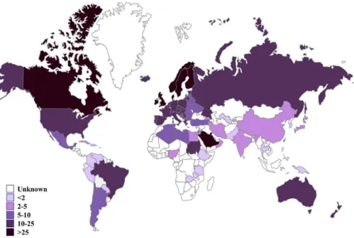
The difference of incidence rate between countries plays a major role in this theory. As Figure 1 demonstrates, throughout Europe, there is a north–south gradient of T1D, exhibiting higher frequency in the Scandinavian countries, and, less so in Southern Europe, and a low presence throughout Africa. Similarly, higher incidences are seen in North America when compared with South America.
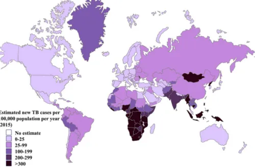
The geographical map of published incidence rates for childhood T1D is the mirror image of that for infectious disease distribution, as seen in Figure 2.
Tuberculosis and childhood diarrhea are more frequent in the southern countries
compared with those of the north [5]. In the case of tuberculosis, a negative correlation between the frequency of the disease and T1D has been shown [5].
However, differences between small geographical areas have also been found.
For example inexplicably, the frequency of T1D is threefold higher in Finland when compared with its geographic neighbor, Russian Karelia [5], although they have the same ethnic background and are separated only by a few dozen kilometers.
Among the several factors implicating an environmental factor, current migration studies exist. Families emigrating from countries with a low T1D incidence to a high T1D incidence country experienced an increase in frequency of the disease in itsfirst-generation migrants, and vice versa. This has been observed in several migrant populations, in particular, Pakistanis now residing in England [5].
One of the more remarkable arguments supporting the hygiene hypothesis is the reverse trend observed between infectious disease incidence and the incidence of autoimmune diseases [5]. In addition, relative and specific evidence also exists.
There is a negative correlation between hygiene condition and T1D, in Northern Ireland, showing that the areas with lowest T1D incidences are in areas with the poorest hygiene conditions [6].
Figure 1.Estimated new cases of type 1 diabetes (<15 years) per 100,000 children per year, 2015.
IDF Diabetes Atlas (2015) published by the International Diabetes Federation was used to produce the estimates
It was also shown in which the T1D incidence rate was higher in children who have had the lowest level of infectious exposure [6].
Animal models
Experimental models offer the superb advantage in allowing deliberate interventions. Non-obese diabetic (NOD) mice spontaneously developed T1D at the age of 2–3 months, and the incidence of the disease and the acceleration of the onset is largely dependent in the way the animals were taken care of, particularly their sanitary conditions [5]. When the sanitary conditions were poor, the fre- quency of the disease has decreased, and incidences increased again by decon- tamination through C-section and raising newborns in isolation [6]. Inversely, it is possible to prevent the onset of diabetes by deliberately infecting mice, which was previously raised in a clean environment with some various infective agents, such as complete Freund’s adjuvant, mouse hepatitis viruses, andSchistosoma mansoni soluble egg antigens [5]. Biobreeding (BB) rats in a germ-free facility also have increased diabetes frequency, and prevention could be achieved by various infections [6].
Figure 2.Estimated TB incidence rates, 2015. Global tuberculosis reports (2016) published by WHO was used to produce the estimates
Human therapeutic trials
Currently, there is no known direct evidence showing that the suppression of infections causes a rise in T1D, yet the available evidence from other studies can prove useful.
Helminthiases treatment performed in countries of high incidence of these parasites demonstrates the disappearance of the parasite and is linked to an increase in atopic disease [6]. Similar observations have been made in Southern Africa, with respect to vaccination againstStreptococcus pneumonia[6]. Clearly, this indicates how a decrease of infection caused an increase of certain diseases and inversely, the question may be asked as to whether an increase of infections can suppress the onset of certain autoimmune or allergic disease in humans [6].
Gut Microbiota
The human intestinal microbiome is a complex, symbiotic ecological community, which influences human health and development, including the development, function, and maintenance of the human immune system [7]. This fact is precisely why the relationship between the gut microbiota and autoimmune diseases has gained an immense amount of interest within the past decade, including its relationship to T1D. The microbiota hypothesis, in contrast to the hygiene hypothesis, focuses more on our ancestral microorganisms rather than cleanliness and postulates the lack of symbiotic microorganisms as being respon- sible for the disease [8].
The microbiome consists of hundreds of trillions of microorganisms, including more than 1014bacteria belonging to 1,000 species, Archaea, bacterio- phage particles, viruses, eukaryotes, and fungi [9, 10].
The microbiota is, at birth, characterized by instability and low diversity.
Its composition is in fact dynamic and influenced by several factors, such as birth delivery mode, breastfeeding and food introduction, geography, immunological status, and the use of antibiotics and hygiene conditions. The early childhood represents the period in which microbiota composition becomes more complex and long-term colonization by most of bacteria is established [9]. For example, it has been shown that the risk and onset of T1D in childhood is higher in children delivered by C-section [11].
External factors influence the function and composition of the microbiota and, in particular, the diet [11]. An example of this can be presented in the study of twins, revealing how obese individuals have reduced the diversity of intestinal microbiome when compared with their lean twin [11].
Although many phyla are represented, and significant variability exists between individuals, recently, molecular techniques identified four major microbial phyla that represent over 90% of the gut microbiota: Firmicutes, Bacteroides, Protobacteria, and Actinobacteria [12]. Most of the commensal bacteria found in human fecalflora are represented by two main groups of Firmicutes, subdivided in Clostridium coccoides (Clostridium cluster XIVa) and Clostridium leptum (Clostridium cluster IV), and the group of Cytophaga–Flavobacterium–Bacteroides [12]. The gut microbiota plays an important role being mainly involved in the development and growth of immune system and in the regulation of several fundamental metabolic pathways. For instance, Clostridium clusters IV and XIVa have an important role in immune gut homeostasis modulating the expression of Foxp3 in CD4+ T cells by production of short-chain fatty acid (SCFA) [12].
Intestinal microbiota and autoimmunity
The intestinal mucosa is a common entry site for pathogens and contains a significant amount of cells of the immune system. An intact mucosa provides the first line of defense against pathogens, and the research continues to define the vital role the intestinal microbiota plays in protecting the mucosa from invading pathogens and influencing the development and maintenance of both the systemic and innate immune systems [11].
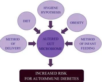
Epidemiological data reveal a high prevalence of autoimmune diseases in developed countries compared with developing countries, creating the original basis for the“hygiene hypothesis.”This hypothesis has been further developed to include concept in which altered intestinal microbiota may be one of several predisposing factors for the development of autoimmunity, as demonstrated in Figure3[11].
In addition to the knowledge in which intestinal microbiota contributes to the development and maintenance of the immune system, several studies demonstrate the relationship between autoimmune diseases and the intestinal flora. Inflammatory bowel diseases (IBDs) are autoimmune diseases of the intestinal tract, and although the exact etiology of IBD is as of yet unclear, it likely involves environmental, genetic, and immune factors [11]. The role of the microbiome in IBD development is becoming increasingly defined, and several studies illustrate a relationship. Animal studies have revealed that the severity of experimentally induced intestinal inflam- mation can be modulated by introduction of anaerobic bacteria [11]. Metagenomic evaluation of fecal samples taken from patients afflicted with Crohn’s disease revealed a reduced diversity of the Firmicutes phyla compared with healthy controls [11].
Patients suffering from Crohn’s disease and ulcerative colitis have a reduced amount
of bacteria in the Firmicutes and Bacteroidetes phyla, and there is an altered intestinal microbiome in individuals with Crohn’s disease compared with their monozygotic twin [11]. Alterations in intestinal flora have been hypothesized to contribute in the development of several other autoimmune diseases, including celiac disease, allergy, multiple sclerosis, rheumatoid arthritis, and ankylosing spondylitis [11]. In addition, an increasing amount of experimental data from humans and animal models has supported the role played by imbalanced gut microbiota and T1D [9].
Considering this evidence, it is only natural to assume that a relationship exists between the gut microbiota and T1D.
Animal models
Flora variation. Peyer’s patches, splenic germinal centers, mesenteric lymph nodes, plasma cells, and CD4+ T cells of germ-free mice are smaller and/or fewer when compared with mice raised in a normal environment [11]. In germ-free animals, reintroduction of normal gutflora can normalize the size and cellularity of lymphoid structures and increase antibody production. It was shown that the introduction of a single organism, segmentedfilamentous bacterium, was suffi- cient to stimulate production of IL-17 producing CD4+ T cells [11].
Studies using NOD mice demonstrate chronic viral infections, mycobacte- rial infections, and stimulus with bacterial antigens decreased the development of
Figure 3.Environmental factors that may directly or indirectly alter the intestinal microbiome and therefore affect the risk of autoimmune diabetes
T1D [11]. However, female NOD mice, affected byBacillus cereus, delayed the onset of T1D [11], suggesting that the incidence of T1D in NOD mice is modulated by restrictedflora, and not germ-free conditions.
Usingfluorescentin situhybridization to target 16S rRNA of Bacteroides, Clostridium, and Lactobacillus species in fecal samples of biobreeding diabetes- prone (BB-DP) rats, it has been demonstrated that the Bacteriodes species were more prominent in those rats which developed diabetes, and further experiments have revealed that there is a higher proportion of Lactobacillus and Bifidobacter- ium genera in biobreeding diabetes-resistant rats compared with BB-DP rats, while BB-DP rats were again found to have a higher amount of the Bacteroides species [11].
Flora modulation.Gut microbiota hydrolyze and ferment the dietary polysac- charides to generate monosaccharides and SCFAs that can be absorbed and utilized by the host for energy. SCFAs, such as acetate, propionate, and butyrate, contribute approximately 5%–10% to human energy resources [10]. These SCFAs have been well characterized as playing a role in gut immune homeostasis. Mice that take acetate through drinking water display suppressed dextran sulfate sodium-induced colitis, inflammatory arthritis, and asthma in a Gpr43-dependent manner [13]. Additionally, NOD mice that consumed acid water lead to a decrease in Firmicutes and an increase in Bacteroidetes, which in turn was associated with an increased onset of diabetes in NOD mice [12].
The initial evidence suggesting a role of nutrition in T1D was accomplished using NOD mice exposed to a diet consisting of wheatflour or hydrolyzed casein, revealing a significant lower rate of incidence of diabetes in mice receiving the hydrolyzed diet. Further studies completely prevented the diabetes by providing antibiotics and hydrolyzed diet to BB-DP rats [11].
Several other studies have been achieved to effectively and precisely pinpoint the components and mechanisms of diet upon T1D. NOD mice lacking exposure to dietary gluten develop diabetes at a significant lower rate when compared with mice on a standard diet [11]. Investigation into the mechanisms of insulitis development and T1D in animals on a gliadin diet suggests these proteins increase small intestine inflammation and intestinal permeability [14]. Further- more, BB-DP rats have a higher proportion of Th1 cells in the mesenteric lymph nodes upon exposure to wheat [11]. Notably, exposure to gliadin also suppresses Treg cell production in NOD mice, which is another potential mechanism by which dietary exposure may enhance T1D risk [11].
Another study, which performed toward examining the effect of a gluten- free diet versus standard diet on bacterial composition in NOD mice, demonstrate how diabetes developed in 47% of the standard-diet mice, compared with only 5%
in the gluten-free diet mice, and examination of the intestinal bacteria revealed a
significantly lower prevalence of aerobic, microaerophilic, and anaerobic bacteria in mice on the gluten-free diet [11]. Much of the difference in bacterial composi- tion was directly attributable to Gram-positive bacteria [11]. An in vitrostudy highlighted how intestinal epithelial cells in culture exposed to gliadin-derived peptides produced inflammatory cytokines, and this process was downregulated when these cells were inoculated with the intestinal bacteria Bifidobacteria, suggesting a mechanism by which intestinal bacteria can protect intestinal integrity [11].
An interesting subject is if manipulation of the gut microbiota can play a role in modulating the onset of T1D. To date, this has only been tested on animal models, where the gut microbiota from a MyD88-deficient NOD mice (which were protected from T1D development) were transplanted in NOD mice. A reduced onset of insulitis and a significant delay of diabetes were observed [12]. Specific probiotic strains of Lactococcus lactis genetically modified to secrete the whole proinsulin autoantigen along with the immu- nomodulator IL-10 was able to revert diabetes in NOD mice [12]. It has also been shown that gender can play an important role in the development of T1D.
Female NOD mice are significantly more susceptible to T1D than males.
Transfer of male microbiota to female NOD prior to disease onset protected against pancreatic islet inflammation, autoantibody production, and the de- velopment of the disease [12]. Another dietary mean by which gut bacteria could be altered includes the ingestion of probiotics– “live microorganisms that, when administered in adequate amounts, confer a health benefit on the host” [8]. Administration of the probiotic mixture VSL#3 containing Streptococcus thermophiles, Bifidobacterium spp., and Lactobacillus spp.
was shown to prevent diabetes development in NOD mice [12]. Additionally, Lactobacillus spp. was investigated in the prevention of T1D. Lactobacillus johnsonii or Lactobacillus reuteri isolated from diabetes-resistant rat was administered to BB-DP rats.L. johnsonii, but notL. reuteri, post-weaned the onset of T1D [12].
Another way to manipulate and alter the gut microbiota is through anti- biotics. Antibiotics have been used in modern societies to treat infections caused by pathogens. As a consequence, the intestinal microbiome is affected by antibiotic usage [8]. Although most data exist from animal models, antibiotic use in mice and in humans induces the proliferation of pathogens in the intestine, including Clostridium difficile and Salmonella enterica [9]. Animal studies demonstrate that the antibiotic-induced modulation of the gut microbiome affects autoimmune diseases [8]. It has been shown that the oral antibiotic treatment of pregnant mice could alter the gut microbiota composition in the offspring, but there was no significant change in the development of T1D [8]. Other studies,
however, demonstrate that the modulation of the gut microbiota composition by antibiotic treatment affects diabetes development in BB-DP rats and NOD mice.
Long-term vancomycin treatment depleted many major bacteria genera, except for the species Akkermansia muciniphila, which became dominant and inhibited diabetes development in NOD mice [8]. However, it is noteworthy that diabetes development was critically affected by the type of antibiotic used as well as the timing of treatment [8]. Another study suggests that targeting Gram-negative bacteria protects NOD mice from diabetes development, whereas depleting Gram-positive bacteria accelerated diabetes in NOD mice. This study also indicates that the most effective time of treatment is early in life, which is consistent with other studies of early-life microbiota disruption and other meta- bolic disorders, such as obesity [8].
Human studies
Flora variation.A study [11] attempting to define the difference in bacterialflora between children who develop T1D and healthy controls revealed that the intestinal microbiota in children who developed T1D had a higher proportion of Bacteroidetes phyla (Gram-negative) and lower proportion of Firmicutes phyla (Gram-positive), whereas control patients exhibited a decrease of Bacteroidetes phyla and an increase of Firmicutes phyla. Within the Bacteroidetes phyla, the Bacteroides ovatusspecies represented 24% of the total increase.
A Spanish cohort study showed that people with T1D had an increased amount of Clostridium, Bacteroides, and Vellionella and a reduced amount of Bifidobacterium and Lactobacillus compared with controls. Interestingly, the latter two organisms have been regarded as beneficial and are used as probiotic candidates [15].
Additionally, it is shown that adequate butyrate production levels are essential for gut integrity [12]. The butyrate-producing bacteria have anti- inflammatory effect both in vivo and in vitro studies [12]. This is interesting since studies show that butyrate-producing bacteria (Eubacterium, Fusibacterium, Anaerostipes, Roseburia, Subdoligranulum, and Faecalibacterium) are more abundant in controls compared with T1D cases, whereas lactate producers (Lactobacillus, Lactococcus, Bifidobacterium, and Streptococcus) were more common in T1D cases [12]. Additional studies showed that streptococci and Bacteroidetes were more represented in diabetic children, while the combined abundance of Clostridium clusters IV and XIVa was higher in healthy controls [12]. These human studies support the notion that alteration in gut microbiota is related toβ-cell autoimmunity and T1D.
Flora modulation.Diet modulates the gut microbiota from birth. For example, breastfeeding seems to protect infants against the development of various diseases and disorders. Breastfeeding is associated with reduced rates of acute infections in infancy and decreased risk of obesity later in life. This protection may be partially due to breast-milk itself, which contains immunologically active compounds, such as antibacterial enzymes and antibodies against pathogens. The transfer of maternal intestinal microbiota capable of educating the neonate’s immune system also contributes to this protection. In addition, human breast milk contains complex prebiotic polysaccharides that promote the colonization of the infant gut and sustain the beneficial microbial communities [16].
Another important factor represented by nutrition is that Lactobacillales and Protobacterias growth is promoted by increased levels of amino acids and short- chain carbohydrates in the intestine that they can metabolize [9]. It is reported that oxygen levels exert strong influence on the bacterial composition, and that high level of oxygen allows for thefirst colonization of the gut by facultative anaerobic bacteria, such as enterococci, enterobacteria, and staphylococci, and aerobic bacteria. At later stages, microbe-derived metabolites and the reduction of oxygen levels caused by the presence of these bacteria make the environment more favorable for the growth of obligate anaerobic bacteria, such as Bacteroides, Clostridia, and Bifidobacterium. The gut microbiota composition of a child after the age of about 2 years resembles that of an adult [9]. As previously mentioned, the colon colonization involves largely Firmicutes and Bacteroidetes, and their growth is favored by their ability to metabolize complex or resistant carbohydrates [9]. It has been shown that the diet has such a strong effect on the microbiota that the modification offiber content can affect its composition in less than 24 h [9].
For example, an elevated content of fiber was associated with the presence of Bacteroides, showing us that the gut microbiota evolves based on changes in the diet composition [9].
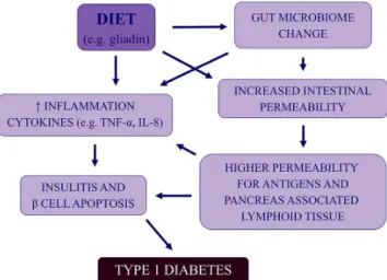
Similar to the animal models, human intestinal integrity appears to be compromised upon gliadin exposure, and due to the association between T1D and celiac disease, it can be proposed that these diseases share specific risk factors, such as gliadin, as demonstrated in Figure4[11]. Samples of human intestine exposed to gliadinin vitroshowed an amplified release of zonulin and increased permeability [11]. The role of gliadin in the pathogenesis of T1D likely extends beyond intestinal integrity, and the small intestine biopsies of T1D patients exposed to gliadin exhibited an unusually high inflammatory response [11]. Production of cytokines TNF-αand IL-8, both involved in the pathogenesis of T1D, is also stimulated by gliadin, distinctively, in subjects with celiac disease [11].
Additionally, islet autoimmunity appears to increase in individuals who are exposed to gliadin-containing foods prior to 6 months of age [11].
Viral Infections
Viruses have, for a long time, been widely suspected as the environmental trigger of T1D and are prime candidates since they are capable of activating both the innate and adaptive immune systems. Their ability to induce strong immune responses and causing inflammation in target organs also make them ideal candi- dates for the initiation of islet autoimmunity. Certain viruses have been linked to T1D, based on epidemiology, serology, histology, and rodent studies; however, viruses have also been shown to prevent autoimmunity, as previously described in the“hygiene hypothesis,” illustrating the potential complexity of the disease.
Epidemiological evidence
Infections with different viruses, such as Epstein–Barr virus, cytomegalovirus, rotavirus, rubella virus, mumps virus, and others [17], have been associated with the development of T1D; however, the strongest evidence in support of viral involve- ment exists from the human enterovirus (HEV) [17–21]. Some of the isolated forms of pancreatic HEV can induce T1D in mice [17] and are also able to infect and destroy human islet cellsin vitro[17]. The RNA of Coxsackievirus B (CVB) has been detected in the blood of recent onset T1D patients [22], and the presence of HEV RNA in the serum constitutes a risk factor for autoimmunity and T1D [23].
Other studies have been able to detect HEV proteins in the Langerhans islets of T1D
Figure 4.General schema outlining the possible relationships between gut microbiota/integrity and development of type 1 diabetes in genetically predisposed individuals
patients [24] and in the DAISY study, it was shown that the progression to T1D was increased in children in the span of time following an enteroviral infection characterized by the presence of viral RNA in blood [25, 26]. In addition, a meta-analytic study demonstrated the odds ratio of 3.7 between HEV infection and T1D-related autoimmunity, and 9.8 between HEV infection and clinical T1D, showing a significant association between HEV infection and T1D [27].
A particularly interesting study, notably the DiViD study, was the first to examine fresh pancreatic tissue at the diagnosis of T1D, for the presence of viruses. Minimal pancreatic tail resection was performed for 3–9 weeks following T1D onset in six adults while non-diabetic organ donors served as controls.
Enteroviral capsid protein 1 (VP1) was detected in the islets of all the T1D patients and in two of the nine controls. Hyperexpression of class I HLA molecules was found in the islets of all the patients, and in one of the nine controls, whereas enterovirus-specific RNA sequences were detected in four of six patients, and in none of the controls [28]. This is consistent with the possibility in which enteroviral infection in the pancreatic islets contributes to disease progression in humans.
Animal models
After inoculating 4-week-old NOD mice with CVB, it was shown that the mice were well protected from developing T1D, but when older NOD mice were used, CVB rapidly induced T1D onset [18]. It was shown that the age of the mouse, correlating to the extent of the islet inflammation, was the key component in this difference, and the virus was only demonstrable in the islets of older mice.
As the NOD mice aged and the insulitis increased, the mice became more susceptible and less protected from the onset of T1D, and this change in status, from a defensive posture to T1D, to increased induction occurred as a function of the extent of the hosts own autoimmune island inflammation. The strain of the virus used to inoculate the mice was also important, illustrating an increase in the rapidly replicating CVB strain, and required fewer infectious particles to induce T1D, than a slower replication strain [29], showing that the virus strain and infectious dose were, as many other infectious agents, linked. These observations have lead to the proposed relationship between HEV infection and T1D. A HEV infection which meets the key criteria to trigger T1D in an at-risk individual might also be understood as an example of another HEV infectious disease, much like a poliovirus (PV) infection which induces polio. In reference to polio, less than 1%
of PV infections lead to paralytic disease; therefore, it can be reasonably assumed that similar infection ratios hold true for other HEV [29].
Discussion
One of the popular theories relating to the environmental triggers of T1D is the hygiene hypothesis, which states that the increased frequency of T1D in several countries can be explained by the decline of infections, generally, as a result of better hygiene, health, and medical conditions [6].
The geographical data suggest an environmental factor, as reviewed in Figure1 and the geographical map of incidence rate for childhood T1D is the mirror image of that for infectious disease distribution, as seen in Figure 2.
Although differences between small geographical areas have also been found, and are difficult to explain, however, may be attributed to other environmental triggers or differences when considering the socioeconomic status [5].
Migration studies support this theory by demonstrating how families who emigrate from countries with low T1D incidence to a high T1D incidence country generally feature an increased frequency of the disease in its first- generation migrants and vice versa. It is difficult to assert that the increase in T1D is due to a loss of a protective factor present in their nation of origin, and not due to the presence of an unfavorable factor in their new home [5, 6].
Animal models have shown that in both BB-rats and NOD-mice, the incidence of the T1D and the rapidity of onset depended particularly on the sanitary conditions, illustrating how poor conditions decreased the disease rate of incidence, and vice versa [5, 6]. Although no human therapeutic trial can prove that a deliberate suppression of infections can trigger a rise in T1D, other studies have been useful. Helminthiases treatment demonstrated that a decrease of the parasite was linked to an increase of atopic disease, and vaccination against S. pneumoniahad a similar result, indicating a decrease of infection can cause an increase of a certain disease [5, 6].
Several mechanisms have been studied to understand how infections can provide defense and protection against certain autoimmune and allergic diseases.
Although there are no definite answers yet, studies have shown that infections occurring earlier in a person’s life generally yields in assuring an increase in defense, and similar results were found in mice [6].
Distinctly, another theory is related to the gut microbiota. A study highlight- ed the risk of T1D onset in childhood and found it is inexplicably higher in children delivered by C-section [11], yet interestingly, the supportive reasoning for the C-section was unknown; therefore, in theory, the method of delivery is largely influential on the gut microbiome composition.
Several studies show a relationship between the intestinalflora and autoim- mune diseases, especially IBD [9, 11], suggesting that a similar relationship between T1D and the gut microbiota is possible.
Some of the more interesting results originating from animal studies highlight how NOD mice, induced withB. cereus, chronic viral infections, and mycobacterial infections or stimulus with bacterial antigens, decreased or delayed the development of T1D [11] leading to the concept that a germ-free environment and/or a restricted flora increase the risk of T1D.
Human studies have began to emerge, implying the intestinal microbiota in children who developed T1D, did indeed, have a higher proportion of Bacter- oidetes phyla and lower proportion of Firmicutes phyla [11], but this was an abbreviated study including eight case subjects and four controls. Similar to animal models, human intestinal integrity seems to be compromised upon gliadin exposure [11], and due to the association between T1D and celiac disease, it can be postulated that these diseases share specific risk factors, as seen in Figure 4.
A very interesting topic is how modulation or manipulation of the gut microbiota can affect the disease. Most of the research today is from animals, and some studies have shown the following interesting results: NOD mice that consumed water lead to an increase in Bacteroidetes and a decrease in Firmicutes [12]. This change in composition has been associated with an increased onset of diabetes in NOD mice [12] and humans [11]. Also, when transplanting gut microbiota from MyD88-deficient NOD mice to NOD mice, a delay of T1D was observed, and transferring male microbiota to female microbiota in NOD mice also showed protection against the disease [12].
Additionally, the administration of the probiotic VSL#3 has shown to prevent diabetes in NOD mice [12], while giving vancomycin was also able to inhibit diabetes development by depleting many bacteria except forA. muciniphilia[8].
This creates a strong suspicion that modulating the flora can be an important therapeutic alternative, although a lot more research must be conducted in relation to how these modulations affect the disease and the immune-mediated mechanisms behind them.
The final environmental trigger of T1D appropriately discussed in this literature review highlights viral infections, specifically, the enterovirus. Several studies from both humans and animals have been reported, and one of the more striking reports originated from a human meta-analytic study shows an odds ratio of 9.8 between HEV infections and clinical T1D [28]. Although there may be some weaknesses to the meta-analytic observational study, a significant hetero- geneity to the different study designs and methods used should be mentioned.
Another interestingfinding is, from the DiViD study, examining fresh pancreatic tissue at the time of T1D diagnosis,finding VP1 and hyperexpression of class I HLA in the islets of all the T1D patients, and enterovirus-specific RNA sequence in four of six patients, with the results being confirmed in various laboratories [18].
This result does not prove the causality between HEV infection and T1D, although it supports the view in which a low-grade enterovirus infection is present in the pancreatic islets at the time of T1D diagnosis; however, when considering there were only six patients and nine controls, similar studies should be accomplished to confirm this in a larger group of patients.
Conclusions
T1D is indeed a complex multifactorial disease, and evidence exists, supporting all the aforementioned potential environmental triggers, although none of them are definite or without shortcomings. In consideration of the hygiene hypothesis and its focus on the lack of infectious agents, the gut microbiota theory focuses on the composition of the intestinalflora and the fact that the enteroviruses transmit generally through the fecal-oral route and replicate primarily in the gut offer us an indication of the close relationship between these theories.
Instead of the disease trigger being one or the other, it can be proposed that all of them, separately or in combination, are capable of triggering T1D in genetically susceptible humans. Like many other diseases, possessing several environmental risk factors, T1D may also be induced by several potential risk factors. The lack of certain infections, the alteration of the gut microbiota, and specific viral infections are all in close relationship and their common denominator is their ability to alter the immune system.
In T1D, a cure for the disease today may seem far-fetched. It can be postulated that any new treatment or cure focusing only on one of the aforemen- tioned potential triggers can, at best, decrease the incidence rate. For example, a vaccine against a virus triggering T1D still leaves the possibility of T1D as the trigger mechanism from factors relating to the gut microbiota or the hygiene hypothesis. Therefore, the key factor lies in understanding in far greater detail how the different potential triggers affect our immune system and precisely, what path the immune system takes leading to the destruction of β-cells. Only when the immune mechanism is accurately mapped, can a complete cure be produced focusing on the common immune pathway the different triggers affect.
Acknowledgements
This scientific contribution is dedicated to the 650th Anniversary of the Foundation of the University of Pécs, Hungary.
Conflict of Interest None.
References
1. Atkinson, M. A., Eisenbarth, G. S., Michels, A. W.: Type 1 diabetes. Lancet383, 69–82 (2014).
2. Patterson, C., Guariguata, L., Dahlquist, G., Soltesz, G., Ogle, G., Silink, M.: Diabetes in the young–A global view and worldwide estimates of numbers of children with type 1 diabetes. Diabetes Res Clin Pract103, 161–175 (2014).
3. Haller, M. J., Atkinson, M. A., Schatz, D.: Type 1 diabetes mellitus: Etiology, presentation, and management. Pediatr Clin North Am52, 1553–1578 (2005).
4. Knip, M.: Pathogenesis of type 1 diabetes: Implications for incidence trends. Horm Res Paediatr76, 57–64 (2011).
5. Bach, J. F.: The hygiene hypothesis in autoimmunity: The role of pathogens and commensals. Nat Rev Immunol18, 105–120 (2017).
6. Bach, J. F., Chatenoud, L.: The hygiene hypothesis: An explanation for the increased frequency of insulin-dependent diabetes. Cold Spring Harb Perspect Med 2, a007799 (2012).
7. Dunne, J. L., Triplett, E. W., Gevers, D., Xavier, R., Insel, R., Danska, J., Atkinson, M. A.:
The intestinal microbiome in type 1 diabetes. Clin Exp Immunol177, 30–37 (2014).
8. Gulden, E., Wong, F. S., Wen, L.: The gut microbiota and type 1 diabetes. Clin Immunol 159, 143–153 (2015).
9. Gianchecchi, E., Fierabracci, A.: On the pathogenesis of insulin-dependent diabetes mellitus: The role of microbiota. Immunol Res65, 242–256 (2017).
10. He, C., Shan, Y., Song, W.: Targeting gut microbiota as a possible therapy for diabetes.
Nutr Res35, 361–367 (2015).
11. Boerner, B. P., Sarvetnick, N. E.: Type 1 diabetes: Role of intestinal microbiome in humans and mice. Ann N Y Acad Sci1243, 103–118 (2011).
12. Bibbo, S., Dore, M. P., Pes, G. M., Delitala, G., Delitala, A. P.: Is there a role for gut microbiota in type 1 diabetes pathogenesis? Ann Med49, 11–22 (2017).
13. Hu, C., Wong, F. S., Wen, L.: Type 1 diabetes and gut microbiota: Friend or foe? Pharmacol Res98, 9–15 (2015).
14. Funda, D. P., Kaas, A., Tlaskalova-Hogenova, H., Buschard, K.: Gluten-free but also gluten-enriched (gluten+) diet prevent diabetes in NOD mice; the gluten enigma in type 1 diabetes. Diabetes Metab Res Rev24, 59–63 (2008).
15. Stewart, C. J., Nelson, A., Campbell, M. D., Walker, M., Stevenson, E. J., Shaw, J. A., Cummings, S. P., West, D. J.: Gut microbiota of type 1 diabetes patients with good glycaemic control and high physical fitness is similar to people without diabetes:
An observational study. Diabet Med34, 127–134 (2017).
16. Knip, M., Siljander, H.: The role of the intestinal microbiota in type 1 diabetes mellitus.
Nat Rev Endocrinol12, 154–167 (2016).
17. Rodriguez-Calvo, T., Sabouri, S., Anquetil, F., von Herrath, M. G.: The viral paradigm in type 1 diabetes: Who are the main suspects? Autoimmun Rev15, 964–969 (2016).
18. Krogvold, L., Edwin, B., Buanes, T., Frisk, G., Skog, O., Anagandula, M., Korsgren, O., Undlien, D., Eike, M. C., Richardson, S. J., Leete, P., Morgan, N. G., Oikarinen, S., Oikarinen, M., Laiho, J. E., Hyoty, H., Ludvigsson, J., Hanssen, K. F., Dahl-Jorgensen, K.:
Detection of a low-grade enteroviral infection in the islets of Langerhans of living patients newly diagnosed with type 1 diabetes. Diabetes64, 1682–1687 (2015).
19. Krogvold, L., Edwin, B., Buanes, T., Ludvigsson, J., Korsgren, O., Hyoty, H., Frisk, G., Hanssen, K. F., Dahl-Jorgensen, K.: Pancreatic biopsy by minimal tail resection in live adult patients at the onset of type 1 diabetes: Experiences from the DiViD study.
Diabetologia57, 841–843 (2014).
20. Ohara, N., Kaneko, M., Nishibori, T., Sato, K., Furukawa, T., Koike, T., Sone, H., Kaneko, K., Kamoi, K.: Fulminant type 1 diabetes mellitus associated with coxsackie virus type A2 infection: A case report and literature review. Intern Med55, 643–646 (2016).
21. El-Senousy, W. M., Abdel-Moneim, A., Abdel-Latif, M., El-Hefnawy, M. H., Khalil, R. G.:
Coxsackievirus B4 as a causative agent of diabetes mellitus type 1: Is there a role of inefficiently treated drinking water and sewage in virus spreading? Food Environ Virol (2017).
22. Elshebani, A., Olsson, A., Westman, J., Tuvemo, T., Korsgren, O., Frisk, G.: Effects on isolated human pancreatic islet cells after infection with strains of enterovirus isolated at clinical presentation of type 1 diabetes. Virus Res124, 193–203 (2007).
23. Schulte, B. M., Bakkers, J., Lanke, K. H., Melchers, W. J., Westerlaken, C., Allebes, W., Aanstoot, H. J., Bruining, G. J., Adema, G. J., Van Kuppeveld, F. J., Galama, J. M.:
Detection of enterovirus RNA in peripheral blood mononuclear cells of type 1 diabetic patients beyond the stage of acute infection. Viral Immunol23, 99–104 (2010).
24. Lonnrot, M., Salminen, K., Knip, M., Savola, K., Kulmala, P., Leinikki, P., Hyypia, T., Akerblom, H. K., Hyoty, H.: Enterovirus RNA in serum is a risk factor for beta-cell autoimmunity and clinical type 1 diabetes: A prospective study. Childhood Diabetes in Finland (DiMe) Study Group. J Med Virol61, 214–220 (2000).
25. Richardson, S. J., Willcox, A., Bone, A. J., Foulis, A. K., Morgan, N. G.: The prevalence of enteroviral capsid protein vp1 immunostaining in pancreatic islets in human type 1 diabetes.
Diabetologia52, 1143–1151 (2009).
26. Willcox, A., Richardson, S. J., Bone, A. J., Foulis, A. K., Morgan, N. G.: Immunohisto- chemical analysis of the relationship between islet cell proliferation and the production of the enteroviral capsid protein, VP1, in the islets of patients with recent-onset type 1 diabetes. Diabetologia54, 2417–2420 (2011).
27. Stene, L. C., Oikarinen, S., Hyoty, H., Barriga, K. J., Norris, J. M., Klingensmith, G., Hutton, J. C., Erlich, H. A., Eisenbarth, G. S., Rewers, M.: Enterovirus infection and progression from islet autoimmunity to type 1 diabetes: The Diabetes and Autoimmunity Study in the Young (DAISY). Diabetes59, 3174–3180 (2010).
28. Yeung, W. C., Rawlinson, W. D., Craig, M. E.: Enterovirus infection and type 1 diabetes mellitus: Systematic review and meta-analysis of observational molecular studies. BMJ 342, d35 (2011).
29. Chapman, N. M., Coppieters, K., von Herrath, M., Tracy, S.: The microbiology of human hygiene and its impact on type 1 diabetes. Islets4, 253–261 (2012).