ORIGINAL ARTICLE
Efficacy of medical care of epileptic pregnant women based on the rate of congenital abnormalities in their offspring
cga_30034..42Ferenc Bánhidy1, Erzsébet H. Puhó2, and Andrew E. Czeizel2
1Second Department of Obstetrics and Gynecology, School of Medicine, Semmelweis University, and2Foundation for the Community Control of Hereditary Diseases, Budapest, Hungary
ABSTRACT
The objective of the present study was to check the efficacy of progress in the medical care of epileptic pregnant women on the basis of the reduction of different con- genital abnormalities (CAs) in their offspring. First, the preva- lence of medically recorded epilepsy was compared in 95 pregnant women who later had offspring with different CAs (case group) and 90 pregnant women who later delivered newborn infants without CA (control group) and matched to cases in the Hungarian Case-Control Surveillance System of Congenital Abnormalities, 1980–1996. Second, the rate of dif- ferent CAs was compared in the offspring of epileptic pregnant women between 1980 and 1989 and 1990–1996. Cleft lip with or without cleft palate, cleft palate, cardiovascular CAs, oesoph- ageal atresia/stenosis, hypospadias and multiple CAs showed a higher risk in the offspring of pregnant women with epilepsy treated with different antiepileptic drugs, explained mainly by polytherapy. There was no higher risk for total CAs after mono- therapy. There was no significantly lower rate of total CAs in the offspring of epileptic pregnant women during the second period of the study. The efficacy of special medical care of epileptic pregnant women was not shown on the basis of decrease in the rate of CAs in the offspring of epileptic pregnant women.Key Words:antiepileptic drug treatment, congenital abnormality, epilepsy, pregnancy
INTRODUCTION
Epilepsy is defined as a disorder of the brain function characterized by the periodic and unpredictable occurrence of seizures and is classified as primary generalized, or focal/partial with or without secondary generalization. Primary general seizures can be divided into absence, myoclonic, atonic, or tonic-clonic (Wyllie 2001).
Epilepsy is one of the most frequently studied maternal diseases during pregnancy (Tompson et al. 1997; Aminoff 2004). Partly most epilepsies had early onset, and therefore, occured in 0.3–0.6%
of pregnant women. Partly the higher rate of structural birth defects (i.e. congenital abnormalities [CAs]) in the children of epileptic women treated with antiepileptic drugs (AEDs) was recognized after the first paper on the topic (Janz and Fuchs 1964) by Meadow (1968) and later confirmed by several studies and completed with minor anomalies, such as broad and low nasal bridge or nail hypo- plasia (Shepard and Lemire 2004; Briggset al. 2005) and func-
tional disturbances (e.g. mental retardation) (Meadoret al. 2009).
There was a long debate whether this higher risk associated with epilepsy itself (genetic predisposition or adverse effect of seizures), AEDs, other (e.g. lifestyle) factors, or their interaction. However, Holmeset al. (2001) showed that pregnant women with a history of epilepsy, but without AED treatment during pregnancy had no higher risk for CA, though it is worth mentioning that untreated women are expected to be affected with less severe epilepsy (Fried et al. 2004).
In the 1980s the preconceptional care of epileptic women was introduced in Hungary (Czeizel 1999; Ero˝set al. 1998) and special prenatal care of epileptic pregnant women was recommended (Tompsonet al. 1997; Aminoff 2004). The aim of the study was therefore to check the efficacy of this recent progress in the medical care of epileptic pregnant women on the basis of the reduction of CA, thus the data in the two periods of the study (i.e. 1980 and 1989 and 1990–1996) were compared in the population-based dataset of the Hungarian Case-Control Surveillance of Congenital Abnormali- ties (HCCSCA) (Czeizelet al. 2001).
MATERIALS AND METHODS
Cases affected with CAs were selected from the dataset of the Hungarian Congenital Abnormality Registry (HCAR), 1980–1996 (Czeizel 1997) for the HCCSCA.
Hungarian Congenital Abnormality Registry
Notification of cases with CA is mandatory for physicians from birth until the end of first postnatal year to the HCAR. Most cases are reported by obstetricians and pediatricians. In Hungary, practi- cally all deliveries take place in inpatient obstetric clinics and the birth attendants are obstetricians. Pediatricians work in the neonatal units of inpatient obstetric clinics, or in various inpatient and out- patient pediatric clinics. Autopsy was mandatory for all infant deaths and common (about 80%) in stillborn fetuses during the study period. Pathologists sent a copy of the autopsy report to the HCAR if defects were identified in stillbirths and infant deaths.
Since 1984, fetal defects diagnosed in prenatal diagnostic centers with or without termination of pregnancy have also been included into the HCAR.
CAs were differentiated into three groups: lethal (if defects cause stillbirths or infant deaths or pregnancies are terminated due to fetal defect in more than 50% of cases), severe (without medical inter- vention CAs cause handicap or death) and mild (CAs require medical intervention but life expectancy is good). Lethal and severe CAs together constitute major CAs. (Minor anomalies or morpho- logical variants without serious medical or cosmetic consequences are excluded from the class of CA.) In addition, two main categories of cases with CAs were differentiated: isolated (only one organ is Correspondence: Andrew E. Czeizel, MD, PhD, Doct. Sci, 1026. Budapest,
Törökvész lejto˝ 32, Hungary. Email: czeizel@interware.hu
Received April 12, 2010; revised and accepted September 21, 2010.
affected) and multiple (concurrence of two or more CAs in the same person affecting at least two different organ systems).
The total (birth+fetal) prevalence of cases with CA diagnosed from the second trimester of pregnancy through the age of one year was 35 per 1000 informative offspring (live-born infants, stillborn fetuses and electively terminated malformed fetuses) in the HCAR, 1980–1996 (Czeizel 1997) and about 90% of major CAs was recorded in the HCAR during the 17 years of the study period (Czeizelet al. 1993). The proportion of live-born infants, stillborn fetuses and electively terminated malformed fetuses was 97.1%, 1.1% and 1.8%, respectively,
Selection of cases and control for the HCCSCA
There were three exclusion criteria of CAs from the HCAR for the dataset of the HCCSCA: (i) cases reported after three months of birth or pregnancy termination (77% of cases were reported during the first three-month time window and the rest included mainly mild CAs); (ii) three mild CAs (congenital dysplasia of hip and inguinal hernia, large hemangioma); and (iii) CA syndromes caused by major mutant genes or chromosomal aberrations with preconcep- tional origin.
Controls were defined as newborn infants without CA and were selected from the National Birth Registry of the Central Statistical Office for the HCCSCA. In general, two controls were matched to every case according to sex, birth week in the year when cases were born, and district of parents’ residence.
Gestational age was calculated from the first day of the last menstrual period.
Collection of exposure data and confounders in the HCCSCA
Prospective medically recorded data
Mothers were asked in an explanatory letter to send us the prenatal maternity logbook and other medical records, particularly discharge summaries concerning their diseases during the study pregnancy and their child’s CA. These documents were sent back within one month.
Prenatal care was mandatory for pregnant women in Hungary (if a pregnant woman did not visit a prenatal care clinic, she did not receive a maternity grant and leave), thus nearly 100% of pregnant women visited prenatal care clinics, on an average of seven times during their pregnancies. The first visit was between the 6th and 12th gestational weeks. The task of obstetricians was to record all preg- nancy complications, maternal diseases (e.g. epilepsy) and related drug prescriptions in the prenatal maternity logbook.
Retrospective self-reported maternal information
A structured questionnaire with a list of medicinal products (drugs and pregnancy supplements) and diseases, plus a printed informed consent form were also mailed to the mothers immediately after the selection of cases and controls. The questionnaire requested infor- mation on pregnancy complications and maternal diseases, on medicinal products taken during pregnancy according to gestational months, and on family history of CAs. To standardize the answers, mothers were asked to read the enclosed lists of medicinal products and diseases as a memory aid before they filled in the questionnaire.
The mean⫾SD time elapsed between the birth or pregnancy termination and the return of the information package (question- naire, logbook, discharge summary, and informed consent form) in our prepaid envelope was 3.5⫾1.2 and 5.2⫾2.9 months in the case and control groups, respectively.
Supplementary data collection
Regional nurses were asked to visit all non-respondent case mothers at home to help them fill in the same questionnaire and evaluate the
available medical records, as well as obtain data regarding smoking and drinking habits through a cross interview of mothers and their close relatives living together, and finally the so-called ‘family consensus’ was recorded. Smoking habit was evaluated on the number of cigarettes per day while three groups of drinking habit were differentiated: (i) abstinent or occasional drinkers (less than one drink per week); (ii) regular drinkers (from one drink per week to one daily drink); and (iii) hard drinkers (more than one drink per day). Regional nurses visited only 200 non-respondent and 600 respondent control mothers in two validation studies (Czeizelet al.
2003; Czeizel and Vargha 2004) using the same method as in case mothers, because the committee on ethics considered this follow-up to be disturbing to the parents of all healthy children.
Overall, the necessary information was available for 96.3% of cases (84.4% from reply to the mailing, 11.9% from the nurse visit) and 83.0% of the controls (81.3% from reply, 1.7% from visit).
Informed consent form was signed by 98% of mothers, and names and addresses were deleted in the remaining 2%.
Herein 17 years of data of the HCCSCA between 1980 and 1996 are evaluated because the data collection has been changed since 1997 (all mothers are visited by regional nurses), and the recent data had not been validated at the time of the analysis.
Diagnostic criteria of epilepsy in pregnant women
Hungarian medical doctors follow the international recommenda- tions at the diagnosis of epilepsy, thus it was based on clinical symptoms, electroencephalograph (EEC) and other examinations.
In the first step of our analysis we selected all pregnant women with the diagnosis of epilepsy with or without AEDs. In the second step we differentiated these pregnant women according to the source of information: (i) prospectively and medically recorded epilepsy in the prenatal maternity logbook or in discharge summaries of hos- pitalized pregnant women; and (ii) epilepsy based on only retro- spective maternal information. Practically all epileptic pregnant women were recorded by obstetricians in the prenatal maternity logbook on the basis of the available medical documents. (Only two pregnant women reported on their untreated epilepsy without record in the prenatal maternity logbook; these two women were excluded from the study.) In general, the type of epilepsy was not mentioned in the prenatal maternity logbook.
Epilepsy-related AED treatments were evaluated according the chemical structure of medicinal products.
Among potential confounding factors, maternal age, birth order, marital and employment status as indicators of socio-economic status (Puhoet al. 2005), other maternal diseases and related drug treatments, as well as folic acid/multivitamin supplementation (Kjaeret al. 2008; Czeizel 2009) were considered.
Statistical analysis of data
SAS version 8.02 (SAS Institute, Cary, NC, USA) was used for statistical analyses. Contingency tables were prepared for the main study variables. First, the characteristics of pregnant women with epilepsy were compared with all pregnant women without epilepsy and c2 test was used for categorical variables, while Student’s t-test was used for quantitative variables. Second, AEDs, other maternal diseases and related drug treatments were evaluated by ordinary logistic regression models and odds ratios (OR) with their 95% confidence intervals (CI) were calculated. Third, we com- pared the prevalence of epilepsy during the study pregnancy in specified CA groups including at least three cases with the preva- lence of epilepsy in their all matched control pairs. Adjusted OR with 95% CI were evaluated in conditional logistic regression models for the prevalence of epilepsy. We examined confounding
variables by comparing the OR for epilepsy in the models with and without inclusion of the potential confounding variables. Maternal age (<20 years, 20–29 years, and 30 years or more), birth order (first delivery or one or more previous deliveries), employment status, and folic acid use (yes/no) were included in the models as potential confounders. Finally, the total prevalence of cases with different isolated CAs and with multiple CAs was compared in epileptic mothers according to the birth year of cases between 1980 and 1989 and 1990–1996.
RESULTS
The total dataset is presented. Of 22 843 cases, 95 (0.42%), while of 38 151 controls without CA, 90 (0.24 %) had mothers with medi- cally recorded epilepsy in the prenatal maternity logbook (OR with 95% CI: 1.8, 1.3–2.4).
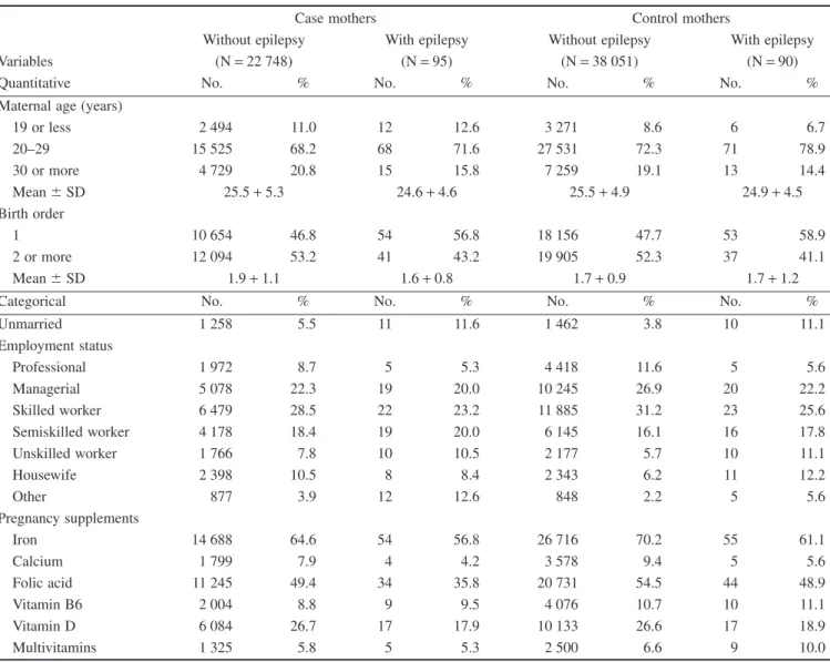
Table 1 summarizes the characteristics of epileptic mothers of cases and controls, in addition to non-epileptic mothers as refer- ence. Epileptic mothers were somewhat younger compared to non-epileptic mothers. The mean birth order was similar in control mothers with or without epilepsy, but was lower in epi-
leptic case mothers than in non-epileptic case mothers. The pro- portion of unmarried epileptic women was larger with a lower proportion of professional/managerial/skilled worker employment status.
The use of folic acid was lower in epileptic mothers than in non-epileptic mothers. In addition, the occurrence of folic acid supplementation was less frequent in epileptic case mothers than in epileptic control mothers (OR with 95% CI: 0.7, 0.5–0.9). Supple- mentation with folic acid-containing multivitamins occurred rarely in the study groups.
Table 2 summarizes the estimated associations between mater- nal epilepsy and different CAs in their offspring; in addition these epileptic pregnant women were differentiated according to without treatment, with mono- and polytherapy. A higher risk of cleft lip with or without cleft palate, cleft palate, cardio- vascular CAs, oesophageal atresia/stenosis (though based on only three cases) and hypospadias was observed in the children of epileptic pregnant women. Thus, the total rate of CAs was also higher.
Only 12 epileptic case mothers were recorded without AED treatment during the study pregnancy, their offspring were affected
Table 1 Maternal characteristics of case and control mothers with epilepsy and without epilepsy as reference
Variables
Case mothers Control mothers
Without epilepsy With epilepsy Without epilepsy With epilepsy
(N=22 748) (N=95) (N=38 051) (N=90)
Quantitative No. % No. % No. % No. %
Maternal age (years)
19 or less 2 494 11.0 12 12.6 3 271 8.6 6 6.7
20–29 15 525 68.2 68 71.6 27 531 72.3 71 78.9
30 or more 4 729 20.8 15 15.8 7 259 19.1 13 14.4
Mean⫾SD 25.5+5.3 24.6+4.6 25.5+4.9 24.9+4.5
Birth order
1 10 654 46.8 54 56.8 18 156 47.7 53 58.9
2 or more 12 094 53.2 41 43.2 19 905 52.3 37 41.1
Mean⫾SD 1.9+1.1 1.6+0.8 1.7+0.9 1.7+1.2
Categorical No. % No. % No. % No. %
Unmarried 1 258 5.5 11 11.6 1 462 3.8 10 11.1
Employment status
Professional 1 972 8.7 5 5.3 4 418 11.6 5 5.6
Managerial 5 078 22.3 19 20.0 10 245 26.9 20 22.2
Skilled worker 6 479 28.5 22 23.2 11 885 31.2 23 25.6
Semiskilled worker 4 178 18.4 19 20.0 6 145 16.1 16 17.8
Unskilled worker 1 766 7.8 10 10.5 2 177 5.7 10 11.1
Housewife 2 398 10.5 8 8.4 2 343 6.2 11 12.2
Other 877 3.9 12 12.6 848 2.2 5 5.6
Pregnancy supplements
Iron 14 688 64.6 54 56.8 26 716 70.2 55 61.1
Calcium 1 799 7.9 4 4.2 3 578 9.4 5 5.6
Folic acid 11 245 49.4 34 35.8 20 731 54.5 44 48.9
Vitamin B6 2 004 8.8 9 9.5 4 076 10.7 10 11.1
Vitamin D 6 084 26.7 17 17.9 10 133 26.6 17 18.9
Multivitamins 1 325 5.8 5 5.3 2 500 6.6 9 10.0
Table2Estimationoftheassociationbetweenmaternalepilepsyduringpregnancyanddifferentcongenitalabnormalities(CAs)onthebasisofcomparisonsofcasesandallmatched controls,aswellasaccordingtothetypeoftreatment Studygroup Grand totalno.
EpilepsyWithouttreatmentMonotherapyPolytherapy No.%OR95%CI†No.%No.%OR95%CI†No.%OR95%CI† Controls38151900.24Reference100.03500.13Reference300.08Reference IsolatedCAs Neural-tubedefects120270.581.90.8–4.410.0840.331.50.3–6.820.172.00.2–14.0 Cleftlip‡1375110.803.41.8–6.400.0020.151.20.3–4.890.658.74.1–18.3 Cleftpalate60150.833.51.4–8.800.0020.332.70.6–11.030.506.62.1–21.7 CardiovascularCAs4480250.562.41.5–3.750.1180.181.40.7–3.0120.273.51.8–6.9 Oesophagealatresia/stenosis21731.385.91.9–18.900.0020.927.41.8–30.710.466.10.8–45.2 Hypospadias3038130.421.81.0–3.330.1070.231.80.8–4.130.101.30.4–4.3 Undescendedtestis205270.341.40.7–3.100.0060.291.30.9–5.410.050.60.1–4.7 Clubfoot242450.210.90.4–2.200.0040.171.30.5–3.610.040.50.1–4.0 Poly/syndactyly174450.291.20.5–3.020.1110.060.50.1–3.320.111.50.4–6.3 OtherisolatedCAs43619§0.210.90.5–1.710.0240.090.80.3–2.140.091.00.3–13.3 MultipleCAs134950.371.60.6–3.900.0010.070.60.1–4.340.303.91.4–11.1 Total22843950.421.81.3–2.4120.05410.181.40.9–2.2420.182.41.5–3.9 †ORadjustedformaternalageandemploymentstatus,birthorder,folicaciduseduringpregnancyinconditionallogisticregressionmodel;‡Cleftlipwithorwithoutcleftpalate; §Congenitalpyloricstenosis2,microtia1,lunghypoplasia1,intestinalatresia1,vaginalatresia1,exomphalos1,torticollis1,pectusexcavatum1. Boldnumbersshowsignificantassociations.
with three different CAs: five cases with cardiovascular CA (crude OR with 95% CI: 3.3, 1.2–9.2), three cases with hypospadias, two cases with poly/syndactyly and one case with anencephaly, thus OR (with 95% CI) was 1.3 (0.7–3.4) for total CA group.
Of 95 epileptic pregnant women, 41 were treated with one AED (Table 2), and this monotherapy associated with a higher risk with oesophageal atresia/stenosis, but this association was based on two cases. However, it is important to mention that there was no sig- nificantly higher risk for the total group of CAs and any other CA group. The offspring of 42 epileptic pregnant women with poly- therapy had a high risk for cleft lip with or without cleft palate, cleft palate, cardiovascular CA and multiple CAs, thus the risk was also higher for total CA group (Table 2).
Table 3 shows the numerical distribution of different CAs according to AEDs used in monotherapy. Of eight cases with car- diovascular CA, four had mothers with carbamazepine, while three were treated with phenytoin. Of seven cases with hypospa- dias, three had epileptic mothers treated with valproate and two were treated with phenytoin. Of six cases with undescended testis, three had epileptic mothers with phenytoin treatment. Of four cases with neural-tube defect, two had mothers with carbam- azepine, one with phenytoin and one with valproate treatment.
However, the most impressive finding is the low number of cleft palate and cleft lip with or without cleft palate after monotherapy.
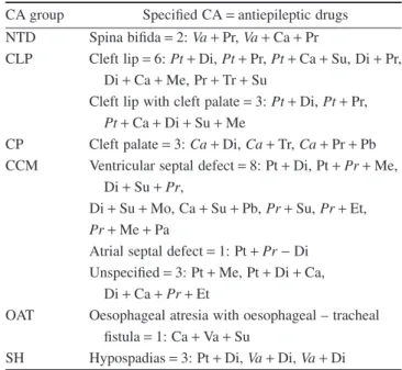
The different combinations of AEDs in polytherapy are shown in Table 4. Most frequently, primidone, phenytoin and carbamazepine were used, followed by diazepam and sultiame. Phenytoin and diazepam occurred most frequently in polytherapy in the group of
Table 3 Controls and cases born to mothers with monotherapy, polytherapy and without treatment according to antiepileptic drug
Antiepileptic Control
Monotherapy
Others MCA Total
NTD CLP CP CCM OAT SH UT CF PY/SY
Carbamazepine (Ca) 10 2 0 0 4 0 1 0 2 0 0 0 8
Clomethiazol (Cl) 0 0 0 0 1 1 0 0 0 0 0 0 2
Clonazepam (Cz) 1 0 0 0 0 0 0 0 0 0 0 0 0
Diazepam (Di) 4 0 0 1 0 0 1 0 0 0 0 0 2
Ethosuximide (Et) 0 0 1 0 0 1 0 0 0 0 0 0 2
Mephenytoin (Me) 2 0 0 0 0 0 0 0 0 0 0 0 0
Morsumide (Mo) 2 0 0 0 0 0 0 0 0 0 0 0 0
Phenacemide (Pa) 0 0 0 0 0 0 0 0 0 0 0 0 0
Phenobarbital (Pb) 0 0 0 0 0 0 0 1 0 0 0 0 1
Phenytoin (Pt) 19 1 1 1 3 0 2 3 1 0 2† 0 14
Primidone (Pr) 6 0 0 0 0 0 0 1 1 0 1‡ 1 4
Sultiame (Su) 0 0 0 0 0 0 0 0 0 0 0 0 0
Trimethadione (Tr) 1 0 0 0 0 0 0 0 0 1 0 0 1
Valproate (Va) 5 1 0 0 0 0 3 1 0 0 1§ 0 7
Total 50 4 2 2 8 2 7 6 4 1 4 1 41
Polytherapy 30 2 9 3 12 1 3 1 1 2 4 4 42
No drug treatment 10 1 0 0 5 0 3 0 0 2 1 0 12
Grand total 90 7 11 5 25 3 13 7 5 5 9 5 95
†Exomphalos, atresia of bile duct; ‡Branchial cyst; §Microtia.
CCM, cardiovascular congenital abnormalities (CAs); CF, clubfoot; CLP, cleft lip with or without cleft palate; CP, cleft palate; MCA, multiple CA; NTD, neural-tube defect; OAT, oesophageal atresia/stenosis; PY/SY, poly/syndactyly; SH, hypospadias; UT, undescended testis.
Table 4 Antiepileptic drugs used in polytherapy according to different congenital abnormalities (CAs)
CA group Specified CA=antiepileptic drugs NTD Spina bifida=2:Va+Pr,Va+Ca+Pr
CLP Cleft lip=6:Pt+Di,Pt+Pr,Pt+Ca+Su, Di+Pr, Di+Ca+Me, Pr+Tr+Su
Cleft lip with cleft palate=3:Pt+Di,Pt+Pr, Pt+Ca+Di+Su+Me
CP Cleft palate=3:Ca+Di,Ca+Tr,Ca+Pr+Pb CCM Ventricular septal defect=8: Pt+Di, Pt+Pr+Me,
Di+Su+Pr,
Di+Su+Mo, Ca+Su+Pb,Pr+Su,Pr+Et, Pr+Me+Pa
Atrial septal defect=1: Pt+Pr-Di Unspecified=3: Pt+Me, Pt+Di+Ca,
Di+Ca+Pr+Et
OAT Oesophageal atresia with oesophageal – tracheal fistula=1: Ca+Va+Su
SH Hypospadias=3: Pt+Di,Va+Di,Va+Di
The abbreviations of different CAs and antiepileptic drugs are shown in Table 3.
The abbreviations of most frequently used drugs are printed in italic.
cleft lip with or without cleft palate. Interestingly, cleft palate showed a different pattern because carbamazepine was the common component in the polytherapy of this CA group. The most frequent components of polytherapy were primidone and phenytoin in the group of cardiovascular CAs with an obvious dominance of ven- tricular septal defect. In the group of neural-tube defects valproate and primidone were components of polytherapy in both cases. In the group of hypospadias, diazepam and valproate were the leading components of polytherapy. Some drugs, such as clonazepam, mephenytoin, morsumide, phenacemide and sultiame were used only in polytherapy.
The combined effect of different AEDs in mono- and polytherapy confirmed the well-known teratogenic effect of valproate, phenytoin and primidone. In addition, our data indi- cated the teratogenic effect of sultiame (OR with 95% CI: 5.7, 1.6–14.4). However, the risk of CAs did not reach the level of significance after the use of carbamazepine (OR with 95% CI:
1.9, 0.9–4.3). The number of pregnant women with trimethadione treatment was too small for evaluation. These Hungarian data did not confirm the teratogenic potential of phenobarbital (OR with 95% CI: 1.3, 0.6–2.6) and diazepam (OR with 95% CI: 1.1, 0.7–1.7).
In the second step of the study we compared the data of cases with CAs between 1980 and 1989 and 1990–1996. There was no significant change in maternal characteristics of epileptic pregnant women between the two study periods.
Table 5 summarized the data of different CAs in the offspring of pregnant women without treatment, as well as with mono- and polytherapy between 1980 and 1989 and 1990–1996. The number of mono- and polytherapy was 28:31 between 1980 and 1989, while this ratio was 13:11 between 1990 and 1996 thus the proportion of polytherapy decreased but it was far from the level of significance (P=0.58). In addition, there was a decrease in the total rate of CAs from 0.44 % to 0.36% (OR with 95% CI: 0.82, 0.56–1.13), that is, this decline was about 20% due to the decrease in their rate both after mono- and polytherapy in the second period of the study. It is worth mentioning the lack of cases with cleft palate in the second period.
The comparison of different antiepileptic drug uses between the two study periods (Table 6) shows that the use of carbamazepine and mainly valproate increased, while the use of phenytoin decreased in epileptic mothers. On the other hand these data indi- cate that the reduction of polytherapy occurred mainly in control mothers.
DISCUSSION
The aim of the study was to check the efficacy of recent progress in the care of epileptic pregnant women. Our data showed some increase in the proportion of recommended monotherapy and a 20%
reduction in the risk for total CAs during the second period of the study. Thus, a real breakthrough in the efficacy of recent medical care of epileptic pregnant women regarding the teratogenic risk of AED has not been found.
In the total material, a higher risk for total CAs was found only after polytherapy, explained mainly by the higher rate of cleft lip with or without cleft palate, cleft palate, cardiovascular CAs and multiple CAs. Thus, the lower teratogenic potential of mono- therapy was confirmed. In addition this study confirmed the ter- atogenic effect of phenytoin, valproate, primidone and completed this list with the teratogenic effect of sultiame (Czeizel et al.
1992). However, this Hungarian material did not confirm the
teratogenic effect of carbamazepine, diazepam and pheno- barbital. The recognition of mild teratogenic potential of carbam- azepine depends on the number of pregnant women (i.e. statistical power).
The teratogenic potential of diazepam and phenobarbital depends on the indication of these drugs (i.e. underlying maternal diseases). The recent Cochrane review of these drugs has not con- firmed their teratogenic potential (Adabet al. 2004). A population- based monitoring system of self-poisoning pregnant women (i.e.
pregnant women who attempted suicide with extremely large doses of drugs) was established in Budapest (Czeizelet al. 2008).
Among 1044 self-poisoned pregnant women, 112 used very large doses of diazepam (25–800 mg) and 37 women attempted suicide between the 4th and 12th gestational weeks, but a higher rate of CAs was not detected (Gidaiet al. 2008). In addition, 88 pregnant women attempted suicide with very large doses of pheno- barbital and 34 women between the 4th and 12th gestational weeks, but a higher risk of CAs was not found (Timmemannet al.
2009).
The strengths of the HCCSCA are that is a population-based large dataset including 185 pregnant women with prospectively and medically recorded epilepsy in an ethnically homogeneous European (Caucasian) population. We were able to differentiate the different groups of AEDs in the study pregnancies. Additional strengths are the matching of cases to controls without CA;
available data for potential confounders, and finally that the diag- nosis of medically reported CAs was checked in the HCAR (Czeizel 1997) and later modified, if necessary, on the basis of recent medical examination within the HCCSCA (Czeizel et al.
2001).
However, this dataset also has limitations: (i) We did not know any details regarding the onset of epilepsy before the study preg- nancy; (ii) Other pregnancy outcomes (e.g. miscarriages) were not known; (iii) Unfortunately, our dataset is not appropriate for the evaluation of functional defects (e.g. valproate associates with the most severe mental retardation-inducing effect); (iv) In general, the doses of AEDs drugs used for the treatment of epileptic preg- nant women were not known, but the results of our validation studies showed that recommended doses were used in most preg- nant women (Czeizelet al. 2003); (v) The data of blood level of AEDs were not recorded in the HCCSCA; and (vi) The major weakness of our study is that the recent data of pregnant women (i.e. after 1996) are not available in the HCCSCA.
The major message of this study and some other recent papers is that pregnancy in epileptic women does not need to be discouraged if these women wish to have babies, but epileptic women need a specific and high standard of medical care.
The first task is to educate epileptic women to understand that it is necessary to prepare their pregnancies before conception.
There are many tasks during this preconceptional period: (i) Check their AEDs and attempt changing the teratogenic drugs (e.g. valproate) to less (e.g. carbamazepine) or non-teratogenic (lamotrigine) drugs under control of specialists. However, at the selection of AEDs for epileptic pregnant women the type of epi- lepsy is the most important aspect, but some other aspects (e.g.
the duration previous seizure-free interval, other diseases) can also be considered; (ii) In general, monotherapy is better because poly- therapy associates with a higher risk of CAs (Källen 1986, Meadoret al. 2008) as in our study; (iii) The use of the lowest effective dose of the given drug is preferable, because higher doses of AEDs associate with a higher risk of specific CAs; (iv) However, it is important to stress that the cluster of seizures during pregnancy in untreated or inappropriately treated epileptic
Table5Comparisonofepilepticpregnantwomenwithouttreatment(notreat),withmonotherapy(mono)andpolytherapy(poly)inthecontrolgroupandinthecasegroupincluding caseswithdifferentcongenitalabnormalities(CAs)duringthetwostudyperiods:1980–1989and1990–1996 StudygroupN
1980–1989 N
1990–1996 NotreatMonoPolyTotalNotreatMonoPolyTotal No.%No.%No.%No.%No.%No.%No.%No.% Controls2534970.03230.09220.09520.211280330.02270.2180.06380.30 IsolatedCAs Neural-tubedefects102300.0040.3910.0150.4917910.5600.010.5621.12 Cleftlip†87300.0010.1170.8080.9250200.0010.2020.4030.60 Cleftpalate41200.0020.4930.7351.2118900.0000.0000.0000.00 CardiovascularCAs266420.0850.19100.38170.64181630.1730.1720.1180.44 Oesophagealatresia/stenosis14600.0021.3700.0021.377100.0000.0011.4111.41 Hypospadias184610.0520.1120.1150.27119220.1750.4210.0880.67 Undescendedtestis126700.0050.1900.0050.3978500.0010.1310.1320.25 Clubfoot161400.0030.1910.0640.2581100.0010.0600.0010.14 Poly/syndactyly104220.1900.0010.1030.2970200.0010.1410.1420.28 OtherisolatedCAs277010.0430.1130.1170.25159000.0010.0610.0620.13 MultipleCAs95100.0010.1130.3240.4239800.0000.0010.2510.25 Total1460860.04280.19310.21650.44823560.07130.16110.13300.36 †Cleftlipwithorwithoutcleftpalate.
Table6Comparisonofusesofdifferentantiepilepticdrugsinthetwostudyperiods:1980–1989and1990–1996 Monotherapy
1980–19891990–1996Total ControlsCasesControlsCasesControlsCases No.%No.%No.%No.%No.%No.% Carbamazepine47.734.6615.8516.71011.188.4 Clomethiazol00.023.100.000.000.022.1 Clonazepam11.900.000.000.011.100.0 Diazepam11.911.537.913.344.422.1 Ethosuximide00.023.100.000.000.022.1 Mephenytoin26.700.000.000.022.200.0 Morsuximide11.900.012.600.022.200.0 Phenacemide00.000.000.000.000.000.0 Phenobarbital00.011.500.000.000.011.1 Phenytoin111.21320.0821.113.31921.11414.7 Primidone23.846.2410.500.066.744.2 Sultiame00.000.000.000.000.000.0 Trimethadione00.011.512.600.011.111.1 Valproate11.911.5410.5620.055.677.4 Total2344.22843.12771.11343.35055.64143.2 Polytherapy2242.33150.0821.11136.73033.34244.2 No.treatment713.569.237.9620.01011.11212.6 Grandtotal52100.065100.038100.130100.090100.095100.0
women associates with an even higher risk of fetal defects (Meadoret al. 2008). Thus, epileptic pregnant women need treat- ment during pregnancy as well.
The second task is the monitoring of health status of epileptic pregnant women and their fetuses. The relation of epilepsy and pregnancy is variable, about 45% of pregnant women have a higher seizure frequency, while about 5% associate with a reduced seizure frequency, thus epilepsy remains unchanged in about 50% of preg- nant women (Knight and Rhind 1975). The higher risk for seizure is explained by generally declined serum levels of AEDs in preg- nancy, therefore an increase in dosage is frequently required during pregnancy to maintain the effective plasma level of AEDs (Tompsonet al. 1997). Another task is the monitoring of the fetal development with high-resolution ultrasound because the principal defects associated with different teratogenic AEDs, such as neural- tube defects (Meador et al. 2008) and fetal hydantoin/dilantin/
phenytoin, fetal trimethadione and fetal valproate syndrome/effect (Jones 1988) are detectable approximately at the 20th gestational week. These defects are rarely diagnosed, but pregnant women have the right to decide to keep or terminate their pregnancies after their diagnosis.
The final task is connected with the preparation of delivery, including maternal ingestion of vitamin K1 (10 mg per day) because clinical or subclinical coagulopathy may occur in newborn infants (Aminoff 2004).
In conclusion, the main experience of this study is that the above principles have not been followed in many Hungarian pregnant women, therefore, the efficacy of recent special medical care of epileptic mothers was not obvious in the reduction of CAs.
REFERENCES
Adab N, Tudor SC, Vinten Jet al. (2004) Common antiepileptic drugs in pregnancy in women with epilepsy.Cochrane Database Syst Rev(3):
CD004848.
Aminoff MJ (2004) Neurologic disorders. In: Creasy RK, Resnik R, Iams JD, eds.Maternal-Fetal Medicine, 5th edn. Saunders, Philadelphia, pp.
1165–1191.
Briggs GG, Freeman RK, Yaffe SJ (2005)Drugs in Pregnancy and Lacta- tion, 7th edn. Lippincott Williams and Wilkins, Philadelphia.
Czeizel AE (1997) The first 25 years of the Hungarian Congenital Abnor- mality Registry.Teratology55: 299–305.
Czeizel AE (1999) Ten years of experience in periconceptional care.Eur J Obstet Gynecol Reprod Biol84: 43–49.
Czeizel AE (2009) Periconceptional folic acid and multivitamin supplemen- tation for the prevention of neural tube defects and other congenital abnormalities.Birth Defects Res (Part A)85: 260–268.
Czeizel AE, Vargha P (2004) Periconceptional folic acid/multivitamin supplementation and twin pregnancy.Am J Obstet Gyencol191: 790–
794.
Czeizel AE, Bod M, Halász P (1992) Evaluation of anticonvulsant drugs during pregnancy in a population-based Hungarian study.Eur J Epidemol 8: 122–127.
Czeizel AE, Gidai J, Petik D, Timmermann G, Puho HE (2008) Self- poisoning during pregnancy as a model for teratogenic risk estimation of drugs.Toxic Indust Health24: 11–28.
Czeizel AE, Into˝dy Z, Modell B (1993) What proportion of congenital abnormalities can be prevented?Br Med J306: 499–503.
Czeizel AE, Petik D, Vargha P (2003) Validation studies of drug exposures in pregnant women.Pharmacoepid Drug Safety2: 409–416.
Czeizel AE, Rockenbauer M, Siffel C, Varga E (2001) Description and mission evaluation of the Hungarian Case-Control Surveillance of Con- genital Abnormalities, 1980–1996.Teratology63: 176–185.
Ero˝s E, Géher P, Gömör B, Czeizel AE (1998) Epileptogenic activity of folic acid after drug induced SLE (folic acid and epilepsy).Eur J Obstet Gynecol Reprod Biol80: 75–78.
Fried S, Kozer E, Nulman Iet al. (2004) Malformation rates in children of women with untreated epilepsy: a meta-analysis.Dug Safety27: 197–
202.
Gidai J, Ács N, Bánhidy F, Czeizel AE (2008) No association found between use of very large doses of diazepam by 112 pregnant women for a suicide attempt and congenital abnormalities in their offspring.Toxic Indust Health24: 29–39.
Holmes LB, Harvey EA, Couil BAet al. (2001) The teratogenicity of anticonvulsants drugs.N Engl J Med344: 1132–1138.
Janz D, Fuchs V (1964) Are anti-epileptic drugs harmful when given during pregnancy?German Med Monogr9: 20–23.
Jones KL (1988)Smith’s Recognizable Patterns of Human Malformation, 4th edn. W.B. Saunders Co, Philadelphia.
Källen B (1986) Maternal epilepsy, antiepileptic drugs and birth defects.
Pathologica78: 757–768.
Kjaer D, Horvath-Puho E, Christensen J et al. (2008) Antiepileptic drug use, folic acid supplementation, and congenital abnormalities:
a population-based case-control study.Br J Obstet Gynaec 115: 98–
103.
Knight AH, Rhind EG (1975) Epilepsy and pregnancy: a study of 153 pregnancies in 59 patients.Epilepsia16: 99–105.
Meador KJ, Baker GA, Browning Net al. (2009) Cognitive function at 3 years of age after fetal exposure to antiepileptic drugs.N Engl J Med360:
1597–1605.
Meador K, Reynolds MW, Crean Set al. (2008) Pregnancy outcome in women with epilepsy: a systematic review and meta-analysis of published pregnancy registries and cohorts.Epilepsy81: 1–13.
Meadow SR (1968) Anticonvulsant drugs and congenital abnormalities.
Lancet2: 1296–1299.
Puho E, Métneki J, Czeizel AE (2005) Maternal employment status and isolated orofacial clefts in Hungary.Cent Eur J Publ Health13: 144–148.
Shepard TH, Lemire RJ (2004)Catalog of Teratogenic Agents, 11th edn.
Johns Hopkins University Press, Baltimore.
Timmemann G, Ács N, Bánhidy F, Czeizel AE (2009) Congenital abnormalities of 88 children born to mothers who attempted suicide with phenobarbital during pregnacy.Pharmacoepid Drug Safety18: 815–
825.
Tompson T, Gram L, Siilanpaa M, Johannenssen SI (1997)Epilepsy and Pregnancy. Wrightson Biomedical Publishing Ltd, Hampshire.
Wyllie E, ed. (2001)The Treatment of Epilepsy, Principles and Practice, 3rd edn. Lippincott Williams and Wilkins, Philadelphia.