Counteracts RETINOBLASTOMA RELATED to Maintain the Root Meristem 1[CC-BY]
Ansul Lokdarshi,a,2Csaba Papdi,b,2,3Aladár Pettkó-Szandtner,cStefan Dorokhov,b Ben Scheres,d,4 Zoltán Magyar,eAlbrecht G. von Arnim ,a,f László Bögre,b,5 and Beatrix M. Horváthb,d,5,6
aDepartment of Biochemistry & Cellular and Molecular Biology, University of Tennessee, Knoxville, Tennessee 37996
bDepartment of Biological Sciences, Centre for Systems and Synthetic Biology, Royal Holloway, University of London, Egham Hill, Egham TW20 0EX, United Kingdom
cLaboratory of Proteomics Research, Biological Research Centre, POB 521, H-6701 Szeged, Hungary
dDepartment of Biology, Utrecht University, 3584 CH Utrecht, The Netherlands
eInstitute of Plant Biology, Biological Research Centre, POB 521, H-6701 Szeged, Hungary
fUT-ORNL Graduate School of Genome Science and Technology, Knoxville, Tennessee 37996
ORCID IDs: 0000-0003-2264-2701 (A.L.); 0000-0003-0640-6098 (C.P.); 0000-0003-2968-8542 (S.D.); 0000-0001-5400-9578 (B.S.);
0000-0001-8376-7220 (Z.M.); 0000-0003-3472-3357 (A.G.v.A.); 0000-0002-6083-992X (B.M.H.).
The ErbB-3 BINDING PROTEIN 1 (EBP1) drives growth, but the mechanism of how it acts in plants is little understood. Here, we show that EBP1 expression and protein abundance in Arabidopsis (Arabidopsis thaliana) are predominantly confined to meristematic cells and are induced by sucrose and partially dependent on TARGET OF RAPAMYCIN (TOR) kinase activity.
Consistent with being downstream of TOR, silencing of EBP1 restrains, while overexpression promotes, root growth, mostly under sucrose-limiting conditions. Inducible overexpression of RETINOBLASTOMA RELATED (RBR), a sugar-dependent transcriptional repressor of cell proliferation, depletes meristematic activity and causes precocious differentiation, which is attenuated by EBP1. To understand the molecular mechanism, we searched for EBP1- and RBR-interacting proteins by affinity purification and mass spectrometry. In line with the double-stranded RNA-binding activity of EBP1 in human (Homo sapiens) cells, the overwhelming majority of EBP1 interactors are part of ribonucleoprotein complexes regulating many aspects of protein synthesis, including ribosome biogenesis and mRNA translation. We confirmed that EBP1 associates with ribosomes and that EBP1 silencing hinders ribosomal RNA processing. We revealed that RBR also interacts with a set of EBP1-associated nucleolar proteins as well as factors that function in protein translation. This suggests EBP1 and RBR act antagonistically on common processes that determine the capacity for translation to tune meristematic activity in relation to available resources.
Cell growth is generally required for cell prolifera- tion, and although these two processes are separately regulated, they are closely connected and coordinated (Fox et al., 2018). The central conserved regulator of the cell cycle is the cyclin-dependent kinase (CDK), the activity of which (1) sets the unidirectional alternation of cell cycle phases, (2) enables cells to tune the cell cycle to match with developmental and environmental cues, or (3) gives a signal to exit from the cycle to allow cel- lular differentiation (De Veylder et al., 2007). A pivotal target of CDK that balances cell proliferation with dif- ferentiation is the RETINOBLASTOMA RELATED (RBR) protein, whose phosphorylation lifts the repres- sion on the cell-cycle regulatory E2F transcription factors (Magyar et al., 2016). In plants, modulation in the levels of these cell cycle regulators, such as the overexpression of Cyclin D3;1 (Dewitte et al., 2003), silencing of the RBR protein (Gutzat et al., 2011), or overexpression of the E2FB transcription factor (Magyar et al., 2005), results in more but smaller cells and stunted and develop- mentally arrested plants. This led to the realization
that acceleration of cell proliferation does not neces- sarily lead to increased plant growth.
The TARGET OF RAPAMYCIN (TOR) kinase path- way is regarded as the central regulator of cell growth in connection with light and sugars by generally boosting anabolic and repressing catabolic processes (Dobrenel et al., 2016a; González and Hall, 2017).
Protein synthesis, being an energy and resource de- manding cellular process, is one of the major cellular functions controlled by TOR signaling (González and Hall, 2017). S6 kinase (S6K) is a key downstream component of TOR signaling that regulates transla- tion and ribosome biogenesis (Deprost et al., 2007;
Schepetilnikov et al., 2013). Thus growth-stimulatory factors such as light, sugar, and auxin activate TOR and affect the translation apparatus in different ways (Liu et al., 2013; Dobrenel et al., 2016b; Chen et al., 2018; Enganti et al., 2018; Schepetilnikov and Ryabova, 2018). Correspondingly, elevated TOR ex- pression boosts plant growth in Arabidopsis (Arabi- dopsis thaliana; Deprost et al., 2007) and increases yield
Plant PhysiologyÒ,February 2020, Vol. 182, pp. 919–932, www.plantphysiol.orgÓ2020 The Authors. All Rights Reserved. 919
in crop plants, specifically in limiting conditions (Bakshi et al., 2019). On the other hand, TOR silencing or chemical inhibition of TOR activity results in re- duced growth (Deprost et al., 2007; Montané and Menand, 2013, 2019).
The growth regulatory TOR signaling drives cell proliferation by controlling the selective translation of key cell-cycle regulatory proteins, such as CLN3 G1 cyclin in yeast (Saccharomyces cerevisiae; Barbet et al., 1996) or Cyclin E in human (Homo sapiens) cells (Dowling et al., 2010). In plants, however, there appears to be an even more direct connection between growth and cell cycle control, as TOR phosphorylates and thus stimulates E2FA and E2FB activities (Xiong et al., 2013;
Wu et al., 2019). More recent research shows that the YET ANOTHER KINASE1(AtYAK1) gene, encoding a member of the dual-specificity Tyr phosphorylation- regulated kinase family, inhibits growth and cell pro- liferation by up-regulating the SIAMESE-RELATED (SMR) proteins when TOR activity is reduced (Barrada et al., 2019; Forzani et al., 2019). Moreover, the principal downstream effector of TOR, AtS6K1, interacts with RBR and facilitates its nuclear localization, which is important to impose repression on cell proliferation in sucrose limiting conditions (Henriques et al., 2013). Finally, S6K1, E2FB, and RBR are inter- connected through negative feedback loops that might be important to toggle between cell prolifera- tion and quiescence (Henriques et al., 2010).
The ErbB-3 BINDING PROTEIN 1 (EBP1) came to the forefront of interest in plants when it was found that it is rapidly induced during early stages of tuber formation, stimulated by sugar in potato (Solanum tuberosum;
Horváth et al., 2006). Overexpression or silencing of EBP1 in potato could dose-dependently tune leaf and tuber growth and thus potato yield without any obvi- ous developmental abnormalities (Horváth et al., 2006).
Studying the cellular basis how EBP1 affects organ growth showed that it boosts cell proliferation in mer- istematic cells, resulting in more but smaller cells, which correlated with elevated expression of cell cycle regulators. In postmitotic cells, EBP1 enhances cell growth leading to a larger final cell size. On the mo- lecular level EBP1 represses RBR protein abundance, whereas RBR negatively influences the EBP1 level. This antagonism suggested that EBP1 provides an important link between a growth driver and cell cycle regulation (Horváth et al., 2006). The initialfinding that EBP1 en- hances growth was verified for a number of EBP1 orthologs identified in different species (Cao et al., 2009;
Cheng et al., 2016; Wang et al., 2016); however, another report described an opposite effect (Li et al., 2016).
Additionally,EBP1expression is induced upon abiotic stress, and when EPB1 levels are elevated, it can confer stress tolerance (Cao et al., 2009; Cheng et al., 2016). In line with being a positive regulator of growth, in a maize hybrid displaying hybrid vigor, EBP1 is expressed in an over dominant fashion (Wang et al., 2016). In a recent study, EBP1 was identified as an interactor with the FERONIA receptor-like kinase with a major function in regulating cell expansion. FER- ONIA directly phosphorylates EBP1, leading to its nu- clear localization to regulate genes responsive to FERONIA signaling (Li et al., 2018).
A clue to understand EBP1’s molecular function in plants was its remarkable coexpression with genes in- volved in the regulation of protein synthesis in prolif- erating cells, the so-calledribiregulon (Horváth et al., 2006). In line with this, several additional supporting lines of evidence have been published. First,EBP1ex- pression is rapidly induced when growth is stimulated in the shoot meristem of dark-grown etiolated seedlings upon light exposure, correlating with the induction of genes involved in protein synthesis (López-Juez et al., 2008; Mohammed et al., 2018). Second, EBP1 is present in nucleoli in both animals (Squatrito et al., 2004; Liu et al., 2006) and in plants (Pendle et al., 2005), as evi- denced by a carboxy-terminal Lys-rich motif (Karlsson et al., 2016) and an N-terminal nucleolar localization signal (Squatrito et al., 2004; Ko et al., 2016). Third, human EBP1 (HsEBP1) has a double-stranded RNA binding activity (Squatrito et al., 2004). It binds to rRNAs and ribosomes (Squatrito et al., 2006) and also supports ribosome biogenesis by interacting with the Pol I transcription factor TIF-IA (Nguyen le et al., 2015;
Nguyen et al., 2019) and possibly with nucleolar nucleophosmin (Okada et al., 2007). Fourth, HsEbp1 also stabilizes several mRNAs by binding their 39UTRs (Bose et al., 2006; Zhou et al., 2010; Pisapia et al., 2015).
1This work was supported by the National Science Foundation (NSF) (grants MCB-1546402 and IOS-1456988), National Institutes of Health (grant R15 GM129672), and the Donald L Akers Jr Faculty Enrichment Fellowship (to A.G.v.A); the European Commission (EC) (Marie-Curie IEF fellowship, FP7-PEOPLE-2012-IEF-330789 to B.M.H.; FP7-PEOPLE-2012-IEF.330713 to C.P.); the Netherlands Or- ganization for Scientific Research (Spinoza grant to B.S.);
Biotechnology and Biological Sciences Research Council (BBSRC) (BBSRC-NSF grant BB/M025047/1 to C.P. and L.B.); and Magyar Tudomanyos Akademia (Hungarian Academy of Sciences) (GI- NOP-2.3.2-15-2016-00032 to A.P.-S. and GINOP-2.3.2-15-2016-00001 to A.P.-S. and Z.M.).
2These authors contributed equally to this work.
3Present address: Institut de Biologie Moléculaire des Plantes, Cen- tre National de la Recherche Scientifique, UPR 2357, Université de Strasbourg, Strasbourg, France.
4Present address: Department of Plant Sciences, Wageningen Uni- versity Research Centre, 6708 PB Wageningen, The Netherlands.
5Senior authors.
6Author for contact: beatrixmhorvath@gmail.com.
The author responsible for distribution of materials integral to the findings presented in this article in accordance with the policy de- scribed in the Instructions for Authors (www.plantphysiol.org) is:
Beatrix M. Horváth (beatrixmhorvath@gmail.com).
B.M.H., A.L., C.P., A.P.-S., and Z.M. designed and performed the experiments; A.L., S.D., A.G.v.A., L.B. and B.M.H. analyzed the data;
B.S. shared material and gave general support; A.G.v.A., L.B., and B.M.H. conceived and designed the research plan, and wrote the manuscript; all authors saw and commented on the manuscripts.
[CC-BY]
Article free via Creative Commons CC-BY 4.0 license.
www.plantphysiol.org/cgi/doi/10.1104/pp.19.00805
920 Plant Physiol. Vol. 182, 2020
Finally, EBP1 has been implicated in translational control inLeishmania major(Norris-Mullins et al., 2014) and, repeatedly, in gene specific translation in cap- independent translation initiation of RNA viruses (Pilipenko et al., 2000) on the androgen receptor mRNA (Zhou et al., 2010; Zhou et al., 2011) by af- fecting eIF2aphosphorylation (Squatrito et al., 2006).
To gain insight into the growth-promoting function of EBP1, it is paramount to identify the molecular partners. To this end, we performed mass spectrometry to identify EBP1 interactors, which uncovered proteins with a broad range of RNA binding and, to a lesser extent, of DNA binding activities, and also proteins involved in ribosome biogenesis, splicing, translation, and transcription. Here, we confirm that EBP1 can as- sociate with ribosomes and is distributed between cy- toplasm, nucleus, and nucleolus, in line with its suggested role in translation and ribosome biogenesis.
We demonstrate that the inhibition ofEBP1expression causes a defect in rRNA processing, which can be res- cued by sucrose. Sugar could also rescue the growth defects during root development in the EBP1 silencing lines, suggesting EBP1 is specifically required to sup- port protein translation in growth limiting conditions.
Strikingly, overexpression of EBP1 can suppress both the loss of meristem activity and precocious differenti- ation that occurs upon induction of elevated RBR ex- pression. In addition, we also found that RBR shares
interacting partners with EBP1, and besides its canon- ical transcriptional repressor function, it potentially acts on common biological processes with EBP1 in ribosome biogenesis and protein translation. Thus, EBP1 and RBR antagonistically regulate translational capacity to determine the balance between meristematic activity and cellular differentiation.
RESULTS
EBP1Is Expressed in Root Meristematic Tissues
When ectopically overexpressed or silenced, EBP1 enhances or reduces the meristematic activity and cor- respondingly organ growth. However, when and where EBP1 is naturally expressed is not well docu- mented (Horváth et al., 2006). To study the regulation of EBP1 expression, we followed the EBP1 promoter activity driving GUS and cyan-fluorescent protein (CFP) expression. For this, we cloned the entire inter- genic region (1193bp) between EBP1(At3g51800) and the nearest upstream gene (At3g51810). Around 25 Arabidopsis transgenic lines were generated for pEBP1:GUS and for pEBP1:CFP. For both, five lines were examined in detail, all showing the same expres- sion profile in root tissues. We detected strong GUS activity in the primary meristem (Fig. 1A), in the region
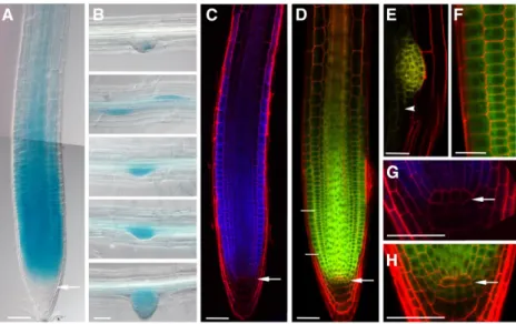
Figure 1.Arabidopsis EBP1 is expressed and localized in actively dividing tissues during root development. Representative differential interference contrast images ofEBP1expression in the primary root (A) and during lateral root development (B) in the transgenicpEBP1:GUSlines after 3h GUS staining. The images of the different stages of lateral root development belong to the same root. C, Confocal microscopy images in the propidium-iodide–stained root sample of thepEBP1:CFPlines. D to F, Confocal microscopy images represent the localization of the EBP1-YFP protein in thepgEBP1:YFPtransgenic line in the primary (D) and secondary (E) meristems. The enlarged sections of the meristematic region in (F) and of a lateral root initial in (E) are showing cytoplasmic localization of the fusion protein (scale bar525mm), whereas (G) and (H) illustrate the lower level of expression in the slowly dividing stem cells and QC. In each case, 5- to 6-d-old seedling were analyzed. White arrow points toward the QC, and the white arrowhead in (E) labels the occasional nuclear localization of the EBP1-YFP protein in the differentiation zone. The merged image in (A) is a composite of two consecutive overlapping images of the same root, taken with the same median focal plane. The enlarged image in (F) comes from the meristematic region, labeled by two white lines in (D). Scale bars550mm, unless stated otherwise.
Plant Physiol. Vol. 182, 2020 921
of the rapidly dividing transit amplifying cells and in the emerging lateral root meristematic cells (Fig. 1B). In the elongation and differentiation zones, we observed expression only at a low level, mainly in the vascula- ture. Similarly, the expression pattern of pEBP1:CFP was largely confined to the meristematic cells (Fig. 1C).
In this aspect, the expression pattern overlaps with the expression pattern published in Arabidopsis.org or Plant eFP at https://bar.utoronto.ca/eplant. To see whether the entire intergenic region is required to reg- ulate the transcription, we studied stepwise-shortened regions of the full promoter. As Supplemental Figure S1 illustrates, the region (-337-0) contains the essential regulatory sequences to induce transcription.
To investigate whetherEBP1promoter activity and protein accumulation correspond, we fused the ge- nomic coding region ofEBP1with the coding region of the yellowfluorescent protein (YFP) and expressed this 39-terminal fusion under its native promoter (pgEBP1- YFP). For this, we used the entire intergenic region (1193bp) containing the 59-upstream region, which also comprises the 59UTR. Of over 30 regenerated transgenic lines, 5 lines were analyzed in detail and showed the same pattern of EBP1-YFP protein accumulation, which over- lapped with the EBP1 promoter activity. EBP1-YFP pro- tein was most abundant in root meristematic cells (Fig. 1D), specifically in the region of the transit ampli- fying cells and emerging lateral root primordia (Fig. 1E).
Lower levels of EBP1-YFP signal were detected in the slowly dividing stem cells and even less in the quiescent center (QC; Fig. 1H). This pattern at the QC region was also closely mirrored in thepEBP1:CFPline (Fig. 1G). The abundance of EBP1-YFP protein also corresponds well with translatomics data, which demonstrate EBP1 mRNA is actively translated in the shoot apex (Tian et al., 2019).
In the transgenic lines, EBP1-YFP signal was most prominent in the cytoplasm of meristematic cells sur- rounding the centrally located nuclei (Fig. 1F). Similar cellular localization patterns were shown indepen- dently by Palm et al. (2019). The amino acid sequence of EBP1 shows high conservation to its human counter- part, which contains a motif for nuclear and nucleolar localization near to its C terminus (Squatrito et al., 2004;
Karlsson et al., 2016). This motif may be more obscured when thefluorescent protein is fused to the C terminus of EBP1. Therefore, we labeled EBP1 both N-and C-terminally using the Cerulean fluorescent protein and transiently expressed it under the control of the 35S promoter. In the transiently transformed Nicotiana benthamiana leaves (Supplemental Fig. S2, A and B), both fusion proteins were found in the nucleoli, the nucleus, and cytosol. Similar localization was observed in onion (Allium cepa) epidermal cells where about 10%
of Cerulean-EBP1 localized in the nucleoli with the re- mainder distributed between the nucleus and cytosol (Supplemental Fig. S2, C and D). Taken together, these data show that both the expression and protein accu- mulation of EBP1 are concentrated in the region of rapidly dividing root meristematic cells. Most of the protein is confined to the cytoplasm in meristematic
cells, but it has the ability to enter into the nucleus and nucleolus in different tissue environments.
EBP1 Supports Root Meristem Activity in Sucrose Limiting Conditions
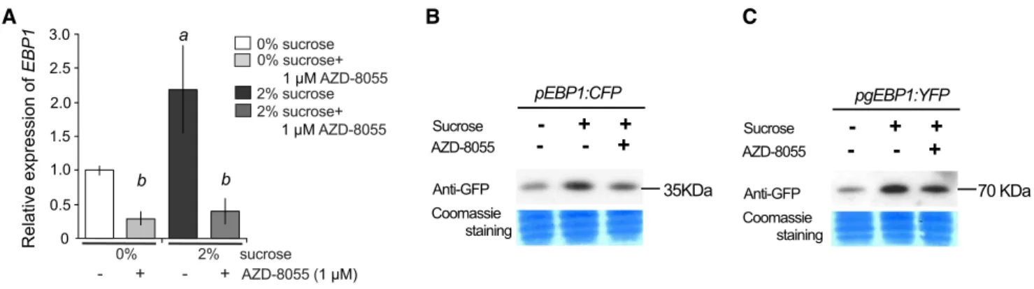
Cell cycle activity in the meristem is reliant on the availability of sugar and regulated by the TOR pathway (Ahmad et al., 2019). Therefore, we examined how the external addition of sucrose and chemical inhibition of TOR activity by AZD-8055 influence EBP1 transcrip- tion, EBP1 promoter activity, and EBP1-YFP protein levels. Endogenous EBP1 mRNA levels were deter- mined in Col-0 seedlings grown on medium without sucrose and then transferred to fresh media with or without sucrose (2% and 0%, respectively) and with or without AZD-8055. EBP1 transcript levels increased upon sucrose addition but were suppressed by AZD- 8055, irrespective of whether sucrose was present in the media (Fig. 2A). To be able to compare the effect of TOR kinase inhibitor in the presence of sucrose on EBP1 promoter activity and protein accumulation, we used ourpEBP1:CFP and pgEBP1-YFPtransgenic lines and followed the amounts of CFP and EBP1-YFP on protein blots. Both EBP1 promoter activity and EBP1-YFP protein levels were induced by sucrose and were af- fected by the TOR inhibitor (Fig. 2, B and C).
To study how EBP1 level influences root growth, we generated inducibleEBP1silencing lines and screened them for growth retardation using 17-b-estradiol on a medium without sucrose. Two independent trans- formants with the EBP1 RNAi construct (iEBP1 line 1 and line 2) showed strongly reduced growth upon 17-b-estradiol treatment as silencing was induced and the EBP1mRNA level diminished (Supplemental Fig.
S3, A and B). However, from the T3 generation onward the retardation in root growth on sucrose-free media was no longer dependent on 17-b-estradiol, and the EBP1level was already reduced without chemical in- duction, indicating that silencing became constitutive (Supplemental Fig. S4A). Therefore, in subsequent ex- periments we did not apply the inducer. To ascertain that EBP1 silencing is not influenced by sucrose, we deter- mined theEBP1level both in the presence and absence of sucrose. Although the level ofEBP1was induced by su- crose as shown above, the silencing remained effective in bothiEBP1lines (Supplemental Fig. S4A).
Next, we studied how seedling growth was influ- enced by the addition of external sucrose whenEBP1 was silenced. Although a large portion of the iEBP1 seedlings (both line 1 and line 2) showed growth arrest and retarded growth on sucrose-free media, this in- hibition was largely suppressed when seeds were germinated on 1% sucrose (Fig. 3, A and B). In an insertional mutant, ebp1-3(CS854731), we observed an even more pronounced growth retardation in the absence of sucrose and recovery on sucrose-free media (Fig. 3, A and B). Leaf development showed similar effects in the absence and presence of sucrose (Supplemental Fig.
922 Plant Physiol. Vol. 182, 2020
S4, B and C). The level of growth retardation closely matched the decline inEBP1 expression in the silencing lines (Supplemental Fig. S4A). To test whether the arrested growth upon EBP1 silencing can be rescued, we trans- ferred seedlings from sucrose-free onto sucrose-containing media and found that root growth indeed recovered (Supplemental Fig. S4, D and E).
EctopicEBP1overexpression can dose-dependently promote growth in potato and Arabidopsis (Horváth et al., 2006). We asked whether this promotion de- pends on sucrose availability. On sucrose-free medium, we found that roots of the two independent EBP1 overexpression lines 1 and 2 (Horváth et al., 2006) grew significantly better than Col-0, but the difference in root length between the transgenic lines and the control was less pronounced on sucrose-containing media (Fig. 3C).
Taken together, the facts that sucrose rescued the growth retardation caused byEBP1silencing and, vice versa, thatEBP1overexpression overcame the sucrose limitation, suggest that EBP1 has a role in coordinating root growth with sugar availability.
EBP1 Supports Maintenance of the Root Meristem and Counteracts Cell Differentiation Induced by RBR
Sugar availability controls cell proliferation through the RBR pathway (Magyar et al., 2016). Silencing of RBR results in excessive cell proliferation, while elevating the RBR level leads to premature differentiation, exhausting the stem-cell pool, and arresting division of the ampli- fying cells. As a result, the root meristem is gradually reduced as cells become differentiated (Wildwater et al., 2005). EBP1 and RBR levels are counter-inhibited in cul- tured cells (Horváth et al., 2006). To follow how the EBP1
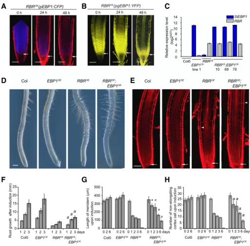
level responds to RBR-induced differentiation in the root, we introgressed pEBP1:CFPandpgEBP1-YFPinto the dexamethasone-inducible RBR overexpression line (RBROE). Upon induction of RBR expression, both the EBP1 promoter activity and EBP1-YFP protein level progressively diminished, together with the meristematic zone, as differentiation progressed (Fig. 4, A and B).
To investigate whether EBP1 has a role in meristem maintenance when RBR is overexpressed, we crossed EBP1OEline 1 and 2 withRBROE. Homozygous offspring of three independent introgressed lines were analyzed and showed elevated levels of bothRBRandEBP1transcripts (Fig. 4C). As expected, induction of RBR overexpression reduced root growth and shortened the meristem, as measured by the distance from the tip to thefirst dif- ferentiating root hair. Strikingly, both effects of RBR overexpression were significantly attenuated by EBP1 overexpression (Fig. 4, D to H; Supplemental Fig. S5A).
Five days after RBR induction the entire root meristem organization was lost and cells appeared vacuolated, but the elevated level of EBP1 prevented this process and supported meristematic function and root growth for a longer period (Supplemental Fig. S5, B and C).
Taken together, the EBP1 level is important to maintain meristematic activity and to counteract the progression of differentiation imposed by RBR overexpression.
EBP1 Predominantly Interacts with Proteins Involved in Ribosome Biogenesis and Protein Translation
To gain insights into the molecular function of EBP1, we searched for EBP1 interacting proteins in a pull-down experiment and identified them by mass spectrometry. To do this we took advantage of the
Figure 2. Both the gene expression and the protein abundance of EBP1 depend on sugar availability and TOR kinase activity. A, EndogenousEBP1transcript level was analyzed in wild-type seedlings (Col0) grown in sucrose free media (0%) for 6 d in con- tinuous light. Subsequently, seedlings were moved to media with or without sucrose and/or the specific TOR inhibitor, AZD-8055 (0% sucrose, 0% sucrose11mMAZD-8055, 2% sucrose, 2% sucrose1AZD-8055). Seedlings were harvested after 3-h treat- ment.EBP1expression was set arbitrarily to 1 in the nontreated sample (0% Suc). Significance was determined using the one-way ANOVA and Tukey’s test; significant differencea:P-value,0.05 between 2% versus 0% sucrose treatment, whileb:P-value, 0.05 between AZD8055 treated versus nontreated samples. Values represent mean of relative expression of three biological repeats (n53) with sample size:n.100, error bars:SD(StDv). The transgenic linespEBP1:CFP(B) andpgEBP1:YFP(C) were germinated and grown on 0% sucrose, then transferred at 6 d after germination to 2% sucrose containing media or 2% sucrose supplemented with 1mMAZD-8055 for 3-h treatment. Protein gel blot analysis was performed using a GFP-specific antibody to detect inEBP1promoter activity (B) and in EBP1-YFP protein level (C).
Plant Physiol. Vol. 182, 2020 923
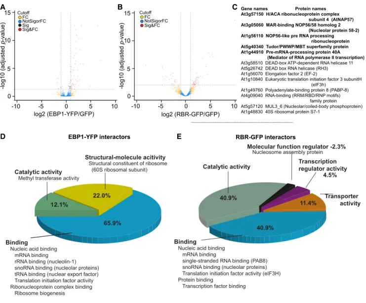
transgenic line pgEBP1-YFP and used p35S:GFP as a control. We identified and quantified EBP1-YFP asso- ciated proteins against proteins associated with the control GFP alone in six replicates each using label-free mass spectrometry (Hubner et al., 2010). To certify interactors with statistical confidence, we computed the false discovery rate and the amount ratio be- tween proteins identified in EBP1-YFP vs GFP pull- downs and established thresholds as visualized in the volcano plot (Fig. 5A; Supplemental Methods).
We identified 55 specific interactors with EBP1-YFP at high confidence (Supplemental Table S1). Gene ontology overrepresentation analysis based on mo- lecular function placed 52 out of the 55 EBP1 inter- actors into different aspects of RNA binding (Fig. 5D;
Supplemental Table S1). Classifying them according to biological processes, ribosome functions and regulation of protein translation, ribosome biogenesis, small nuclear RNA synthesis, and modifications and RNA splicing were the only prominent processes (Supplemental Table S1). Taken together, mass spectrometry identification of interactors suggested that EBP1 has the potential to reg- ulate multiple aspects of ribosome biogenesis and protein translation.
Intrigued by the molecular and functional antago- nism between EBP1 and RBR, we also searched for RBR-interacting proteins (Fig. 5, B and E; Supplemental
Table S2) using the pgRBR-GFP line (Magyar et al., 2012). The list of RBR-interactors (55) included known partners, such as E2FB and DPB and additional, pre- viously unidentified interactors. Among which 13 were annotated with the molecular function of RNA- binding, involved in ribosome biogenesis and protein translation (Fig. 5C). Five of these also interacted with EBP1, including members of the H/ACA ribonucleo- protein complex, known to catalyze the site-specific pseudo-uridylation and most of the methylation of rRNA (Watkins and Bohnsack, 2012); NOP56, which operates in trans to assist with the maturation of rRNAs (Lykke-Andersen et al., 2018); and finally the pre- mRNA-processing protein 40A, which binds the carboxyl-terminal domain of the largest subunit of RNA polymerase II and functions as a scaffold for RNA processing (Morris and Greenleaf, 2000). This set of common interactors points toward an as yet unknown aspect of RBR function that converges with EBP1 and explaining their opposing functions in the meristem.
EBP1 Is Associated with the Ribosome and Supports Global Translation in Sucrose Limiting Conditions
Given that EBP1 was found to interact with a number of ribosomal proteins by mass spectrometry, we asked
Figure 3. Sugar can compensate for the lack of EBP1expression and an elevated level ofEBP1 can suppress sugar-dependent root growth defi- ciencies. A, Root growth of two independent EBP1 RNAi (iEBP1) lines and the insertional mutant, ebp1-3 compared with Col0, germi- nated and grown (12 d) in the absence (0%) and presence of sucrose (1%). Scale bars51 cm. B, Boxplot analyses show the quantification and distribution of the root length (in millimeters) of seedlings grown on 0% and 1% sucrose media at the given time points after sowing. The boxplot gives the mean line and the mean marker (cross);
the quartile calculation was done exclusive of the median on root-length measurements from n53,n.35 in each repeat. Significance was determined by Studentttesta:P-value,0.01.
C, Quantitative analysis of root growth of the transgenicEBP1OElines (referred as lines 19.2 and 43.2, respectively in Horva´th et al., 2006) compared with Col0, germinated and grown on media with or without sucrose (0%, 1%). Box- plot analysis was carried out as in (B);n53, n.35 in each repeat; significance was deter- mined by Studentttesta:P-value,0.01. The level of EBP1 expression upon silencing and overexpression is shown in Supplemental Figure S4A and Figure 4C, respectively.
924 Plant Physiol. Vol. 182, 2020
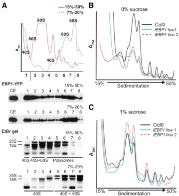
whether EBP1 is preferentially associated with single ribosomes, their subunits, or with polysomes. In a high- density sucrose gradient (15% to 50%), EBP1 fusion protein cofractionated with polysomes and ribosomal subunits (Fig. 6A), in keeping with its association with 60S subunit proteins in the pull-down experiments (Fig. 5). After separating 40S, 60S, and 80S subunits in a low-sucrose gradient (7% to 20%), EBP1 also appeared in fractions containing only 40S, as well as in the non- ribosomal fraction (Fig. 6A). Taken together, these data
confirm that EBP1 has the potential to interact with the ribosome in polysomal and nonpolysomal contexts.
To test whether EBP1 supports translation, we obtained polysome profiles of 12-d-old EBP1 RNAi seedlings grown without sucrose supplementation and after phenotypic rescue using 1% sucrose. When poly- somes were normalized by fresh weight, the EBP1 RNAiplants grown on 0% sucrose contained fewer ri- bosomes overall than the wild-type control, whereas at 1% sucrose, this difference disappeared (Fig. 6, B and
Figure 4. Elevated level ofEBP1delays differentiation upon induction of RBR expression. A, Confocal microscopy images of the transgenic lines,RBROE(pEBP1:CFP) andRBROE(pgEBP1:YFP). B, Upon induction of RBR with dexamethasone (dex) treatment (1 mM), at the given time points. Note, the loss of meristem structure and progressive differentiation due to RBR induction and the concomitant drop inEBP1transcription and reduction of EBP1-YFP abundance. C, To generate theRBROE;EBP1OElines, the transgenic linesRBROE(Wildwater et al., 2005) andEBP1OE(line 1; Horva´th et al., 2006) constitutively overexpressing theSo- lanum tuberosum EBP1(StEBP1) were introgressed. The expression levels forRBRandEBP1were analyzed by quantitative re- verse transcription-PCR in three independent introgressed lines (RBROE;EBP1OEline 10, 68, 78; F3 generation) usingRBR- and StEBP1-specific primers. As a control, the parental lines (RBROEandEBP1OE) and Col0 were tested. For the introgressed line 10, the transgenic lineEBP1OEline 1 was used as a parent, while for the lines 68 and 78,EBP1OEline 2. D, Representative root samples of Col0,EBP1OE,RBROEandRBROE;EBP1OElines upon 24 h dex (1mM) induction. See also Supplemental Figure S5A. E, Confocal images of propidium iodide–stained root samples from Col0,EBP1OE, RBROEandRBROE;EBP1OElines upon 72 h dex (1mM) treatment. In (D) and (E), the arrowhead shows the position of the first differentiating epidermal cell, while in (E) the arrow points to the QC. Scale bars51 mm (D) and 50mm (E). F, Root growth (millimeters) after 24, 48, and 72 h dex (1mM) treatment.
Induction with dex started on 6-d-old seedlings. G, Meristem length and number of non-elongating cortical cells (H) of Col0, EBP1OE, RBROEandRBROE;EBP1OElines after 0, 24, 48, 72, and 144 h dex (1mM) induction. The region of the transit amplifying cells in theRBROEroot meristem is fully differentiated around 96 h dex treatment;n.3,n.15 seedlings at each repeat for each genotype. In (F), (G), and (H), values represent means with StDv.a:P-value,0.01 shows the significance of the measured values ofRBROE;EBP1OEline compared withRBROEat the given time point.
Plant Physiol. Vol. 182, 2020 925
C). However, the polysome-to-monosome (80S) ratio was not obviously affected by sucrose. Thus, the defi- ciency in ribosomes was rescued by sucrose, matching with the effect of sucrose on the growth kinetics shown earlier (Fig. 3; Supplemental Fig. S4).
EBP1 Supports Ribosome Biogenesis
To further delineate the potential molecular functions of EBP1 in plants, especially in light of its association
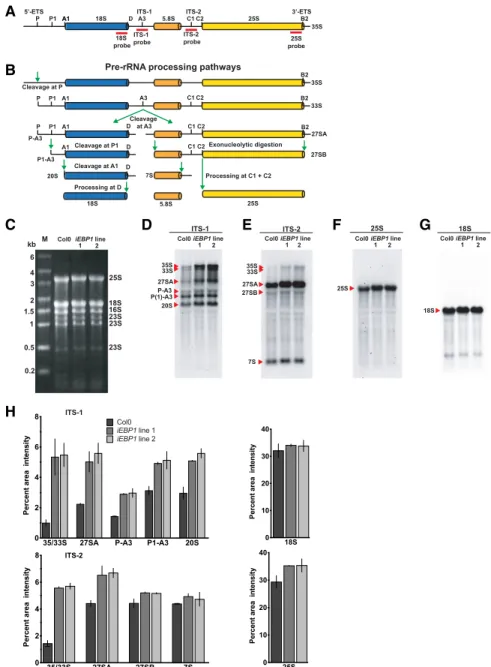
with proteins involved in ribosome biogenesis in Ara- bidopsis (Supplemental Table S1) and in human (Squatrito et al., 2004), we examined theiEBP1lines for defects in rRNA processing. We conducted northern blot analyses of rRNA processing intermediates with probes against the internal transcribed spacers ITS- 1 and ITS-2, as well as the mature 18S and 25S rRNA (Fig. 7, A and B). Despite equal loading of all three samples (Fig. 7C), the immature forms of the nuclear rRNAs were elevated in the twoiEBP1lines compared with the wild-type control (Fig. 7. D and E). In contrast,
Figure 5. Functional classification of the EBP1 and RBR interacting proteins identified by mass-spectrometric analysis. Volcano plots showing the enrichment of EBP1-interacting (A) and RBR-interacting proteins (B) recovered from seedlings expressing the EBP1-YFP (A) and RBR-GFP (B) fusion proteins compared with the proteins identified from the transgenic lines Col0(p35S:GFP).
Thexaxis shows the fold enrichment (FC) between the fusion protein and GFP alone (log2); whereas theyaxis illustrates sig- nificance (Sig), the adjustedP-value (2log10) of the pulled-down proteins with anti-GFP antibody from EBP1-YFP (A) and RBR- GFP (B) seedlings compared with GFP-expressing Col0 transgenic plants. The vertical and horizontal stippled lines demarcate a 2-fold enrichment and an adjustedp-value of 0.05, respectively; proteins in the top right portion of the graph are considered to be the candidates for EBP1- (A) and RBR (B)-interactors. C, The table summarizes the list of RBR interactors involved in RNA binding (13) and highlights those that are common in both sets, the RBR and EBP1 interactors (5). D and E, The pie charts represent the major categories by their molecular functions using the gene ontology classification tool PANTHER version 14.1. Notable subcategories are also listed. For details on protein names and functions refer to Supplemental Tables S1 and S2.
926 Plant Physiol. Vol. 182, 2020
the abundance of the mature 18S and 25S rRNAs was indistinguishable between the Col-0 and RNAi lines (Fig. 7, F and G). These data suggest that the early processing events take place rapidly, as indicated by small pool sizes of the processing intermediates, whereas the later events (e.g. P[1]-A3 to 20S) appear to be slower, as shown by larger pool sizes (Fig. 7, D and E). Through quantification of the signals (Fig. 7H), we confirmed that the early processing events were par- ticularly sensitive to the reduction in the EBP1 level, suggesting that EBP1 affects early processing steps in rRNA maturation. However, as the later processing intermediates (e.g. P[1]-A3 and 20S) were not depleted in the EBP1 RNAi lines, we suggest that EBP1 also supports the efficiency of processing steps at later stages. In conclusion, these data implicate EBP1 in ri- bosome biogenesis and, either directly or indirectly, in
rRNA processing. The molecular defects in ribosome biogenesis may explain the slow growth of the EBP1 RNAilines. In the rRNA processing experiments, we did not observe any imbalance in the ratios of specific rRNAs when samples containing equal amounts of total RNA were compared (Fig. 7C). The requirement for EBP1 for rRNA processing was rescued by growing the seedlings on sucrose-containing media (Supplemental Fig. S6, A to E,2S to1S). This result is in keeping with the conclusion that the slow growth ofEBP1 RNAiplants is due, at least in part, to rRNA processing defects.
DISCUSSION
In plants, EBP1 attracted attention as a growth driver both in Arabidopsis and in crop species, where it was shown to increase potato yield (Horváth et al., 2006) and to associate with hybrid vigor in maize (Wang et al., 2016). However, the molecular function and how EBP1 acts to increase plant growth and crop yield remained unknown. Here we show that EBP1 associ- ates with proteins that bind RNA and are involved in a broad range of functions from ribosome biogenesis to transcriptional and translational regulation. Specifi- cally, (1) plant EBP1 can localize to nucleoli, (2) EBP1 is found in cellular fractions containing ribosomal subunits as well as polysomes, and (3) silencing of EBP1 causes imbalances in the pattern of rRNA processing interme- diates. In addition, wefind that EBP1 antagonizes RBR action to maintain proliferation in the meristem, and sustains root growth, specifically in sucrose-limiting conditions.
Nutrient limiting conditions as well as abiotic stress constrain growth. It is of particular importance that plants maintain the proliferation potential in the meri- stem under these conditions (Julkowska and Testerink, 2015). For example, drought triggers jasmonate signal- ing, which counteracts the decline ofEBP1expression as leaf development progresses and cell proliferation grad- ually ceases (Kim et al., 2017). The same regulation is also observed for the large majority of genes coregulated with EBP1in theribiregulon (Noir et al., 2013). Thus, drought perception through jasmonate signaling has been sug- gested to establish a “ready-to-go” state that enables rapid recovery of meristematic functions after the stress subsides. Correspondingly, elevated EBP1 expres- sion confers resistance to abiotic stresses such as cold (Cao et al., 2009) and drought (Cheng et al., 2016).
Under stress, survival and growth are separate events, and it is likely that EBP1 is involved in the latter (Skirycz et al., 2011).
Here we show that EBP1 is important to maintain root meristem activity and growth in sucrose-limiting conditions. Thus, as during abiotic stress, EBP1 is also important to sustain growth when assimilates are lim- ited. Consistent with previous data (Deprost et al., 2007), we show here that EBP1 mRNA and protein levels are stimulated by TOR kinase. sucrose stimulates
Figure 6. EBP1 associates with cytosolic ribosomes and supports ri- bosome biogenesis. A, Absorbance profile of a sucrose density gradient after fractionation of whole tissue from Col0(pgEBP1-YFP) seedlings (12 d). Thexaxis represents the sedimentation distance in a 15% to 50%
(red trace) or 7% to 20% (blue stippled trace) sucrose gradient, while the yaxis representsA254(A254). Peaks corresponding to 40S, 60S subunits, and 80S ribosomes are indicated on the individual profiles; polysomes are to the right of the 80S. Under the graph, the immunoblot analysis (EBP1-YFP) illustrates the distribution of EBP1-YFP in the total cell ex- tract (CE) and in the eight fractions of the different gradients, while the ethidium bromide-stained agarose gels (EtBr gel) show the distribution of 18S (40S subunit) and 25S (60S subunit) rRNAs also in the eight fractions from the different gradients as shown in the graph. B and C, Polysome profiling of EBP1 RNAi lines (iEBP1 line 1 and line 2).
Seedlings were grown for 12 d without sucrose (B) and with 1% sucrose (C). Total cell extracts from equal amounts of whole seedlings from Col0 and EBP1 RNAi transgenics were fractionated on 15% to 50% sucrose density gradients.
Plant Physiol. Vol. 182, 2020 927
cell proliferation in multiple ways, which include TOR-mediated (Xiong et al., 2013; Dobrenel et al., 2016a) and TOR-independent pathways. For exam- ple, sugar controls CDK-driven RBR phosphoryla- tion, which lifts RBR-repression on cell proliferation (Magyar et al., 2012). This event may be TOR-mediated, because TOR, acting through YAK1 kinase, inhibits the expression of a class of CDK inhibitors, the SMRs, to promote root growth (Barrada et al., 2019). We now understand better how EBP1 assists in these events.
Specifically, while RBR overexpression halts root growth by depleting stem cells and enhancing dif- ferentiation (Wildwater et al., 2005), EBP1 counter- acts this RBR-driven repression and allows the maintenance of root meristem activity and growth.
Moreover, overexpression of EBP1 bypasses the re- quirement for sugar to support root growth. Taken together, TOR activity is relayed to control growth by
regulating both the level of EBP1 and the repressor activity of RBR. A remarkable result is that EBP1si- lencing specifically leads to sensitivity under limited sugar availability, whereas addition of sucrose to the media largely compensates for the loss of EBP1. This result confirms that sucrose, while stimulatingEBP1expression, also supports growth in other EBP1-independent ways, such as the TOR-dependent pathways regulating E2F and RBR (Magyar et al., 2012; Xiong et al., 2013; Barrada et al., 2019).
Both EBP1expression and EBP1 protein are largely confined to meristems, such as the root apical and lat- eral root meristems. Translatomic data also demon- strate that EBP1 mRNA is actively translated in the shoot apex (Tian et al., 2019). Furthermore, EBP1ex- pression progressively diminishes as the meristematic cells are exhausted when RBR overexpression is in- duced. This shows that EBP1 level is tightly linked with
Figure 7. EBP1 RNAitransgenic plants accu- mulate rRNA processing intermediates. Sche- matic representation of the Arabidopsis full length 35S pre-rRNA transcript indicating processing sites and the position of the probes used for northern blot analysis (red bars; A) and pre-rRNA processing pathway based on Zakrzewska-Placzek at al. (2010) (B). ETS, external transcribed spacer; ITS, internal tran- scribed spacer. C, Ethidium bromide-stained 1% (w/v) agarose gel showing equal loading of total RNA (4mg) from Col0 and two inde- pendentEBP1 RNAitransgenic lines 1 and 2 grown without sucrose. Molecular weight marker (M) is shown on left, and the position of the rRNAs is indicated on the right. D to G, northern blot analysis of total RNA from Col0 and EBP1RNAi lines using digoxigenin- labeled specific probes against ITS-1 (D), ITS- 2 (E), 25S (F), and 18S (G), visualized by chemiluminescence. Positions of the full length pre-rRNA transcript (35S) and the pro- cessed products are indicated on the left. H, Quantification of the signal intensity of the various rRNA transcript species shown in (D) to (G). Thexaxis indicates the precursors and processed rRNAs, while theyaxis shows rel- ative signal intensity. Values represent means and error bars stand forSEfrom two northern blot experiments carried out with two inde- pendent biological materials.
928 Plant Physiol. Vol. 182, 2020
meristematic activity. Fluorescently tagged EBP1 could be detected throughout the cytosol, nucleus, and nucleolus, although the nuclear and nucleolar accu- mulation were largely masked when EBP1 was tag- ged at its C terminus. We conclude that EBP1 is likely found in all three compartments as suggested by the N-terminally tagged version, because (1) growth factor signaling promoted nuclear accumulation of EBP1-GFP (Li et al., 2018), (2) others have detected tagged EBP1 proteins in plant nuclei of various cell types (Zhang et al., 2005; Cao et al., 2009; Li et al., 2016; Palm et al., 2019), and (3) native Arabidopsis EBP1 was detected in a purified nucleolar prepara- tion (Pendle et al., 2005).
In line with the localization of EBP1 in the cytoplasm, nucleus, and nucleolus, we identified EBP1-interacting proteins broadly distributed in the cell but remarkably almost exclusively involved in various aspects of pro- tein synthesis. We identified EBP1 in association with ribosomal proteins of the 60S subunit, as well as the eukaryotic translation initiation factor eIF5B. EBP1 was detected in sucrose gradient fractions that contain pol- ysomes, suggesting association of EBP1 with mature cytosolic ribosomes as well as ribosomal subunits, similar to the association pattern of human EBP1 (Squatrito et al., 2004; Squatrito et al., 2006). Related to the nucleolar localization of EBP1, we found EBP1 in- teraction with ribosome biogenesis factors, such as multiple subunits of the H/ACA ribonucleoprotein complex involved in pre-rRNA processing (Watkins and Bohnsack, 2012), with nucleolar S-adenosyl-L- Met-dependent methyltransferases, with other rRNA- processing proteins (Lykke-Andersen et al., 2018) and the multifunctional protein nucleolin (Jia et al., 2017).
Similar to EBP1, nucleolin is also most abundant in meristematic tissues and, in parallel withD-type cyclin, it is rapidly induced when cell proliferation resumes after nutrient starvation (Bögre et al., 1996). Besides the proteins with nucleolar functions, EBP1 also interacted with proteins involved in transcriptional and post- transcriptional regulation. In conclusion, similarly to human EBP1 with RNA binding activity (Squatrito et al., 2004; Squatrito et al., 2006), the common feature of EBP1 interacting proteins is that they function as part of ribonucleoprotein complexes.
Although the precise role of EBP1 in ribosome bio- genesis remains unclear, the related metallopeptidase- fold protein Arx1 is a canonical 60S biogenesis factor (Greber, 2016). Seedlings with reduced EBP1 level showed a defect in rRNA processing that affected spe- cifically the early processing intermediates. Just as the growth phenotype was rescued by sucrose, so was the rRNA processing delay in the EBP1 silencing lines.
These results are consistent with the assumption that EBP1 has a role in ribosome biogenesis. Nevertheless, we cannot rule out that Arabidopsis EBP1 also supports rRNA transcription, as was described in human cells (Nguyen le et al., 2015). However, our data point to a defect during rRNA processing, because a transcrip- tional defect uponEBP1silencing would reduce rather
than increase the abundance of rRNA processing intermediates.
In vertebrates EBP1 is expressed in two isoforms;
the longer p48 isoform is generally associated with cell proliferation and tumorigenesis, specifically by inhibiting cell death and promoting Pol I-dependent rRNA expression. The shorter p42 isoform lacks the N terminus of p48 and is considered to have a tumor suppressor role. Together with Rb, E2F1, and chro- matin modifying proteins, HsEBP1 p42 represses cell proliferation (Liu et al., 2006; Ko et al., 2016; Nguyen et al., 2018; Nguyen et al., 2019). The internal methi- onine that initiates translation of the p42 isoform is not conserved in plants. Thus, there is no evidence that the shorter suppressive form exists in plants. In line with thesefindings, the plant EBP1 promotes cell growth and division (Horváth et al., 2006; Cheng et al., 2016).
In addition, in this study we show that EBP1over- expression can compensate for the pro-differentiation activity of RBR in the root stem cell niche. Inducible overexpression of RBR downregulated EBP1 expres- sion and rapidly abolished proliferation competence. In contrast, overexpression ofEBP1countered RBR’s pro- differentiation activity and rescued the maintenance of cell proliferation around the stem cell niche. Thus, EBP1 expression is in an antagonistic relationship with the pro-differentiation factor, RBR. Our data illustrate how stem cell fate is maintained by a balance between pro- differentiation signals and the pro-growth agenda of the ribosome biogenesis machinery.
The strikingfinding that EBP1 and RBR share com- mon interactors involved in ribosome biogenesis and that RBR associated with a number of proteins in the cytoplasmic translation machinery may shed light on how EBP1, in conjunction with RBR, regulates meri- stem activity. Future research will elaborate on our hypothesis that RBR and EBP1 counteract to regulate meristem function through acting on a common bi- ological process to regulate the capacity for protein synthesis. In conclusion, our findings on the role of EBP1 in root meristem activity and growth could be important in breeding to improve yield and yield stability in crops.
MATERIALS AND METHODS Plant Material and Growth Conditions
Arabidopsis (Arabidopsis thaliana) seeds were sterilized, incubated at 4°C for 2 d, then germinated and grown vertically on half-strength Mur- ashige and Skoog salt plant media (Sigma, cat #2633024) with different concentration of Phytoagar under a long day cycle of 16 h light (806 10mmol m22s21)/8 h dark at 22°C and 50% humidity. Sucrose was added at 1%, 2% (w/v), or omitted, as specified. Arabidopsis ecotype Columbia (Col-0) was used as the wild-type control as the genetic background to generate transgenic lines and as a source of DNA to clone either promoter or coding regions. Transgenic lines,EBPOElines 1 and 2, overexpressing the potato (Solanum tuberosum) EBP1 (StEBP1, Horváth et al., 2006 referred as line 19 and 43), andRBROE(Wildwater et al., 2005) were described earlier. The insertional line, CS854731, named asebp1-3(Li et al., 2018), was obtained from the Salk Institute (http://signal.sal.edu).
Plant Physiol. Vol. 182, 2020 929
Generating Transcriptional and Translational Fusion Proteins
Both transcriptional and translational fusions were constructed by the Multisite Gateway System (Invitrogen) by amplifying the putative regulatory and genomic coding sequences with gene-specific primers. To generate the p35S:Cerulean-EBP1reporter construct and theEBP1inducible silencing lines, EBP1 RNAi, Arabidopsis EBP1 cDNA was amplified and cloned in the Gateway compatible vectors. The plant binary vector system,pGREENwas used in the Agrobacterium tumefaciensC589(pMP90)-mediated plant transformation with thefloral dip method. Details are described in Supplemental Data S1 and primer sequences summarized in Supplemental Table S3.
Root Measurement and Microscopy
Photographs of vertically or horizontally grown seedlings were taken with a digital camera (Canon). The primary root length was measured using ImageJ version 1.41 software (http://rsb.info.nih.gov/ij/index.html) and analyzed statistically with GraphPad Prism 7.0a (GraphPad Software). The root meri- stematic zone was determined by measuring the distance from the QC to the first elongating cortex cells; the same region was used to count the number of cortex cells with no signs of rapid elongation. For measurements and to follow tissue-specific expression, either light (Zeiss Axioscope with Nomarski optics) or confocal microscopy (Zeiss LSM710 and Leica SP8) was carried out. To take differential interference contrast images, chloral-hydrate treated roots were used; starch granules were visualized with 1% (w/v) lugol solution. Seedlings (T3 generation) were stained for GUS activity for 3–6 h at 37°C in 0.5 mg/mL X-gluc (Biosynth AG), dissolved according to the manufacturer’s recommen- dation. For confocal microscopy, to visualize cell walls, 5–10 mg/mL propidium iodide (Sigma-Aldrich) solution was applied. For analysis, longitudinal, single sections were taken with the same median focal plane.
Thefinal images were built and merged from partially overlapping sec- tions with Adobe Illustrator CS6.
Chemical Treatments
The introgression lines,EBP1OE;RBROE(F3 generation) were germinated and grown on vertical plates (1% sucrose media). The 5- to 6-d-old seedlings were transferred to 1mMdexamethasone-containing plates, and growth was scored over a time series (24 h–144 h period).RBROE,RBROEbackcrossed to Col-0, EBP1OE, and Col-0 were used as a control to follow root growth, meristem size, and level of gene expression. To screen transformants of the silencing lines, EBP1 RNAi, seeds (T1 and T2) were germinated and grown on 5mM17- b-estradiol-containing medium supplied with 1% sucrose or no sucrose.
Next, the candidate lines were grown in normal growth conditions and transferred to 17-b-estradiol medium with or without sucrose. The induction and level of silencing was measured by quantitative reverse transcription PCR.
Immunoblot and AZD Treatment
For immunoblot analysis, the transgenic lines Col-0(pgEBP1-YFP) and Col- 0(pEBP1:CFP) were germinated and grown for 5 d on sucrose-free medium in 12-h light/12-h dark conditions. On the 5th day, at ZT0 timepoint, seedlings were transferred to medium containing either 2% sucrose, 2% sucrose11mM AZD8055 or to fresh sucrose-free medium. Samples were harvested 3 h later by flash-freezing the whole seedlings. Total protein was extracted from samples using extraction buffer containing 75 mMNaCl, 25 mMTris-HCl (pH57.5), 15 mMb-glycerolphosphate, 15 mMEGTA, 15 mMp-nitrophenylphosphate, 10 mMMgCl2, 1 mMdithiothreitol, 1 mMNaF, 0.5 mMNa3VO4, 0.5 mMphe- nylmethylsulfonylfluoride, and 0.1% Tween 20 (Magyar et al., 2005). The total protein samples were separated in a 10% acrylamide gel using SDS-PAGE and transferred to polyvinylidene difluoride membrane. Antibodies used for de- tection: Anti-GFP mouse monoclonal antibody (ROCHE) in 1:1000 dilution and goat anti-mouse IgG-HRP Secondary Antibody (Sigma) in 1:10000 dilution.
Northern Blot Analysis, Polysome Profiling, and Protein Fractionation
Northern blot analyses were performed with 4mg of total RNA from seedlings grown under the indicated conditions. Blots were probed with digoxigenin-labeled probes corresponding to the internal transcribed spacer
1 and 2, 18S and 25S rRNAs, and signal was detected with chemiluminescent reagent, imaged on a BioRad ChemiDoc instrument, and quantified with ImageJ version 1.41 software (http://rsb.info.nih.gov/ij/index.html). Poly- some profiles were obtained from 12-d-old Col-0 orEBP1 RNAiwhole seedlings grown without or with 2% sucrose essentially as described (Kim et al., 2007).
Then 100ml supernatant was layered on either a 2-mL 15-50% or a 2 mL 7% to 20% linear gradient prepared with a Hoefer gradient maker and centrifuged at 50,000 rpm (Beckmann TLS55 rotor) for 1 h 10 min at 4°C. After the UVA254and fractionation into eight equal fractions were monitored, EBP1 protein was detected by immunoblotting with GFP-specific antibody (Lokdarshi et al., 2016). For detailed procedures see Supplemental Data S1.
Protein Complex Isolation, LC-MS/MS Identification, and Statistical Analysis
A detailed description and link to the raw datasets are given in the Supplemental Data S1.
Accession Numbers
Accession numbers are At3g51800,EBP1; At3g12280,RBR; the other ac- cession numbers are listed in Supplemental Table S3.
Supplemental Data
The following supplemental materials are available.
Supplemental Figure S1.EBP1is expressed in actively dividing tissues during root development.
Supplemental Figure S2.EBP1 localizes to nucleoli, nuclei and cytosol.
Supplemental Figure S3.Reduced level of EBP1 results in retarded growth in the absence of sucrose.
Supplemental Figure S4.Root and leaf growth deficiency in the absence of sugar and recovery of root growth defects upon sucrose supplementa- tion in theEBP1transgenic lines.
Supplemental Figure S5.Elevated level ofEBP1delays the differentiation induced byRBRoverexpression.
Supplemental Figure S6.sucrose supplementation alleviates the require- ment for EBP1 in rRNA processing.
Supplemental Table S1.List of EBP1 interacting proteins.
Supplemental Table S2.List of RBR interacting proteins.
Supplemental Table S3.Primers used in this study.
Supplemental Data S1.https://doi.org/10.6084/m9.figshare.10069247.
ACKNOWLEDGMENTS
We thank Dr. Patrick Giavalisco (Max Planck Institute Cologne) for initial discussions on EBP1, linking it with protein translation; Dr. Tessa Burch-Smith and Dr. Daniel M Roberts (University of Tennessee) for valuable advice with northern blot analysis and for providing anti-GFP antibody, respectively; Imma Perez-Salamo for valuable discussions (Royal Holloway, University of Lon- don); and Christian Bachem (Wageningen University and Research) for critical reading of the manuscript.
Received July 2, 2019; accepted November 22, 2019; published December 9, 2019.
LITERATURE CITED
Ahmad Z, Magyar Z, Bögre L, Papdi C(2019) Cell cycle control by the target of rapamycin signalling pathway in plants. J Exp Bot 70:
2275–2284
Bakshi A, Moin M, Madhav MS, Kirti PB(2019) Target of rapamycin, a master regulator of multiple signalling pathways and a potential can- didate gene for crop improvement. Plant Biol (Stuttg)21:190–205
930 Plant Physiol. Vol. 182, 2020