FORMATION, PHOTOPHYSICS AND PHOTOCHEMISTRY OF WATER-SOLUBLE LANTHANIDE(III) PORPHYRINS
PhD dissertation written by
Muhammad Imran
Supervisors:
Dr. Ottó Horváth, DSc Dr. Zsolt Valicsek, PhD
University of Pannonia
Doctoral School of Chemical and Environmental Sciences
Institute of Chemistry
Department of General and Inorganic Chemistry
Veszprém 2016
DOI:10.18136/PE.2016.612
ii
Értekezés doktori (PhD) fokozat elnyerése érdekében Írta:
Muhammad Imran
Készült a Pannon Egyetem Kémiai és Környezettudományi Doktori Iskolája keretében.
Témavezetők:
Dr. Horváth Ottó
Elfogadásra javaslom (igen / nem) ………..
(aláírás) Dr. Valicsek Zsolt
Elfogadásra javaslom (igen / nem) ………..
(aláírás)
A jelölt a doktori szigorlaton ...%-ot ért el, Az értekezést bírálóként elfogadásra javaslom:
Bíráló neve: …... …... igen /nem
……….
(aláírás) Bíráló neve: …... …... igen /nem
……….
(aláírás) A jelölt az értekezés nyilvános vitáján …...%-ot ért el.
Veszprém, ……….
a Bíráló Bizottság elnöke
A doktori (PhD) oklevél minősítése…...
………
Az EDHT elnöke
iii
Abstract
Porphyrins are the most widely studied macrocyclic compounds because of their several important roles in nature as well as their special coordinative, spectral and redox features.
The cavity of the porphyrins containing four pyrrolic nitrogen is ideally suited for binding the vast majority of metal ions of the periodic table. If the metal ion is too big to fit co-planarly into the cavity of porphyrin, it is located out of the ligand plane and distorting it. This structure results in kinetic lability as well as peculiar photophysical and photochemical behavior. Lanthanide(III) ions, due to their large ionic radius and contraction upon increasing the atomic number, proved to be suitable for the fine tuning of the out-of-plane (OOP) distance of the metal center. In the presence of acetate ion as a potential axial ligand mainly the lanthanide(III) monoporphyrin species were formed, while, in the case of perchlorate ion, bisporphyrins (3:2 = metal ion : porphyrin) too. The stability constants increased with the atomic number in accordance with the stronger metal-ligand interaction due to the shorter OOP distance. The dome distortion of the macrocycle in these complexes manifested in the redshift of the visible absorption bands and the decrease of the S1-fluorescence intensity compared to those of the free-base porphyrin. The moderate effect of dimerization on the absorption and emission properties indicated a weak interaction of the monomers in the bisporphyrins, which suggested a tail-to-tail structure connected through the peripheral sulfonate groups by a metal-ion bridge. Irradiation of the Ln(III) porphyrins at Soret- and Q- band maxima resulted in three types of photoinduced reactions, namely redox degradation, dissociation, and transformation between the Ln(III) mono- and bisporphyrins. Two types of photoproducts depending on the excitation wavelength were detected; Soret-band irradiation generated a short-lived intermediate, while Q-band excitation produced a stable end-product.
The individual quantum yields of all the photochemical reactions were determined by the concentration distribution method for the lanthanide(III) porphyrins studied. For both the mono- and the bisporphyrins, the overall quantum yields, in which the efficiency of the redox degradation is predominant, display a decreasing trend (following a small increase) as a function of the atomic number of the metal center, as the consequence of the diminution of the out-of-plane distance and, thus, the chance for the charge separation following the photoinduced LMCT process.
iv
Kivonat
A porfirinek a legszélesebb körben tanulmányozott makrociklusos vegyületek, mivel fontos szerepet játszanak a természetben, továbbá különleges koordinációs, spektrális és redoxi tulajdonságokkal rendelkeznek. A porfirinek négy pirrol-nitrogénnel rendelkező ürege ideális a fémionok többségének megkötésére. Ha a fémion mérete túl nagy ahhoz, hogy a porfirin koordinációs üregébe koplanárisan illeszkedjen, a ligandum síkja fölött helyezkedik el és azt torzítja. Ez a szerkezet kinetikai labilitást, valamint sajátos fotofizikai és fotokémiai viselkedést eredményez. A lantanoida(III)ionok – nagy ionsugaruk és a rendszám növekedésével járó méretcsökkenésük következtében – alkalmasnak bizonyultak a központi fémion síkon kívüli (OOP) távolságának finomhangolására. Acetátion mint potenciális axiális ligandum jelenlétében főként lantanoida(III)-monoporfirin képződött, míg perklorátionok mellett biszporfirin (3:2 = fémion:porfirin) is. A stabilitási állandók növekedtek a lantanoidák rendszámával – összhangban a rövidebb OOP távolságból adódó erősebb fém-ligandum kölcsönhatással. A ligandum dómos torzulása ezekben a komplexekben a látható tartományban a szabad porfirin elnyelési sávjainak vöröseltolódásában és az S1- fluoreszcenciája intenzitásának csökkenésében nyilvánult meg. A dimerizáció csak mérsékelten befolyásolta az abszorpciós és emissziós tulajdonságokat, ami a monomerek gyenge kölcsönhatását jelezte a biszporfirinekben, “farok-farok” szerkezetet sugallva, melyben a kapcsolódás a szulfonát szubsztituensek közt valósul meg fémion-híd révén. A Ln(III)-porfirinek besugárzása a Soret- és Q-sávok maximumán háromféle fotoindukált reakciót idézett elő; redoxi lebomlást, disszociációt, valamint a mono- és a biszporfirinek közötti átalakulást. Emellett a besugárzási hullámhossztól függően kétféle, eddig nem észlelt fototerméket sikerült detektálni; a Soret-sávon a besugárzás egy rövid-élettartamú köztiterméket eredményezett, míg a Q-sávos gerjesztés egy stabil végterméket. A vizsgált lantanoida(III)-porfirinek valamennyi fotokémiai reakciójának egyedi kvantumhasznosítási tényezőjét meghatároztam a koncentráció-eloszlás módszerével. Mind a mono-, mind a biszporfirinekre vonatkozó bruttó érték, melyben a redoxi bomlás hatékonysága a meghatározó, a központi fémion rendszámának növekedtével – egy kis emelkedést követően – csökkenő tendenciát mutat, együtt a síkon kívüli távolsággal, s így a fotoindukált LMCT folyamatot követő töltésszétválás valószínűségével.
v
Zusammenfassung
Porphyrine sind die bestuntersuchten makrozyklischen Verbindungen, da sie in der Natur eine wichtige Rolle spielen und dafür spezielle Koordinations- und Redoxeigenschaften besitzen. Der Koordinationshohlraum der Porphyrine, die vier Pyrrol-Stickstoffmoleküle haben, ist ideal für die Bindung von vielen Metallionen. Wenn das Metallion zu groß ist, um sich in den Koordinationshohlraum des Porphyrins koplanar einzufügen, placiert es sich über der Ebene des Liganden und deformiert ihn dadurch. Diese Struktur verursacht kinetische Labilität, sowie besonderes photophysikalisches und photochemisches Verhalten.
Lanthanoid(III)-Ionen, erwiesen sich, infolge ihrer großen Ionenradien und der Lanthaniodenkontraktion, als geeignet für die Feinabstimmung des “out-of-plane” (OOP) Abstandes. In Anwesenheit von Acetat-Ionen als potenzielle axiale Liganden bilden sich bevorzugt Lanthanoid(III)-Monoporphyrine, jedoch bei Anwesenheit von Perchlorat-Ionen werden auch Bisporphyrine (3:2 = Metallion : Porphyrin) gebildet. Die Stabilitätskonstanten nahmen mit der Ordnungszahl der Lanthanoiden zu – übereinstimmend mit der stärkeren Metall–Ligand-Wechselwirkung, die aus dem kürzerem OOP Abstand resultiert. Die kuppelförmige Deformation vom Ligand in diesen Komplexen äußerte sich als die Rotverschiebung in den Absorptionsbanden im sichtbaren Bereich und der Abnahme der Intensität der S1-Fluoreszenz – im Vergleich mit den entsprechen Werten des freien Porphyrins. Die Dimerisierung beeinflusst die Absorptions- und Emissionseigenschaften nur mässig, was die schwache Wechselwirkung der Monomeren in den Bisporphyrinen zeigt, suggerierend eine “Schwanz-zu-Schwanz” Struktur, in der die Bindung zwischen den Sulfonat-Substituenten durch eine Metallion-Brücke gegeben ist. Die Bestrahlung der Ln(III)-Porphyrine in den Soret- und Q-Banden verursacht dreierlei photochemische Reaktionen; Redox-Zersetzung, Dissoziation, sowie Umwandlung zwischen Mono- und Bisporphyrinen. Daneben gelang es, zwei, bisher noch nicht beobachtete Photoprodukte zu bestimmen, die von der Anregungswellenlänge abhängen. Die Soret-Band-Bestrahlung ergab ein kurzlebiges Zwischenprodukt, während die Q-Band-Anregung zu einem stabilen Endprodukt führt. Die individuellen Quantenausbeuten für alle photochemischen Reaktionen der untersuchten Komplexe wurden durch die Methode der Konzentrationsverteilung bestimmt. Der Bruttowert, bei dem die Redox-Zersetzung maßgeblich ist, für Mono- als auch die Bisporphyrine zeigt – nach einer kleinen Zunahme – einen Abwärtstrend mit wachsender Ordnungszahl des Metallzentrums, infolge der Abnahme des OOP Abstands und somit der Möglichkeit der Ladungstrennung nach einem photo- induzierten LMCT Prozess.
vi
Acknowledgments
Many thanks are extended to my supervisors, Prof. Ottó Horváth and Dr. Zsolt Valicsek for their continued guidance and advice throughout the research work in both practical and theoretical respects, and for the appropriate experimental conditions.
I am very grateful to the colleagues at the Department of General and Inorganic Chemistry of University of Pannonia for their help and competent assistance in any respects, and for their hospitality.
I am indebted to Prof. Günter Grampp and his colleagues at the Institute of Physical and Theoretical Chemistry at Graz University of Technology for their extensive help and assistance in the ESR measurements.
Many thanks are due to my family, first of all my parents and my wife, whose efforts, love, understanding and patience made my studies possible in a country far from our home.
I also appreciate the financial background of this work supported by the Hungarian Research Fund (NN107310), the Hungarian Government and the European Union, with the co-funding of the European Social Fund (TAMOP-4.2.2.A-11 /1/KONV-2012-0071) and the Austrian- Hungarian Action Foundation (86öu3).
1
Contents
Abstract ... iii
Chapter 1: Introduction ... 3
1. Introduction of porphyrin chemistry ... 3
2. Nomenclature and isomerism of porphyrins ... 4
3. Electronic properties of porphyrins ... 5
3.1. Light absorption ... 5
3.2. Light emission ... 8
4. Occurrence and preparation of porphyrins ... 10
4.1. Natural porphyrins ... 10
4.2. Synthetic porphyrins ... 12
5. Metalloporphyrins ... 13
5.1. Natural metalloporphyrins ... 14
5.2. Synthetic metalloporphyrins ... 20
5.3. Applications of metalloporphyrins... 32
6. Water-soluble porphyrins... 34
Chapter 2: Objectives ... 37
Chapter 3: Experimental ... 38
1. Materials ... 38
2. Preparation of Ln(III) porphyrin complexes ... 39
3. Instrumentation and methods ... 39
3.1. UV-Visible absorption spectrophotometry ... 39
3.2. Fluorescence spectroscopy and lifetime measurements ... 42
3.3. Photolysis instrumentation and procedures ... 43
3.4. ESR measurements ... 45
Chapter 4: Results and discussion ... 46
1. Formation, structure and absorption spectra of Ln(III) porphyrins ... 46
1.1. Trend in stability constants ... 52
2. Photophysics of lanthanide(III) mono- and bisporphyrins ... 54
2.1. Trends in photophysical properties ... 63
3. Photochemistry of lanthanide(III) mono- and bisporphyrins ... 64
2
3.1. Mechanistic investigations of the photochemical reactions ... 71
3.2. Trends in photochemical properties of lanthanide(III) porphyrins ... 76
3.3. Applicability of lanthanide(III) porphyrin systems ... 77
Summary ... 80
Thesis points ... 82
References ... 84
Appendix ... 96
List of figures ... 96
List of tables ... 97
Publications: ... 98
Presentations ... 98
3
Chapter 1: Introduction
1. Introduction of porphyrin chemistry
Porphyrins are the most plentiful chemical compounds found in nature which are biochemically important, medically useful, and synthetically fascinating compounds.
Porphyrins that found in nature are playing a vibrant role on this planet to sustain life by assorted ways like oxygen transport (hemoglobin), its storage (myoglobin), chlorophyll for natural photosynthesis, enzymes like catalases, peroxidases, cytochromes and vitamins are of prime importance for living organisms [1]. Synthetic porphyrins and their metal complexes possess stimulating physical, chemical and biological properties and are significant candidates for utilization of solar energy, photodynamic therapy, electrooptics, mimicking of enzymes and as industrial catalysts. The electronic spectra of porphyrins have been extensively studied due to their important role in the natural biochemical processes and special absorption, emission, charge transfer, complexing properties [1]. The word porphyrin was coined first time by ancient Greeks, they used the word porphura for the intense purple color and have been used as a dye to color the cloths [2].
The structure of porphyrin (
Figure 1.1) is composed of four pyrrolic units that are linked by methine bridges at their α- positions. The four pyrrolic units after linking with each other give a planar structure to the porphyrin molecules with an extended conjugated 18 π-electron system being responsible for the aromatic behavior of porphyrins [3, 4, 5].
NH N
N HN
meso
Figure 1.1. Structure of unsubstituted porphyrin [6,7]
Porphyrins being tetradentate ligand molecules can easily accommodate metal ions. In deprotonated form, they offer a square planar environment and a rigid cavity of a 0.6-0.7-Å
4
radius, which is ideally suited for metal incorporation [8, 9]. As in most of cases, the porphyrin dianion acts as a tetradentate ligand towards metal ions; the minimum coordination number of the metal is four.
The presence of highly conjugated π-electronic systems is responsible for the intense color and other distinctive electronic and redox properties of porphyrins. Compounds like chlorin and bacteriochlorin show resemblances in structure with porphyrin [10].
2. Nomenclature and isomerism of porphyrins
The first effort of giving names and directions for drawing the different isomers of porphyrins was made by Hans Fischer [11].
He numbered the eight outer carbon atoms belonging to the pyrrolic fragments from 1 to 8, while the four bridging C atoms as α, β, γ, and δ. The Fisher’s nomenclature works only for simple porphyrins, but when the complexity of the substituents attached to the pyrrolic nitrogen increases, this system has limitation to assign them a systematic name [12]. To overcome the shortcomings in the Fischer’s system, IUPAC gave a new organized and comprehensive system of naming for porphyrins and their derivatives, which was completed in 1987. In the IUPAC system all atoms in the porphyrinic macrocycle was numbered as shown in Figure 1.2.
Figure 1.2. Numbering of atoms in porphyrin macrocycle according to IUPAC.
By given numbering to all the atoms was also useful because it helped give some structural information about the compound. In IUPAC system the positions of the principle groups determine the numbering of the substituted porphyrins and the name of the substituents (e.g.
propionic and acetic acid) comes after the name of porphyrins, and an alphabetical ascending order was used for naming the substituents attached to the macrocycle periphery.
NH N
N HN
1 2
3 4
5 6
7 8 9
20 11
12 14 13
16 15 17 18
19
20 21 22
24 23
5
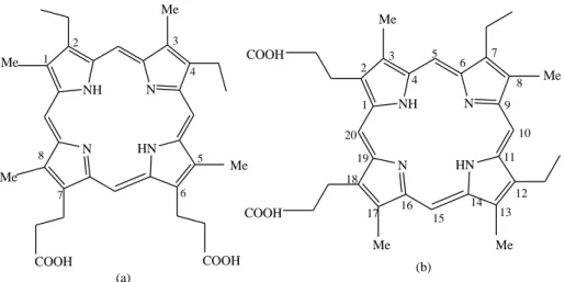
Figure 1.3 (a) 2, 4-diethyl-1, 4, 5, 8-tetramethylporphyrin-6, 7-dipropionic acid (mesoporphyrin) (b) 7, 12-diethyl-3, 8, 13, 17-tetramethylporphyrin-2, 18-dipropionic acid
(mesoporphyrin) [4]
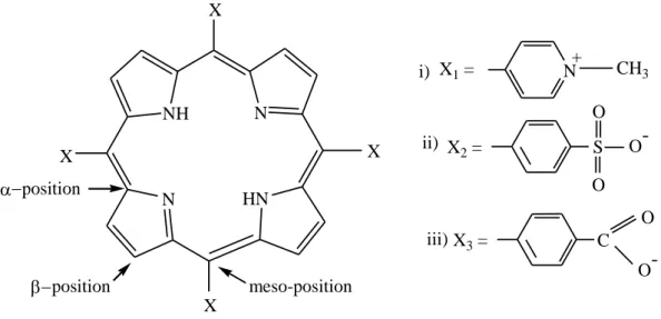
The name of mesoporphyrin according to the Fischer’s nomenclature is 2,4-diethyl-1,4,5,8- tetramethylporphyrin-6,7-dipropionic acid and 7,12-diethyl-3,8, 13,17-tetramethylporphyrin- 2,18-dipropionic acid is given to the same compound by the IUPAC system as shown in figure 1.3(a) and (b) respectively. In this work an anionic porphyrin was used, with four sulfonatophenyl groups which are attached at 5, 10, 15 and 20 position of the porphyrin macrocyclic ring and its name is 5,10,15,20-tetrakis(4-sulfonatophyenyl)porphyrin abbreviated as H2TSPP4- or simply H2P.
3. Electronic properties of porphyrins
3.1. Light absorption
Porphyrins are the strongest light absorbing materials in nature and they are also called pigments of life. Hence the UV-Vis absorption spectroscopy is most fundamental and suitable analytical technique for the elucidation of electronic structure of porphyrins and their metal derivatives even at very low concentrations [13]. By monitoring the change in the intensity and wavelength of absorption spectra of porphyrins, a very substantial information on their excited states and a variation in the peripheral substituents of the porphyrin ring could be gained. The study of the photophysical properties, electronic structure, excited state and deactivation of porphyrins and their metal complexes is of prime importance to evaluate the possible applications. A brief explanation on the spectra of porphyrins is given in the following.
NH N
N HN
Me
Me
Me Me
COOH COOH
1
2 3
4
5 7 6
8
NH N
N HN
COOH
COOH
Me
Me
Me Me
1 2
3 4
5 6 7
8 9
10 11
12 13 14 15 17 16
18 19 20
(a) (b)
6
The extended conjugation of 18-π electrons on the frontier orbitals is responsible for their highly intense color. Porphyrins possess characteristic UV-visible spectra because of their fourfold symmetry and four nitrogen atoms directed towards the center of the electronic heart. The characteristic absorption spectra of porphyrins undergo perturbation by different factors like conjugation pathway, symmetry and other chemical variations [14]. They are helpful to distinguish between free-base porphyrins and their metal complexes. Upon the insertion of metal ion into the porphyrin cavity the D2h symmetry changes to D4h. [15]. Most of the porphyrins show two sets of distinct region or bands in their electronic absorption spectrum. The ranges of the first and second band sets are 350-500 nm and 500-750 nm, respectively. The first sets of band are called Soret- or B-bands with molar absorption coefficients of 105 M-1cm-1 and involve the electronic transition from the ground state to the second singlet exited state (S0
→
S2). The second set of band is called the Q-band with molar absorption coefficients of 104 M-1 cm-1 and involve the transition from the ground state to the first singlet exited state (S0→
S1). The conjugation of 18-π-electrons gives advantageous spectroscopic features to porphyrins, which is supportive to monitoring the binding of diverse hosts to the porphyrin by using UV-visible spectroscopy [5, 16]. A UV-Vis spectrum within a wide range of Q-bands extended from 480 to 700 nm is shown in Figure 1.4 [17]. The electronic absorption spectra of porphyrins show different types of band. Depending on the type and intensity of the bands, a valued evidence could be obtained regarding the possible substitution or metalation, and classification of porphyrin spectra has also been made.The four types of Q-bands could be seen in the absorption spectra of a free base porphyrin which are represented by roman numbers and are placed according to the increasing wavelength as IV, III, II and I. If the intensities of the Q bands are in order of IV ˃ III ˃II ˃ I
0 0.5 1 1.5 2
490 550 610 670 730
molar absorbance /104 M-1cm-1
wavelength / nm III
IV
II
I
Figure 1.4 UV-Vis spectrum of porphyrin in the Q-region of 480-750 nm [17]
7
then the spectra will be of etio-type; and porphyrins will be called as etioporphyrins. This type of spectra is exhibited by all those porphyrins in which all the eight β-positions of macrocyle are substituted by the groups that do not possess π-electrons.
The relative intensities of the Q-bands show a slight modification when groups with π- electrons are attached at the β-positions (i.e., at the outer pyrrolic carbon atoms) of the macrocycle and the order of Q-bands are III ˃ IV ˃II ˃ I. The resulting spectra are called rhodo-type and the porphyrin will be rhodoporphyrin. In the third case, if the two groups with π-electrons are attached at the two pyrrol units that are opposite to each other, it gives rise to oxo-rhodo-type spectrum with an intensity diminution in the series of III ˃ II ˃IV ˃ I. At last, when the β-positions are unsubstituted, Q-band intensity series becomes IV ˃ II ˃III ˃ I, and the spectrum type is known as phyllo-type [4, 18]. Because of crucial significance of porphyrins and their derivatives in important life processes, many scientists have tried over the years to unveil more feature concerning the position and multiplicities of Q- and B-bands in the UV-Visible spectra of metal-free porphyrins.
Figure 1.5 HOMOs (bottom) and LUMOs (top) [adapted from 19]
Notably, an American scientist, Martin Gouterman, developed a very well-known theory named as “four-orbital” model about the spectra of porphyrins [20]. The Gouterman’s model illustrated in
8
Figure 1.5, helps us rationalize the electronic density in orbitals. According to Gouterman’s four-orbital model, porphyrins have two highest occupied π orbitals titled as HOMOs and two lowest unoccupied π* orbitals termed as LUMOs. The transitions between these two sets of orbitals are responsible for the advent of the absorption bands of porphyrins [18]. HOMOs own a1u and a2u orbitals, while LUMOs are identified as a degenerate eg set of orbitals, which are localized on the macrocycle ring. The HOMO a2u orbital is localized on pyrrolic nitrogens and meso carbons, while HOMO-1 a1u is localized mainly on Cα and Cβ atoms [21]. The Gouterman’s four-orbital model pays a special attention to the electronic transition between filled bonding MO’s levels a1u, a2u and antibonding MO’s levels (eg*). In porphyrin spectra HOMO to π* transition results in the appearance of four absorption peaks. Out of four absorption bands two are from x and two from y component of Q-bands. The Qx and Qy
components are also composed of two types of vibrational excitations which are Q(0, 0) at lower energy and Q(1,0) at higher energy. Therefore, the four absorptions lines can be written as Qx (0, 0), Qx (1, 0), Qy (0, 0) and Qy (1, 0) in the increasing order of energy. Upon complexation porphyrins show the appearance of a very intense red-shifted absorption band in the Soret-region at 420-425 nm and two weak Q bands at 540-600 nm. These bands are assigned to intra ligand π-π* transitions of porphyrin [3, 16].
3.2. Light emission
Fluorescence and phosphorescence processes involves the relaxation of excited state molecules to ground state through photon emission which is just the opposite process to absorption. These photonic processes involve transition between electronic and vibrational states of molecules. Light emission is termed as fluorescence if no change of spin state accompanies the electronic transition [22, 23].
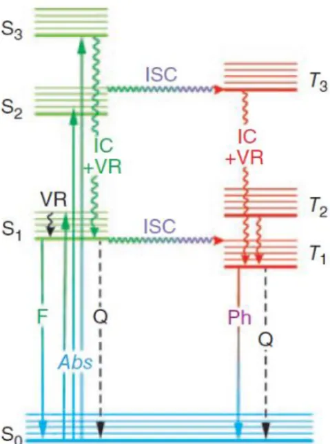
The Jablonski diagram (Figure 1.6) offers a good opportunity to explain the mechanism of the excitation and relaxation processes in molecules. The molecules are able to occupy higher energy singlet electronic states like S1, S2...Sn depending upon the energy of light absorbed [24]. According to the Kasha’s rule, the relaxation process which takes place with emission of light principally starts from S1, in solution, which is the lowest-energy excited electronic state, while in the case of higher-energy electronic excited state, like S2 and S3, there will be internal conversion prior to the emission [25]. The energy of emission is less than that of
9
absorption, thus, fluorescence occurs at longer wavelength as compared to the wavelength at which the molecule was excited. The difference between positions of the band maxima of the absorption and emission spectra of the same electronic transition is called Stokes-shift [26].
Figure 1.6 The Jablonski diagram: Abs, absorption; F, fluorescence; IC, interval conversion;
ISC, intersystem crossing; Ph, phosphorescence; Q, quenching; VR, vibrational relaxing; S0 is the ground singlet electronic state; S1 and S2 are higher-energy excited singlet electronic
states; T1 is the lowest-energy triplet state [27]
In most of the ground-state molecules with even number of electrons, the electrons occupy the low-energy-level orbitals with opposite spins and have zero for the sum of the spin quantum numbers; this state is called as singlet electronic state. When a molecule is excited, an electron moves from an occupied to an unoccupied orbital where it may keep its original spin state (singlet excited state) or changes it to the opposite orientation (triplet excited state).
The return of this electron from a singlet excited state to the lower orbital takes place more than 100 ns. and this process is called fluorescence. If the electron returns from a triplet excited state to the singlet ground state, the electron undergoes a spin-reversal, which is a forbidden process and is the basis of phosphorescence, which is a less rapid process and may usually last from milliseconds to seconds [28].
The molecules in their singlet excited state may undergo a vibrational relaxation process to the lowest vibronic level of the corresponding excited state, and this process is completed by the releasing of the thermal energy to the surroundings. Beside the biological importance, porphyrins have interesting luminescence and non-radiative features [29]. The vibrational
10
relaxation of singlet-excited porphyrins is often relatively slow compared to their radiative and intersystem crossing processes. The overall quantum yield of fluorescence and intersystem crossing in formation of triplet states is over 95%. Due to this property, porphyrins are efficient in photosensitization [21]. Results from our research group on lanthanum(III) and mercury(II) porphyrin have shown that coordination of a metal ion into a porphyrin ligand results in a blueshift or hypsochromic effect on the emission bands and a significant decrease of fluorescence intensity and lifetime of monoporphyrins. Emission spectra of previously studied metalloporphyrins have indicated that the structure of the originally flat (free base) ligand is distorted in metalloporphyrins and the decrease in quantum yield is the consequence of distortion of ligand, which promotes other energy dissipation processes than light emission. Other out-of-plane metalloporphyrins, like Hg(II) TSPP4-, Cd(II) TSPP4- and Bi(III)TSPP3-, have shown the similar emission tendencies of the band-shift and quantum yield [30, 31].
4. Occurrence and preparation of porphyrins
Many types of metal-free porphyrins have been successfully isolated and modified from natural resources. A few examples of porphyrins obtained from natural resources and synthesis of metal-free porphyrins in laboratory are given below [32].
4.1. Natural porphyrins
Nature gives us a huge accumulation of porphyrins from which many other types of porphyrins can also be developed. Chlorophyll from plants and blood of animals are main sources to produce porphyrins [33].
Such a type is protoporphyrin. Scientists have developed numerous methods for the preparation of protoporphyrin from hemoglobin. In commonly adopted methods globin is first removed from hemoglobin to get hemin. [34, 35]. To obtain protoporphyrin, the solution of hemin or hematin is reduced in the presence of organic acid. According to Grinstein procedure as compared to protoporphyrin, protoporphyrin dimethyl ester can be easily obtained from hemin by simultaneous removal of iron and its esterification [36, 37].
11
Hematoporphyrin was prepared by treatment of blood with sulfuric acid [38]. It can also be isolated by treatment of hemin with hydrogen bromide in the presence of acetic acid [39].
Mesoporphyrin has been synthesized by the reduction of protohemin in the presence of hydrogen iodide [40, 41]. The most convenient way of obtaining large quantity of mesoporphyrin was developed by Caughey. In this method hydrogen was bubbled through the formic acid solution of hemin over PdO and no side product were obtained [42].
Harderoporphyrin is found in the harderian gland that is present in orbital cavity of Wister rats. It has the ability to synthesize harderoporphyrin. Various derivatives such as esters can be prepared from the harderian gland of rats [43].
Pemptoporphyrin was isolated in 1964, from feces of patients suffering from intestinal malabsorption [44, 45]. NMR studies were used to elucidate the structure of pemptoporphyrin as 4-vinyldeuteroporphyrin [45].
Uroporphyrin I and coproporphyrin I have been isolated from urine and feces of human and birds that have congenital erythropoietic porphyria disease [36, 46]. For isolation of uroporphyrin and coproporphyrin, urine sample is acidified with acetic acid and extracted with ether [47, 48].
Chlorophyll a and b is a rich source of different types of porphyrin. By following appropriate procedures and conditions a variety of porphyrins and chlorins could be obtain from chlorophyll as starting material [49, 50]. In 1907 Willstatter and Hocheder first time documented the sensitivity of chlorophyll a and b to change magnesium(II) ion to protons, forming pheophytin [51]. They treated chlorophyll obtained from spinach with acetic acid (Figure 1.7).
When chlorophylls are treated with strong acids then phytyl groups may be also hydrolyzed to pheophorbides with carbomethoxy groups [50]. Pheophytin has also been isolated from a neurodifferentiation compound that is present in marine brown alga known as Sargassum fulvellum [52].
12
R= CH3 pheophytin a, R= CHO pheophytin b
Figure 1.7 Formation of pheophytin from chlorophyll [53]
4.2. Synthetic porphyrins
The synthetic history of porphyrins initiated from 1930. Until now, porphyrins of various types have been prepared and characterized by adopting diverse range of chemical strategies.
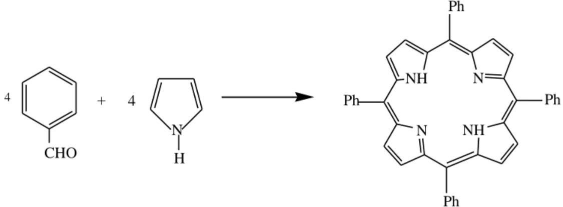
The molecules like pyrrol, linear tetrapyrroles, tripyrranes, aldehydes and dipyrromethanes have been used as starting materials for the synthesis of porphyrins [18].
Fischer and Gleim in 1935 first time obtain 17 mg of porphyrin by reaction of pyrrole and aldehyde in the presence of formic acid under reflux condition [54]. In the same year Rothemund also reported the formation of porphyrin from pyrrol and aldehyde (formaldehyde and acetaldehyde) [55, 56]. His synthetic approach involves a one-step reaction between pyrrole and benzaldehyde (Figure 1.8).
N N
N N
Mg
H
R
O CH2
COOCH3 CH2
CO2 Phytyl
NH N
N HN
H
R
O CH2
COOCH3 CH2
CO2 Phytyl
CH3COOH
(Chlorophyll) (Pheophytin)
-CH2CH C CH3
-(CH2-CH2-CH2CH)3-CH3 CH3 Phytyl:
13 CHO
4 +
H N
NH N
N NH
Ph
Ph
Ph
Ph 4
Figure 1.8 Rothemund synthesis of 5, 10, 15, 20-tetrapheny porphyrin
5. Metalloporphyrins
Metalloporphyrins are formed when one of the electron lone pairs existing on the central nitrogen atoms of the porphyrin ring is shared with the metal center acting like a Lewis acid [36, 57]. Because of their acidic character, the protons attached to the nitrogen atoms of the porphyrin can be easily deprotonated to produce tetradentate anionic porphyrinato ligand with superior coordinative properties. Thus, the dianion species having a planar framework with a cavity of specific size display an incredible property of bond formation with almost all metals and some metalloids, resulting in the formation of a wide variety of porphyrin derivatives [58]. Porphyrins behave as dibasic acids, the metal ion in porphyrin complexes exist in +2 oxidation state unless additional anionic ligands are attached at the axial location [59]. The metalloporphyrin formation process completes in different steps like protonation deprotonation equilibria of porphyrin, release of metal ion from metal salt, charge neutralization and completion of coordination sphere. Stability of metalloporphyrins depends upon the comparability of the size of the ionic radius of the metal ion and the cavity of the ligand; metalloporphyrins with comparable size of metal and ligand cavity are more stable and vice versa [59]. A generalized reaction for the formation of metalloporphyrins can be written as follows.
M
2++ H
2P PM + 2H
+In the above generalized reaction, H2P represents the neutral porphyrin and PM is the resulting metalloporphyrin upon metal incorporation. The metal incorporation may adopt one of the following two possible routes given as A and B. In route A metal-porphyrin complex
14
forms before the loss of two protons while in route B in first step the porphyrin transforms into its monoanion or dianion form before reacting with the metal ion [60].
Route A
M2+ +H2P H2PM2+ PM +2H+
Route B
H2P HP-
+H+ P2-
+ H+
Because of tetradentate nature of anionic porphyrinato ligand, in metalloporphyrins the minimum possible coordination number of metals is four, a divalent metal ion gives a neutral complex with square planar geometry. Porphyrins as ligands have the ability to stabilize different metal ions in their unusual oxidation states [10].
Porphyrins and their metal complexes are of prime importance in biological field (e.g.
chlorophyll, heme, vitamin B12,) and are of equal importance in chemical and technological point of view [61, 62]. Silver porphyrin is the first metal porphyrin system that has documented in 1935 [63].
5.1. Natural metalloporphyrins
Chlorophylls are Mg(II) derivatives of tetrapyrroles; these molecules are present in the subcellular structure called chloroplast of all plant cells. They give green color to leaves and stems and their presence is essential for photosynthesis. Chlorophylls have the ability to absorb light at longer-wavelength range [64, 65]. A minor change in the porphyrin moiety of the basic magnesium(II) porphyrin system gives rise to different types of chlorophylls like chlorophyll a, b, c or bacteriochlorophyll (Figure 1.9), each of them possesses a different catalytic activity.
P2- + M2+ PM
15
Figure 1.9 (a) Chlorophyll a, (b) Chlorophyll b (c) Bacteriochlorophyll a [33]
Chlorophyll a is present in algae and all other higher plants, hence this pigment is present in the largest amount on Earth. Some algae, mosses and other higher plants also have chlorophyll b. Bacteriochlorophyll a is present in a few photosynthetic bacteria [33].
Chlorophylls play significant roles in the light-driven reactions of photosynthesis. In this process chlorophylls harvest the light energy and utilize it in generation of reducing agents, ATP (adenosine triphosphate) as energy storing compound, and O2 by oxidation of water. In the dark reactions reducing agents and ATP promote the formation of carbohydrates [4, 64, 66, 67].
5.1.1. Hemoglobin and myoglobin
In all higher animals the oxygen is transported and stored by hemoglobin and myoglobin.
Hemoglobin transport oxygen from lungs to the cellular level of the organisms, where myoglobin stores it and facilitates oxygen for use in respiration process [68]. Hemoglobin and myoglobin are heme-containing proteins which are composed of two components; one is a metalloporphyrin (iron(II) porphyrin) known as heme prosthetic group and the second one is a polypeptide chain known as apoprotein [33]. The prosthetic group in hemoglobin and myoglobin is protoheme as shown in Figure 1.10.
N N
N N
Mg
H
CH3
O CH2
CH2
CO2 Phytyl
N N
N N
Mg
H
CHO
O CH2
COOCH3 CH2
CO2 Phytyl
N N
N N
Mg
H
CH3
O CH2
COOCH3 CH2
CO2 Phytyl
C O
CH3
(a) (b) (c)
COOCH3
16
Figure 1.10 Structure of protoheme [33]
In the prosthetic group of hemoglobin and myoglobin, the iron coordinated to the pyrrolic nitrogen is in its + 2 oxidation state. When iron is in +3 state, the methemoglobin and metmyoglobin are formed which are unable to bind with dioxygen molecule. The protein portion of hemoglobin and myoglobin is helpful in stabilizing the Fe(II) by creating hydrophobic environment and by folded around the heme. These two factors only allow the coordination of dioxygen molecule and minimize the chance of water molecule to coordinate with heme [64, 68].
In myoglobin, the protein component is a single strand of 152 amino acids, while hemoglobin is composed of four globins; two of them contain 141 amino acids and called as α chains, the remaining two chains have 146 amino acids and represented as β chains [4]. Hemoglobin and myoglobin possess different helical regions; heme is squeezed between these regions, and oxygen binds on the distal side of porphyrin [69]. Myoglobin is easily converted to oxymyoglobin as compared to hemoglobin at lower oxygen concentration and affects the oxygen transport at cellular level [70]. At low oxygen pressure the detachment of oxygen molecules from hemoglobin which is fully saturated with O2 occurs very easily [71].
After carrying oxygen in blood for a certain time period the red blood cells are transformed to liver where they are degraded into open chain tetrapyrroles known as bile pigments, accompanied by the release of iron. There are two common bile pigments named as biliverdin and bilirubin which are of greenish and yellowish color, respectively [12, 72].
N N
N N
COOH COOH
Fe
17
In humans, the simultaneous breakdown and release of energy from heme results in the formation of bilirubin. To solubilize and make feasible for excretion into the intestine the body esterifies bilirubin with different kinds of sugar. Some of the bilirubins are also produced as a result of degradation of other hemoproteins like P450 [73, 74]. The structure of biliverdin and bilirubin are given in Figure 1.11.
NH HN
N HN
COOH COOH
O O
NH HN
NH HN
COOH COOH
O O
H H
a b
Figure 1.11 Structure of biliverdin (a) and bilirubin (b) [adopted from ref. 4]
5.1.2. Cytochromes
The cytochromes are iron containing metalloporphyrins which perform different functions in cells of animals and plants depending on the class of cytochromes. Some cytochromes transport electrons and are involved in cell respiration and photosynthesis, while others like cytochromes c and P450 are involved in oxidation-reduction reactions [8]. The cytochromes have the ability to make a reversible change between Fe (II) and Fe (III) oxidation states during a catalytic process [75]. Figure 1.12 shows that the iron porphyrin portion of cytochrome c is connected with amino acids of protein via cysteine and thio groups by covalent bonds, while in all other cytochromes have non covalent bonds [69].
18
N N
N N
Fe
CH3 CH2
CH2COOH O C
H
CH2 CH2COOH H3C
H3C
C H
CH3 S
C H
H3C S Cys
Protein
Cys Protein
Figure 1.12 Structure of cytochrome c [adapted from 4]
5.1.3. Cofactor F430
Cofactor F430 is a naturally occurring metalloporphyrin (Figure 1.13), which was first time obtained in 1978 from methyl-coenzyme M reductase (MCR) present in all methanogenic archaea. It is a nickel-containing tetrapyrrole and acts as a prosthetic group in methyl- coenzyme M reductase. This cofactor is of yellowish color with non-fluorescent behavior, and the name 430 was given because of its intense absorption band at 430 nm [69, 76, 77].
During the catalytic process of methane generation nickel ion acts as a catalytically active site and it changes its oxidation state while reaction is in progress [78]. This process results in a large, non-planarity change in the metal core size, which may affect its axial ligand affinity [79].
19
N N
N N
Ni COOH
H H2NOC
HOOC H
H
O COOH
COOH N COOH
H O
Figure 1.13 Structural representation of Cofactor F 430 [adapted from ref 80]
5.1.4. Metalloporphyrins in petroleum
Metalloporphyrins found in petroleum are called petroporphyrins. Alfred Treibs was the first researcher who discovered the vanadyl deoxophylloerythroetionporphyrin (VODPEP) the major metalloporphyrins found in petroleum. His discovery suggested the biological origin of coal and oil. Beside vanadium in VODPEP nickel porphyrins are found in petroleum and oil shale. Nickel is present in its +2 oxidation state and bound in the plane of four pyrrole rings of the porphyrin macrocycle, while vanadium is in its +4 oxidation state and exists as vanadyl VO2+. In vanadyl group the oxygen atom is situated perpendicularly to the plane of porphyrin macrocycle and vanadium atom lies about 0.48 Å overhead the plane of porphyrin in VODPEP as shown in Figure 1.14 [4, 81].
N N
N N
V
CH3
H3C
CH3 H3C
O
Figure 1.14 Structure of vanadyl DPEP [adapted from ref. 4]
20
Beside the above discussed, famous types of metalloporphyrins, there are many other naturally occurring metal porphyrin derivatives which includes vitamin B12 (cobalamins) and variety of other naturally occurring enzymes like oxygenases, peroxidases and catalase etc.
[80].
5.2. Synthetic metalloporphyrins
The synthetic history of metalloporphyrins started in 1902. Owing to the diverse range of applications, especially in solar energy conversion, photocatalytic reactions and their understandable relevance as biological models metalloporphyrins have gained a significant importance. All the known metalloporphyrins are very stable and have been comprehensively investigated both theoretically and experimentally. Porphyrin complexes are recognized for most of the elements except for the rare gases, nitrogen and the halogens. In metalloporphyrins, the porphyrin macrocycle has the ability to act as bi-, tri-, tetra-, and hexadentate ligand, while the metal ions may possess 2-, 3-, 4-, 5-, 6- or 8-coordination. The square-planar coordination environment of the porphyrin ligand has the ability to leave vacant axial positions for the binding of further ligands which can assume cis or trans positions with respect to each other [82]. Trans ligation is preferred when the metal centre is situated in the porphyrin plane otherwise cis coordination has been observed. It has been noticed that two ligand at the trans position strive for the stronger bond formation to the metal centre. When the size of metal ions are large, then the another porphyrin molecule attaches its self as a second ligand and results in the development of double-decker complexes in which the metal ion is sandwich between two porphyrin molecules detail of such type of complexes will be discussed below. Metalation of porphyrin is also a vital biosynthetic reaction in which the insertion of the metal ions into the porphyrin cavity is assisted by enzymes [83, 84]. In metalloporphyrin synthesis generally, in first step, porphyrins are synthesized without metal ion and in the second step the metal ions are inserted into the cavity of porphyrins.
Complexation of metal ion with porphyrin results in color change and alteration of the UV- Vis spectrum in the Soret- and Q-region. Synthetic metalloporphyrins can be classified into two types, depending upon the comparability of the ionic radius of the metal ions with the size of the π-macrocyclic hole of the porphyrin [36, 85, 86].
21
5.2.1. In-plane or coplanar metalloporphyrins
In addition to the versatile coordinative properties, the radius of the deprotonated porphyrinic macrocyle cavity is from 0.6 to 0.7 Å [9] created by four pyrrolic nitrogen atoms is ideally well-matched to bind nearly all metal ions. Because of these properties, a large variety of metalloporphyrins could be synthesized by the insertion of metals into the center of the macrocycles, which are of vital importance in many biochemical processes. The position of the metal center in the porphyrin cavity depends on its spin multiplicity, charge, and size.
When the cationic radius of the coordinating metal is in the approximate range of 55-80 pm, the resulting metalloporphyrins is called in-plane/normal/regular or coplanar. In such type of metalloporphyrins the metal centers are situated within the plane of the porphyrin ring, and fit perfectly into the ligand cavity as represented in Figure 1.15 [18].
Figure 1.15 Representation of a regular metalloporphyrin [18]
Metal ions like Zn(II), Cu(II), Ni(II), Co(III) etc. are able to fit perfectly into the cavity of the porphyrin macrocycle and result in the formation of kinetically inert in-plane or coplanar metalloporphyrins. Most of the metalloporphyrins found in natural systems are of regular or co-planar type. The rate of formation of in-plane or normal metalloporphyrins is rather slow because of the rigidity of porphyrins. The symmetry of free-base porphyrins is D2h, which is due to the presence of two protons attached to the diagonally located pyrrolic nitrogen atoms.
Upon complexation the symmetry changes to D4h [18]. Some examples of in-plane metalloporphyrins from literature are AlIIITSPP3– with ionic radius of 53.5 pm for Al(III) [87], FeIIITSPP3– with ionic radius of 60 pm for Fe(III) [88, 89], and PdIITSPP4– with ionic radius of 86 pm for Pd(II) [87, 90] (H2TSPP4- = 5,10,15,20-tetrakis(4- sulfonatophenyl)porphyrin).
Porphyrins have the ability to form stable metal complexes without big structural change. The non-planarity of porphyrins play a crucial role in biological functions, for example hemoproteins such as peroxidases and cytochromes have distorted structures [80, 91].
Therefore, a considerable attention has been paid to the different types of porphyrin distortion on the property and reactivity of porphyrin complexes [92]. In biological systems, during the formation of metalloporphyrins, the amino acids of the metal ion inserting enzymes like
22
ferro-, nickel-, cobalt-chelatase distort the porphyrins to a saddle shape to enhance the metal incorporation. Distortions in porphyrin and their metal derivatives affect their catalytic activity, reactivity, and redox potentials. As a result of distortion, the symmetry decreases and changes in the various regions of the electromagnetic spectrum have been observed [80, 93].
a) b)
Figure 1.16 Distortions of in-plane metalloporphyrins a) saddle and b) ruffled type [92]
In-plane or coplanar metalloporphyrins may exhibit ruffled or saddle types of distortion as shown in Figure 1.16. These types of distortion arise from the congested substitution on the periphery of the porphyrin macrocyle, the protonation of alkylation of the pyrrolic nitrogens, and too short metal nitrogen bonds (shorter than 2Å) which results in the contraction of the coordination cavity. The examples of these types of metalloporphyrins are low-spin nickel(II) [94, 95], chromium(III), titanium(IV), and manganese(III) porphyrins [96, 97]. Ruffled and saddle distortions result in a stronger deviation from the plane, which can be confirmed by the N-Cα-Cmeso-Cᾱ dihedral angles [17] Distortion in the planar geometry of porphyrins may tune the chemical and photochemical characteristics of metalloporphyrins. In natural biochemical processes the distortion in the porphyrin ring realizes in different ways for example by axial ligation. The effect of distortion in metalloporphyrins on their chemical reactivity can be explained by an enzymatic reaction in methanogenic bacteria, which requires a highly reduced nickel tetrapyrrole cofactor F430 for the production of methane by reducing methyl sulfide. The highly distorted F430 controls the reactivity of this enzymatic reaction since nickel protoporphyrin IX which is a planar type are ineffective for this reaction. Drain et al. has explained their findings on nickel porphyrin, their results demonstrated that how intensely electronic properties and excited state dynamics are regulated by distortions in metalloporphyrins [98, 99]. Jentzen et al. and other researches confirm the presence of ruffled and saddle type of distortion in zinc and nickel metalloporphyrin. They also explained that the nonplanar distortions have the ability to modify the photophysical and photochemical properties of metalloporphyrins. The nonplanar
23
distortion in porphyrins has spectroscopic consequences which have been observed as a red shift in the absorption band in UV-visible spectrum. The size of red shift depends on the magnitude of distortion [80, 100, 101]. Researchers have developed numerous in-plane metal porphyrin complexes for variety of applications [102, 103, 104].
5.2.2. Out-of-plane or SAT metalloporphyrins
Out-of-plane (OOP) metalloporphyrins are formed when the metal centers are unable to fit into the porphyrin cavity. Metal ions of ionic radius greater than 80-90 pm are too large to fit into the porphyrin hole and they are located out of the porphyrin macrocycle plane, distorting it and results in the formation of sitting-atop (SAT) or out-of-plane complexes. Different names like allo, exoplanar, dome metalloporphyrins, roof, sitting above the plane of ligand have been used by researchers for the SAT or OOP complexes. [105, 106, 107, 108]. But the SAT term has been used for out-of-plane products of complexation. [109] In order to avoid misunderstanding, I shall use the out-of-plane (OOP) or sitting-atop (SAT) term in my dissertation. These metal porphyrin complexes exhibit special properties originating from the non-planar structure caused by the size of the metallic cation. A simple representation of out- of-plane metalloporphyrins is shown in Figure 1.17 [18].
Figure 1.17 Representation of SAT complexes [18]
The out-of-plane metalloporphyrins are kinetically labile, thermodynamically less stable compounds and possess distinguishing structural and photoinduced features which are different from those of the normal or in-plane metalloporphyrins. The OOP complexes formed at faster rate and are more reactive [110]. The out-of-plane position of the metal in SAT complexes induces superior photochemical and photophysical features to all of this class of compounds. The symmetry of SAT complexes is C4v to C1, which is lower than those of the free-base porphyrins (D2h) and in-plane metalloporphyrins complexes (D4h), in which the metal ion fits into the porphyrin cavity. Because of the inflexibility of porphyrins, the rate of the formation of sitting-atop metalloporphyrins is much faster as compared to the in-plane or
24
normal metalloporphyrins. In SAT complexes, the distortion in the porphyrin macrocycle produced by the out-of-plane locality of the metal center makes the two diagonal pyrrolic nitrogens more accessible on the other side of the ligand due to the sp3 hybridization.
Structural representations of the out-of-plane metallo-TSPP are shown in Figure 1.18, clearly indicating that the metal ion lies in an out-of-plane position of the porphyrin cavity [18, 30].
Figure 1.18 Structural model of out-of-plane metallo-TSPP {TSPP=5, 10, 15, 20-tetrakis (4- sulfonatophenyl) porphyrin} [87]
In out-of-plane metalloporphyrins, due to the large radius of the metal center or its coordination ability not preferring square planar arrangement and may result in a distortion known as dome distortion as shown in Figure 1.19. This type of distortion can be observed when the M-N bonds are much longer than half of the diagonal N-N bond distance in the free-base porphyrin. In some special case small metal ions may have out-of-plane position which cause dome distortion; this happens if it coordinates a ligand in axial position [87, 90].
Figure 1.19 Dome distortion in SAT complexes (left) the deviation of atom from the mean plane of the 24-atom porphyrin core: above (∆) and below (▼) (right)[92]
This dome distorted structure in SAT metalloporphyrins imparts peculiar photochemical characteristics. They can undergo photoinduced charge transfer from the porphyrin macrocyclic ligand towards the metal center. The emission and absorption features of SAT complexes are dissimilar from the in-plane metalloporphyrins [30]. Some out-of-plane
25
metalloporphyrins of heavy metal ions, like Hg2+, Cd2+, and Pb2+, have the ability to catalyze the synthesis of in-plane complexes via exchangeability through the formation of SAT complexes as intermediates as shown in Figure 1.20 [87, 111]. A small amount of a larger- sized metal ion can accelerate the insertion of smaller metal ions into the ligand cavity.
Figure 1.20 Synthesis of in-plane metalloporphyrins [87]
J. H. Wang et al. have also reported the formation of sitting atop the flat porphyrin molecule complexes as a reaction intermediate during the generation of normal or in-plane metalloporphyrin [82]. According to the published literature, photolysis of normal or in-plane metalloporphyrins does not lead to photooxidation of the porphyrin macrocycle for example in palladium(II) [112] and aluminium(III) porphyrins [113]. The reason for this type of behavior is their planar structure and kinetic stability (inertness), which hinder an efficient ligand-to-metal charge-transfer (LMCT) reactions. Contrary to in-plane complexes, out-of- plane metalloporphyrins like tin(II) [114] and (di)(thallium (I) [115] and some border-line complexes [110], for example magnesium(II) and zinc(II) complexes, display a characteristic photoredox chemistry due to the irreversible photodegradation of the porphyrin ligand [31, 87, 116]. This photochemical behavior is caused by an efficient separation of the reduced metal center and the oxidized macrocyle, following the LMCT reaction, which leads to irreversible ring-cleavage giving open-chain dioxo-tetrapyrrol bilindions [118, 119]. The free-base ligand may undergo a photoinduced ring-oxidation but with very low quantum yields and metallation can increase the efficiency of this process [14, 120].
Scientists have successfully carried out the experiments for the insertion of metal ions into the porphyrin cavity. The out-of-plane or sitting-atop position of different metals in porphyrin plane has been fully elucidated on the basis of X-ray structural analysis data, characteristics
26
absorption bands and proton NMR data [108, 121, 122]. Water-soluble sitting-atop (SAT) ferrous porphyrin (FeIITPPS) has been reported [88]. Some more examples of water-soluble SAT metalloporphyrins are AgIITSPP4- with ionic radius of 94 pm [63], HgIITSPP4- with ionic radius of 102 pm [31, 123], and BiIII TSPP3- with 103-pm ionic radius [124].There are different types of out-of-plane complexes, depending on the number of porphyrin or phthalocyanine ligands and metal ions involved in a species. The first type contains mononuclear monoporphyrin complexes. The examples of this type of OOP complexes are zirconium(IV) and hafnium(IV) porphyrins also having two acetate ligands in axial position.
The second type of complex in this category is mononuclear bisporphyrins or phthalocyanines. Tin(IV) phthalocyanines are typical examples of such a structure, X-ray analysis has elucidated that tin(IV) is out of both phthalocyanine planes on S2 axis [125, 126].
Porphyrins have the ability to coordinate two metal centers, forming dinuclear monoporphyrins. Tsutsui has prepared such kind of dirhenium and ditechnetium complexes [127, 128].
In sitting-atop complexes, the out-of-plane position of metal in the porphyrin plane is vulnerable for the probability of aggregation by different bonding modes. These complexes can interconnect by head-to-tail interaction through peripheral substituents on the porphyrin molecule. Moreover, one out-of-plane metal ion may coordinate simultaneously to the cavities of two porphyrin macrocycles, resulting the formation of sandwich shaped structure.
Researchers have reported the different possible modes of bonding in out-of-plane complexes and the formation of sandwich type structures [129-132]. Examples of sandwiched or stacked polymer type metalloporphyrins are trinuclear bisporphyrin mercury complexes in which three metal ions are bonded with two macrocycles, representing the third class of out-of-plane complexes. In these sandwich complexes the π-π interactions between porphyrin macrocycles can result in interesting electronic, steric, as well as photophysical and photochemical consequences [125, 129].
In the literature several lanthanide monoporphyrinates and sandwich complexes have been reported. In sandwich type complexes two or three macrocycles are linked with one or two lanthanide ions; in these type of complexes strong electronic interactions between the porphyrin macrocycles impart unique properties to these systems. Because of too large size of lanthanides to fit into the porphyrin cavity, a considerable out-of-plane displacement of the
![Figure 1.4 UV-Vis spectrum of porphyrin in the Q-region of 480-750 nm [17]](https://thumb-eu.123doks.com/thumbv2/9dokorg/873723.47006/12.892.259.641.747.974/figure-uv-vis-spectrum-porphyrin-q-region-nm.webp)
![Figure 1.5 HOMOs (bottom) and LUMOs (top) [adapted from 19]](https://thumb-eu.123doks.com/thumbv2/9dokorg/873723.47006/13.892.293.602.568.933/figure-homos-lumos-adapted.webp)
![Figure 1.12 Structure of cytochrome c [adapted from 4]](https://thumb-eu.123doks.com/thumbv2/9dokorg/873723.47006/24.892.287.606.102.476/figure-structure-cytochrome-c-adapted.webp)
![Figure 1.13 Structural representation of Cofactor F 430 [adapted from ref 80]](https://thumb-eu.123doks.com/thumbv2/9dokorg/873723.47006/25.892.261.632.102.400/figure-structural-representation-cofactor-f-adapted-ref.webp)
![Figure 1.20 Synthesis of in-plane metalloporphyrins [87]](https://thumb-eu.123doks.com/thumbv2/9dokorg/873723.47006/31.892.242.708.273.512/figure-synthesis-of-in-plane-metalloporphyrins.webp)