This article has been accepted for publication and undergone full peer review but has not been through the copyediting, typesetting, pagination and proofreading process which may
Discovery and characterization of ORM-11372, a unique and positively inotropic sodium-calcium exchanger/inhibitor
Short running title: Discovery and characterization of the NCX inhibitor ORM-11372
Leena Otsomaa1, Jouko Levijoki1, Gerd Wohlfahrt1, Hugh Chapman1, Ari-Pekka Koivisto1, Kaisa Syrjänen1 Tuula Koskelainen1, Saara-Elisa Peltokorpi1, Piet Finckenberg2, Aira Heikkilä, Najah Abi- Gerges3, Andre Ghetti3, Paul E. Miller3, Guy Page3, Eero Mervaala2, Norbert Nagy4,5, Zsófia Kohajda4, Norbert Jost4,5, László Virág5, András Varró4,5, Julius Gy. Papp4,5
1Orion Pharma R&D, Orionintie 1, P.O.Box 65, 02101 Espoo, Finland
2Department of Pharmacology, Faculty of Medicine, Helsinki, Finland
3ANABIOS Corporation, 3030 Bunker Hill Street #312, San Diego, CA 92109
4MTA-SZTE Research Group of Cardiovascular Pharmacology, Hungarian Academy of Sciences, Szeged
5Department of Pharmacology and Pharmacotherapy, Interdisciplinary Excellence Centre, Faculty of Medicine, University of Szeged, Szeged, Hungary
Word count: 3956
Abstract
Background and purpose
The lack of selective sodium-calcium exchanger (NCX) inhibitors has hampered the exploration of physiological and pathophysiological roles of cardiac NCX 1.1. We aimed to discover more potent and selective drug like NCX 1.1. inhibitor.
Experimental approach
A flavan series-based pharmacophore model was constructed. Virtual screening helped us identify a novel scaffold for NCX inhibition. A distinctively different NCX 1.1 inhibitor, ORM-11372, was discovered after lead optimization. Its potency against human and rat NCX 1.1 and selectivity against other ion channels was assessed. The cardiovascular effects of ORM-11372 were studied in normal and infarcted rats, and rabbits. Human cardiac safety was studied ex-vivo using human ventricular trabeculae.
Key results
ORM-11372 inhibited human NCX 1.1 reverse and forward currents; IC50 values were 5 and 6 nM, respectively. ORM-11372 inhibited human cardiac sodium 1.5 (INa) and hERG KV11.1 currents (IhERG) in a concentration-dependent manner; IC50 values were 23.2 and 10.0 µM. ORM-11372 caused no changes in action potential duration; short term variability and triangulation were observed for concentrations of upto 10 µM.
ORM-11372 induced positive inotropic effects in 18 ± 6% and 35 ± 8% anesthetized rats with myocardial infarctions and rabbits, respectively; no other haemodynamic effects were observed, except improved relaxation at the lowest dose.
Conclusion and implications
ORM-11372, a unique, novel, and potent inhibitor of human and rat NCX 1.1, is a positive inotropic compound. NCX inhibition can induce clinically relevant improvements in left ventricular contractions without affecting relaxation, heart rate, or blood pressure, without pro-arrhythmic risk.
Keywords:
Sodium-calcium exchanger; NCX; ORM-11372; positive inotropic effect; cardiac safety
Abbreviations
Amax Maximum amplitude of action potential AP Action potential
APD90 Action potential duration 90%
ARRIVE Animal research: reporting in vivo experiments Cav Voltage dependent Ca2+ channel
CHO-K1 Chinese Hamster Ovary cells, subtype cLogP Calculated logarithm of partition coefficient cLogS Calculated logarithm of solubility
CM Cardiomyocyte
ECC Exitation-contraction coupling
FCCP Carbonyl cyanide 4-(trifluoromethoxyl) Fluo-4 Ca2+ dye
[3H]NE Tritium labelled norepinephrine HAM F-12 Ham’s Nutrient Mixture F12
HIPAA Health Insurance Portability and Accountability Act
HR Heart rate
hSCN5 Human sodium voltage-gated channel subunit 5 ICAL L-type Ca2+ channel current
IhERG Human ether-á-go-go-related gene-encoded voltage dependent potassium channel current
IMR-32 Human neuroblastoma cell line INa Human cardiac sodium 1.5 current INCX Na+/Ca2+ exchanger current
hiPSC Human induced pluripotent stem cell IRB Institutional Review Board
KATP ATP dependent potassium channel
KCNH2 Potassium Voltage-Gated Channel, Subfamily H (Eag-related), Member 2 Kv Voltage dependent potassium channel
Kv11.1 Alpha subunit of a potassium ion channel
LQT Long QT time
LVP Left ventricular pressure LV+dP/dtmax Left ventricular inotropic effect LV-dP/dtmin Left ventricular relaxation m- Meta-position
MEM Minimum Essential Medium MI Myocardial infarction mOsm Milliosmoles
MRSA Methicillin-resistant Staphylococcocus aureus
Na2ATP Adenosine 5′-triphosphate disodium salt hydrate NaBH4 Sodium borohydride
Nav1.5 Voltage-gated sodium channel, alpha subunit 5
NC3Rs National centre for replacement, refinement, reduction of animals in research NCX Sodium-calcium exchanger
NCXIF Intrinsic factor inhibiting NCX NIH National Institute of Health PMCA Plasma membrane Ca2+ ATPase p- Para-position
RMP Resting Membrane Potential rpm rounds per minute
SAR Structure Activity Relationship
SCN5A Voltage-gated sodium channel alpha subunit 5 (i.e.Nav1.5) Sf9 Insect cell Spodoptera frugiperda
SKCa Small conductance calcium dependent potassium channel SLC8 Solute carrier transporter gene family
SP Systemic blood pressure
STV Short term variability analysis of action potential duration TEACl Tetraethylammonium Chloride
TNM-FH Insect cell culture medium XIP Exchanger inhibiting peptide
Summary:
What is already known
NCX plays a pathological role in heart failure, cardiac ischemia, and arrhythmia.
Known NCX modulators are unselective small molecules or peptides.
What this study adds
The newly-discovered NCX inhibitor was the most potent and selective.
Positive inotropic effect in in vivo rabbits and no pro-arrhytmic risk in human cardiac tissue.
Clinical significance
Positive inotropic effect without effects on other haemodynamic parameters.
Introduction
Sodium-calcium exchanger (NCX) has dynamic role in exitation-contraction coupling (ECC) in cardiomyocytes. The driving force of NCX depends on sodium and calcium concentrations across cell membrane cell as well as membrane potential. NCX operates dominantly in the forward mode (Ca2+
extrusion and inducing depolarizating current) during systole in all species. Therefore, selective NCX inhibitors have only minor effect on peak [Ca2+]i in mice and dogs (Kohajda et al., 2016; Kormos et al., 2014; Oravecz et al., 2018). In addition, both mechanical relaxation and [Ca2+]i decay remained unchanged (Kormos et al., 2014). Selective NCX inhibitors either slightly shorten action potential duration or have no effect in normal oxygen and ionic conditions. Overall it seems that selective NCX inhibition has minimal effect on intracellular Ca2+ or APD under normal conditions.
In heart failure, the intracellular Ca2+ concentration balance is changed, and the role of NCX becomes even more important (Bers et al., 2006). In addition, NCX and intracellular Ca2+ regulate each other and affect cardiac remodeling, as described in a very recent study (Primessnig et al., 2019). ORM- 11035, a selective NCX inhibitor, attenuated cardiac hypertrophic remodeling and prevented cardiac dysfunction in rats exhibiting heart failure. NCX also reportedly controls the heart rate through its effects on the sinus and atrioventricular nodes (Kaese et al., 2017). In sinus node, funny current (If) and NCX current (INCX) together establish a strong depolarization capacity providing important safety factor for stable pacemaking (Kohajda et al., 2019). NCX also plays a role in blood pressure control in normotensive and hypertensive individuals (Zhang, 2013).
The SLC8 gene family encodes several Na+/Ca2+ exchanger subtypes. SLC8A1 gene overexpression and NCX1.1 protein upregulation is linked to many pathological conditions that lead to reduced contractility and arrhythmias (Khananshvili, 2013). NCX is organized into ten transmembrane segments and is localized in the sarcolemmal membrane (Jost et al., 2013; Shattock et al., 2015).
Two Ca2+ binding domains are known currently. Their activation is regulated by Ca2+ binding at these sites, whereas Na+ binding leads to NCX inactivation (Hilgemann et al., 1992).
Several potent NCX inhibitors have been reported (Table S1). In 1996, KB-R7943 was the first NCX inhibitor to be discovered. SN-6 (Iwamoto et al., 2004) and SEA0400 (Matsuda et al., 2001) were reported to be more selective NCX inhibitors than KB-R7943. YM-244769 (Iwamoto et al., 2006), which was discovered in 2006, was reported to be a novel NCX inhibitor with higher selectivity. ORM-10103 (Koskelainen et al., 2003) was discovered in 2013, followed by ORM-10962 (Otsomaa et al., 2004) and GYKB-6635 (Geramipour et al., 2016) in 2016; they were highly selective NCX inhibitors (Jost et al., 2013; Kohajda et al., 2016). Despite their higher selectivity, their poor solubility as positive inotropic tool compounds in in vivo experiments (solubility of SEA0400 is less than 10 µg/ml into pH 7.4 phosphate buffer i.e. solubility class insoluble) prevented their use. ORM-10962 is an exception from previously reported selective NCX inhibitors, as it exhibits reasonable solubility in in vivo studies.
Here, we describe the discovery of a new type of positive inotropic compound, ORM-11372, exhibiting high NCX1.1 selectivity and pharmacological profiling in vitro, in vivo, and ex-vivo in human ventricular trabeculae. ORM-11372 was developed for acute short-term use with fast clearance.
Materials and Methods
Discovery of a novel chemical series
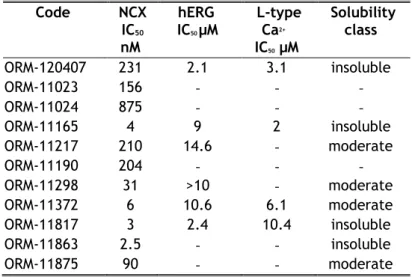
A novel and unique chemical series was discovered using ligand-based pharmacophores for virtual screening with Catalyst (Accelrys) (Figure 1A). Pharmacophore features used for virtual screening were derived from previously discovered NCX1 inhibitor flavan structures, such as ORM-10103 (Koskelainen et al., 2003) and ORM-10962 (Otsomaa et al., 2004). Based on the results of virtual screening, a proposed library with 636 commercially available compounds was selected for testing in the fluorescence-based assay, at concentrations of 10 µM. The original hit ORM-120407 inhibited NCX1.1 by 87% and had an IC50 value of ~200 nM. It inhibited hERG and L-type Ca2+ channels, with IC50
values of 2.1 µM and 3.1 µM, respectively. These results indicated that the scaffold exhibited
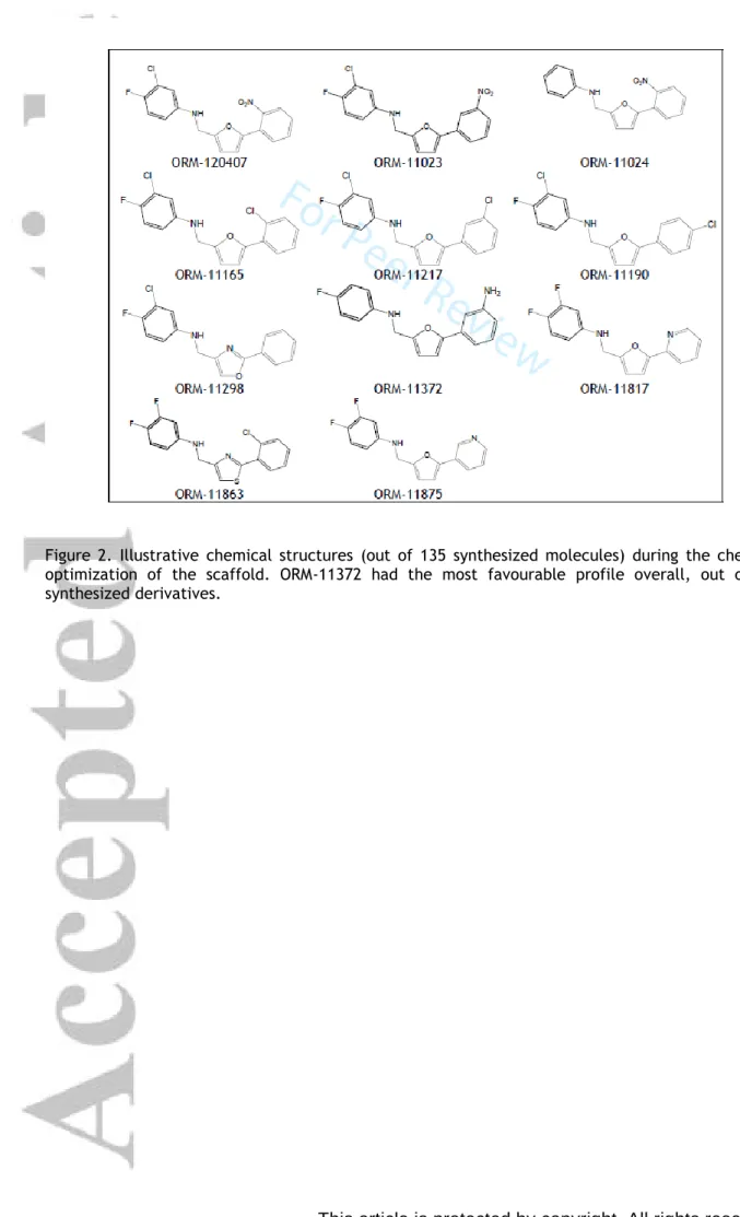
optimization potential. The SAR of ORM-120407 was explored; thus, ORM-11372 (Table 1 and Figure 2) was discovered during medicinal chemistry optimization.
An analysis of 250 previously discovered NCX inhibitor compounds by Orion (Koskelainen et al., 2003;
Otsomaa et al., 2004) resulted in a 5-feature pharmacophore, which was used for the virtual screening of Chambridge and Spcs compound libraries. In-silico hits were further filtered based on predicted activity, clogS, clogP, and diversity. The substructure features were optimized in parallel processes and beneficial structural features were merged, in order to identify the potential overall synergistic effects for NCX1.1 inhibition. Representative samples of 135 synthesized derivatives are presented in Figure 2.
The hydrogen bond donor property of aniline in the original hit molecule ORM-120407 was proven to be important for NCX1.1 activity in the scaffold. The addition of polar substituents was not tolerated in the A ring, but halogen substitution on the p- and m- positions were tolerated, and resulted in improved NCX1 inhibition. However, neither position was favoured over the other, and none showed synergistic or additive effects. Their selectivity towards the hERG channel was found to be its differentiating property.
SAR tolerated 5-membered B-ring systems better than 6-membered systems. Ring heteroatoms i.e.
oxygen or nitrogen molecules at position one (of furan) in combination with a 1-, 5- substitution was proven to be critical for binding, i.e. a carbon at position 1 abolished activity. This finding indicates that the presence of a hydrogen bond acceptor at that position is important. B-ring optimization provided three potential ring systems that were tolerated; furans, oxazoles, and thiazoles. Two latter systems tolerated 4,2- and 2,4- substitutions, while the three heteroatoms in the B-ring reduced NCX1 inhibition activity. Though ORM-120407 showed good inhibitory activity towards NCX1, its selectivity towards hERG (2.1 µM) and L-type calcium (3.1 µM) channels needed to be improved. ORM- 120407 also had poor solubility (<10 µg/ml). ORM-11298 exhibited better inhibitory activity towards NCX1 and good selectivity towards hERG channels. The oxazole series also exhibited good selectivity towards L-type calcium channels such as ORM-11298, but many derivatives were chemically unstable.
The substitution of the C-ring phenyl was unnecessary, but some substituents such as aniline and chlorine were tolerated. The replacement of the phenyl ring with different heteroaromatic ring systems was mainly tolerated.
Synthesis
Most compounds of were prepared via reductive amination (Figure 1C, route 1), except for ORM- 11298 and ORM-11863, which were prepared by aniline alkylation (Figure 1C, route 2). The reaction between the aniline and carbaldehyde derivatives could occur in the presence a strong acid at elevated temperatures. The reduction of the imine intermediate could be carried out using a suitable reducing agent, such as NaBH4. The alkylation reaction in route 2 is performed in the presence of a base. The products were isolated from the reaction mixture by extraction with ethyl acetate, followed by evaporation. The starting materials used in the processes were either commercially available or could be prepared via synthetic routes (Parry et al., 2003; Ye et al., 2010). ORM-11372
was synthesized via the reductive amination of 5-(3-nitrophenyl)furan-2-carbaldehyde with the corresponding fluoroaniline, followed by the hydrogenation of the nitro group under mild conditions.
Details of experiments can be found in the supplementary material (Figures S1-S14).
Cell lines and cell culture
Spodoptera frugiperda (Sf9; RRID:CVCL_0549) cells are widely used for the transient and stable expression of recombinant proteins. Here Sf9 cells were stably transfected with human NCX 1.1 and maintained in spinner flasks with TNM-FH insect medium, supplemented with 10 % foetal bovine serum, Antibiotic-Antimycotic solution, and 50 µg/ml blasticidin at 28 oC in a non-humidified, CO2- free atmosphere. The Sf9 cell suspension was subcultured thrice a week.
Human induced pluripotent stem cell (iPSC)-derived cardiomyocytes (Cor.4U cardiomyocytes from Ncardia, Germany; RRID:CVCL_Y550) were thawed, seeded onto gelatine coated coverslips, and maintained, as described in the manufacturer’s protocol. Cor4U cells express cardiac proteins including NCX1 and exhibit the relevant electrophysiology, demonstrating a potential to model human cardiac responses to drugs (Blinova et al., 2017; Huo et al., 2017).
Chinese hamster ovary (CHO) cells stably expressing either hERG1a (KCNH2; RRID:CVCL_H512; from Sophion Biosciences, Denmark) or human 5-HT2B receptors (from Euroscreen, Belgium) were cultured at 37 oC in a 5 % CO2/95 % air atmosphere in the HAM F-12 medium supplemented with 10 % foetal bovine serum (heat inactivated), 100 μg/ml hygromycin B (Invitrogen), and 100 μg/ml geneticin or 100 IU mL-1 penicillin and 100 IU mL-1 streptomycin, 25 mM HEPES, 500 μg/ml geneticin, and 250 μg/ml Zeocin®. Adhered cells were detached using either Detachin® solution or trypsin and replated twice a week. HEK 293 (RRID:CVCL_0045) and IMR-32 (from American Type Culture Collection, CCL- 127; RRID:CVCL_0346) were cultured similarly, but maintained instead in Dulbecco’s modified Eagle’s medium or Eagle’s minimum essential medium, supplemented with 10 % foetal bovine serum (heat inactivated), 100 IU mL-1 penicillin and 100 IU mL-1 streptomycin, and 25 mM HEPES, and in the case of the IMR-32 cell line, with MEM non-essential amino acids. IMR-32 cells are derived from a human neuroblastoma and endogenously express L-type calcium channels (Sher et al., 1988).
The IhERGcells to be studied were harvested with Detachin solution and either diluted with a volume of CHO-cell serum-free media (supplemented with 100 IU mL-1 penicillin/streptomycin and 25 mM HEPES) to obtain cells, for which the cell density was ~4 million cells mL-1 or plated on glass coverslips and used on the following 1-2 days. HEK 293 cells were transiently transfected using lipofectamine (Invitrogen, USA), with an hSCN5A (transcript variant 2)-containing plasmid, in Optimem medium. After a 5-hour incubation period, the transfection medium was replaced with the normal growth medium. The cells were harvested by trypsinisation, centrifuged, and resuspended in the appropriate extracellular solution for performing studies the next day. CHO-5-HT2B cells were plated into 96-well plates on the previous day, at a density of 40 000 cells/well, with a modified version of the growth media.
Rat ventricular cardiomyocytes
Ethical statement. All experiments were in compliance with the Guide for the Care and Use of Laboratory Animals (USA NIH publication No 85–23, revised 1996), and were approved by the Csongrád County Governmental Office for Food Safety and Animal Health, Hungary (approval No.:
XIII/1211/2012). The ARRIVE guidelines were adhered to during the study (NC3Rs Reporting Guidelines Working Group, 2010).
Animals, housing, and husbandry. Six-week old male Wistar rats (200-250; RRID:RGD_13508588, obtained from a licensed supplier Toxi-coop Ltd. Hungary) were used in the study. The rats were maintained in standard rat cages (380 mm X 270 mm X 200 mm; ~1025 cm2). The number of cage companions was 4 animals/cage. Cages were equipped with external bottle-top type lids with half- pocket wire bar lid feeders. The bedding of the cage floor was composed of aspen chips (Innovo Ltd.
Hungary). The room temperature of the animal house was kept constant at 23 °C, with a humidity of 40-65%. Twelve hours of dark-light cycle was applied with a low light intensity. Food (obtained from Innovo Ltd. Hungary) and tap water were provided ad libitum to the animals. The tap water is regularly checked for any pathogens.
Cell preparation: Rats were anaesthetised with sodium thiopental (0.1 g/kg, i.p.) and injected with heparin sodium (500 IU i.v.). Hearts were rapidly excised, mounted via the aorta on a Langendorff apparatus, and retrogradely perfused at 37 oC with the Krebs-Henseleit solution for 5 min. This solution contained (in mM) 118.5 NaCl, 4 KCI, 2 CaCl2, 1 MgSO4, 1.2 NaH2PO4, 25 NaHCO3, and 11.1 glucose with a solution whose pH was maintained at 7.4, when saturated with a mixture of 95% O2 and 5% CO2. The perfusion of the heart was continued using the Ca2+-free Krebs-Henseleit solution for 10 min, and completed by the addition of 0.05% collagenase (type I), 0.05% hyaluronidase, and 200 µM CaCl2, for a further 10 min. Subsequently, the left ventricular myocardium was minced and gently agitated. Dissociated cells were stored at room temperature in a solution containing (in mM) 89 KOH, 70 glutamate, 15 taurine, 30 KCI, 10 KH2PO4,10 HEPES, 0.5 MgCl2, 11 glucose, and 0.5 EGTA and the pH was set to 7.3 using KOH. Cells were rod shaped and showed a clear striation, when external calcium levels were restored. One drop of the cell suspension was placed in a recording chamber, mounted on the stage of an inverted microscope (Olympus IX51, Olympus, Japan), and the individual myocytes were allowed to settle and adhere to the bottom of the chamber for at least 5 minutes before initiating superfusion.
Fluorescence screening assay for hNCX 1.1 inhibition
NCX reverse mode activity was stimulated via a 50% dilution of the extracellular Na+ level using the internal pipettor of FLEXstation, a fluorescence imaging plate reader (Molecular Devices, USA), while simultaneously monitoring intracellular calcium levels. After dilution concentration of other ions remained the same, but Na+ concentration was diluted to the half of original concentration of 172 mM to 86 mM. For Sf9 insect cells normal extracellular Na+ ion concentration is 172 mM. Typically, 50 µl of a solution containing 150000 – 200000 Sf9-NCX cells was loaded onto each well of a 96-well plate. Cells were preincubated with the intracellular calcium dye Fluo-4 and 6, and the concentrations of the test compound (3 replicates) were determined for about 1 h at room
temperature before experimentation. The extracellular solution contained (in mM) 172 NaCl, 10 HEPES, 1 CaCl2, 1.2 MgCl2, 0.33 NaH2PO4, 5 glucose, and 5 probenecid. The pH was adjusted to 7.4 with NaOH. The extracellular Na+ was diluted by the addition of a Na+-free extracellular solution (containing in mM: 147 NMDG, 10 HEPES, 1 CaCl2, 1.2 MgCl2, 0.33 NaH2PO4, and 5 glucose with the pH adjusted by HCl to 7.4) induced robust and reproducible intracellular calcium elevations. The osmolarity of both solutions was adjusted to 340 – 355 mOsm in order to match the Sf9 cell media osmolarity. In each plate, the IC50 determination for each test compound was based on relative fluorescence changes in comparison to control and 5 mM nickel acetate-induced NCX inhibition.
Confirmatory fluorescence assay for hNCX 1 inhibition
A confirmatory hNCX1 inhibition assay was performed in Charles River Laboratories Cleveland. Briefly HEK293 cells stably expressing hNCX1 were plated in 384-well black wall, clear bottom microtiter plates and the next day loaded with Fluo-8 for 30 min at 37 ºC. A plate was inserted into a FLIPRTETRA and a baseline was recorded during a pre-incubation period where test compound or vehicle in a Na+- free HB-PS containing thapsigargin (6 µM) and FCCP (30 µM) was added to each well for ~5 min. Next the NCX1 stimulation period was recorded in which Na+-containing HB-PS (with 2 µM thapsigargin and 10 µM FCCP) was added. The experiments were performed at room temperature. The kinetic data generated was reduced for each well to maximum relative fluorescence units (RFU) minus minimum RFU after subtracting bias based on the first sample. The mean of the max-min RFU values during the stimulation period for the 3-4 replicates at each concentration on a plate were then plotted versus concentration and fitted to a Hill equation.
Fluorescence secondary screening assays
L-type calcium channel inhibition. Undifferentiated IMR-32 cells were harvested, centrifuged and resuspended in a probenecid ringer consisting (in mM) 150 NaCl, 3 KCl, 1.2 MgCl2, 1 CaCl2, 20 HEPES, 5 glucose, and 2.5 probenecid (pH 7.4 adjusted with NaOH, osmolarity 320-324 mOsm). We added 0.04% Pluronic F-127 and Fluo-4 to this solution, and after incubating the cells for 30 min at room temperature, these additives were removed from the probenecid ringer via centrifugation and resuspension. The cell suspension was pipetted (250,000 cells in 75 μl per well) into a 96-well plate, into which the test compound (75 μl per well at 2,667 x the final concentration; 5 concentrations with 8 replicates) had already been added. The plate was centrifuged once (5 min, 1200 rpm) to ensure that the cells were moved to the bottom of the wells. FLEXstation was then used to measure the increase in intracellular calcium at 37 ºC, following depolarisation, which was induced by the addition (50 μl per well) of a KCl ringer containing (in mM) 200 KCl, 20 CaCl2, 1.2 MgCl2, 20 HEPES, and 2.5 probenecid, respectively (pH adjusted to 7.4 with KOH, with an osmolarity of 320-324 mOsm).
Serotonin 2B receptor. Changes in the intracellular calcium concentration of CHO-5-HT2B cells in the probenecid ringer were measured, using Fluo-4 or Calcium 3 dyes and the FLEXstation, at 37 ºC.
ORM-11372 was applied at 7 concentrations (4 replicates), 10 s after the initiating the measurement process in the agonist assay, but was preincubated (at 8 concentrations with 3 replicates) in the antagonist assay, with 10 nM 5-HT added at the 10 s time point.
Electrophysiology
Manual patch-clamp recordings15 of membrane currents (INCX, ICaL, INa and IhERG) were undertaken using either an Axopatch 200B or Axopatch ID amplifier (Molecular Devices, USA), using the whole cell configuration. Cells were at least initially (except when recording INa) perfused with an extracellular solution consisting (in mM) 143 NaCl, 4 KCl, 1.8 CaCl2, 1.2 MgCl2, 5 glucose, and 10 HEPES (pH 7.4 with NaOH; osmolarity adjusted to 301 ± 3 mOsm). A slightly modified version was used for the rat ventricular cardiomyocytes (i.e.in mM: 144 NaCl, 0.4 NaH2PO4, 4.0 KCl, 1.8 CaCl2, 0.53 MgSO4, 5.5 glucose, and 5.0 HEPES). Patch pipettes were pulled with a P-2000 or P-97 micropipette puller (Sutter Instruments, USA) from borosilicate glass capillaries and had resistances of between 2 and 4 M when filled with any of the pipette solutions (see below). The data was filtered at 1 or 2 kHz and digitised at 10 kHz with acquisition and analysis performed by use of pClamp software (versions 8-10; Molecular Devices; RRID:SCR_011323).
Measurement of NCX current (INCX). After the establishment of the whole cell configuration with hiPSC derived cardiomyocytes or rat ventricular myocytes the extracellular solution was switched to a K+-free bath solution as described earlier (Hobai et al., 1997; Jost et al., 2013). The solution was composed (in mM) 135 NaCl, 10 CsCl, 1 CaCl2, 1 MgCl2, 0.2 BaCl2, 0.33 NaH2PO4, 10 TEACI, 10 HEPES, and 10 glucose supplemented with 20 µM ouabain, 50 µM lidocaine, and 1 µM nisoldipine or 2 µM nitrendipine at pH 7.0 (osmolarity adjusted to 301 ± 3 mOsm). The pipette solution contained (in mM) 140 CsOH, 75 aspartic acid, 20 TEACI, 5 MgATP, 10 HEPES, 20 NaCl, 20 EGTA, and 10 CaCl2 (pH adjusted to 7.2 with CsOH; osmolarity adjusted to 290 ± 2 mOsm). The free intracellular Ca2+ level in the patch pipette was 140 nM according to Maxchelator (RRID:SCR_000459) software (Bers et al., 2010).
The measurement of current in the K+-free bath solution using a ramp voltage protocol at 20 s intervals represented the first control, after which values were measured in the presence of one or more increasing concentrations of ORM-11372, and finally upon exposure to 10 mM NiCl2. The voltage protocol consisted of voltage ramps (at a rate of 100 mV/s) with a holding potential of -40 mV to 60 mV, which changed to -100 mV and then reverted back to -40 mV. INCX was defined as the Ni2+-sensitive current value; however, for rat ventricular myocytes, the magnitude of the inward current (forward mode) was particularly small and variable. Therefore, to enhance the inward current and enable a solution with a certain concentration of ORM-11372 to be measured, the concentrations of the K+- free and pipette solutions were altered. In the bath solution, the CaCl2 concentration was reduced to 0.5 mM, while in the pipetted solution, the NaCl and EGTA concentrations were decreased to 5 and 10 mM, respectively. All experiments were performed at 35-37 oC. The outward human INCX inhibition current was measured at the following ORM-11372 concentrations: 3 (n = 4), 10 (n = 6), 30 (n = 4), and 100 (n = 3) nM. The concentrations used for human inward INCX current measurement were 3 (n = 4), 10 (n = 4), 30 (n = 3), and 100 (n = 2) nM. The outward INCX current in rat cardiomyocytes was measured at concentrations ranging from 1 to 1000 nM (n = 5). The inward INCX current in rats was measured in additional experiments under changed conditions, to enhance the inward INCX
current. In these experiments (n = 3), the effect of ORM-11372 was tested at a concentration of 10 nM, at which the IC50 and reverse INCX current values were approximately equal.
Measurement of L-type calcium current (ICaL). ICaL was recorded from hiPSC-derived cardiomyocytes (at room temperature, n = 2) and rat ventricular myocytes at 1 µM (n = 3; 5 cells) and at 10 µM (n = 4; 6 cells) in an extracellular solution supplemented with 3 mM of 4-aminopyridine at 37 oC. The pipette solution used for hiPSC-derived cardiomyocytes consisted (in mM) (pH 7.2; osmolarity adjusted to 293 mOsm) 110 KCl, 40 KOH, 20 TEACI, 3 MgATP, 10 EGTA, and 5 HEPES. The composition of the pipette solution used for rat cardiomyocytes was (in mM) 125 CsCl, 20 TEACI, 5 MgATP, 10 EGTA, and 10 HEPES; the pH was adjusted to 7.2 using CsOH. ICaL was evoked by a 400 ms depolarization process to 0 mV from a holding potential of -40 mV every 5 s, or in the case of the rat cardiomyocytes, by 400 ms; depolarizations to potentials ranging from -35 mV to 55 mV occurred after a prepulse to -40 mV, from the holding potential of -80 mV.
Measurement of NaV1.5 current (INa). Recordings were performed on HEK cells (n = 3) that transiently expressed SCN5A at room temperature, in a bath solution containing (in mM) (pH 7.4 with N-methyl- D-glucamine; osmolarity adjusted to 300 ± 2 mOsm) of 40 NaCl, 97 L-aspartic acid, 4 KCl, 1.8 CaCl2,
1 MgCl2, 10 glucose, and 10 HEPES. The pipette solution contained (pH 7.2 with CsOH; osmolarity adjusted to 270 ± 3 mOsm) 130 caesium methane sulfonate, 5 MgCl2, 5 EGTA, 0.1 GTP, 4 Na2ATP, and 10 HEPES. The voltage protocol, which was repeated after each second, included of the process of hyperpolarisation, from a holding potential of –80 mV to -120 mV for 200 ms, followed by that of depolarisation to -15 mV for 10ms. The peak current values observed while applying the test pulse at -15 mV were used for analysis.
Measurement of KV11.1 current (IhERG). Values from hERG expressing cells (n = 4) were recorded with the standard extracellular solution at the physiological temperature and a pipette solution containing (in mM) 130 KCl, 7 NaCl, 5 EGTA, 1 MgCl2, 5 Na2ATP, and 5 HEPES (pH was set to 7.2 with KOH; osmolarity was adjusted to 290 ± 3 mOsm). IhERG was evoked using a voltage protocol, which was repeatedly performed every 10s, consisting of a depolarisation step from the holding potential of –75 mV to 10 mV for 500 ms, followed by a repolarisation step to –40 mV for 500 ms. The peak tail current values at –40 mV were used for analysis.
In addition, after loading the CHO-hERG cell suspension, whole cell voltage-clamp values were recorded at room temperature on a QPatch 16x automated patch-clamp (Sophion Biosciences), in the single-hole mode. ORM-11372 solutions with concentration of 0.3 (n = 4), 1 (n = 5), 3 (n = 8), 10 (n = 8), and 30 µM (n = 8) were studied. The extracellular solution contained (in mM) 145 NaCl, 4 KCl, 2 CaCl2, 1 MgCl2, 10 glucose, and 10 HEPES (pH 7.4 with NaOH; osmolarity adjusted to 305 mOsm). The intracellular recording solution contained (in mM) 120 KCl, 1.75 MgCl2, 5.37 CaCl2, 4 Na2ATP, 10 EGTA, and 10 HEPES (pH 7.2 with KOH; osmolarity adjusted to 295 mOsm).The voltage protocol, which was repeated every 10 s, included a 200 ms step to change the holding potential from –80 to -50 mV, to measure the leak current, and further depolarization to + 20 mV for 2 s, followed by a repolarization to –50 mV for 2 s. The hERG tail current was measured as the difference between the peak tail current amplitude during the repolarization step and leak current measurement step. Subsequently, the
voltage-dependence of the block was also assessed, by carrying out depolarization for 4 s, from –80 mV to +60 mV in 10 mV steps, before and after the addition of 20 µM ORM-11372 (n = 7). The peak tail current elicited during the 5 s repolarization step to –50 mV was measured and plotted against the preceding depolarization step voltage values.
Action Potentials. Action potentials were recorded in spontaneously beating Cor.4U cardiomyocytes in the current-clamp using the perforated patch technique and voltage-sensitive dye (di-4-ANEPPS), in a 96-well plate format. The latter ratiometric optical measurements were performed on the CellOPTIQ platform at Clyde Biosciences (Newhouse, UK). The patch-clamp measurements used the standard extracellular solution at the physiological temperature and a pipette solution containing (in mM) 122 K-gluconate, 30 KCl, 1 MgCl2, and 5 HEPES (pH 7.2 with KOH; osmolarity adjusted to 290 ± 3 mOsm), and 0.24 mg/ml of amphotericin B. The patch-clamp data shown is from cardiomyocytes (n = 4) that exhibited ventricular-like action potentials, i.e. a ratio (time difference between APD30
and APD40/time difference between APD70 and APD80) >1.5 (Ma et al., 2011).
Selectivity Panel
. Radioligand binding assays were performed at Eurofins Cerep SA (Celle L’Evescault, France), for testing the inhibition capacity of ORM-11372 (at 10 µM with 2 replicates), in over 75 targets. These included receptors (e.g. human A1, A2A, and A3 adenosine receptors; α1 and α2 adrenergic receptors; human β1-β3 adrenergic receptors; human D1, D2S,D3, D4.4, and D5 dopamine receptors; EGF and VEGF receptors; human M1-M5 muscarinic acetylcholine receptors; human 5-HT1A, 5-HT2A, 5-HT2B, 5-HT2C, 5-HT4e, 5-HT5A, 5-HT6, and 5-HT7 serotonin receptors), ion channels (5-HT3, Ca2+, KATP, KV, SKCa, Na+, and Cl−), and transporters (adenosine, norepinephrine, dopamine and serotonin). Follow-up functional studies (with 5 concentrations and 2 replicates) were performed for 5-HT2A and the norepinephrine transporter. Agonism and antagonism at h5-HT2A was explored in recombinant HEK 293, and intracellular calcium changes were detected using fluorimetry.Norepinephrine uptake was measured by carrying out scintillation counting of [3H]NE incorporated into rat hypothalamus synaptosomes.
Design and analysis for isolated heart preparations and in vivo experiments
Donor Heart Procurement: All the human hearts used for this study were obtained after legal consent was provided by organ donors in the US. The policies for donor screening and consent were the same as those established by the United Network for Organ Sharing (OPTN, 2019). Organizations supplying human tissues to AnaBios follow the standards and procedures established by the US Centers for Disease Control, and are inspected biannually by the Department of Health and Human Services.
Tissue distribution is governed by internal IRB procedures and was compliance with HIPAA regulations (Edemekong et al., 2019) regarding patient privacy. All organ donor transfers to AnaBios are fully traceable and periodically reviewed by US Federal authorities. In general, AnaBios obtains donor hearts from adults aged 17–60 years old. Though some donors were trauma victims, donors with the following conditions were excluded: ejection fraction < 45%, HIV, cardiac death, HBV, congenital LQT syndrome, HCV, LOT syndrome, MRSA, downtime >20 min, ongoing infections, positive blood cultures without treatment and 48-h result data. Donor hearts from 1 male and 1 female (both 57 years old,
Table S5) were harvested using AnaBios’ proprietary surgical techniques and tools, and were shipped to AnaBios via dedicated couriers. Upon arriving at AnaBios, each heart was assigned a unique identifier number that was reproduced on all relevant medical history files, data entry forms, and electronic records.
Recording of Action Potentials in Human Ventricular Trabeculae: Procedures used for tissue dissection and recording were similar to those described previously (Page et al., 2016). Briefly, the human heart was transferred into a dissection vessel containing a cold (4°C), fresh proprietary dissection solution. The heart was completely submerged into the dissection solution. Ventricular trabeculae were dissected and transferred to the recording chamber. The approach used to record action potentials (APs) is similar to that described by Page et al (Page et al., 2016). Briefly, a single tissue was mounted into the experimental chamber filled with oxygenated Tyrode’s external solution, containing (in mM) 136 NaCl, 4 KCl, 0.5 MgCl2, 12 NaHCO3, 0.35 NaH2PO4, 11.1 dextrose, 1.8 CaCl2, and 10 HEPES (pH 7.4). The temperature of the solution was maintained at 37°C, at a flow rate of 5 mL per minute. The tissue was allowed to equilibrate for 30-60 min, while providing stimulation (3 V, 3 ms) at a frequency of 1.0 Hz. High impedance borosilicate microelectrodes were prepared with a tip resistance of 10-20 MΏ, filled with 3 M KCl. Upon tissue impalement, the membrane potential was allowed to stabilize (typically, around -85 mV). Tissues with resting membrane potentials more positive than −75 mV were rejected. Bipolar stimulation at 1.5x threshold was applied, and recordings were obtained in the continuous mode with sampling at 20 kHz, using ADInstruments and LabChart Software. Tissue exclusion criteria included the following: (1) interruption of perfusion/oxygenation;
(2) absence of APs following stimulation at baseline; (3) time frame of drug exposure not respected;
(4) unstable response to stimulation at baseline; (5) resting membrane potential (RMP) > −75 mV; (6) maximal amplitude of AP (AMAX) < 70 mV; and (7) AP duration at 90% repolarization (APD90) < 200ms or >450 ms. ORM-11372 was evaluated at 3 concentrations in 4 ventricular trabeculae derived from 2 donor hearts (n=2, two replicates). Testing concentrations were 0.1, 1, and 10 mM. Following the stabilization of each tissue, APs were collected and assessed for 31 min in the vehicle control solution (Tyrode with 0.1% DMSO), at stimulation frequencies of 1 Hz for 25min, 2Hz for 3 min, and 1Hz for 3 min. Following this vehicle control period, 3 concentrations of ORM-11372 were applied sequentially and cumulatively. Each concentration was applied for 31 min with the same stimulation sequence as in the vehicle controls.
Animal experimental design and analysis
All animal experiments were performed according to European Community Guidelines for the use of experimental animals and approved by the Finnish National Animal Experiment Board. All studies complied with the ARRIVE guidelines (NC3Rs Reporting Guidelines Working Group, 2010).
Selection of animal species: The rat is the most widely used rodent species in toxicology studies performed for drug development. The Sprague-Dawley rat strain (RRID:RGD_70508) is commonly used in the rat myocardial infarction (MI) model (Fishbein et al., 1978). However, the functional similarity between the Ca2+ handling proteins (including NCX) in rats (Bassani et al., 1994) and human hearts is rather low. The Ca2+ circulation balance in the guinea-pig and rabbit hearts are more similar to that
of a human (Milani-Nejad et al., 2014). Rabbits and humans are also known to react similarly to medication; hence, the rabbit was selected as a nonrodent species.
Housing and husbandry: Animals were monitored daily by laboratory personnel. If the general health status of an animal was significantly worsened, the animal was sacrificed with an overdose of pentobarbital. Human endpoints included no spontaneous movements and inability to drink or eat during the 24-h observation period, massive bleeding, spontaneous inflammation, missing anatomical features or swelling, and breathing difficulties. Specific pathogen-free animals were housed in half- barrier rooms where special protective clothing was required by personnel. Animal rooms were cleaned regularly 3 times per week and cages and bottles were changed at regular intervals once a week. The temperature was maintained at 22 ± 2°C and humidity at 55 ± 15%. In the light-dark cycle, lights were kept on from 06.00 to 20.00 h. Body weights of all animals were measured weekly.
Randomization and blinding: Randomization was used whenever feasible. Guinea-pigs were randomized into vehicle and ORM-11372 groups for in vitro contraction force measurement from papillary muscle. In hemodynamic studies involving rabbits, only one study group was used, according to the 3R principle. Because the experimental setup involved ascending doses in one study group, randomization was impossible. Baseline values were used as controls to reduce variability. However, the MI rat model required the inclusion of two study groups, to eliminate the effect of surgical operations. The MI study setup enabled the use of randomization. The study duration for anestetised animals is limited; hence, higher doses had to be administered, which required blinding to be performed in all in vivo studies. The blinding was not considered relevant, because measured absolute values and derivated parameters are not sensitive to biased interpretations.
In vitro contraction dynamics measurement
Guinea-pigs (both sex, body weight ranging 342-565 g, Dunkin Hartley, M&B A/S, Ry, Denmark) were used in the study. The animals were housed in solid bottom polycarbonate cages (Makrolon®
IV, 385 x 590 x 200 mm) with stainless steel wire mesh lids, up to three animals of same sex per cage. Autoclaved aspen chips (Tapvei Ky, Kaavi, Finland) were used as bedding. A commercially available rodent SDS FD1 (P) SQC pellet diet (Special Diet Services Ltd, Witham, England) and tap water from the public supply (Espoon vesilaitos, Espoo, Finland) were available ad libitum in polycarbonate bottles (800-1000 ml) with plastic cabs (Scanbur A/S, Ejby, Denmark).
Guinea-pigs will be killed by a blow on the skull and the heart will be excised. Right ventricular papillary muscle will be dissected and rinsed in ice-cold Tyrode solution. Thereafter, the papillary muscle will be mounted for the measurement of twitch tension in an organ bath containing
modified Tyrode solution (at 37°C) bubbled with carbogen (95% O2, 5% CO2). The composition of the Tyrode solution will be the following (mM): 135 NaCl, 1 MgCl2 x 6H2O, 5 KCl, 2 CaCl2 x 2H2O, 15 NaHCO3, 1 Na2HPO4 x 2H2O, 10 glucose at pH 7.3-7.4.
The signal was acquired with a validated acquisition system ACFO v1.0 (Fision Oy, Finland). The following parameters were measured: twitch tension (Tw), rest tension (RT), time to peak tension
(TTP), and half relaxation time (HRT). At steady state, 15 twitch tensions were acquired and averaged was used for statistics.
The baseline twitch tensions were measured after a stabilization period of 20-40 min. Thereafter, vehicle (1.1% DMSO) or ORM-11372 in the Tyrode solution were superfused into the cuvette for 15 min and the effects were measured from the steady state level. All experiments were carried out at 37°C.
Experimental of myocardial infarction model
Male Sprague-Dawley rats (Harlan, Netherlands B.V.) were housed in polycarbonate cages (Makrolon®
IV with stainless steel wire mesh lids). A maximum of 5 rats were housed per cage with aspen chip bedding (Tapvei Ky, Kaavi, Finland). Rats had free access to water (twice filtered, Espoon Vesi) and a rodent diet (SDS RM1 (P) SQC pelleted diet, Special Diet Services Ltd, Witham, England). The acclimation period before experiments was at least 5 days. In the myocardial infarction model, rats (7-8 weeks, 200-250 g) were randomised into either the myocardial infarction or sham groups. In this weight range, a rat is considered a young adult, when its evolution phase is characterized by slow growth and its surgical mortality is lower than that of older animals. Male rats were used to minimize the variability of the cardiac response to several stimuli (Zornoff et al., 2009).
Rats were anesthetized with a combination of ketamine (Ketalar® 50 mg/kg i.p.) and medetomidine (Domitor 250 µg/kg i.p.), intubated, and artificially ventilated with air (Ugo Basile 7025). A myocardial infarction (MI) was induced by the occlusion of the left coronary artery under aseptic conditions (Levijoki et al., 2001; Selye et al., 1960). The left coronary artery was ligated at a distance of about 2-3 mm from the origin of the aorta with a silk suture and the heart was repositioned into the chest. After ligation, the wound was closed carefully. The muscle layer was closed with soluble stitches and skin layers with insoluble stitches, followed by a partial reversal of anesthesia via the intraperitoneal administration of atipamezole (Antisedan® 0.3 -1 mg/kg i.m.). The trachea tube was removed, and the trachea and underlying muscle and skin layers were closed immediately. To prevent dehydration, rats were subcutaneously administered 5 ml of 0.9% NaCl. After surgery, rats were pain- medicated with buprenorphine for at least two days (0.05 mg/kg s.c. twice a day). For sham-operated rats, the same procedure was performed, without ligation. Rats were analysed for 7 days after the myocardial infarction (MI), for severe acute heart failure development e.g. oedema or breathing difficulties.
Hemodynamic measurements of anesthetized animals
All animals were administered multiple ascending infusion doses of ORM-11372; blinding was not possible. Systemic blood (SP) and left ventricular pressure values (LVPs) were measured with pressure transducers (SP for femoral artery: Isotec, Hugo Sachs Elektronic, Germany; a miniature pressure transducer Mikro-Tip transducer SPR-249, Millar Instruments was inserted into the left ventricle via the right carotid artery). The signal was amplified (DC-bridge amplifier type 660, Hugo Sachs Elektronik, Germany), digitised (I/O connector block type SCB-68, National Instruments, USA),
recorded, and analysed (IHME 1.0.9, Fision Ltd, Finland). Left ventricular inotropic effect (LV +dP/dtmax), relaxation (LV -dP/dtmin), and heart rate (HR) values were analysed from LVP signals.
At the end of the hemodynamic experiments, rats and rabbits were killed with an overdose of pentobarbital. Small laboratory equipments (forceps, scissors, scalpels, etc.) were sterilized in the glass bead sterilizer at 300°C for 10 sec (10 sec).
The blood pressure waveform was measured continuously in milliseconds. The blinding was not considered relevant, because the absolute value was not sensitive to biased interpretations, and all parameters are derived from of them.
Haemodynamics in MI rats: Haemodynamics were assessed 7 days after a myocardial infarction (n=6) or sham operation (n=6) as follows. Rats were anesthetized with isoflurane (2.25-2.5 %, Baxter) in carbogen (95 % O2 and 5 % CO2) and nitrous oxide (1:1), using a small rodent ventilator (Ugo Basile 7025, ~10 ml/kg, 60 strokes/min). The rats were infused with 0.9 % NaCl in the carotid vein at the stabilization and baseline levels. ORM-11372 was administered into MI (n=6) and sham (n=6) rats in ascending infusion doses of 1.7, 17, 167, and 417 µg/kg/min. Infusions were administered into the jugular vein of MI rats at the infusion rate of 5 ml/kg/h (Terumo TE-311, Belgium). Doses were selected based on a pilot study with healthy rats (Figure S20). The total number of animals in the study was 12+2, due to the replacement of two MI rats. One MI rat died while administering anesthesia, as infarcted animals are sensitive to anesthesia, and another MI rat was excluded due to the dosing error indicated by the bioanalysis of plasma samples (no ORM-11372 concentration observed in plasma).
After hemodynamic assessments, the anesthetized rats were killed with an overdose of pentobarbital (2 ml of Mebunat® vet 60 mg-ml per rat). The entire heart of each animal was fixed in buffered 4%
formaldehyde solution, trimmed, processed, and embedded in paraffin. The hearts were cut horizontally at three different levels: apex, mid part, and base. Sections with a thickness of 4 µm were cut from each level stained with haematoxylin and eosin, for general histopathological analysis.
Infarct sizes were determined using picrosirius red staining as an indicator of cardiac fibrosis, and the size was measured using Analysis Pro software. The infarct area was calculated as the percent of fibrotic tissue in the total myocardial area (mean of three levels).
Haemodynamics in rabbits: Male New Zealand white rabbits (Harlan, Netherlands B.V.) were housed individually in polycarbonate cages (Scanbur number 8 plastic cages with stainless steel door) with aspen bedding (Aspen Bricks M, Tapvei Ky, Kaavi, Finland). The acclimation period before experiments was at least 21 days. Rabbits weighing 2.0-2.3 kg (age 10-12 weeks) were used for hemodynamically assessing ORM-11372 (no randomisation, baseline values as own control). The diet provided to rabbits was SDS Stanrab (P) SQC pelleted (Special Diet Services Ltd, Witham, England).
Water was provided ad libitum in polycarbonate bottles (750 ml).
Rabbits (n=5) were sedated with intravenously (marginal vein) injected diazepam (2 mg/kg, Diapam®, Orion Pharma). Anesthesia was inducted with intravenous s-ketamine (10-20 mg/kg Ketanest-S®, Pfizer) and maintained with a s-ketamine intravenous infusion (15-80 mg/kg/h). Animals were placed on a heating table (+38°C) and tracheas were cannulated. Rabbits were ventilated via a rodent
ventilator (Ugo Basile type 7025, Hugo Sachs Elektronik, Germany; respiratory volume 10 ml/kg, 30 strokes per min for rabbits). ORM-11372 was administered into the jugular vein at an infusion rate of 10 ml/kg/h (Terumo TE-311, Belgium), at infusion doses of 17, 167, and 833 µg/kg/min.
Blood sampling for bioanalysis
At the end of each infusion process, blood samples (300-500 µl) added to a chilled EDTA polypropylene tube (CapiJect®, Terumo) and centrifuged (4000 rpm, 10 min, at 4°C). Plasma was immediately frozen in polypropylene tubes and stored at –20°C. ORM-11372 was extracted from plasma samples using a liquid–liquid extraction process and analysed using liquid chromatography–tandem mass spectrometry (Agilent Technologies series 1100 liquid chromatographic system and a Sciex LC-MS/MS API 4000 mass spectrometry). The lower limit of quantification for the plasma concentration of ORM- 11372 was 32.0 ng/ml. Plasma protein binding in ORM-11372 was assessed using the TRANSIL High Sensitivity kit, as per the manufacturer’s instructions (Sovicell, GmbH, Germany).
Materials
Study substance: ORM-11372 was synthetized by Orion Pharma. To perform in vitro experiments, ORM-11372 was dissolved in dimethyl sulphoxide (DMSO), to obtain a 10 mM stock solution. Spiking solutions were prepared daily by diluting the stock solution with DMSO, and used to prepare the final drug solutions with certain concentrations by diluting them with extracellular solutions (the vehicle concentration was either 0.1 or 0.3 %). The solubility of ORM-11372 in saline and 5% glucose solutions used for in vivo dosing were were 5.3 mg/ml and 5.4 mg/ml, respectively. ORM-11372 was soluble in 10 – 100 µg/ml of phosphate buffer (in pH 7.4). ORM-11372 has the following drug like properties:
the number of hydrogen bond acceptors is 1 (<10) and donors is 3 (<5), molecular weight of free base is 282 (<500), and the predicted logP value is less than 5 (ADMET Predictor: 3.95, MOE: 3.413, QikProp:
3.974).
Chemicals: Isoflurane (Forane®, Baxter), atipamezole (Antisedan®, Orion Pharma), medetomidine (Domitor®, Orion Pharma, ketamine (Ketalar®, Pfizer Oy Animal Health, lipofectamine (Invitrogen, USA), hygromycin B (Invitrogen), buprenorphine (Temgesic®, INDIVIOR UK Ltd). All other chemicals and cell culture media were obtained from Sigma-Aldrich.
Cell lines and cell culture: NCX 1.1 was cloned and plasmid was constructed by Orion Pharma Spodoptera frugiperda (Sf9) cells (ATCC), human induced pluripotent stem cell (iPSC) derived cardiomyocytes (Cor.4U cardiomyocytes from Ncardia, Germany), DetachinTM Cell Detachment Solution, Genlantis), HEK 293 cells and IMR-32 human neuroblastoma cells (ATCC), CHO cells hERG1a (KCNH2; Sophion Biosciences, Denmark), CHO cells human 5-HT2B receptors (Euroscreen, Belgium), hSCN5A plasmid (OriGene Technologies Inc., Rockville, MD, USA).
Data and Statistical Analysis
Data Analysis of human trabeculae: For each frequency tested, the last 30 APs acquired at the end of the period were averaged for vehicle controls, for each ORM-11372 solution with a certain concentration. Analysis at 1 Hz included only the last 30 APs from the initial 25 min incubation period.
The following AP parameters and pro-arrhythmia variables were analysed offline upon the completion
of recordings: RMP (mV), AMAX (mV), AP duration at percent repolarization (APD30, APD50, APD90) (ms), short term variability analysis of AP duration (STV), triangulation (APD90-APD30).
STV was calculated as the beat-to-beat variability in repolarization from APD90 Poincare plots over a 30 sec duration. STV for APD90 was calculated as follows: STV= ⅀|APDn+1−APDn|/(30×√2), where APD (n) and APD(n+1) are the APDs for the nth AP and the following AP, respectively.
The effects of ORM-11372 were quantified relative to the data collected during the vehicle control period. Threshold values for changes over the baseline control for APD30, APD50, APD90, triangulation, and STV at 1 and 2 Hz pacing frequencies have been determined in a previous validation study (Page et al., 2016). Results are expressed as mean ± standard error values of the mean (SEM).
The data and statistical analysis of in vitro and in vivo studies comply with the recommendations for experimental design and analysis in pharmacology.(Curtis et al., 2018). Statistical analysis were done only where number of independent samples was 5 or more. A p-value of <0.05 was considered to be significant. Two-way repeated measures ANOVA were used for the analysis of the papillary muscle and myocardial infarction models. One-way repeated measures ANOVA was used for the rabbit hemodynamic study. The Sidak posthoc-test was run only if the F-value was statistically significant, and there was no significant variance in homogeneity. Data is presented as mean ± SEM. Prism 8.0.2 (GraphPad Software Inc., San Diego, CA, USA; RRID:SCR_002798) was used for statistical analysis.
Nomenclature of Targets and Ligands
Key protein targets and ligands in this article are hyperlinked to corresponding entries in http://www.guidetopharmacology.org, the common portal for data from the IUPHAR/BPS Guide to PHARMACOLOGY (Harding et al., 2018), and are permanently archived in the Concise Guide to PHARMACOLOGY 2019/20 (Alexander et al., 2019).
Results
ORM-11372 concentrations dependently inhibited the increase in intracellular calcium in insect cell line expressing human NCX1.1. The insect cell line was used as the screening assay. The IC50 for the inhibition of NCX in the reverse mode was 6.2±0.4 nM.
The potency of ORM-11372 for NCX was subsequently studied using the whole-cell patch-clamp technique in human iPS-derived cardiomyocytes (hiPSC-CMs; Figure 3) and rat ventricular cardiomyocytes (Figure 4). The solutions and experimental protocol used to measure the bidirectional NCX current (INCX) was identical for both preparations. As shown in Figure 3A, the current was recorded in response to repetitive voltage ramp pulses first in a K+-free bath solution after blocking Na+, Ca2+, K+, and Na+/K+ pump currents, to yield the baseline, and during the addition of ORM-11372.
Finally, when the highest concentration of ORM-11372 had reached a steady state, 10 mM NiCl2 was added to completely block INCX. Examples of individual trace currents under different conditions are shown for a hiPSC-CM in Figure 3B and for a rat cardiomyocyte in Figure 4A. ORM-11372 decreased both the outward and inward currents. The IC50 values for the outward and inward INCX (i.e. the Ni2+- sensitive current) of hiPSC-CMs were 4.8 and 5.6 nM, respectively (Figure 3C). In rat primary ventricular cardiomyocytes, the magnitude of outward INCX (the reverse mode) with an IC50 of 11.3 nM (Figure 4B) was reduced in an ORM-11372 concentration-dependent manner. It was difficult to
record inward INCX (the forward mode) from rat cardiomyocytes; hence, the experimental conditions were altered, to enhance its magnitude (see Methods). At 10nM, the approximate IC50 on the outward INCX, ORM-11372 inhibited inward INCX by 53.8±3.9 %.
Comparison with known NCX inhibitors
Further confirmation of the inhibitory effects of ORM-11372 was sought in the hNCX1 fluorescence calcium flux assay available at Charles River. In experimental conditions increasing intracellular calcium (by the presence of thapsigargin and FCCP) and eliciting NCX forward mode activity (switching from Na+-free to Na+-containing buffer), ORM-11372 concentration-dependently inhibited the calcium efflux signal (Figure 5A) with an IC50 of 142 nM and 164 nM (0.3 % and 1 % DMSO respectively; NS). The hNCX1 antagonist of five other compounds was also assessed in this assay.
ORM-11372 was the most potent inhibitor (Figure 5B) followed by ORM-10962 (Figure 5C), ORM-10103, SEA0400, KB-R7943, and lastly SN-6 whose IC50 appears >100 µM.
Selectivity
The functional selectivity of ORM-11372 towards the L-type Ca2+ channel was first tested with the IMR-32 neuroblastoma cell line, using fluorometric images of intracellular changes in calcium levels.
Depolarisation of undifferentiated IMR-32 cells by KCl addition induced an increase in intracellular calcium levels, which can be suppressed by preincubation with verapamil or the 1,4-dihydropyridine nicardipine (Sher et al., 1988). L-type calcium channels mediated changes in intracellular calcium levels, and these were concentration dependently inhibited by an ORM-11372 solution with an IC50 of 6.1 µM. Further electrophysiological studies were then performed with hiPSC-CMs and rat ventricular cardiomyocytes. The inward ICaL was reduced by 1 µM ORM-11372 at 0 mV by 14.1 % in hiPSC-CMs (Figure 6A), and a 7.8 % inhibition was observed in rat cardiomyocytes (Figure S16A-B; Individual inhibition values at 1 and 10 µM are shown in Table S3 and S4, respectively). However, when rat ventricular cardiomyocytes were exposed to ORM-11372 at a concentration of 10 µM, ICaL was significantly decreased (Figure S16C). ICaL inhibition exhibited voltage dependency, and was greater (59-69 % versus 36-39 %) at more negative activating test potentials (-35 to -20 mV versus 0 to 40 mV).
The effect of ORM-11372 on other cardiac ion channel currents were investigated. The reduction in the human KV11.1 current (IhERG) was initially examined using stably expressing cells on an automated patch-clamp instrument, and then with the manual patch-clamp. ORM-11372 concentration dependently inhibited IhERG (Figure 6B); IC50 values were 19.2 and 10.0 µM, using the automated and manual patch-clamp methods, respectively (Figure S15A). ORM-11372 significantly altered the voltage dependency of the IhERG activation curve (Figure S15C) and displayed significant voltage dependent blockage. While IhERG inhibition was minimal at –30 and –20 mV, inhibition increased markedly at –10 and 0 mV, and reached a steady state between 10 and 60 mV, i.e. at potentials where the channels were fully activated (Figure S15D). Additionally, ORM-11372 concentration dependently inhibited human the NaV1.5 current (Figure 6C), with an IC50 value of 23.2 µM.
ORM-11372 increased twitch tension by 146 ± 32% vs. 7 ± 5% in vehicle group (Figure 6D), but did not increase rest tension (-8 ± 2% vs. -10 ± 1% in vehicle, Figure 6E), did not affect time to peak (-4
± 1% vs. -5 ± 2% in vehicle, Figure 6F) nor half relaxation time (8 ± 9% vs. 11 ± 4% in vehicle, Figure 6G).
The selectivity profile of ORM-11372 was additionally evaluated in binding assays for a diverse panel of targets. While minor effects such as those for the Na+/K+-ATPase or ryanodine receptor (11 % and 15 % inhibition) were seen, with a screening concentration of 10 µM, specific ligand binding inhibition was significant(i.e. >50 %) at the peripheral benzodiazepine receptor, imidazoline I2 receptor, serotonin 2A and 2B receptors, and norepinephrine transporter. The latter three targets were studied functionally. The uptake of norepinephrine into rat hypothalamus synaptosomes was inhibited by ORM-11372 in a concentration-dependent manner; the IC50 value was 1.7 µM. Neither agonism nor antagonism was seen with ORM-11372 (maximum concentration: 10 µM) at 5-HT2A. Similarly, no agonism was evident at 5-HT2B; however, 10 µM ORM-11372 moderately inhibited (38 %) the 5-HT2B- mediated increase in intracellular calcium evoked by 10 nM serotonin.
Human ventricular trabeculae
ORM-11372 caused no significant changes in APD30, APD50, and APD90 values up to a concentration of 10 µM, at both 1 and 2 Hz pacing frequencies. However, there was a trend in the reduction in APD, particularly APD30, at 1Hz (Figure S17); this seemingly contrasts with the significant shortening of hiPSC-CM APD by ORM-11372 (at 0.1 or 0.3 µM; Figure S18). As expected, the effect of the positive control (0.1 μM dofetilide) was observed in trabeculae tested with ORM-11372 (Figure 7; individual values are shown in Figure S17 (Page et al., 2016; Qu et al., 2017). Moreover, ORM-11372 showed no effects on STV and triangulation, two pro-arrhythmia markers that are used to identify pro- arrhythmic risk (Page et al., 2016; Qu et al., 2017), at a pacing rate of 1 or 2 Hz, at any tested concentration (Individual values in Figure S19). However, the addition of 0.1 μM dofetilide to the trabeculae tested with ORM-11372 increased STV and triangulation (Figure S18). Taken together, these data clearly suggest that ORM-11372 can be classified as devoid of pro-arrhythmic risk.
In vivo haemodynamics in rat myocardial infarction (MI) model
Infarcted areas were 14.5±1.8% and 1.1±0.6% in the MI and sham-operated groups, respectively (Figure S20). Percentage changes are calculated from absolute values. Infarctions caused the baseline of positive inotropic effect and relaxation to be reduced by 38% and 23%, respectively. Heart rates (- 3%) were unaffected, but systemic arterial pressure reduced by 20%.
In MI rats, ORM-11372 improved maximally positive inotropic effect by 18 ± 6% and by 31 ± 6% in sham rats (Figure 8A). Maximal effects were observed at the highest infusion dose of 417 µg/kg/min, corresponding to free plasma concentrations of 6.7 ± 2.5 and 4.9 ± 0.4 nM. At the lowest infusion dose of 1.7 µg/kg/min, ORM-11372 improved relaxation levels in the MI group by 24 ± 2% (Figure 8B).
ORM-11372 maximally reduced the heart rate by 14 ± 4% (SHAM -2 ± 2%; Figure 8C). ORM-11372 had no systemic arterial blood pressure effects in the MI or SHAM groups (Figure 8D).
In vivo haemodynamics in rabbits
ORM-11372 induced a 35 ± 8% (calculated from absolute values) statistically significant increase in cardiac contractility in healthy rabbits, at an infusion rate of 833 µg/kg/min (Figure 9A). At rates of
17-833 µg/kg/h, ORM-11372 infusions did not have any effects on cardiac relaxation (Figure 9B), heart rate (Figure 9C), or systolic blood pressure (Figure 9D) in healthy rabbits.
Discussion
Although the first NCX inhibitor, XIP, was a peptide, it enabled us to study the role of NCX under physiological and pathological conditions in cells and isolated tissues (Hobai et al., 1997). However, it took 20-30 min for NCX to be inhibited; it also inhibited SERCA and PMCA (Enyedi et al., 1993). The next important step was the isolation and characterization of the naturally occurring NCX inhibitor (NCXIF) (Hiller et al., 2000). It was the first specific, cell membrane-penetrating NCX inhibitor (Shpak et al., 2003), but the structure of NCXIF was not published.
The first generation compounds (Table S1) that were specifically targeted to inhibit NCX were used as pharmacological tool to assess the inotropic mechanisms of various drugs like cardiac glycosides (Ruch et al., 2003; Tanaka et al., 2007) and insulin (Hsu et al., 2006). The potency of the first generation NCX inhibitors was modest and selectivity between NCX and other membrane currents was poor, especially towards the L-type Ca2+ current (Birinyi et al., 2005). SEA0400 was positively inotropic in rats, but not in rabbits (Farkas et al., 2008), in which the role of NCX is similar to that of humans. For the second generation NCX inhibitor ORM-10103 (Koskelainen et al., 2003), the selectivity between NCX and other currents were improved (Kormos et al., 2014). The following molecule ORM-10962 (Otsomaa et al., 2004) exhibits even better potency and more improved solubility (Kohajda et al., 2016). Discovery of ORM-11372 created the ground for the third generation NCX inhibitors with the unique structure combined with improved drug likeness.
ORM-11372 inhibited NCX 1.1 reverse and forward mode currents with a similar potency in both human iPS and rat cardiomyocytes. Furthermore, the inhibition of human and rat NCX 1.1 currents were shown to be of the same magnitude (IC50 were 5 vs 10 nM). NCX 1.1 expression has been demonstrated in iPS-CMs (Fine et al., 2013; Kodama et al., 2019). To our knowledge, the presence or absence of the other NCX subtypes in hiPS has not yet been reported. Furthermore, NCX 1.1 expression is tissue specific (Lee et al., 1994); it can be stated that ORM-11372 is a potent and selective NCX 1.1 inhibitor compared to other cardiac ion channels, but NCX subtype selectivity is unknown. NCX have specialized role in excitation-contraction coupling. Cell specific regulation of NCX1 expression is due to cardiac specific promoter which leads to very high expression of NCX1.1 in cardiac tissue (Nicholas et al., 1998). Hence, the effect of ORM-11372 is cardiac specific. In earlier studies, it was speculated that positive inotropic effects could be attributable to the asymmetrical blocking of NCX (Oravecz et al., 2018). ORM-11372 induced positive inotropy in healthy and myocardial infarcted rats, although it symmetrically inhibited both inward and reverse NCX currents. In rat plasma, the effective free concentration range of ORM-11372 was from 4 to 300 nM (Fig S20), corresponding to the NCX inhibition range of IC25 to IC80 in rat cardiomyocytes (Fig 4C). ORM-11372 induced positive inotropy even in vivo, in the healthy rabbits, in contrast to SEA0400 (Farkas et al., 2008). The fact that the ORM-11372 NCX inhibitor is over 10 times more potent than SEA0400 explains its positive inotropic effect in rabbits.
This was also confirmed in hNCX1-HEK assay (Fig 5). ORM-11372 increased twitch tension (Figure 6D)