Docosahexaenoic acid normalizes QT interval in long QT type 2 transgenic rabbit models in a genotype-specific fashion
Alessandro Castiglione
1,2,3†, Tibor Hornyik
1,2,3,4,5†, Eike M. Wu ¨ lfers
4, Lucilla Giammarino
2,3, Iask Edler
6, Jessica J. Jowais
7, Marina Rieder
1,2,3, Stefanie Perez-Feliz
1,4, Gideon Koren
8, Zsuzsanna B} osze
9, Andra´s Varro´
5, Manfred Zehender
1, Michael Brunner
1,10, Christoph Bode
1, Sara I. Liin
6, Hans Peter Larsson
7, Istva´n Baczko ´
5, and Katja E. Odening
1,2,3*
1Department of Cardiology and Angiology I, University Heart Center Freiburg, Medical Faculty, University of Freiburg, Freiburg, Germany;2Translational Cardiology, Department of Cardiology, Inselspital, Bern University Hospital, University of Bern, Buehlplatz 5, CH-3012 Bern, Switzerland;3Department of Translational Cardiology/Electrophysiology, Institute of Physiology, University of Bern, Bern, Switzerland;4Institute of Experimental Cardiovascular Medicine, University Heart Center Freiburg—Bad Krozingen, Medical Faculty, University of Freiburg, Freiburg, Germany;5Department of Pharmacology and Pharmacotherapy, University of Szeged, Szeged, Hungary;6Department of Biomedical and Clinical Sciences, Linko¨ping University, Linko¨ping, Sweden;7Department of Physiology and Biophysics, University of Miami, Miami, FL, USA;8Division of Cardiology,
Cardiovascular Research Center, Brown University, Providence, RI, USA;9Animal Biotechnology Department, NARIC Agricultural Biotechnology Institute, Go¨do¨llo, Hungary; and}
10Department of Cardiology and Medical Intensive Care, St. Josefskrankenhaus, Freiburg, Germany Received 2 June 2021; accepted after revision 11 August 2021
Aim Long QT syndrome (LQTS) is a cardiac channelopathy predisposing to ventricular arrhythmias and sudden cardiac death. Since current therapies often fail to prevent arrhythmic events in certain LQTS subtypes, new therapeutic strategies are needed. Docosahexaenoic acid (DHA) is a polyunsaturated fatty acid, which enhances the repolariz- ingIKscurrent.
...
Methods and results
We investigated the effects of DHA in wild type (WT) and transgenic long QT Type 1 (LQT1; loss ofIKs), LQT2 (loss of IKr), LQT5 (reduction of IKs), and LQT2–5 (loss of IKr and reduction of IKs) rabbits. In vivo ECGs were recorded at baseline and after 10mM/kg DHA to assess changes in heart-rate corrected QT (QTc) and short-term variability of QT (STVQT). Ex vivo monophasic action potentials were recorded in Langendorff-perfused rabbit hearts, and action potential duration (APD75) and triangulation were assessed. Docosahexaenoic acid significantly shortened QTcin vivoonly in WT and LQT2 rabbits, in which botha- andb-subunits ofIKs-conducting channels are functionally intact. In LQT2, this led to a normalization of QTc and of its short-term variability.
Docosahexaenoic acid had no effect on QTc in LQT1, LQT5, and LQT2–5. Similarly, ex vivo, DHA shortened APD75in WT and normalized it in LQT2, and additionally decreased AP triangulation in LQT2.
...
Conclusions Docosahexaenoic acid exerts a genotype-specific beneficial shortening/normalizing effect on QTc and APD75and reduces pro-arrhythmia markers STVQT and AP triangulation through activation ofIKsin LQT2 rabbits but has no effects if either a- or b-subunits to IKs are functionally impaired. Docosahexaenoic acid could represent a new genotype-specific therapy in LQT2.
䊏 䊏 䊏 䊏 䊏 䊏 䊏 䊏 䊏 䊏 䊏 䊏 䊏 䊏 䊏 䊏 䊏 䊏 䊏 䊏 䊏 䊏 䊏 䊏 䊏 䊏 䊏 䊏 䊏 䊏 䊏 䊏 䊏 䊏 䊏 䊏 䊏 䊏 䊏 䊏 䊏 䊏 䊏 䊏 䊏 䊏 䊏 䊏 䊏 䊏 䊏 䊏 䊏 䊏 䊏 䊏 䊏 䊏 䊏 䊏 䊏 䊏 䊏 䊏 䊏 䊏 䊏 䊏 䊏 䊏 䊏 䊏 䊏 䊏 䊏 䊏 䊏 䊏 䊏 䊏 䊏 䊏 䊏 䊏 䊏 䊏 䊏 䊏 䊏 䊏 䊏 䊏 䊏 䊏 䊏 䊏 䊏 䊏 䊏 䊏 䊏 䊏 䊏 䊏 䊏 䊏 䊏 䊏 䊏 䊏 䊏 䊏 䊏 䊏 䊏 䊏 䊏 䊏 䊏 䊏 䊏 䊏 䊏 䊏 䊏 䊏 䊏 䊏 䊏 䊏 䊏 䊏 䊏 䊏 䊏 䊏 䊏 䊏 䊏 䊏 䊏 䊏 䊏 䊏 䊏 䊏 䊏 䊏 䊏 䊏 䊏 䊏 䊏 䊏 䊏 䊏 䊏 䊏 䊏 䊏 䊏 䊏 䊏 䊏 䊏 䊏 䊏 䊏 䊏 䊏 䊏 䊏 䊏 䊏 䊏 䊏 䊏 䊏 䊏 䊏 䊏 䊏 䊏 䊏 䊏 䊏 䊏 䊏 䊏 䊏 䊏 䊏 䊏 䊏 䊏 䊏 䊏 䊏 䊏 䊏 䊏 䊏 䊏 䊏 䊏 䊏 䊏 䊏 䊏 䊏 䊏 䊏
Keywords Docosahexaenoic acid
•
Repolarization•
Action potential duration•
Long QT syndrome•
QTnormalization
•
Ion currents•
Rabbit models•
Genotype-specific therapy...
*Corresponding author. Tel:þ41 31 684 8711. E-mail address: katja.odening@unibe.ch and katja.odening@insel.ch The first two authors contributed equally to the study.
Published on behalf of the European Society of Cardiology. All rights reserved.VCThe Author(s) 2021. For permissions, please email: journals.permissions@oup.com.
Downloaded from https://academic.oup.com/europace/advance-article/doi/10.1093/europace/euab228/6380593 by University of Szeged user on 06 October 2021
Introduction
Long QT syndrome (LQTS) is a genetic channelopathy with impaired cardiac repolarization that can lead to Torsades de Pointes (TdP) ventricular tachycardias, arrhythmic syncopes, or sudden cardiac death (SCD).1In >80% of LQTS patients one of the main ventricular repolarizing potassium currents,IKsand IKr, is reduced or absent due to loss-of-function mutations in genes encoding for the correspond- ing potassium channel subunits such asa-subunit toIKs-conducting potassium channel (KCNQ1) [long QT Type 1 (LQT1)],a-subunit to IKr-conducting potassium channel (KCNH2) [long QT Type 2 (LQT2)], orb-subunit toIKs-conducting potassium channel (KCNE1) [long QT Type 5 (LQT5)].2Beta-blockers, such as nadolol or pro- pranolol, represent the first-line therapy in LQT1 and 2, but offer a rather unspecific protection from arrhythmic events by reducing the triggering sympathetic stimuli. Reports suggest a 5-year risk of cardiac events up to 32% in symptomatic LQTS patients despite an appropri- ate beta-blocker therapy.3Therefore, there is still an urgent need for new therapeutic strategies, which reduce the incidence of arrhyth- mias in LQTS.
Docosahexaenoic acid (DHA) is an omega-3 fatty acid of marine origin, which contributes to various biological activities such as (i) the modulation of gene expression, (ii) the regulation of the physical properties of membranes, and (iii) the production of eicosanoids.
Importantly, DHA was described to increaseIKscurrents and may thereby affect cardiac repolarization.4 Interactions between DHA and the channel complex constituted by KCNQ1 and KCNE1 in guinea pig cardiomyocytes have been studied by Morenoet al.,5sug- gesting that DHA exerts an activating effect onIKsby altering the re- ciprocal electrostatic interactions between KCNQ1 and KCNE1. In this regard, Liinet al.4suggested that KCNE1 may influence the sensi- bility of KCNQ1 to DHA or other fatty acids due to a protonation of KCNQ1. Apart from cellular electrophysiology experiments, recent ex vivowhole heart andin vivoexperiments in guinea pig models dem- onstrated that DHA may indeed shorten cardiac repolarization/QT interval in healthy and drug-induced LQTS guinea pig models.6
In this study, we take these experiments a step further, investigat- ing potentially beneficial shortening effects on QT and action poten- tial duration (APD) in genetic rabbit models for LQTS, also
investigating potential genotype-specific effects. The rabbit is a spe- cies that plays an important role in LQTS-related arrhythmia re- search: the function and gating kinetics of the underlying cardiac repolarizing ion-channels/currents, the shape of action potential (AP), and cardiac responses to pharmacological interventions show very close resemblance to human cardiac physiology.7 Transgenic rabbit models for various subtypes of LQTS have been developed and mimic the human disease phenotypes.8,9
The LQT1 and LQT2 rabbit models lackIKsorIKrcurrents, respec- tively due to loss-of-function mutations inKCNQ1orKCNH28; LQT5 rabbits have reduced IKs current due to a mutation in the beta- subunitKCNE19; and LQT2–5 rabbits lackIKrand have reduced activ- ity ofIKs.10These LQTS rabbit models thus provide the opportunity to look into the effects of DHA on ventricular cardiac repolarization in a genotype-specific fashion.
Methods
A more detailed method section is found inSupplementary material online.
Ethical aspects
All animal experiments were performed in compliance with EU legislation (directive 2010/63/EU) and the German (TierSchG and TierSchVersV) animal welfare laws, after approval by the local Institutional Animal Care and Use Committees in Germany (Regierungspraesidium Freiburg; ap- proval number G14/111). The experimental use ofXenopus laeviswas reviewed and approved by the regional board of ethics in Linko¨ping, Sweden (Case no 1941). Animal housing and handling were in accor- dance with good animal practice as defined by the Federation of European Laboratory Animal Science Association. Animal studies were reported in compliance with the ARRIVE guidelines.
Rabbit models
The study was conducted on wild type (New Zealand White, WT, n= 11) and different transgenic LQTS rabbit models overexpressing hu- man mutant KCNQ1-Y315S (LQT1, lack ofIKs, n= 9), KCNH2-G628S (LQT2, lack ofIKr, n= 11), KCNE1-G52R (LQT5, impairedIKs, n= 12) and KCNH2-G628SþKCNE1-G52R (LQT2–5, lack ofIKrand impairedIKs, n= 12) in the heart.8,9Both male and female animals were equally in- cluded in the study.
Telemetric ECG monitoring
To study the potential beneficial genotype-specific repolarization short- ening effect of DHA in awake, free-moving, non-sedated rabbitsin vivo,6 WT, 8 LQT1, 6 LQT2, 8 LQT2–5, and 8 LQT5 rabbits were subjected to subcutaneous telemetric ECG transmitter implantations as described.10. 24-H telemetric ECG recordings (representing standard ECG limb leads I–III) were carried out at baseline (drug-free) and following 10mM/kg BW IM administration of DHA on the subsequent day. Conventional ECG parameters (RR, PR, QT, and QTc intervals) were measured. At baseline, pairs of RR and QT intervals were assessed every 30 min as averaged val- ues over 5 s to collect in total 48 QT-RR pairs for each animal. Similarly, ECGs were analysed within the first 90 min following the administration of DHA (starting at 5 min post-injection at which time-point DHA effects were already observed) every 4–5 min to obtain at least 20 datasets of RR, QT, PR, and QRS values. For each rabbit, an individual heart rate cor- rection formula was used to calculate QTc. The individual correction for- mula was created by plotting the baseline QT and RR pairs (48 pairs for
What’s new?
• IKs-activator docosahexaenoic acid shortens QTc/APD75 and reduces short-term variability of QT in transgenic long QT Type 2 (LQT2) and in wild-type rabbits.
• This docosahexaenoic acid (DHA) effect leads to a complete normalization of QT, STVQT, and APD75in LQT2.
• In contrast, DHA does not exert any relevant effects on ventricular repolarization in transgenic LQT1, LQT5, and LQT2–5 rabbits with impairedIKsfunction.
• These genotype-specific effects of DHA on repolarization suggest that botha- andb-subunits toIKs(KCNQ1 and KCNE1) need to be functionally intact forIKs-activation by DHA.
2 A. Castiglioneet al.
Downloaded from https://academic.oup.com/europace/advance-article/doi/10.1093/europace/euab228/6380593 by University of Szeged user on 06 October 2021
each rabbits) on a Cartesian co-ordinate system and fitting a linear regres- sion curve. The steepness (a) of the acquired individual QT-RR regres- sion curve [QT (y) =aRR (x)þb] was used for the individual heart rate correction formula [QTc = QTa(RR250)].10
In vivo 12-lead ECG recording in anaesthetized rabbits
To study DHA effectsin vivo,12-lead ECGs were recorded at baseline and (within 20 min) after IV administration of 10mM/kg BW DHA in WT (n= 6), LQT1 (n= 8), LQT2 (n= 5), LQT 2–5 (n= 4), and LQT5 (n= 7) rabbits anaesthetized with ketamine (12.5 mg/kg BW) and xylazine (3.75 mg/kg BW) IM. All parameters were stable within 10–15 min after DHA bolus. We analysed DHA effects in the stable phase at 20 min post- bolus. DHA effects on conventional ECG parameters and on pro- arrhythmia markers such as QT dispersion and short-term QT variabil- ity11were assessed. The heart rate correction of the QT was performed as described in Telemetric ECG monitoring section.
Monophasic action potential recording
To investigate the effect of DHAex vivo, monophasic action potentials (MAP) were recorded in Langendorff-perfused WT (n= 7), LQT1 (n= 7), LQT2 (n= 6), LQT 2–5 (n= 9), and LQT5 (n= 9) rabbit hearts as described before.10The duration of the MAPs at 75% of repolarization as well as the MAP triangulation (APD90–APD30)—a marker of arrhythmo- genicity—were measured at baseline and after 10 min perfusion with 20mM DHA.
Two-electrode voltage-clamp experiments on Xenopus oocytes
Oocytes were isolated from the African claw frogX. laevisthrough in- house frog surgery, following RNA injection and two-electrode voltage- clamp experiments as described in detail.12,13In brief, 50 ng of comple- mentary RNA of human KCNQ1 (NM_000218), human KCNE1 (NM_000219), rabbit KCNQ1 (NM_008252197.2), and rabbit KCNE1 (NM_001109822) were injected into defolliculatedXenopusoocytes (at a 3:1 KCNQ1/KCNE1 ratio for co-expression). Xenopus oocytes were recorded in the two-electrode voltage-clamp configuration using a Dagan CA-1B high-performance oocyte clamp amplifier (Dagan, MN, USA).
Control or DHA-supplemented control solutions (2–20mM) were per- fused extracellularly to the oocyte chamber using a Minipuls 3 peristaltic pump (Gilson, WI, USA) with a perfusion rate of 1 mL/min. Solution was perfused until a stable effect on current amplitude was observed (about 5–10 min of perfusion). Electrophysiological recordings were obtained using Clampex 10.7 software (Molecular Devices, CA, USA).
Measurements were performed with a holding voltage of80 mV fol- lowed by test voltages ranging90 toþ80 mV for 5 s each in 10 mV increments, followed by a tail voltage to20 mV. The DHA effect on the maximum conductance (V50) and maximal conductance (Gmax) and the relative current at 0 mV was determined as previously described.13
In silico modelling
Computational simulations of single cells and tissue strands were per- formed using the electrophysiological rabbit cardiomyocyte model by Shannonet al.eRef1Model parametersgKsand gKr(maximum conductances ofIKsand IKr) were adjusted to obtain APD90values consistent with APD90measured in Langendorff-perfused hearts of WT and LQT2 rab- bits14instead of isolated WT cardiomyocytes as in the original model. As IKr is absent in LQT2 cardiomyocytes harbouring the KCNH2-G628S loss-of-function mutation,8in LQT2 models,gKrwas set to 0 mS/cm2and gKswas fitted to be consistent with APD90in LQT2 hearts. DHA effects
on (human and rabbit) KCNQ1/KCNE1IKswere included into the LQT2 model by increasinggKsto 200%, in accordance with the patch clamp data.
To quantify DHA effects on the cellular repolarization reserve, we conducted single-cell simulations, in which we additionally variedgKsand gCa, L(the maximum conductance of L-type calcium channel) in the range of 0–100% and 100–500%, respectively. These changes are designed to increase likelihood of pro-arrhythmic behaviour, such as early afterdepo- larizations (EADs), n: 1 blocks, or permanent depolarization.eRef2 For each model variant and at different pacing frequencies, we quantified, which fraction of the parameter space produced pro-arrhythmic APs or blocks.
Statistics
All data that support the findings of this study are available from the cor- responding author upon reasonable request. Data are expressed as mean
± standard error of the mean (SEM). Statistical and Power analyses were performed by Prism 8.0 (Graphpad, San Diego, CA, USA), Microsoft Excel 2016 (Microsoft Corporation, Redmond, WA, USA), and Prism StatMate. Graphs were created by Prism 8.0. After verification of normal distribution, comparisons between different genotype groups were per- formed using one-way ANOVA. Comparisons between values recorded before and after DHA administration within the same groups were car- ried out using two-tailed paired Student’sT-tests. The acceptable maxi- malaerror was set at 5%.
Results
Docosahexaenoic acid activates human and rabbit KCNQ1/KCNE1 channels expressed in Xenopus oocyte
Docosahexaenoic acid-induced activation of human KCNQ1 alone is observed as a shifted voltage dependence of channel opening (DV50) towards more negative voltages (Figure1A) and an increasedGmax
(Figure1B).13,14While DHA did not shiftV50 of human KCNQ1/
KCNE1, (Figure1D), DHA increasedGmaxof human KCNQ1/KCNE1 (Figure1E) to a larger extent. The total activating effect of DHA, caused by the combined effect of shifting theV50and increasing the Gmax, at a voltage close to the systolic plateau voltage during a cardiac AP can be estimated by quantifying the relative increase in Kþcurrent (IDHA/ICtrl) at 0 mV.eRef3On average, for human KCNQ1 alone the rel- ative increase ofIDHA/ICtrlwas 1.3 ± 0.09 (n= 6) for 20mM of DHA (Figure1C), while it was larger for human KCNQ1/KCNE1 withIDHA/ ICtrlof 2.0 ± 0.6 (n= 4) for 20mM of DHA (Figure1F, data from Ref.13).
Similar experiments on rabbit KCNQ1/KCNE1 expressed in Xenopusoocytes showed that DHA shiftsV50towards more negative voltages (Figure1G), increasesGmax(Figure1H), and increases the rel- ative KþcurrentIDHA/ICtrlat 0 mV (Figure1I) by 2.1 ± 0.2 (n= 7) for 20mM of DHA. Altogether, the data summarized inFigure1show that DHA activates human KCNQ1, human KCNQ1/KCNE1 and rabbit KCNQ1/KCNE1, with the largest activating effect at a physio- logically relevant voltage for the cardiac plateau phase for the two KCNQ1/KCNE1 channels and with less effect on human KCNQ1 channels.
Downloaded from https://academic.oup.com/europace/advance-article/doi/10.1093/europace/euab228/6380593 by University of Szeged user on 06 October 2021
Baseline electrocardiographic characteristics in vivo
The average heart rate was similar in animals from the different geno- types (RR-intervals, WT: 253 ± 19 ms; LQT1: 264 ± 6.2 ms; LQT2:
259 ± 17 ms; LQT2–5: 272 ± 39 ms; and LQT5: 272 ± 39 ms).
The average heart-rate corrected QTc interval duration, however, dif- fered between WT and transgenic LQTS rabbit models (Figure2A). In line with previous data,9,10LQT1, LQT2, and LQT2–5 rabbits showed a significantly longer QTc than WT rabbits (allP-values < 0.01), while LQT5 rabbits did not show any QT changes compared with WT. As the QT prolongation was particularly pronounced at slower heart rates, the
QT/RR ratio was steeper in LQT2 and LQT2–5 rabbits than in WT con- trols (P-values < 0.001) (Supplementary material online,FigureS1A).
Baseline ex vivo recordings on
Langendorff-perfused isolated rabbit hearts
At a stimulation rate of 2 Hz, mean left ventricular APD75recorded in WT rabbit hearts was 135.0 ± 7.02 ms. In transgenic LQTS rabbits, mean APD75was longer only in LQT2 rabbit hearts (LQT2, 162.3 ± 7.44 ms, DAPD75compared with WT,þ27.2 ± 7.01 ms,P-value = 0.022). No dif- ference was found in mean APD75 of LQT1 (mean APD75 = Figure 1Effect of DHA on human and rabbit KCNQ1/KCNE1 channel expressed inXenopusoocytes. Effect of 2–20mM of DHA onV50(A),Gmax
(B), and the current at 0 mV (C) for human KCNQ1. All data are presented as mean ± SEM.n= 3–6 depending on concentration. (D–F) Same as in (A–C) but for human KCNQ1/KCNE1. All data are presented as mean ± SEM.n= 4. (G–I) Same as in (D–F) but for rabbit KCNQ1/KCNE1. All data are presented as mean ± SEM.n= 7. (Data presented in panelsA, D, E, andFhave been previously published in Refs.12,13.) DHA, docosahexaenoic acid;KCNE1,b-subunit toIKs-conducting potassium channel; KCNQ1,a-subunit toIKs-conducting potassium channel; SEM, standard error of the mean.
4 A. Castiglioneet al.
Downloaded from https://academic.oup.com/europace/advance-article/doi/10.1093/europace/euab228/6380593 by University of Szeged user on 06 October 2021
131.9 ± 4.92 ms), LQT2–5 (mean APD75= 144.3 ± 6.07 ms), or LQT5 rabbit hearts (mean APD75= 120.7 ± 4.59 ms) compared with WT rab- bit hearts (Figure2B), similarly as in previous studies.10,eRef4AP triangula- tion—a marker for the prolongation of phase 3 repolarization—was more pronounced in LQT2 and LQT2–5 than in WT rabbit hearts (WT: 75.2 ± 2.34 ms; LQT2: 103.8 ± 2.83 ms,P-value < 0.001 vs. WT;
and LQT2–5: 91.5 ± 3.39 ms,P-value = 0.002 vs. WT;Figure 2C). No sig- nificant differences in AP triangulation were apparent between LQT1, LQT5, and WT rabbits.
Effects of docosahexaenoic acid on telemetric ECG in conscious rabbits
In LQT1 rabbits, DHA administration shortened mean RR intervals from 264.0 ± 6.24 to 235.5 ± 5.00 ms (P-value = 0.006), thus inducing a slight increase in average heart rate. In all other genotypes only mar- ginal, non-significant changes in RR interval were observed.
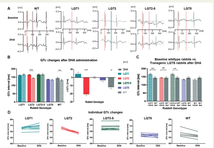
Docosahexaenoic acid influenced cardiac repolarization in a genotype-specific fashion: In WT and—more pronouncedly—in LQT2 rabbits, DHA shortened the absolute QT and heart-rate corrected QTc intervals (DQTc: WT,12.0 ± 1.88 ms,P-value = 0.001; LQT2, 20.7 ± 1.71 ms,P-value < 0.001), while no changes were observed in LQT1, LQT5, and LQT2–5 rabbits, whose IKs-function is impaired (Figure 3A and B). Important to note, this QTc-shortening effect in WT and LQT2 rabbits was seen in each individual rabbit (Figure 3D).
In LQT2 rabbits, the QT shortening effect of DHA could be seen at all heart rates and flattened the QT/RR ratio curve (P-value = 0.01) (Supplementary material online,Figure S1B).
To investigate whether DHA may normalize the prolonged QTc to physiological values, we compared QTc of transgenic LQTS rab- bits following DHA administration to QTc of WT rabbits at baseline (Figure 3C). DHA administration normalized QTc in LQT2 rabbits completely (P-value for difference between WT and LQT2þDHA
= 0.950). QTc intervals in LQT1 and LQT2–5 rabbits, in contrast, Figure 2Baseline electrical differences between different LQTS genotypesin vivo and ex vivo. (A) Heart-rate corrected QTc interval. (B) Action po- tential duration at 75% of repolarization. (C) Action potential triangulation in transgenic LQT1. (D) Short-term variability of QT. (A–D) LQT1: green, in vivo n= 8,ex vivo n= 7; LQT2: red,in vivo n= 6,ex vivo n= 6; LQT2–5: violet,in vivo n= 8,ex vivo n= 9; and LQT5: blue,in vivo n= 8,ex vivo n= 9; WT littermates: grey,in vivo n= 6,ex vivo n= 7. Differences are indicated as * forP-value < 0.050, ** forP-value < 0.010, and *** forP-value < 0.001. All data are presented as mean ± SEM. The WT (grey) bars are shown next to each LQTS genotype bar to better indicate the difference to WT for each individual genotype. LQTS, long QT syndrome; LQT1, long QT Type 1; LQT2, long QT Type 2; LQT5, long QT Type 5; LQT2–5, combined form of long QT Types 2 and 5; SEM, standard error of the mean; WT, wild type.
Downloaded from https://academic.oup.com/europace/advance-article/doi/10.1093/europace/euab228/6380593 by University of Szeged user on 06 October 2021
remained prolonged after DHA administration (LQT1, P-value = 0.003; LQT2–5,P-value = 0.008). In LQT5 rabbits without any overt QT prolongation, QTc remained unchanged after DHA.
Effects of docosahexaenoic acid on 12- lead ECG in anaesthetized rabbits
Docosahexaenoic acid administration accelerated heart rate (short- ened RR interval) only in LQT1 (RRbaseline: 347.42 ± 17.04 ms, RRDHA: 371.75 ± 17.37 ms,P-value < 0.001) and not in the other ge- notype groups. DHA had no effects on PR or QRS in any of the genotypes.
After intravenous administration of DHA in anaesthetized rabbits, a statistically significant shortening of QTc was observed in WT
(DQTc: 7.31 ± 1.52 ms, P-value = 0.005) and in LQT2 (DQTc:
11.35 ± 1.77 ms,P-value = 0.003), while no changes of QTc were observed in LQT1, LQT2–5, and LQT5 rabbits (Supplementary ma- terial online,Figure S2A).
At baseline, temporal QT variability [characterized by the short-term variability of QT interval (STVQT)] was significantly higher in LQT2 rab- bits compared with LQT1 or WT rabbits (baseline STVQTLQT2: 8.46 ± 1.90 ms, baseline STVQTLQT13.45 ± 0.32 ms, baseline STVQTWT
4.75 ± 1.00 ms; One-way ANOVAP-value = 0.036) (Figure 2D). Due to a significant reduction of STVQT upon DHA-infusion in LQT2 rabbits (DSTVQTLQT2:2.33 ± 0.58 ms,P-value = 0.016), after 20 min adminis- tration of DHA STVQT was similar in all genotypes (one-way ANOVA P-value = 0.566), indicating a normalization of STVQT in LQT2 (Figure 4A–CandSupplementary material online,Figure S3).
Figure 3 Effects of DHA on QT intervalin vivoin telemetric ECGs. (A) Representative examples of telemetric ECGs recorded in WT, LQT1, LQT2, LQT2–5, and LQT5 rabbits before (above) and after (below) administration of DHA. Q and the end of the T-wave are indicated with vertical lines. (B) Left panel: QTc comparison before (fully coloured columns) and after (dotted columns) administration of DHA in WT, LQT1, LQT2, LQT2–5, and LQT5 rabbits. Right panel: changes in QTc in the different genotypes are indicated asDQTc. (C) Comparison of QTc in LQTS rabbits treated with DHA vs. baseline QTc in WT animals (grey), indicates a normalization in LQT2 (lack of differences between LQT2þDHA and WT at baseline). Grey bars are repeated to better show the difference to WT for each individual genotype. (D) Representation of DHA-induced QTc changes in the individual rabbits (left: QTc values before and right: after administration of DHA). (B–D) Sample numbers: LQT1 (green)n= 8, LQT2 (red)n= 6, LQT2–5 (violet)n= 8, LQT5 (blue)n= 8, and WT rabbits (grey)n= 6. Differences are indicated as * forP-value < 0.050, ** forP-value <
0.010, and *** forP-value < 0.001. All data are presented as mean ± SEM. DHA, docosahexaenoic acid;LQTS, long QT syndrome; LQT1, long QT Type 1; LQT2, long QT Type 2; LQT5, long QT Type 5; LQT2–5, combined form of long QT Types 2 and 5; SEM, standard error of the mean; WT, wild type.
6 A. Castiglioneet al.
Downloaded from https://academic.oup.com/europace/advance-article/doi/10.1093/europace/euab228/6380593 by University of Szeged user on 06 October 2021
Spatial QT dispersion neither showed relevant genotype-related dif- ferences at baseline nor was it significantly influenced by DHA in any of the genotype groups (Supplementary material online,Figure S2B).
Effects of docosahexaenoic acid in perfused whole hearts ex vivo
Docosahexaenoic acid shortened mean APD75 in WT (DAPD75: 12.3 ± 2.22 ms, P-value < 0.001) and LQT2 rabbits (DAPD75: 18.1 ± 3.54 ms,P-value = 0.004) (Figure 5A–B). The DHA-shortening in LQT2 was so pronounced that in LQT2 hearts perfused with DHA no longer any prolongation of APD75was observed in comparison to WT hearts at baseline conditions (APD75-LQT2-DHA: 144.12 ± 5.50 ms, APD75-WT: 135.03 ± 7.02 ms,T-testP-value = 0.342), indicating a DHA- induced normalization of APD75in LQT2 (Figure 5C). Importantly, the APD-shortening effect of DHA in WT and LQT2 rabbits was seen in each individual rabbit (Figure 5D). No relevant changes of the APD75
were recorded in LQT1, LQT2–5, or LQT5 rabbit (Figure 5A–B).
We further analysed whether DHA’s APD75-shortening effects in LQT2 and WT rabbits differed in the various LV regions. At 2 Hz
pacing rate, APD75shortening effects were observed in both LQT2 and WT rabbits in the LV mid-lateral wall and in LV lateral base regions.
However, only in WT but not in LQT2 rabbits APD75-shortening was also significant at the LV apex and LV medial base regions (Figure 6A).
Notably, spatial APD75dispersion was not altered by DHA neither in WT nor in LQT2—similarly as observed with QT dispersion.
Action potential triangulation was significantly reduced after DHA administration only in LQT2 rabbits (from baseline 103.8 ± 2.83 to 96.9 ± 2.41 ms;P-value < 0.001;Figure 6B), though not in WT, LQT1, LQT2–5, or LQT5 rabbits. Despite its reduction, in DHA-treated LQT2 rabbits AP triangulation remained more elevated than in WT rabbits at baseline (Figure 6C).
Effects of docosahexaenoic acid on action potential duration and arrhythmogenesis in silico
The modelled APs (Figure 7A) demonstrate a longer APD in LQT2 than in WT and a pronounced DHA-induced shortening of APD in LQT2, supporting our experimental observations. Single-cell Figure 4Effects of DHA on STVQTin vivo. (A) Representative examples of short term variability of QT measured in WT and LQT2 rabbits before (left) and after (right) administration of DHA. (B) Histograms showing STVQT before (fully coloured columns) and after (dotted columns) administra- tion of DHA in WT, LQT1, LQT2, LQT2–5, and LQT5 rabbits. Differences are indicated with * forP-value < 0.050. All data are presented as mean ± SEM. (C) Histograms showing a comparison between STVQT in transgenic LQTS rabbits treated with DHA vs. baseline STVQT in WT animals (grey); a normalization in LQT2 can be observed (lack of significant differences between LQT2þDHA and WT at baseline). Differences are indicated as * forP-value <0.050, and as ** forP-value <0.010. All data are presented as mean ± SEM. Grey bars are repeated to better show the difference to WT for each individual genotype. DHA, docosahexaenoic acid; LQTS, long QT syndrome; LQT1, long QT Type 1; LQT2, long QT Type 2; LQT5, long QT Type 5; LQT2–5, combined form of long QT Types 2 and 5; STVQT, short-term variability of QT interval; SEM, standard error of the mean;
WT, wild type.
Downloaded from https://academic.oup.com/europace/advance-article/doi/10.1093/europace/euab228/6380593 by University of Szeged user on 06 October 2021
simulations with varied conductivities ofgKsand gCa,Laiming at quanti- fying (potential positive) DHA effects on the cellular repolarization reserve were performed. The analysis of the parameter space ofgKs
and gCa,Lrevealed that the LQT2 model produce EAD, blocks, and permanent depolarization for many more combinations of parame- ters than the WT model (Figure 7B for pacing at 1.0–2.5 Hz, Supplementary material online,Figure S4for pacing at 1.0–4.5 Hz), and importantly, that DHA exerted an anti-arrhythmic effect by re- ducing these arrhythmogenic AP compared with LQT2, reaching a level very close to WT cells.Figure 7Cshows the prevalence for arrhythmogenic behaviour over all tested pacing frequencies, expressed as the ratio of parameter combinations that do not result in EAD, blocks, or permanent depolarization, compared with WT at 1 Hz. Again, LQT2þDHA cells had consistently lower arrhythmic behaviour than LQT2 cells over all frequencies, nearly reaching the level of WT cells, particularly at faster stimulation frequencies.
Discussion
In this study, we investigated the potential beneficial APD/QT- shortening effects of theIKs-activator DHA, a natural polyunsaturated fatty acid (PUFA),5in vivo and ex vivoon the whole heart level in four different transgenic rabbit models of LQTS.
In WT and more pronouncedly in transgenic LQT2 rabbit models, DHA significantly shortened QTc and reduced the beat-to-beat vari- ability of repolarization, quantified as STVQTin vivo, and shortened APD75and reduced AP triangulationex vivo. Notably, in LQT2 rabbits the effect of DHA led to a normalization of QTc, STVQT, and APD75 to the level observed in healthy WT rabbits, suggesting that DHA may exert a beneficial therapeutic effect in LQT2.
Several experimental studies have investigated the effects of differ- ent PUFAs on cardiac electrophysiology. We have chosen DHA, which increased the magnitude ofIKscurrent in KCNQ1/KCNE1- Figure 5Effects of DHA on action potential durationex vivo. (A) Representative examples of recorded MAPs before (in grey) and after administra- tion of DHA in perfused WT (black), LQT1 (green), LQT2 (red), LQT2–5 (violet), and LQT5 (blue) rabbit hearts. The APD shortening appears during phase 3, which corresponds to the phase in whichIKsis conducted. (B) Left panel: APD75in WT, LQT1, LQT2, LQT2–5, and LQT5 rabbits before and after DHA administration. Right panel: changes in APD75in the different genotypes are indicated asDAPD75. (C) Comparison of APD75in LQTS rab- bits treated with DHA vs. baseline QTc in WT animals (grey) indicates a normalization in LQT2 (lack of differences between LQT2þDHA and WT at baseline). Grey bars are repeated to better show the difference to WT for each individual genotype. (D) Representation of DHA-induced APD75
changes in the individual rabbits (left: APD75values before; right: after administration of DHA). (B–D) Sample numbers: LQT1 (green)n= 7, LQT2 (red)n= 6, LQT2–5 (violet)n= 9, LQT5 (blue)n= 9, and WT rabbits (grey)n= 7. Differences are indicated as * forP-value < 0.050, ** forP-value <
0.010, and *** forP-value < 0.001. All data are presented as mean ± SEM. DHA, docosahexaenoic acid; LQTS, long QT syndrome; LQT1, long QT Type 1; LQT2, long QT Type 2; LQT5, long QT Type 5; LQT2–5, combined form of long QT Types 2 and 5; MAP, monophasic action potential; SEM, standard error of the mean; WT, wild type.
8 A. Castiglioneet al.
Downloaded from https://academic.oup.com/europace/advance-article/doi/10.1093/europace/euab228/6380593 by University of Szeged user on 06 October 2021
transfected COS7 cells and in guinea pig cardiomyocytes more than, for example, eicosapentaenoic acid, another omega-3 fatty acid of marine origin. These findings have been further supported by the groups of Liin, Bentzen, and Larsson,4,6,12who have observed that natural and modified PUFAs activate the IKs-conducting channel through a lipoelectric interaction between the negatively charged PUFA head group and positively charged aminoacidic residues in the alpha-subunit KCNQ1.
Along this line, they also described a significant shortening of the prolonged QT and prolonged APD in a guinea pig model of drug-
induced LQTS.9Our results confirm this observation of QT/APD- shortening in wild-type rabbits—and importantly—in transgenic LQTS rabbit models. Moreover, as we used several different trans- genic rabbit models for different LQTS subtypes, in which different al- pha- and beta-subunits of repolarizing ion channels are impaired (namely, KCNQ1 in LQT1, KCNH2 in LQT2, KCNE1 in LQT5, and KCNQ1 and KCNE1 in LQT2–5), important information regarding the mechanistic action of DHA onIKscould be obtained.
Morenoet al.5 suggested that the DHA-induced acceleration of ventricular repolarization was based on a modification of the Figure 6Effects of DHA on regional APD and AP triangulationex vivo. (A). Effects of DHA on APD75in apex, mid lateral wall, lateral basis, and me- dial basis in WT, LQT1, LQT2, LQT2–5, and LQT5 rabbits. (B) AP triangulation before (fully coloured columns) and after (dotted columns) adminis- tration of DHA in WT, LQT1, LQT2, LQT2–5, and LQT5 rabbits. (C) Comparison between APD75in LQTS rabbit hearts after administration of DHA and AP triangulation in WT rabbit hearts before administration of DHA. In contrast to QTc and APD75,a normalization of AP triangulation can- not be observed in LQT2 after DHA administration; sample numbers: LQT1 (green)n= 7, LQT2 (red)n= 6, LQT2–5 (violet)n= 9, LQT5 (blue) n= 9, and WT rabbits (grey)n= 7. Differences are indicated as * forP-value < 0.050, ** forP-value < 0.010, and *** forP-value < 0.001. All data are presented as mean ± SEM. AP, action potential; APD, action potential duration; DHA, docosahexaenoic acid; LQTS, long QT syndrome; LQT1, long QT Type 1; LQT2, long QT Type 2; LQT5, long QT Type 5; LQT2–5, combined form of long QT Types 2 and 5; MAP, monophasic action potential;
SEM, standard error of the mean; WT, wild type.
Downloaded from https://academic.oup.com/europace/advance-article/doi/10.1093/europace/euab228/6380593 by University of Szeged user on 06 October 2021
interaction between the alpha- and beta-subunits KCNQ1 and KCNE1 that leads to an enhancement ofIKscurrent. Accordingly,IKs- activating effects of DHA could not be observed, when KCNQ1 channels were expressed alone,5 suggesting that the presence of functional KCNE1 subunits was essential for the observed electro- physiological DHA-effects. We now demonstrated inXenopusoo- cyte experiments that DHA activates human KCNQ1, human KCNQ1/KCNE1 and rabbit KCNQ1/KCNE1IKsto different extents, with the largest activating effect in the two KCNQ1/KCNE1 channels and with less effect on human KCNQ1 channels (when the beta- subunit KCNE1 is missing), indicating an importance of both subunits for a pronounced DHA-inducedIKsactivation. The larger relative in- crease in Kþcurrent at 0 mV for human KCNQ1/KCNE1 compared with KCNQ1 can be largely explained by the different voltage depen- dence of the two channels. Half maximal conductance is reached at
aboutþ20 mV for human KCNQ1/KCNE1 and at about30 mV for KCNQ1,eRef5 which means that at 0 mV KCNQ1 has already approached its maximal conductance, giving less possibility for DHA to induce combined activating effects. In line with these hypotheses, no QT/APD-shortening effects were observed neither in LQT5 and LQT2–5 rabbits with impaired KCNE1-subunits nor in LQT1 rabbits with impaired KCNQ1—while pronounced QT/APD-shortening effects were observed in LQT2 and WT with intact KCNE1 and KCNQ1 function. This strongly suggests that DHA may exert a genotype-specific beneficial effect only in LQTS subtypes with intact IKsfunction.
Recently, possible PUFA interaction sites have been identified at the voltage-sensor and the pore region of the KCNQ1 channel, using coarse-grained and all-atom molecular dynamics simulations.15 Although DHA is a protonable PUFA, which allows it to cross the Figure 7In silicomodelling of DHA effects. (A) Simulated APs at 2 Hz based on Shannon model for WT, LQT2 (gKr= 0%), and LQT2þDHA (gKs
þ100%) cardiomyocytes. (B) Results of single-cellin silicosimulations for combinations of parametersgKsandgCa, Lat 1.0–2.5 Hz. Every square repre- sents one simulation. Shaded squares represent parameters where pacing was successful (shade encodes APD90). Pink and purple squares represent simulations where 2:1 block and EAD occur, respectively. Black squares represent simulations where the cell was unable to repolarize. Numbers in bottom right corners of each plot indicate simulations resulting in block, EAD or permanent depolarization. (C) Normalized repolarization reserve cal- culated from the number of single-cell pacing simulations in the parameter space 0 <gKs/gKs,orig< 1 (step 0.1) and 1 <gCaL/gCaL,orig< 5 (step 0.2) that did not result in EAD, block or permanent depolarization. Reference for normalization is the number of simulations for WT at 1 Hz that did not show these pro-arrhythmic features. AP, action potential; DHA, docosahexaenoic acid; EAD, early after depolarization; LQT2, long QT Type 2; WT, wild type.
10 A. Castiglioneet al.
Downloaded from https://academic.oup.com/europace/advance-article/doi/10.1093/europace/euab228/6380593 by University of Szeged user on 06 October 2021
cell membrane and to exert effects through intracellular or extracel- lular interactions, the functional PUFA sites in KCNQ1 seem to be lo- cated in the outer membrane leaflet.15 Along those lines, it is important to note, that the KCNQ1 mutation expressed in our trans- genic LQT1 rabbit models is a dominant negative pore mutation with complete loss of functionalIKs, in which no DHA-induced APD short- ening can be expected. This may of course be different in other KCNQ1 variants in other regions of the gene and raises the possibil- ity that some clinically relevant mutations may influence the DHA ef- fect (e.g. when located in PUFA-interacting regions); and in some LQT1 variants one might still see some DHA-induced rescue of the phenotype. Hence, it is very important to stress that DHA may—in addition to the genotype-specific beneficial DHA effect in LQTS gen- otypes with normal KCNQ1 and KCNE1 function—also have mutation-specific beneficial effects in some LQT1 and/or LQT5 mutations.
The fact that not only global QTc and APD were shortened/nor- malized by DHA in LQT2 rabbits but also classicalin vivo and ex vivo pro-arrhythmia markers such as STVQT and AP triangulation, sug- gests that DHA might also exert anti-arrhythmic effects. STVQT, which characterizes temporal instability in cardiac repolarization, has been validated as a novel marker regarding arrhythmic risk also in hu- man LQTS16—and it was also most pronounced in transgenic LQT2 rabbits with the highest risk for spontaneous TdP arrhythmias.8,10 Importantly, DHA normalized STVQT to the level observed in WT rabbits, strongly suggesting that not only overall cardiac repolariza- tion was stabilized but also the pro-arrhythmic temporal instability of repolarization was reduced. Similarly, the arrhythmogenicity marker AP triangulation, which is particularly increased in LQT2 rabbits with a marked pro-arrhythmic phenotype,8,10was also reduced by DHA in LQT2. The failure to normalize AP triangulation completely may be due to the fact that the extent of AP triangulation is strongly de- pendent onIK1and IKr, and somewhat less onIKs.17
To further investigate potential anti-arrhythmic effects of DHA in LQT2, we have conductedin silicomodelling experiments incorporat- ing the observed DHA-induced increase inIKs. In these we identified a reduction in EAD formation and in 2:1 blocks as well as an improved repolarization reserve in LQT2 cells with shortened APD due to an increase inIKsbyþ100% that corresponds to the experimentally ob- served DHA-effect onIKs. Despite such encouraging evidence, the potential anti-arrhythmic effects certainly need to be validated in larger, long-term studies directly investigating DHA-effects on spon- taneous (and provoked) TdP ventricular arrhythmias and SCD in LQTS.
In both WT and in LQT2 rabbits, an APD-shortening was ob- served in all investigated left ventricular regions, despite not reaching statistical significance in the apex and in the medial base in LQT2.
This may be the reason why DHA had no effect on APD dispersion and—particularly—did not increase APD dispersion. This is impor- tant as it has previously been shown that the activation of one specific potassium channel may exert pro-arrhythmic effects by increasing the dispersion of repolarization or by causing excessive regional APD-shortening effects, because of underlying regional heterogeneity in the expression of cardiac potassium channels.18Why the pharma- cological activation of IKr increased APD dispersion,18 while the
activation ofIKsby DHA did not, is currently unclear. One might spec- ulate that the underlying regional differences inIKsandIKrthat are unmasked by the activation of the reciprocal current might differ in their extent. The fact that DHA also affects other channel proteins, such as voltage-gated Naþand Ca2þchannels that are heteroge- neously expressed throughout the ventricles may counteract some of the regional effects ofIKsactivation. DHA effects on other (un- known) protein that alter excitability in pacemaker cells which coun- teract the decrease in excitability by DHA’s activating effect onIKs, might also underlie the observation that DHA impacts on heart rate in LQT1 but not in WT and LQTS rabbits with intactIKs.
As rabbits show pronounced similarities to humans in terms of cardiac electrophysiology, and—particularly—as the transgenic LQTS rabbits mimic all major aspects of the human LQTS disease phenotypes, it stands to reason that the observed beneficial DHA effects in LQT2 rabbits could have a translational impact on future genotype-specific treatment approaches in LQTS. Thus far, no stud- ies have investigated DHA effects in human LQTS patients.
Moreover, the available literature offers only limited data regarding the effects of PUFA and more specifically of DHA on cardiac electro- physiology—as the focus of these studies was more on prevention of cardiovascular diseases—where PUFA supplementation seems to be beneficial.19,eRef6Recently, though, Yagiet al.20observed that low lev- els of DHA were associated with cardiogenic syncope in patients af- fected by Brugada syndrome, suggesting that DHA may play an important role in preventing ventricular fibrillation in this cohort of patients. Our observations in transgenic LQTS rabbit models may open the door for a translational, clinical evaluation of DHA as novel genotype-specific therapy in LQTS. To this aim, additional experi- ments directly demonstrating anti-arrhythmic effects in animal mod- els and—importantly—first confirmatory studies in human patients are still warranted.
Conclusion
We demonstrated that DHA exerts a genotype-specific benefi- cial shortening/normalizing effect on QTc, STVQT, APD75, and AP triangulation through activation ofIKsin LQT2 rabbits but has no effects if either KCNQ1 (a-subunit toIKs) or KCNE1 (b-subu- nits to IKs) are functionally impaired (as in LQT1, LQT5, and LQT2–5). Thus, DHA could represent a new genotype-specific therapeutic option in LQT2 syndrome (or other LQTS subtypes with intacta- andb-subunits toIKs).
Supplementary material
Supplementary materialis available atEuropaceonline.
Funding
This work was supported by a grant from the German Heart Foundation (F/02/14) to K.E.O., by a grant of the National Heart Lung and Blood Institute (NHLBI R01-HL131461) to H.P.L. and K.E.O, the Hungarian National Research, Development, and Innovation Office (NKFIH K-128851) to I.B., and the European Research Council under
Downloaded from https://academic.oup.com/europace/advance-article/doi/10.1093/europace/euab228/6380593 by University of Szeged user on 06 October 2021
the European Union’s Horizon 2020 research and innovation pro- gram (grant agreement no. 850622) to S.I.L.
Conflict of interest:none declared.
Data availability
All data are available on request at the Institute for Physiology of the University of Bern.
References
1. Priori SG, Blomstrom-Lundqvist C, Mazzanti A, Blom N, Borggrefe M, Camm Jet al.2015 ESC Guidelines for the management of patients with ventricular arrhyth- mias and the prevention of sudden cardiac death.Europace2015;17:1601–87.
2. Schwartz PJ, Crotti L, Insolia R. Long-QT syndrome: from genetics to manage- ment.Circ Arrhythm Electrophysiol2012;5:868–77.
3. Cho Y. Management of patients with long QT syndrome.Korean Circ J2016;46:
747–52.
4. Liin SI, Yazdi S, Ramentol R, Barro-Soria R, Larsson HP. Mechanisms underlying the dual effect of polyunsaturated fatty acid analogs on Kv7.1.Cell Rep2018;24:
2908–18.
5. Moreno C, de la Cruz A, Oliveras A, Kharche SR, Guizy M, Comes Net al.
Marine n-3 PUFAs modulate IKs gating, channel expression, and location in mem- brane microdomains.Cardiovasc Res2015;105:223–32.
6. Skarsfeldt MA, Liin SI, Larsson HP, Bentzen BH. Polyunsaturated fatty acid- derived I(Ks) channel activators shorten the QT interval ex-vivo and in-vivo.Acta Physiol2020;229:e13471.
7. Nerbonne JM. Molecular basis of functional voltage-gated Kþchannel diversity in the mammalian myocardium.J Physiol2000;525 Pt 2:285–98.
8. Brunner M, Peng X, Liu GX, Ren XQ, Ziv O, Choi BRet al.Mechanisms of car- diac arrhythmias and sudden death in transgenic rabbits with long QT syndrome.
J Clin Invest2008;118:2246–59.
9. Major P, Baczko´ I, Hiripi L, Odening KE, Juha´sz V, Kohajda Zet al.A novel trans- genic rabbit model with reduced repolarization reserve: long QT syndrome caused by a dominant-negative mutation of the KCNE1 gene.Br J Pharmacol 2016;173:2046–61.
10. Hornyik T, Castiglione A, Franke G, Perez-Feliz S, Major P, Hiripi Let al.
Transgenic LQT2, LQT5, and LQT2-5 rabbit models with decreased repolarisa- tion reserve for prediction of drug-induced ventricular arrhythmias. Br J Pharmacol2020;177:3744–59.
11. Orosz A, Baczko´ I, Nagy V, Gavalle´r H, Csana´dy M, Forster Tet al.Short-term beat-to-beat variability of the QT interval is increased and correlates with parameters of left ventricular hypertrophy in patients with hypertrophic cardio- myopathy.Can J Physiol Pharmacol2015;93:765–72.
12. Liin SI, Silvera˚ Ejneby M, Barro-Soria R, Skarsfeldt MA, Larsson JE, Starck Ha¨rlin F et al.Polyunsaturated fatty acid analogs act antiarrhythmically on the cardiac IKs channel.Proc Natl Acad Sci U S A2015;112:5714–9.
13. Bohannon BM, Wu X, Wu X, Perez ME, Liin SI, Larsson HP. Polyunsaturated fatty acids produce a range of activators for heterogeneous IKs channel dysfunc- tion.J Gen Physiol2020;152:e201912396.
14. Bodi I, Sorge J, Castiglione A, Glatz SM, Wuelfers EM, Franke Get al.Postpartum hormones oxytocin and prolactin cause pro-arrhythmic prolongation of cardiac repolarization in long QT syndrome type 2.Europace2019;21:1126–38.
15. Yazdi S, Nikesjo¨ J, Miranda W, Corradi V, Tieleman DP, Noskov SY et al.
Identification of PUFA interaction sites on the cardiac potassium channel KCNQ1.J Gen Physiol2021;153:e202012850.
16. Hinterseer M, Beckmann B-M, Thomsen MB, Pfeufer A, Dalla Pozza R, Loeff Met al.Relation of increased short-term variability of QT interval to congenital long- QT syndrome.Am J Cardiol2009;103:1244–8.
17. Romero L, Pueyo E, Fink M, Rodrı´guez B. Impact of ionic current variability on human ventricular cellular electrophysiology.Am J Physiol Heart Circ Physiol2009;
297:H1436–45.
18. Bentzen BH, Bahrke S, Wu K, Larsen AP, Odening KE, Franke G et al.
Pharmacological activation of Kv11.1 in transgenic long QT-1 rabbits.J Cardiovasc Pharmacol2011;57:223–30.
19. Parish S, Mafham M, Offer A, Barton J, Wallendszus K, Stevens W et al.;
ASCEND Study Collaborative Group. Effects of omega-3 fatty acid supplements on arrhythmias.Circulation2020;141:331–3.
20. Yagi S, Soeki T, Aihara K-I, Fukuda D, Ise T, Kadota Met al.Low serum levels of eicosapentaenoic acid and docosahexaenoic acid are risk factors for cardiogenic syncope in patients with Brugada syndrome.Int Heart J2017;58:720–23.
The eReferences 1–6 can be found in the online eReference list in the supple- mentary material.
12 A. Castiglioneet al.
Downloaded from https://academic.oup.com/europace/advance-article/doi/10.1093/europace/euab228/6380593 by University of Szeged user on 06 October 2021