N EW T R E N D S IN T H E D E SC R IP T IO N O F T H E G EN ER A L M EC H A N IS M
A N D R EG U LA TI O N O F EN ZY M ES
Symposia Miologiea Mu ugarira 21
NEW TRENDS
IN THE DESCRIPTION OF THE
GENERAL MECHANISM AND REGULATION
OF ENZYMES
NEW TRENDS IN THE DESCRIPTION OF THE GENERAL MECHANISM
AND REGULATION OF ENZYMES Symposium on Enzyme Action 9—12 July 1978, Debrecen, Hungary
Edited by
S. DAMJANOVICH, P. ELŐDI and B. SOMOGYI
(Symposia Biologica Hungarica 21) The purpose of this volume, based on papers presented at a joint Summer Symposium of the Medical Univer
sity School of Debrecen and the Bio
logical Division of the Hungarian Academy of Sciences, is to give a representative survey of current re
search activities in the field of enzyme energetics and regulation. The range of topics covered are as follows:
Subunit catalytic cooperativity; math
ematical analysis of heterotropic and homotropic interaction; thermo
dynamic approach to enzyme action, regulation and evolution; role of thermodynamic fluctuation in enzyme action; microenvironment and organ
ized multienzyme systems; evolu
tionary adaptation; contribution of non-active site regions; electronic theory of enzyme action; secondary interactions; adsorptive enzyme sys
tems; role of hydrogen ions.
The contributing authors have all adopted a true multidisciplinary ap
proach to the problems of enzyme catalysis and the participation of the whole enzyme in catalysis.
.1828.
AKADÉMIAI KIADÓ
Publishing House of the Hungarian Academy of Sciences
BUDAPEST
ISBN 963 05 1881 3
Symposia Biologica Hungarica
R e d ig u n t S. DAM JANOVICH
P. ELŐDI B. SOMOGYI
Vo]. 21
AKADÉMIAI KIADÓ, BUDAPEST 1978
Symposia Biologica Hungarica
21
NEW TRENDS
IN THE DESCRIPTION
OF THE GENERAL MECHANISM AND REGULATION OF ENZYMES
Symposium on Enzyme Action. 9—12 July 1978
Debrecen, Hungary
E d ite d b y
S. DAMJANOVICH P. ELŐD I B. SOMOGYI
AKADÉMIAI KIADÓ, BUDAPEST 1978
ISBN 963 05 1881 3
Akadémiai Kiadó, Budapest 1978
Printed in Hungary
LIST OF PARTICIPANTS
ANTONOV, V. Institute of Bioorganic Chemistry, Academy of Sciences of the USSR, Moscow, USSR
BARDSLEY, W. G. Department of Obstetrics and Gynaecology at St. Mary’s Hospital, University of Manchester, Manchester, England
BATKE, J. Enzymology Department, Institute of Biochemistry, Hungarian Academy of Sciences, Budapest, Hungary
BOYER, P. D. Molecular Biology Institute and Department of Chemistry, University of California, Los Angeles, California, USA
CARERI, G. University di Roma, Institute of Physics “Guglielmo Marconi”, Rome, Italy
DAMJANOVICH, S. Department of Biophysics, Debrecen University School of Medicine, Debrecen, Hungary
ELÖDI, P. Department of Biochemistry, Debrecen University School of Medicine, Debrecen, Hungary
ERNSTER, L. Department of Biochemistry, Arrhenius Laboratory, University of Stockholm, Stockholm, Sweden
FRIEDEN, C. Department of Biological Chemistry, Washington University School of Medicine, St. Louis, Missouri, USA
KELETI, I. Enzymology Department, Institute of Biochemistry, Hungarian Academy of Sciences, Budapest, Hungary
KESZTHELYI, L. Department of Biophysics, Biological Research Institute of the Hungarian Acade
my of Sciences, Szeged, Hungary
KURGANOV, B. I. All-Union Vitamin Research Institute, Moscow, USSR
LOW, P. S. Department of Chemistry, Purdue University, West Lafayette, Indiana, USA
5
POLGÁR, L. Enzymology Department, Institute of Biochemistry of. the Hungarian Academy of Sciences, Budapest, Hungary
SALERNO, C. Ilnd Department of Chemical Biology, Faculty of Medicine and Surgery, University of Rome, Rome, Italy
SIMON, I. Enzymology Department, Institute of Biochemistry of the Hungarian Academy of Sciences, Budapest, Hungary
SOMERO, G. N. Scripps Institution of Oceanography, University of California, San Diego, La Jolla, California, USA
SOMOGYI, B. Department of Biophysics, Debrecen University School of Medicine, Debrecen, Hungary
STRAUB, F. B. Enzymology Department, Institute of Biochemistry of the Hungarian Academy of Sciences, Budapest, Hungary
SZABOLCSI, G. Enzymology Department, Institute of Biochemistry of the Hungarian Academy of Sciences, Budapest, Hungary
VOLKENSTEIN, M. V. Institute of Molecular Biology, Academy of Sciences of the USSR, Moscow, USSR
WELCH, G. R. Department of Biological Sciences, University of New Orleans, New Orleans, Louisiana, USA
WIEKER, H.-J. Max-Planck-Institut für Ernährungsphysiologie, Dortmund, FRG
6
CONTENTS
OPENING ADDRESS 9
PAUL D. BOYER, R. LEE HUTTON, JEFFREY CARDON, MASAHIRO ARIKI:
Subunit catalytic cooperativity in enzyme catalysis 13
W. G. BARDSLEY:
The theory and graphical tests for homotropic and heterotropic effects 35 V. K. ANTONOV:
Secondary interactions in the enzymic catalysis 63
B. I. KURGANOV, S. V. KL1N0V,N. P. SUGROBOVA:
Regulation of enzyme activity in adsorptive enzyme systems 81
T. KELETI:
Thermodynamic approach to enzyme action, regulation and evolution 107 M. V. VOLKENSTEIN:
Theoretical approaches to the physics of enzymatic catalysis 131 G. CARERI:
Statistical physical aspects of enzyme action 151
S. DAMJANOVICH, B. SOMOGYI:
A possible role of thermodynamic fluctuation in the overall enzyme action 159 G. R. WELCH:
On the nature of enzyme catalysis in the “living state” : the role of organized
multienzyme systems 185
P. S. LOW:
Protein hydration changes during catalysis: an important contribution to the properties
of enzyme catalyzed reactions 217
7
G. N. SOMERO, P. H. YANCEY:
Evolutionary adaptation of Km and Kcat values: fitting the enzyme to its environment through modifications in amino acid sequences and changes in the solute composition
of the cytosol 249
H. -J. WIEKER:
Regulation of erzyme cooperativity by hydrogen ions 277
INDEX 307
8
OPENING ADDRESS
Research in the biological sciences is not void of fashic trends either. To put it more elegantly: the primary trend or research always tends to such problems whose solution is de
manded by advances in science as well as by the development of production and by the efficiency of health care, on the one hand, and whose solving rests on progress of other disciplines and of the instrumentation, on the other hand. This is undoubt
edly true in most fields of research.
There exists, however, another, rarely mentioned phenome
non: the effect of fashionable trends, as it is called for lack of a better expression. Some problems arise, become the centre of interest and are adopted by several research teams as their target problem. In this way many valuable data provide reveal
ing insight into parts of diverse problems, but the entire problem is rarely understood completely because continuously arising new problems divert attention. Thus many researchers change their old "love" for a new one.
The history of enzymology is a good example of the above tendency. The structure and function of enzymes were studied extensively in many institutes and university departments in the fifties and sixties. Several enzymes have been studied from various aspects with the most diverse methods. Besides their chemical structure, i.e. amino acid sequence, much has been learnt about enzymes as a result of the introduction of physico-chemical and physical methods, especially X-ray stud
ies, in addition to the traditional techniques applied in en
zymology. The discovery of the regulation of enzyme action and 9
\
the interpretation of the phenomenon by different models were basic results concerning the biological function of enzymes.
At present enzymology is not considered as one of the most stimulating, most fashionable, or most exciting topics of biochemistry and biophysics. Nucleic acids, the molecular com
ponents and the mechanism of information processing are in the focus of attention. Molecular biology, as today studies in molecular genetics are called, however, cannot be understood if only the structure and properties of nucleic acids are in
vestigated. Detailed knowledge of enzymes is essential in mo
lecular biology as it is essential not only for the understand
ing of metabolism, membrane transport, energy exchange, con
traction, reproduction and the molecular mechanism of many other biological processes, but for the understanding of bio
logical information, as well.
Moreover, it is also true that even the basic phenomena of enzyme action are not quite clear. Very often only hypotheses are created from the experimental data, for example, what kind of side chains form the active center of an enzyme, but hardly could we answer the question on how they contribute to the de
crease of activation energy. Nor would we be more certain about the role of the rest of the protein part of the enzyme molecule in enzymatic catalysis. Although many good X-ray data are a- vailable on enzyme-substrate complexes, more than 60 years after the publication of the Michaelis-Menten -formula hundreds of papers are published yearly on enzyme-substrate interaction, on the kinds and number of ES complexes. We are rather guessing but not quite sure about the real, intrinsic energetics of en
zyme-catalyzed reactions and about the intimate details of en
zymatic catalysis. We cannot give the exact description of such trivial problems either, as the interaction between enzymes and the surrounding aqueous medium, the energetic results of the interaction, the energy content of the protein molecule and the fluctuation of its structure, as well as the effect of these parameters on catalysis. Although these phenomena and interac
tions are discussed in some details in every text-book and hand-book.
1 0
I
The aim of this meeting is to discuss such and similar topics in the hope of advancing the knowledge of the biological sciences concerning the above topics. We thought that the lim
ited number of participants, the less strictly organized pro
gram and the pleasant atmosphere of the place would provide more and much pleasant opportunity than a bigger meeting with a much more rigid time table could.
Welcome to everybody. I wish you success and I also wish you a good time.
P.Elődi
Editors' note:
It is pointed out that the camera-ready technique has been used in the production of this volume as this ensures rapid publication. Thus, editori
al work has been limited to the correction of any misprints in the original papers which might otherwise have interfered with the meaning.
Illustrations have been reproduced as submitted.
1 1
Sym posia Biologica Hungarica 21 (1978)
SUBUNIT CATALYTIC COOPERATIVITY IN ENZYME CATALYSIS
PAUL D. BOYER, R. LEE HUTTON, JEFFREY CARDON, MASAHIRO ARIKI Department of Chemistry and Molecular Biology Institute, University of California
Los Angeles 90024, USA
This paper presents some recent experimentation in my laboratory that has made us much more aware of the p o t e n t i a l i t i e s of c ata lyt ic cooperativity between subunits of multisubunit enzymes. More spe
c i f i c a l l y , I want to raise for your consideration and for discussion the pos si bi li ty of alternating s i t e cooperativity in c atalys is . As defined in a recent paper by David Hackney and myself (1) "Alternating s i t e cooperativity occurs when two or more identical c ata lyt ic s it es part ici pate in sequence in a manner so that acceleration of c ata lyt ic events at one s i t e resul ts from binding of substrate(s) a t another c ata lyt ic s i t e . " Such a concept has been considered by others, but has not gained any widespread acceptance. Recent findings in my laboratory with mitochondrial ATPase give what appears to be the most convincing data yet obtained for the occurrence of such c ata lyt ic cooperativity. These and other results encourage me to suggest that the phenomenon may be much more prevalent than recognized. Indeed, subunit c ata lyt ic cooperativity could be the most important advantage nature gains from multisubunit enzymes.* 1
Some background. A quite brief historical sketch might be useful.
In a provocative paper published in 1968 Harada and Wolfe (3) pre
sented r esults with mal ate dehydrogenase indicative that binding at one subunit promoted c ata lyt ic events on the other identical subunit.
Their i nterpretation has not been regarded as convincing and t heir paper has received l i t t l e attention in the ensuing years. In this period, the p os s i b i l i t i es of some type of cooperation between subunits has been suggested for several enzymes, including alkaline phosphatase (see 4), amino acyl tRNA synthetase (5) and adenosine triphosphophatases (6, 7). The recognition of negative cooperativity of substrate binding by Koshland and associates (see 8) was an important related development.
* Researches reported herein were supported by grants from the I n s t i tute of General Medical Sciences, U.S. Public Health Service, and the U.S. National Science Foundation.
1 For a suggestion that multisubunits function principally to confer s t a b i l i t y see Chan (2).
\
1 3
However, this phenomenon has been interpreted largely in terms of possible control mechanisms with quite limited mention that the negative cooperativity of substrate binding might r e f l e c t a more basic c ata lyt ic cooperativity between, subunits (8). Koshland has also suggested that the principal advantage of multisubunits could be in evolution of enzyme function (9).
Subunit cooperativity in Fl ATPase. I nterest of my laboratory in subunit interaction in catalysis is a di rect outgrowth of studies on how ATP is synthesized in mitochondria and chloroplasts. These researches were reviewed a t a symposium of the recent FEBS meeting and will not be further discussed here. A brief review of the energy- linked conformational coupling that is involved has been presented elsewhere (10). ATP formation is regarded as involving an energy- linked conformational change that promotes binding of ADP and Pi at one s i t e in a mode competent for ATP formation and, concomitantly,
ATP>E*ADP
Conformational .changes in
ATPase complex
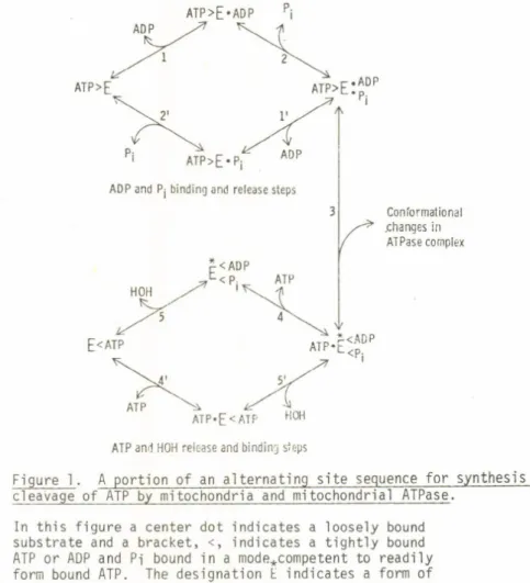
Figure 1. A portion of an alternating s i t e sequence for synthesis and cleavage of ATP by mitochondria and mitochondrial ATPase.
In this figure a center dot indicates a loosely bound substrate and a bracket, <, indicates a t ight ly bound ATP or ADP and Pi bound in a mode*competent to readily form bound ATP. The designation E indicates a form of the enzyme containing such competently bound ADP and Pi.
1 4 I
favors release of a t r a n s i t o r i l y t i gh tl y bound ATP from an alt ernat e s i t e . I will present in more detail recent unpublished data with the purified Fl ATPase coupling factor as isolated from beef heart mitochondria. The application of some newer methods developed by Dr. Hackney in my laboratory (11, 12) to t his enzyme has added con
siderably to the evidence for alternating s i t e cooperativity in ATP synthesis or cleavage. A sketch depicting one-half of a complete cycle (1, 13) is given in Fig. 1.
INTERMEDIATE P ii* H O H EXCHANGE WITH F, ATPase AT LOW ATP CONCENTRATIONS
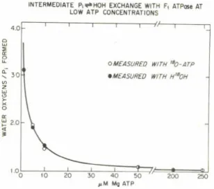
Figure 2. The effect of ATP concentration on the ATPase and the intermediate Pj^OH exchange a c t i v i t i e s of FI. The 1.0 ml of final reaction mixture a t 30“ and pH 7.6 contained 30 mM K-acetate, 30 mM Tr is-acetate, 3 mM phosphoenolpyruvate, 5 mM M<J + , 200 yg pyruvate kinase, and the indicated amount of ATP. 180 was present as H180H (0.74 atom % excess) or in the terminal phosphoryl group of ATP (60 atom % excess). With H180H present, ATPase concentration ranged from 9 to 230 yg/ml at the highest to lowest ATP concentrations, respectively, and incubation time was 20 min. With [ 180]ATP present, ATPase was 0.35 to 2.4 yg/ml with a 15 min. incubation. The ordinate gives the number of water oxygens present in each Pi formed, and not the total extent of oxygen exchange as corrected for approach to isotopic equilibrium.
As mentioned briefly in an e a r l i e r symposium (14), Dr. Glenda Choate during her Ph.D. thesis research made the striking observation that beef heart ATPase acquires a capacity for intermediate Pi ^ H0H . exchange as the ATP concentration is lowered. More recent data obtained by Lee Hutton are shown in Fig. 2 (13). At higher ATP concentration, l i t t l e or no oxygen exchange occurs and the Pi formed contains only
1 5
the one oxygen required for hydrolytic cleavage. As ATP concentration is decreased in the number of water oxygens incorporated in each pro
duct Pi formed as the concentration of medium ATP is lowered. These observations are readily explainable by the alternating catalyt ic s i t e model. In retrospect we should have done such experiments much e a r l i e r . Mention should be made that in mitochondria and submito- chondrial p a rt i c l e s, intermediate exchange accompanying ATP cleavage occurs as much higher ATP concentrations, resulting from additional rate limitation imposed by requisite conformational interactions that couple ATP cleavage to membrane energization.
The simple explanation offered by the alternating s i t e model is t hat at low ATP concentration ADP and P-j are formed at a c ata lyt ic s i t e (step 5, Fig. 1), but cannot be released until a medium ATP binds at the alt ernat e s i t e (step 4, Fig. 1). At low ATP concentra
t ions, the t r a n s i t o r i l y t ig ht ly bound ADP and P-j reversibly form bound ATP, and because of the equivalence of oxygens of bound P i , i n t e r mediate oxygen exchange occurs.
Other possible explanations. Although this explanation was a t t r a c t i v e , such r esults were not conclusive. As with other sugges
tions for subunit c at al yt i c cooperativity, additional reasonable explanations could also account for the data. These are as follows:
1. Control s ite s : At high ATP concentrations control s it e s are f i l l e d and modify the catalysis so that no intermediate exchange occurs.
2. Enzyme heterogeneity: Two types of c at al yt i c si te s are present. Most of the ATPase has a rel at ive ly high Km and forms Pi without any oxygen exchange. An ATPase present in smaller amount but with a low Km forms Pi with extensive oxygen exchange.
This ATPase cleaves a larger portion of the ATP as the ATP con
centration is lowered. 3
3. Enzyme h ysteresis: This is also referred to as enzyme memory.
Possible relationships to enzyme regulation have been given by Frieden (15). As applied to ATPase, the enzyme i n i t i a l l y cleaves ATP with extensive intermediate Pi ^ HOH exchange, but is converted to a more active form. I f high ATP is present, t his modified enzyme cleaves subsequent ATP molecules without exchange. At low ATP concentration, the enzyme reverts to the original form, and each Pi formed thus shows extensive exchange. This model may be diagrammed as follows:
1 6
s]o_w - ' 'step E
V
S ES -A new approach based on measurement of 1B0-Pj species. Fortunately, as mentioned e a r l i e r , a means of discerning among the various explanations for the experimental resul ts with the ATPase has recently become a v a i l able (11, 12). The approach is based on the mass spectrometric measure
ment of the di st ribut ion of 180 in various Pi species. Some explanatory material is necessary to understand the approach. Consider an enzyme, such as muscle myosin, catalyzing a medium Pi ^ HOH exchange starti ng with Pi containing 180 in all four oxygens. As exchange proceeds the following five 180-Pi species might be present:
Product formed at low (S) with exchange
Product formed at high (S) without exchar
0 0 0 01 »1
S p e c i e s
| O - P - O
i
1 0 — P — 0
j
o — P — •
1 • - P - *
1 - r
0
1
• •1 •I •
D e s i g n a t i o n P 180o Q_ co o
p18o2 CL. COO ro p l 8 |
The Pi formed may be converted to the v ol at il e trimethysilyl derivative, and the r el ati ve amounts of the five species determined by mass spec
trometry (11).
An ATP, fully labeled with 180 in the y-phosphoryl group, that is hydrolyzed enzymically may undergo intermediate Pi -- HOH exchange as indicated by Equation 1.
HOH
ATP + E J i L - » E - A T P < ^ k^ - > E - A D P - P i —ÍSL -> E + ADP + P i ( 1 ) (exchange step)k-2
The partitioning of enzyme bound Pi between reformation of ATP and release of products is given by the following:
Partition coeff ici ent , Pc = K- 2 + Ki— .3
2 New Trends 1 7
Two extremes for 180-P-j species formation are as follows:
Pi with all oxygens replaced by HÜH
Pi containing only one water oxygen required for hydrolysis
The number of water oxygens present in each Pi formed the 0/P r atio or designated O' (11), would be 1 for the upper limiting case and 4 for the lower. Intermediate 0/P rat ios could arise i f one enzyme were present with a c ha racter is tic amount of intermediate exchange
(k- 2 44 or 44 kä). The total amount of intermediate_exchange, where the 0/P rat io is designated as R, is given by R = 40' -_4
4 - O'
Application to muscle myosin ATPase. The approach was f i r s t applied to c la ri fy a bothersome discrepancy in r esults obtained with Mcf-activated myosin ATPase. Measured values of 0/P rat ios reported
in the l i t e r a t u r e averaged around 3 and showed considerable variation
100 Myosin MgATPase
Observed
4->O
VS/A Theory f = .66
Figure 3 . Distribution of 180-Pj species following hydrolysis of 180-enriched ATP by muscle myosin.
(McJ-activated rabbit muscle myosin, 25°, 92.7%
180 in y-P03 of ATP, average 180 content of
P-j o 0/P r at io of 2.86).
1 8
(see 12). Vet estimations, from kinetic measurements of bound ADP levels, of the amount of reversal of cleavage of bound ATP that had occurred indicated that 0/P rat ios approaching 4 should be observed, t hat i s , very extensive intermediate exchange should'have occurred.
This was also made probable by the observations that with the myosin subfragment 1 ATPase, a more purified preparation, 0/P rat ios of about 3.9 were observed (16). The dist ribut ion of 180-Pi species for a
myosin preparation giving an O.P r at io of 2.86 were measured, and results as indicated in the upper portion of Fig. 3 were obtained (12). Shown in the lower portion of Fig. 3 is the expected s t a t i s t i c a l d i st r i b u tion of species i f the product Pi had been produced by a single cata
l y ti c s i t e with a Pc t hat would give r is e to an 0/P r at io of 2.86.
Clearly the experimental results do not correspond to cleavage of the ATP by a single type of c ata lyt ic s i t e . However, i f i t is assumed that close to 66% of the cleavage occurs by myosin with an 0/P r atio of 3.9 (near total exchange of Pi oxygens with water) and the remainder by a contaminant a c ti v it y that gives product Pi with an 0/P r at io of 1 (no exchange), a theoretical di st ribut ion pattern close to the experi
mental resul ts is obtained (upper portion, Fig. 3). I t was thus con
cluded t hat myosin preparations were heterogeneous with respect to ATPase a ct iv it y (12). Examination of the product Pi had revealed that two d iff ere nt types of catalyt ic s it e s had participated in the ATP cleavage.
TABLE I
180-Pi Species Formed by F]-ATPase when 5-6 yM 180-ATP (85 a . p . e . ) Hydrolyzed
Application to mitochondrial ATPase. Armed with this new approach, Lee Hutton in our laboratory has examined the 180-Pi species formed by the purified mitochondrial ATPase. For t his ATP was cleaved at a concentration where moderate increase in intermediate exchange had
occurred. If enzyme heterogeneity or enzyme hysteresis were responsible for the- increased oxygen exchange, two types of cleavage would be occurring, one with extensive exchange and one with no exchange, to give the observed overall 0/P r at io.
Table I gives r esults from an early experi
ment. These data show that the di stribution of
180- Pi species is t hat predicted for cleavage by one type of c ata lyt ic s i t e . This r es ul t has been confirmed
(30° , pH 7.7, 5 mM MgCl2 )
Pi Sample P180, Ple02 P; s 03
Theory, all Pi
with 0/P = 1.6 0.46 1.00a 1.16
Experimental,
average 0/P = 1 7 0.52 O o Q)
1.06 Theory, al'l Pi
with 0/P = 1.8 0.60 1.0 0a 0.86 aNormalized for 1802 at 1.00.
2* 19
in l a t e r , more extensive experiments a t differing 0/P r at ios and with higher 180 levels. The clear conclusion can be reached t hat the heterogeneity and hysteresis models do not explain the r e su l ts . In addition, control s i t e s , such that part of the cleavage would be by the enzyme with the control s i t e vacant, giving exchange, and part with the control s i t e occupied by ATP, giving no exchange, can be eliminated.
Another type of control s i t e could be visualized, in which the equilibration of ATP between medium and the control s i t e s and the changes in enzyme structure accompanying ATP binding or release at the control s i t e were rapid compared to the cleavage reaction. Then each enzyme might behave as i f i t had only one type of c at al yt i c s i t e . This appears to us to be a quite unlikely sequence of events, and one that can be rendered even more unlikely by additional data. Binding s it e s on the ATPase have been carefully examined in a number of l aboratories, and s i t e s for binding of ATP t i gh t l y do e xi st (see 17).
Indeed, the isolated ATPase contains both bound ADP and bound ATP.
Evidence in our laboratory indicates that most or all of these long
term t i gh tl y bound nucleotides are not a t c at al yt i c s it e s (14, unpub
lished data). Other workers have suggested that such s i t e s might correspond to control s i t e s (see 17). Their occupancy appears to be necessary for the ATPase to act as a coupling factor. However, i f
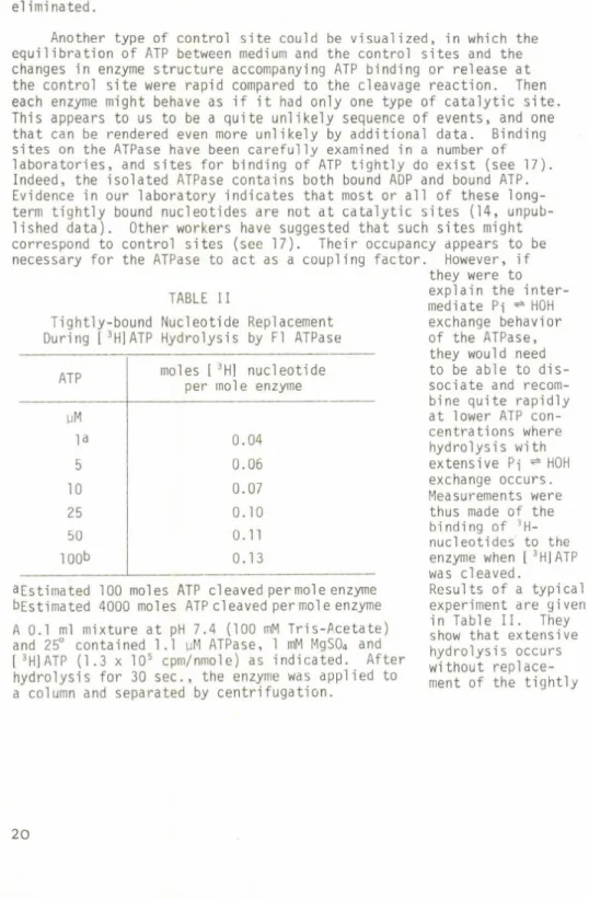
A 0.1 ml mixture a t pH 7.4 (100 mM Tris-Acetate) and 25° contained 1.1 pM ATPase, 1 mM MgS04 and [ 3H]ATP (1.3 x 10⁵ cpm/nmole) as indicated. After hydrolysis for 30 s ec. , the enzyme was applied to a column and separated by centrifugation.
aEstimated 100 moles ATP cleaved per mole enzyme bEstimated 4000 moles ATP cleaved per mol e enzyme
TABLE II
Tightly-bound Nucleotide Replacement During [ 3H]ATP Hydrolysis by Fl ATPase
ATP pM la 5 10 25 50 100b
moles [ 3H] nucleotide per mole enzyme
0.04 0.06 0.07 0.10 0.11 0.13
they were to explain the i n t e r mediate Pi ^ H0H exchange behavior of the ATPase, they would need to be able to d i s sociate and recom
bine quite rapidly at lower ATP con
centrations where hydrolysis with extensive Pi ^ HOH exchange occurs.
Measurements were thus made of the binding of 3H- nucleotides to the enzyme when [ 3H] ATP was cleaved.
Results of a typical experiment are given in Table II. They show that extensive hydrolysis occurs without replace
ment of the tight ly
2 0
bound nucleotides. With elimination of possible control s i t e s , hetero
geneity and hysteresis the alternating c at al yt i c s i t e model remains as the only reasonable explanation for the data.
Taken together with the extensive observations that favor a l t e r nating s i t e action of the ATPase on submitochondrial part icl es during ATP synthesis (1, 18) or hydrolysis (19), and other di rect measurements of Pi binding,2 the evidence in favor of the alternating catalyt ic s it es appears to me to become compelling. This remarkable phenomenon, where a bound Pi continues to undergo covalent interconversions but cannot be readily released until ATP binds another- s i t e , gives the
f i r s t well-documented example of alternating s i t e c ata lyt ic cooperativity.
Possible subunit interactions with glyceraldehyde 3-phosphate dehydrogenase. As mentioned e a r l i e r , i t seemed likely that c ata lyt ic cooperativity with multisubunit enzymes might be considerably more common than recognized. One enzyme chosen for further study is glycer
aldehyde 3-phosphate dehydrogenase. Results of several studies with t his enzyme, including the prominent negative cooperativity of NAD*
binding to the four identical subunits (21), appeared to me to point to overlooked c ata lyt ic cooperativity with the muscle enzyme. More s p eci f ic al ly , i t seemed plausible NAD* binding to a c ata lyt ic s i t e on one subunit might promote an otherwise rat e-l imi ti ng release of NADH from an alt ernat e s i t e .
The muscle enzyme is known to c r y s t a l l i z e with 2 to 3 t ight ly bound NAD* molecules present per mole of enzyme (22). To explore the p o s s ib i l i t i e s of c ata lyt ic cooperativity, Jeffrey Cardon and I have i n i t i a t e d a series of experiments on effects of medium NAD* and NADH on the release of t ig ht ly bound nucleotides. For t his we have made use of the simple column technique described by Penefsky for measurement of Pi binding to mitochondrial ATPase (20). A small column of Sephadex G-50 in the barrel of a 1 ml. p l as tic syringe is c e n t r i fuged b rie fly to remove liquid and pack the column. Then a small volume, such as 100 p i , of enzyme solution is placed on the column and exposed briefly to a centrifugal force. The enzyme with t ight ly bound components appears in the eluate remarkably free of medium solutes.
Some experimental r esul ts that have been obtained are summarized in Tables III and IV. What they show is outlined in the following para
graphs .
Presence of tightly-bound NAD* on the enzyme. The i n i t i a l desalted enzyme had an A2 8o/A2 6o indicative of approximately 2 moles of t i gh t ly - 2 Penefsky has presented evidence that Pi binds t ig ht ly to a catalyt ic
s i t e on the ATPase (20). In our laboratory, R. L. Hutton has shown that t his Pi is rapidly released when ATP is added, an observation obviously consistent with the alternating s i t e model.
2 1
TABLE III
Exchanges Between Tightly Bound NAD* and NADH on Glyceraldehyde 3-Phosphate Dehydrogenase
Fi rs t Second Third Moles/enz.yme
Column Column Column NAD* NADH
3 mM glyceraldehyde 3-phosphate
250 pM NAD* — — 0.3
1 mM
[ 3H] NAD* only buffer — 1.8
1 mM
[ 3H] NAD* 1 mM
NADH - - - 0.8 1.0
1 mM 1 mM 1 mM 2.1
[ 3H] NAD* NADH [ 3H] NAD*
60 pM enzyme exposed to additions indicated j u s t before column separa
t ions, 50 mM Tris-S04 , pH 7.5, 0°C.
TABLE IV
Replacement of Tightly Bound [ 3H]NAD* on Glyceraldehyde 3-Phosphate Dehydrogenase
Additions [ 3H] NAD* /enzyme
( af ter second column separation)
none 2.3
1 mM NAD* 0.13
1 mM NADH 0.42
0.2 mM NADH 1.0
0.05 mM NADH 1.4
20 pM enzyme, exposed to 0.5 mM [ 3H]NAD* and rapidly passed through Sephadex G-50 column, had 2.3 t i ght ly bound [ 3H]NAD+
per mole, pH 7.5, 50 mM Tris-S04 , 0°C.
bound NAD* per mole of enzyme. The f act that the NAD* was not removed by passage through the Sephadex column shows that i t is not only thermodynamically t i gh tl y bound but has a rel at iv e ly low off constant.
This was to be expected in view of the low dissociation constant
2 2
estimated by Conway and Koshland (21f for the binding to the f i r s t 2 NAD* to the enzyme and the upper limit of diffusion control for the rate of NAD* binding.
Conversion of tightly-bound NAD* to t ig ht ly bound NADH. I t is important to know i f the tight ly bound NAD* is capable of the rapid reduction t hat would be required i f such a step were an intermediate in the catalysi s. After addition of Pi and glyceraldehyde 3-phosphate the tightly-bound NAD* was converted to NADH. As shown in Table I I I , much of t his NADH was also s uff ici en tl y t i gh tl y bound to be retained on the enzyme a ft e r passage through an additional column. In rapid mixing experiments-, Peczon and Spivey (23) have demonstrated the impor
tant point that under conditions similar to those used for Table I I I , and with insuf fici ent medium NAD* present to bind to the loose s i t e s , NAD* reduction occurs considerably f as t er than the rate of overall catalysi s.
Replacement of tightly-bound NAD* by radioactive NAD* . A logical step in a c ata lyt ic cooperativity by glyceraldehyde 3-phosphate dehy
drogenase would-be for rapid interconversion of t ight and loose binding s it es to occur when both types of s it e s are occupied. Were t his the case, then addition of s uf fi ci en t radioactive NAD* to bind to the loose s it e s should r es ul t in interchange of the t ight ly bound NAD*
with radioactive NAD*. Experiments to assess t his probability are summarized in Tables III and IV. They show ready replacement of the t ig ht ly bound NAD* by medium radioactive NAD*. Clearly in some manner the rate of dissociation of the bound NAD* must be promoted by binding of medium NAD*.
As noted in Table IV, i f enzyme with t ig ht ly bound radioactive NAD* is prepared, such NAD* is also readily replaced by medium NAD*.
Replacement of t ig ht ly bound NADH by medium NAD*. If c atalyt ic cooperativity occurred during net oxidation of glyceraldehyde 3-phosphate then a change must obviously occur such that t ight ly bound NADH becomes a loosely bound NADH with an off constant commensurate with the overall c ata lyt ic rat e. Results shown in Table III demonstrate that tight ly bound NADH is readily replaced when the enzyme is exposed to medium NAD*. Clearly in some manner the rate of dissociation of the bound NADH must be promoted by binding of medium NAD* .
Discussion of resul ts with glyceraldehyde 3-phosphate dehydrogenase.
These and other experimental r esults are readily accommodated by a 3 3 Results of Bell and Dalziel (24) indicate higher dissociation con
stants for the t ig ht NAD* than those of Conway and Koshland (21).
However, i t appears uncertain to us that the l a t t e r measurements were free from complications of enzyme dissociation a t low concen
t ra tion (25-27) or inactivation of the NAD* free enzyme (22).
2 3
SU B U N I T INTERACTION IN NAD B I NDI NG
Apoenzyme, equivalent sites o r readily interconvertible sites
Asymmetric enzyme, tight NAD site occupied.
S u b u n its stay in o n e conform ation.
NAD • E< NAD*
NAD :> E • NAD"
NAD > E •
(
Enzyme with both loose and tig h t sites occupied. S u b u n it conform ations rapidly in te rc o n v e rt. Formation of tight binding at one s u b u n it drives loosening of binding at a lte rn a te s u b u n it.A lternate tig h t site occupied.
Figure 4 . An alternating s i t e scheme for coenz.yme binding and release by muscle glyceraldeh.yde 3-phosphate dehydrogenase.
scheme as depicted in Fig. 4. This scheme depicts behavior for one subunit pair, each with a t i gh t and a loose s i t e . Evidence for disso
ciation of the enzyme into dimers (25-27) makes i t likely that prin
cipal , but not necessarily a l l , c ata lyt ic interactions will be expressed between subunit pairs. Central to this scheme is the suggestion of subunit structural asymmetry in the enzyme with only one or two NAD* 's or other reagent bound per mole. Evidence that asymmetry associated with h al f- si t es rea ct ivi ty has been noted by a number of workers
(28-31) including convincing X-ray analysis of abortive ternary complex of the lobster muscle enzyme (31). Preexisting asymmetry (see 29) is neither essential nor excluded.
Further, the r esults in this paper and e a rl i e r data support the concept that when both t i gh t and loose s it es are occupied the subunits are in rapid conformational interconversion (Fig. 4). This has the consequence of allowing all four subunits to participate equally in the catalysis by alternating between the t i ght and loose binding conformations. The model readily accommodates the observation of Peczon and Spivey (23, see also Table I) t hat the rate of conversion of t ig ht ly bound NAD* to t ig ht ly bound NADH is catalytical ly competent.
Indeed, i t appears quite unlikely that both t ight or loose NAD* would have equivalent reaction c h ar ac te ri st ic s, and the demonstration of the reduction of t ight NAD* thus leads to the suggestion 'that the oxidation-reduction reaction occurs only with the subunit in the con
formation that t ig ht ly binds NAD* .
If formation of t ig ht ly bound NADH is a step in the c atalys is , obviously something must happen to rapidly change the tendency for
2 4
NADH to dissociate. Various p os s i b i l i t i e s could be suggested, but the resul ts shown in Table III indicate the simple and appealing pos sibi li ty that this is accomplished by conformational change driven principally by binding of NAD* to an a lt erna te c ata lyt ic s i t e . Indeed, even i f an NAD* regenerating system were present net oxidation of glyceraldehyde 3-phosphate under conditions of Table III would be severely rate-limited by the NADH dissociation rate. This appears analogous to the s ituation with mitochondrial Fl ATPase, where bound ADP and Pi cannot be released until a medium ATP binds a t an alt ernate catalyt ic s i t e . The projected NADH-release sequence is depicted in equation 2.
E<NADH + NAD* NAD* • E<NADHNAD* >E • NADH— »NAD* >E + NADH (2) (slow NADH (rapid conformational (enzyme ready for off constant) interconversion) substrate binding
and NAD* reduction) The r equisite displacement of NADH by NAD* as required for the cooperative catalysis sequence also gains support from the ease of replacement of tightly-bound NAD* by radioactive medium NAD* (Table I I I ) . This experiment was foreshadowed by the early classic studies of Velick et a l . (22) who demonstrated that addition of [ 32P] NAD* , from yeast grown with 32Pi followed by ammonium sulfate precipitation and enzyme i sol ati on, gave enzyme with t ig ht ly bound [ 32P]NAD .
A variety of other factors likely interplay with the confor
mational changes involved in loosening NADH binding. Experiments of Smith (32) indicated that enzyme acylation interferes with release of NADH in presence of medium NAD*. Later data of Trentham (33) also show that deacylation by phosphorolysis may precede NADH release.
With glyceraldehyde 3-phosphate dehydrogenase, a reluctance to accept c ata lyt ic cooperativity models has arisen from the valuable experiments of Peczon and Spivey (23) and of Trentham (33) and Armstrong and Trentham (34) that indicated that all bound nucleotides were
c a t a l yt i ca l ly equivalent. However, examination of t hei r data shows that i t can readily be accounted for by the scheme of Fig. 4. A rapid interconversion of c ata lyt ic s it e s when all s it e s are occupied readily induces equivalent rea ct iv i ty of all bound nucleotides. But the equivalent rea ct iv i ty does not mean that t ight ly bound and loosely bound NAD* both undergo reduction with equal ease. I t means that the loosely bound form is rapidly converted to the t i ght ly bound form as a requi sit e step in the c atalys is .
The r es ul ts with glyceraldehyde 3-phosphate dehydrogenase are obviously not su ff i ci en tl y complete or compelling to allow firm con
clusions a t this stage. Additional experiments with various approaches are underway in my laboratory. Also we have i n it i a t ed experiments
2 5
with other mul ti subunit enzymes where l i t e r a t u r e evidence indicates the po ss ib il it y of overlooked c ata lyt ic cooperativity.
Some r esults with sarcoplasmic reticulum ATPase. Before closing, I would l i ke to mention briefly some studies with another enzyme system. The important Ca-pumping ATPase of the muscle sarcoplasmic reticulum can be isolated as a 100,000 dalton monomer. But in the membrane there is suggestive evidence that cooperativity between dimers or oligomers may occur, although such evidence is by no means defi ni ti ve (see 35). In a recent study by myself and Leopoldo DeMeis (35) we suggested that an intermediate Pi ^ H0H exchange might occur with binding of ATP promoting Pi release. This pos sibi li ty has been explained further by Masahiro Ari k i , who has made the important obser
vation that as ATP concentration is decreased the intermediate Pi ^ H0H exchange per Pi formed is increased. Again, alternation of catalyt ic si te s on adjacent enzymes could be involved. Assessments of hetero
geneity and hysteresis models using the 180-Pi di st ribut ion technique are now in progress.
26
REFERENCES
1. Hackney, D. D., and Boyer, P. D. (1978) J. Biol. Chem., 253, 3164-3170.
2. Chan, W. W. -C. (1976) Trends Biochem. Sei. X, 258-260.
3. Harado, K. and Wolfe, R. G. (1968) J. Biol. Chem. 243, 4131-4137.
4. Lazdunski, M. (1974) Progress in Bioorganic Chem. 82-140.
5. Fersht, A. R. (1975) Biochem. 14, 5-12.
6. Repke, K. R. H., and Schon, R. Tl974) Acta Biol. Med. Germ. 33, K27-K38.
7. Adolfsen, R., and Moudrianakis, E. N. (1976) Arch. Biochem Biophys.
172, 425-433.
8. Levitzki, A. and Koshland, D. E. J r . (1976) Curr. Topics in Cell Reg.
JO, 1-40.
9. Koshland, D. E. Jr. (1976) Fed. Proc. 35, 2104-2111.
10. Boyer, P. D. (1977) Trends in Biochemical Sei. 2, 38-41.
11. Hackney, D. D. and Boyer, P. D. (1978) Proc. Natl. Acad. Sei. U.S., in press.
12. Sleep, J. A., Hackney, D. D. and Boyer, P. D. (1978) J. Biol. Chem., in press.
13. Choate, G., Hutton, R. L., and Boyer, P. D. Submitted for publication.
14. Boyer, P. D., Gresser, M., Vinkler, C. , Hackney, D., and Choate, G. (1977) in Structure and Function of Energy-Transducing Membranes (K. van Dam and B.. F. van Gelder eds.) Elsevier/North-Holland Biomedical Press, 261-274.
15. Frieden, C. (1970) J. Biol. Chem. 245, 5788-5799.
16. Sleep, J. A. and Boyer, P. D. (1978) Submitted for publication.
17. Harris, D. A. (1978) Biochem. Biophys. Acta 463, 245-273.
18. Smith, D. J. and Boyer, P. D. (1976) Proc. Natl. Acad. Sei. USA 73, 4314-4318.
19. Kayalar, C., Rosing, J. and Boyer, P. D. (1977) J. Biol. Chem. 252, 2486-2491.
20. Penefsky, H. S. (1977) J. Biol. Chem. 252, 2891-2899.
21. Conway, A. and Koshland, D. E. J r. (1968) Biochem. 7_, 4011-4022.
22. Velick, S. F., Hayes, J. E. J r . and Harding, J. (1953) J. Biol. Chem.
203, 527-544.
23. Peczon, B. D. and Spivey, H. 0. (1972) Biochem. 1_]_, 2209-2217.
24. Bell, J. E. and Dalziel, K. (1975) Biochem. Biophys. Acta 391 , 249-258.
25. Hoagland, V. D. J r. and Teller, D. C. (1969) Biochem. 8, 594-602.
26. Lakatos, S. and Zavodszky, P. (1976) FEBS Lett. 63, 145-148
27. Nagradova, N. K., Golovina, T. 0. and Mevkh, A. TI X1974) FEBS Lett.
49, 242-245.
28. Stallcup, U. B. and Koshland, E. E. J r. (1973) J. Mol. Biol. 80, 77-91.
29. Seydoux, F. , Malhotra, 0. P. and Bernhard, S. (1974) Critical Reviews of Biochem. 2, 227-257.
30. Bode, J . , Blumenstein, M. and Raftery, M. A. (1975) Biochem. J4, 1153-1160.
31. Garavito, R. M., Berger, D. and Rossman, M. G. (1977) Biochem. 16, 4393-4398.
32. Smith, T. E. (1966) Biochem. 5, 2919.
33. Trentham, D. (1971) Biochem. J. 122, 71-77.
34. Armstrong, J. M. and Trentham, D. R. (1976) Biochem. J. 159, 513-527.
35. De Meis, L. and Boyer, P. D. (1978) J. Biol. Chem. 253, 1556-1559.
2 7
D I SCUSSI ON
ERNSTER:
Your finding that the isolated, soluble ATPase ca
talyses P^-HOH exchange only at low ATP concentrations is very striking. How do you explain that with the membrane-bound enzyme, the critical ATP concentration required for P^-HOH exchange shifts to a higher range?
BOYER:
You have raised a very important question which, as you are aware, could be related to the energy-linked conformational changes involved in ATP synthesis. In the membrane-bound enzyme the reaction step of
ATP.E <ADP
<P . < —s"~
i ^ 4
> ATP > ■ADP
• P.
l
is readily reversible. That is, the conversion of
"loose" to "tight" ATP and the concomitant "tight" to
"loose" state of ADP and P^ is opposed and reversed by the energization represented by "squiggle" /--— — -«•/ . This may be energy-requiring conformational change or developing trans-membrane protonmotive force. In con
trast, with the purified ATPase there is neither oppo
sition nor reversal of the step leading to tight bind
ing of ATP and release of products. Thus at higher ATP concentrations oxygen does not accompany ATP cleavage.
ANTONOV:
What is the chemical nature of the tightly bound P^-ATPase complex? Is the phosphate bound covalently?
2 8
BOYER:
Present evidence indicates that the P. is not l
covalently bound. It is released as P^ when protein is denaturated by perchloric acid or by guanidine hydro
chloride at neutral pH, or when protein is removed by phenol extraction at acid, neutral or alkaline pH. Thus if a covalent bond is present, it is more labile than protein phosphoryl derivatives.
It is attractive to consider that the tightly bound P^
may be in a region of low water activity. Also, it is possible that some anhydrous derivative such as meta
phosphate might be present.
KELETI:
Have you performed single step kinetics of oxygen- exchange in the absence of ADP?
BOYER:
Such experiments would be valuable. Unfortunately they are made difficult by the relatively insensitive 180 technique. Present methodology is not sufficiently sen
sitive to measure oxygen exchange in a single step or enzyme turnover.
KELETI:
By the stepwise reduction and gradual stripping of the coenzymes of GAPDH we have also observed that the firmly bound coenzyme is reduced. However, using iso- topically labelled coenzyme we found very high off and on rate constants for all binding sites.
BOYER:
Your results showing that firmly bound NAD+ is reduced by substrate is important. This appears to have been independently demonstrated in several laboratories.
Clearly the tightly bound NAD+ appears to qualify as a potential catalytic intermediate.
29
In your and other measurements of exchange of bound and free radioactive coenzymes, a relatively long time elapses for the separation of protein from the free co
enzymes. According to our model, exchange between free and bound coenzymes would be expected in a few seconds even at micromolar concentrations. The important point from our experiments is that in the absence of free co
enzymes in solution the "off" constant for the tightly bound coenzyme is very low. In the same manner the binding of a coenzyme from solution promotes release of the
tightly bound coenzyme.
SOMERO:
How widespread is catalytic cooperativity among en
zymes? If this is an important catalytic mechanism, i.e.
for rate-enhancement, would not Nature "use" this mech
anism in all multi-subunit enzymes?
BOYER:
As a working hypothesis I suggest that catalytic coop
erativity is very widespread. However, this has not been established experimentally. Indeed, as briefly mentioned in my talk, such cooperativity has not been established for any enzyme and in some instances, as with glyceraldehyde 3-phosphate dehydrogenase, experi
ments have been interpreted to indicate absence of such cooperativity. Our results with ATPase and glycer
aldehyde 3-phosphate dehydrogenase, and preliminary data with some other enzymes suggest that nature may indeed make widespread use of subunit catalytic coop
erativity . SOMERO:
Multimeric enzymes containing heterologous subunits would seem to be promising study systems for study of catalytic cooperativity, e.g. with lactate dehydrogenase containing both M- and H-type subunits.
3 0
BOYER:
I concur.
31 LOW:
Do the experimental data clearly suggest that the weak NAD+ binding site is another catalytic site rather than simply an allosteric site which affects NAD+ affinity?
If one starts with the apoenzyme of GAPDH, does one observe a conformational change in GAPDH /via a tryptophan difference spectrum, etc./ upon titrating GAPDH with NAD+?
BOYER:
Evidence for conformational change on NAD+ binding has been obtained but I do not recall whether measurements have been made using difference spectral measurements or protein fluorescence.
I» POLGÁR:
Prof. Rossman /personal communication/ has recently shown that the structure of the apo and holo D-glycer- aldehyde-3-phosphate dehydrogenase is very similar, which is quite surprising in the light of the consider
able effect of coenzyme on the protein structure.
BOYER:
This is an interesting observation.lt may be that the crystallization has selected or favoured formation of a protein conformation like that when coenzyme is bound.
WIEKER:
I think, one should not only look whether the principle of catalytic cooperativity is realised by enzymes act
ing on the same coenzymes as suggested by Dr. Somero, but also on enzyme reactions where similar transition states occur. For instance, the substrates of triose- phosphate isomerase and pyruvate kinase are quite
different, but in both reactions tricarbon-enol- structures occur which are quite similar. Thus, the structures at the active site should be similar, and this is consistent with X-ray data of Muirhead showing nearly identical structures for both enzymes in the active site region, but differences in the rest of the enzyme molecules.
BOYER:
Your point is an interesting one. At some future time it might be possible to make generalizations about structure involved in catalytic cooperativity.
SIMON:
Many years ago I investigated GAPD by small-angle X-ray scattering, so it was an X-ray investigation in sol
ution. I found conformation change in the subunit struc
ture of GAPD during the NAD binding. I think this con
formation change during the stepwise coenzyme binding makes the difference between the coenzyme binding site, which is probably very similar in the apoenzyme. My X-ra data showed that the 1st and the 3rd coenzyme bound near the center of the gravity of GAPD and the 2nd and the 4th in the surface of the macromolecule. It may be the origin of the difference in the equilibrium con
stants of coenzyme binding.
BOYER:
Thank you for your comment. Such conformational changes are obviously consistent with my suggestions. But also, quite obviously they do not prove the existence of sub
unit catalytic cooperativity.
ANTONOV:
What do you think on the possibility of the tightly bound intermediates on the monomeric enzymes?
32
BOYER:
If the enzyme remains monomeric during catalysis then of course no cooperativity between catalytic sites oc
curs. However, the formation of tightly bound inter
mediates is quite plausible in monomeric enzymic ca
talysis. Tight binding of a transition state is a fre
quent suggestion, but is not readily experimentally accessible. There is increasing experimental evidence that product release is a frequent rate limiting step.
In many enzymic catalysis, monomeric or multimeric, there may be conversions of a tightly bound reactant to a loosely bound one as a rate-limiting or non-rate- limiting step in catalysis.
WELCH:
I have a few general comments. First, I would like to echo the possible importance of the "catalytic coop- erativity" phenomenon more generally to heteropolyrneric enzyme systems /e.g., multienzyme complexes/. This may partially explain the unique catalytic properties of certain of these enzyme aggregates. Second, let us consider the situation in vivo, where life imposes non
equilibrium /steady-state/ conditions on enzymatic pro
cesses. A few years ago, Wyman /Proc. Nat. Acad. Sei.
USA 7_2;3983; 1975/ proposed a steady-state "turning wheel" model for the behaviour of a large cooperative enzyme system. This represents a generalization of the type of "catalytic cooperativity" which you are dis
cussing here. Third, I would like to mention that this type of homotropic cooperativity, even in a relatively simple enzyme, can - under non-equilibrium conditions - give rise to the kinds of biochemical oscillations which are seen, for example, in glycolysis.
BOYER:
An interesting comment. These may well indeed be ex
pressions of catalytic cooperativity at higher organ
izational levels.
3 New Trends 3 3
Symposia Biologica Hungarica 21 (1978)
THE THEORY AND GRAPHICAL TESTS FOR HOMOTROPIC AND HETEROTROPIC EFFECTS
W. G. BARDSLEY
Department of Obstetrics and Gynaecology, University of Manchester, at St. Mary’s Hospital, Manchester, M13 OJH, UK
SUMMARY
It is shown that the current definitions of positive and negative co-operativity are confused since they are ambiguous and based upon a misunderstanding of the mapping of geometrical features between alternative
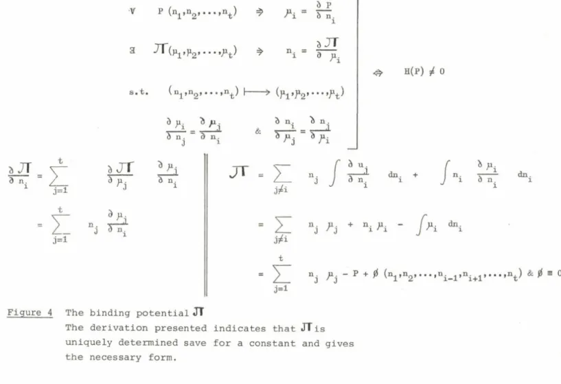
spaces. Also it is concluded that, while co-operativity cannot be defined in steady-state systems, a rigorous definition is possible in binding systems due to the existence of a binding potential tTT . Heterotropic effects can then be uniquely defined by reference to the degeneracy of this potential while homotropic effects can be defined by a special function, T , known as a tact invariant, the sign of which gives the precise graph shape test for co-operativity in any axes whatsoever. This applies even for systems where the macromolecule aggregates and there are no Adair constants.
In a recent literature survey (Hill et al., 1977), we discovered that over eight hundred enzymes are now known to show deviations from Michaelis Menten kinetics and were led to suggest that perhaps truly linear double reciprocal plots are the exception rather than the rule. From this search it became clear that there is widespread ambiguity in the definition of positive and negative co-operativity since some authors refer to phenomenological effects
3 5
and some attempt to relate co— operativity to specific mechanistic features. We commence by examining some of the definitions used for positive co-operativity.
i) Graph shape arguments Numerous authors have expressed the belief that positive co-operativity gives rise to sigmoid saturation curves which plot in double reciprocal axes as concave up curves. Negative co-operativity is supposed to produce concave down double reciprocal plots and mixed
co-operativity can give rise to stair step curves or inflected reciprocal or Hill plots (Koshland, 1970; Levitski and Koshland, 1976; Hammes and Wu,
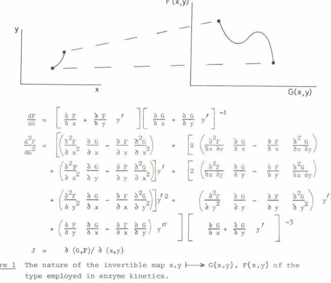
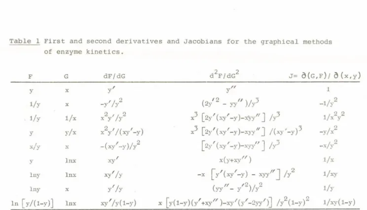
1974; Cornish-Bowden and Koshland, 1975; Teipel and Koshland, 1969). The most serious criticism of these beliefs follows from an examination of the geometry of the mapping operations used in enzyme kinetics and summarised in Figure 1 and Table 1. It was shown that sigmoid curves do not necessarily produce uniformly concave up plots and inflected double reciprocal plots are almost always given by complex binding or velocity models. Also, it was concluded that the reason for the present misunderstanding was attempting to extrapolate from simple models such as the Hill equation or 2:2 function, where these rules have some relevance, to realistic high degree cases where they do not. (Bardsley and Childs, 1975? Bardsley, 1976; Bardsley, 1977a;
Bardsley, 1977b).
ii) Arguments based on statistical ratios between adjacent binding constants When macromolecules do not aggregate, then the binding of ligand may be described by a binding polynomial of the form
N = 1 + nK1x + ^ ( n - l j K ^ x 2 + ... + K ^ ... K^x“
where here the stepwise binding constants are corrected for statistical
factions and a co-operativity coefficient, ^ , can be defined as representing deviation of these adjacent constants from the value they would have if N were a perfect n'ic i.e. of the form (l+kx)n . (Wyman, 1948; Wyman, 1972).
3 6