Biological hydropersulfides and related polysulfides – a new concept and perspective in redox biology
Jon M. Fukuto1, Louis J. Ignarro2, Peter Nagy3, David A. Wink4, Christopher G. Kevil5, Martin Feelisch6, Miriam M. Cortese-Krott7, Christopher L. Bianco7, Yoshito Kumagai8, Adrian J. Hobbs9, Joseph Lin10, Tomoaki Ida11 and Takaaki Akaike11
1 Department of Chemistry, Sonoma State University, Rohnert Park, CA, USA
2 Department of Molecular and Medical Pharmacology, Center for the Health Sciences, UCLA School of Medicine, Los Angeles, CA, USA 3 Department of Molecular Immunology and Toxicology, National Institute of Oncology, Budapest, Hungary
4 Tumor Biology Section, Radiation Biology Branch, National Cancer Institute, Bethesda, MD, USA 5 Department of Pathology, Louisiana Statue University Health Sciences Center, Shreveport, LA, USA
6 NIHR Southampton Biomedical Research Center, University Hospital Southampton NHS Foundation Trust, Southampton, UK
7 Cardiovascular Research Laboratory, Department of Cardiology, Pneumology and Angiology, Medical Faculty, Heinrich Heine University, Dusseldorf, Germany
8 Environmental Biology Section, Faculty of Medicine, University of Tsukuba, Ibaraki, Japan
9 William Harvey Research Institute, Bart & London School of Medicine, Queen Mary University of London, Charterhouse Square, London, UK 10 Department of Biology, Sonoma State University, Rohnert Park, CA, USA
11 Department of Environmental Medicine and Molecular Toxicology, Tohoku University Graduate School of Medicine, Sendai, Japan
Correspondence
J. M. Fukuto, Department of Chemistry, Sonoma State University, Rohnert Park, CA 94928, USA
Fax: +1 707 664 3378 Tel: +1 707 664 2187 E-mail: fukuto@sonoma.edu and
L. J. Ignarro, Department of Molecular and Medical Pharmacology, UCLA School of Medicine, Center for the Health Sciences, Los Angeles, CA 90095, USA
Fax: +1 310 825 6267 Tel: +1 310 859 3980 E-mail: LIgnarro@gmail.com and
T. Akaike, Department of Environmental Medicine and Molecular Toxicology, Tohoku University Graduate School of Medicine, Sendai 980-8575, Japan
Fax: +81 22 717 8219 Tel: +81 22 717 8101
E-mail: takaike@med.tohoku.ac.jp (Received 25 April 2018, accepted 30 April 2018)
doi:10.1002/1873-3468.13090 Edited by Wilhelm Just
The chemical biology of thiols (RSH, e.g., cysteine and cysteine-containing proteins/peptides) has been a topic of extreme interest for many decades due to their reported roles in protein structure/folding, redox signaling, metal ligation, cellular protection, and enzymology. While many of the studies on thiol/sulfur biochemistry have focused on thiols, relatively ignored have been hydropersulfides (RSSH) and higher order polysulfur species (RSSnH, RSSnR, n> 1). Recent and provocative work has alluded to the prevalence and likely physiological importance of RSSH and related RSSnH. RSSH of cysteine (Cys-SSH) has been found to be prevalent in mammalian systems along with Cys-SSH-containing proteins. The RSSH functionality has not been examined to the extent of other biologically relevant sulfur derivatives (e.g., sulfenic acids, disulfides, etc.), whose roles in cell signaling are strongly indicated. The recent finding of Cys-SSH biosynthesis and translational incorporation into proteins is an unequivocal indication of its fundamental importance and necessitates a more profound look into the physiology of RSSH. In this Review, we discuss the currently reported chemical biology of RSSH (and related species) as a prelude to discussing their possible physiological roles.
Keywords:cysteine persulfides; hydropersulfides; translational polysulfida- tion
Abbreviations
3MST, 3-mercaptopyruvate sulfurtransferase; CARS, cysteinyl tRNA synthetases; CBS, cystathionine beta-synthase; CN, cyanide ion;
CSE, cystathionine gamma-lyase; GSH, glutathione; GSSH, glutathione hydropersulfide; GSSSG, glutathione trisulfide; GSSSSG, glutathione tetrasulfide; MST, mercaptopyruvate sulfur transferase; SeCys, selenocysteine.
1 FEBS Letters (2018)ª2018 The Authors.FEBS Letterspublished by John Wiley & Sons Ltd on behalf of Federation of European Biochemical Societies.
The chemical biology of thiols (RSH, e.g., cysteine and cysteine-containing proteins/peptides) has been a topic of extreme interest for many decades due to their reported roles in protein structure/folding, redox sig- naling, metal ligation, cellular protection, and enzy- mology [1–5]. That is, oxidation of thiols to covalently linked disulfides (RSSR) is a critical aspect of protein folding and structure, modification (typically oxidative) of critical thiols can result in alteration of protein function/activity, thiol sulfur atoms are used in the binding of metals in numerous metalloproteins, thiol peptides scavenge otherwise toxic electrophiles, and thiolates (RS) serve as potent nucleophiles important in the catalytic activity of numerous enzymes. More- over, oxidized thiol species, such as thiyl radicals (RS), sulfenic acids (RSOH), and sulfinic acids (RS (O)OH) also possess important chemical properties that Nature has exploited for various purposes. For example, RSis used as an oxidant in the biosynthesis of DNA [6] and RSOH/RS(O)OH bind specific metals in metalloproteins, conferring activity [7]. Thus, the diverse chemistry of thiols and derived species are fun- damental to various aspects of cell biochemistry, func- tion, and signaling.
In the recent past, most studies of thiol/sulfur bio- chemistry have focused primarily on RSH/RS, RS, RSSR, RSOH, nitrosothiols (RSNO), RS(O)OH, and sulfonic acids (RS(O)2OH). Relatively ignored have been hydropersulfides (RSSH) and higher order poly- sulfur species (i.e., RSSnH, RSSnR, n>1, note: the term “hydropersulfide” and the abbreviation “RSSH”
will be used to denote both the protonated and depro- tonated species unless otherwise specified). Indeed, until very recently, the discussion of the chemical biology of hydropersulfides and polysulfides has been primarily rel- egated to studies of alliums and other food sources rich in these species [8] and to scant and specific examples of proteins with presumed hydropersulfides (of unknown function) [9–11]. Also, hydropersulfides have been implicated in the biochemistry of several specific enzymes, such as mercaptopyruvate sulfur transferase (MST) [12], NIFS (a bacterial enzyme involved in met- allocluster biosynthesis [13]) and rhodanese [14], among others [15], some of which with no established physio- logical function. In spite of these examples, the idea that hydropersulfides are fundamental signaling/bio- chemical intermediates with important physiological functions (akin to previously mentioned sulfur species, such as sulfenic acids, disulfides, or S-nitrosothiols) has not been generally considered. To be sure, important papers by Toohey, Wood and others, some of them published more than 25 years ago [16–18], indicated the
possible general importance of hydropersulfides in bio- logical systems. In spite of these somewhat prophetic reports, the idea that hydropersulfides (and related/
derived polysulfides) are important and general biologi- cal mediators/effectors has remained unappreciated.
However, there has been a resurgence of this idea due to recent and substantive reports of their biological prevalence and likely importance. In particular, a new and watershed report demonstrates translational cys- teine hydropersulfide incorporation into proteins (vide infra), consistent with the idea of fundamentally important physiological roles for hydropersulfides. This finding, along with other recent work on the unique chemical and biological properties of hydropersulfides and related polysulfides, serves as the impetus for pro- viding a perspective in redox biology.
It is the tenet of this article that hydropersulfides and related polysulfides represent a greatly underappreciated and fundamentally important aspect of sulfur-based bio- chemistry/signaling that is involved in a diverse array of physiological and/or biochemical functions. Moreover, future elucidation of these effects and functions will lead to a more profound and complete understanding of the general utility of sulfur chemistry in biological/mam- malian systems. Herein are discussed some of the recent reports indicating that hydropersulfides and polysulfides can play a role in mammalian biology, as well as the possible future directions of this field.
The generation and prevalence of hydropersulfides and polysulfides in eukaryotes
The past and apparent lack of general interest in the biology of per- and polysulfides likely stemmed from the fact that their natural existence was only indirectly alluded to (based on nonspecific assays) and limited to only a select number of enzymes/proteins such as xan- thine oxidase [10] and aldehyde oxidase [11]. Until recently, the ubiquitous and highly prevalent nature of polysulfur species in mammalian systems was unknown. Recent and significant interest in the biolog- ical actions and signaling of hydrogen sulfide (H2S) [19]
spurred interest in H2S-related and/or derived species as possible biological mediators/effectors. One of the most important early studies regarding H2S pharmacology and physiology was by Blackstone and coworkers [20]
who reported that pharmacological administration of H2S induced a reversible hibernative state in mice. This study revealed that low levels of H2S can have dramatic physiological effects and is not just a toxic species (which is observed at higher levels). An important
aspect of the presence of H2S in biological systems is the possibility that RSSH and higher order polysulfides can be formed [21,22].
One of the earliest and seminal studies drawing attention to the possible prevalence of polysulfur spe- cies (specifically hydropersulfides) in proteins was by Mustafa and coworkers [23] who utilized a modified biotin-switch assay to detect protein hydropersulfides and suggested that 10–25% of numerous liver proteins contained protein hydropersulfides (this assay, as con- ducted, may not have been specific for hydropersul- fides [2,24]). These investigators also reported that protein “sulfhydration”1 led to enhanced activity for glyceraldehyde 3-phosphate dehydrogenase (although brought into question [25,26]) and promoted actin polymerization. Subsequent reports generally support the idea of the ubiquitous and highly prevalent nature of hydropersulfides and polysulfides in biological (i.e., eukaryotic) systems. For example, Ida and coworkers [27] utilized electrophilic trapping followed by LC-MS analysis to find that small molecule hydropersulfides such as cysteine hydropersulfide (Cys-SSH) and glu- tathione hydropersulfide (GSSH) as well as higher order (additional sulfur atoms, e.g., RSSSH) hydroper- sulfides were present in mammalian cells, tissue, and plasma. Indeed, levels of GSSH were found to be greater than 50 micromolar in mouse heart and liver and greater than 150 micromolar in mouse brain.
Levels of Cys-SSH, although significantly lower, were found to be in the low micromolar range in mouse heart, liver, and brain. Using a tag-switch-tag method [27], Ida and coworkers also detected numerous pro- tein hydropersulfides, confirming the results of Mus- tafa and coworkers that the existence of protein
hydropersulfide is widespread. Assuming the value of GSSH levels in mouse brain to be accurate (i.e.,>150 micromolar), GSSH may be the most prevalent cys- teine-containing sulfur species behind only glutathione (GSH) itself (which is typically between 1 and 10 mM
in cells) in certain tissues.
The Nagy laboratory also developed a hydropersul- fide-specific assay, referred to as the protein hydroper- sulfide detection protocol (ProPerDP method), amenable to whole cells and tissues and confirmed the existence and prevalence of protein hydropersulfides in cells at levels in a steady-state range of approximately 1–12 micrograms/milligram protein [28]. An advantage of this method compared to others developed previously is that it utilized an alkylation step prior to cellular lysis, thus precluding cell lysis-mediated artifactual oxi- dation/persulfidation. Further work by Longen and coworkers [29], also using a mass spectrometry-based assay (entitled quantitative PerSulfide Site Identification or qPerS-SID), found that cells treated with H2S resulted in the generation of numerous protein hydrop- ersulfides (with significant identification overlap with the above-mentioned work by Ida and coworkers [27]).
They also found that H2S-mediated protein hydroper- sulfide formation appeared to exhibit specificity among numerous potential “sulfhydration” sites. That is, among numerous thiols on a single protein, only certain thiols were subject to hydropersulfide formation. Thus, there appears to be little doubt of the biological preva- lence of hydropersulfide formation in peptides and pro- teins. This contention is supported by the finding by the Akaike group [30] of translational incorporation of cys- teine hydropersulfide into proteins. Indeed, it is this major discovery that provides much of the impetus for writing this perspective, which will hopefully draw attention to this provocative discovery and its relevance to the field of sulfur biochemistry and physiology.
Up until 2017, hydropersulfide formation in peptides and proteins was thought to result primarily from a post-translational modification of existing cysteine resi- dues in proteins. That is, reaction of an oxidized pro- tein cysteine (possibly a disulfide or sulfenic acid, among others) could react with H2S giving the corre- sponding hydropersulfide (Reactions 1 and 2).
R-S-S-RþH2SRSSHþRSH ð1Þ RSOHþH2S!RSSHþH2O ð2Þ
These reactions have been studied extensively [21,22,31,32] and they represent possible routes of endogenous hydropersulfide formation. It needs to be
1The term “sulfhydration” was used to denote the addition/inser- tion of a sulfane sulfur (basically atomic sulfur) to/into a mole- cule. That is, the conversion of RSH to RSSH was referred to as
“sulfhydration” of RSH. This term may not be the most accurate description of the chemistry since “hydration” describes reactions that are simply represented by the addition of water. For exam- ple, the addition of water to an olefin to generate the correspond- ing alcohol is a hydration reaction. The term “sulfhydration”
may have been derived from the term “sulfhydryl”, which has been used to describe an -SH group (note that “sulf” denotes a sulfur atom and “hydryl” denotes a hydrogen and is distinct from “hydration”). Thus, it may be tempting to invoke the term
“sulfhydrylation”. However, this terminology is also not an accu- rate description of the chemistry since conversion of RSH to RSSH is not the addition of an -SH (sulfhydryl) group but, as described above, the addition of a sulfur atom. Therefore, as Toohey discusses and recommends [33], “sulfhydration” should be replaced by “sulfuration” for these reactions since the conver- sion of RSH to RSSH is equivalent to the addition of a sulfur atom.
mentioned, however, that formation of hydropersul- fidesviaReaction 1 has been questioned on the basis of the low reducing power of H2S, its relatively low con- centration and the high concentrations of GSH [33].
Regardless, since H2S is biosynthesizedviaa variety of enzymes (vide infra) and the presence of oxidized sul- furs (i.e., disulfides, sulfenic acids) in biological systems is well established, generation of hydropersulfides via Reactions 1 or 2 is clearly possible. Indeed, hydroper- sulfides are likely involved in numerous equilibria with other sulfur species and, although at low steady-state levels, are biologically accessible via these equilibria [34]. However, it remains to be determined whether hydropersulfide formation via these reactions (or others) is significant and serves a biological function.
That is, does Nature generate hydropersulfides as a response or reaction to some physiological state, cellu- lar stress, or biochemical requirement? Do hydropersul- fides serve a biological function and are, therefore, generated in a regulated manner?
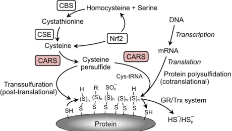
These questions have recently been answered by the finding that protein hydropersulfides are incorporated during translation via the actions of cysteinyl tRNA synthetases (CARS) [30] (Fig.1). The Akaike group has provided strong evidence that CARS are capable of converting Cys-SH to Cys-SSH via a pyridoxal phosphate-dependent process using a second Cys-SH as the sulfur atom donor (independent of ATP and tRNA). The CARS-synthesized Cys-SSH can then form a tRNA-bound Cys-SSH adduct (alsoviaCARS catalysis) resulting in the incorporation of Cys-SSH
into proteins, thus generating a protein containing a hydropersulfide functionality. This process is reminis- cent of, although not mechanistically analogous to, the incorporation of selenocysteine (SeCys) into selenopro- teins [35,36]. Figure 2 schematically illustrates and compares the mechanisms of Cys-SSH and SeCys gen- eration via translational machinery. A fundamental difference between the incorporation of Cys-SSH and SeCys into proteins is that SeCys is biosynthesized from a tRNA-bound serine while Cys-SSH is premade from Cys-SH by CARS prior to association with the tRNA, which is also catalyzed by CARS. Regardless, both SeCys and Cys-SSH are biosynthesized by trans- lational “machinery” prior to incorporation into pro- teins. The fact that hydropersulfides are both biosynthesized and attached to tRNA by CARS and subsequently translationally incorporated into proteins indicates they have a potential biological action and are not merely an artifact of H2S biosynthesis.
Considering the analogy between SeCys and Cys- SSH, it is worthwhile to further compare these two amino acids. SeCys is considered to be Nature’s 21st amino acid since it is biosynthesized pretranslationally (i.e., is not a post-translational modification), specifi- cally encoded (using UGA, which is normally the stop codon) and incorporated into proteins to serve a specific function. Analogously, Cys-SSH may repre- sent Nature’s 22nd amino acid (in eukaryotes) for most of the same reasons (pyrrolysine is considered to be the 22nd amino acid in Archaea [37]). However, the case for Cys-SSH as the 22nd amino acid in
Transsulfuration (post-translational)
Translation
Protein polysulfidation (cotranslational) Cys-tRNA
GR/Trx system Transcription DNA
mRNA
HS–/HSn – S
(S)n
SH S S (S)n
H S (S)n
R
S (S)n
Protein SOx–
S (S)n
H
SH CARS Homocysteine + Serine Cystathionine
Cysteine persulfide CARS
CSE Nrf2 CBS
Cysteine
Fig. 1.A schematic drawing for the cotranslational formation of Cys-SSH and protein polysulfidation as catalyzed by CARSs. Cysteine persulfide (Cys-SSH) formation and protein polysulfidation are mediated by the novel Cys-SSH-producing enzyme CARS. CBS and CSE are involved in the cysteine biogenesis in canonical transsulfuration pathway. Cystine transport by cystine-glutamate transporter system mediated by xCT is also sustaining the intracellular cysteine. Cysteine biosynthesis and cystine uptake are known to be up-regulated by Nrf2 (NF-E2- related factor 2), and the persulfides and polysulfides formed are reductively metabolized by glutathione reductase/thioredoxin (GR/Trx) system.
eukaryotes is not yet as strong as it is for SeCys since the nature of the codon, the regulation of the process and the mechanism(s) that allows for specific incorpo- ration are not yet established. Interestingly, based on their chemical properties, SeCys and Cys-SSH can be considered to be functionally similar. Comparing these amino acids to Cys-SH, they are both superior nucleophiles, more potent reductants, and more acidic (i.e., exist primarily in the anionic form). In the case of SeCys, all of these properties have been reported to be important with respect to its biological func- tions [38]. For example, the high nucleophilicity and reducing capabilities of SeCys are critical to the activ- ities of the redox proteins glutathione peroxidase and thioredoxin reductase. The same would be expected for the biological utility of protein hydropersulfides.
However, an important distinction between SeCys and Cys-SSH is the fact that Cys-SSH can be easily converted to Cys-SH, thus generating a “normal” cys- teine protein (i.e., Reaction 3 or Reactions 4 and 5), whereas SeCys is essentially a permanent functional group on selenoproteins. Thus, Cys-SSH can be viewed as a potentially fleeting SeCys-like functional- ity allowing proteins containing hydropersulfides to
change their chemical properties depending on, for example, the cellular redox environment.
ProteinSSHþRSH!ProteinSHþRSSH ð3Þ
ProteinSSHþRSH!ProteinSSRþH2S ð4Þ
ProteinSSRþRSH!ProteinSHþRSSR ð5Þ Previous to the discovery of Cys-SSH formationvia CARS, the biosynthesis of Cys-SSH was known to occur primarily via the actions of two enzymes, cys- tathionine beta-synthase (CBS) and cystathionine gamma-lyase (CSE). These enzymes appear to be responsible for at least a portion of the Cys-SSH found in cells since, for example, CSE knock out cells were significantly lower in intracellular hydropersul- fides (and polysulfides) and CSE overexpressing cells possessed high levels [27]. Previous work by Sen and coworkers using an improved maleimide-based assay also showed that CSE-delete mice lacked the ability to A
B
Cys-SH CARS (PLP) Cys-SH Alanine?
Cys-SSH Other functions
tRNA
CARS
ATP AMP, PPi
tRNA
Cys-SSH
Translational incorporation of Cys-SSH into proteins
tRNA SARS
Ser-OH AMP
ATP PPi
tRNA PSTK
ADP ATP
Ser-SSH
tRNA
Ser-O-P
2Pi
tRNA
SeCys-Se−
Translational incorporation of SeCys into proteins SEPSECS
H2SePO3– SEPHS2
AMP, Pi ATP
HSe−
Fig. 2.Schematics of the biosynthetic pathways for A) Cys-SSH and B) SeCys. Cysteine tRNA synthetase (CARS); Serine tRNA synthetase (SARS); Phosphoseryl-tRNA kinase (PSTK); Selenophosphate synthetase 2 (SEPHS2); Sep (O-phosphoserine) tRNA; SeCys (Selenocysteine) tRNA synthetase (SEPSECS).
form hydropersulfides on the p65 subunit of NF-jb [39], affecting activity and Mustafa et al. [23] also reported that CSE expression led to hydropersulfide formation in proteins such as GAPDH,b-tubulin, and actin. The idea that CSE and/or CBS are generally responsible for the Cys-SSH found in cells has been questioned since the levels of enzymatic substrates and the reported kinetics of the enzymes would predict that H2S is the major product and not Cys-SSH. That is, both CSE and CBS are somewhat promiscuous enzymes capable of catalyzing numerous reactions and based on the kinetic constants for these reactions and the approximate steady-state levels of all the pos- sible enzymatic substrates and reactive species, it has been argued that Cys-SSH is merely a fleeting inter- mediate. It should be noted, however, that this treat- ment may oversimplify the system and does not, for example, account for other factors, such as Cys-SS- Cys uptake and redox regulation of the enzymes. Of significant importance, however, is the recent finding by Akaike and coworkers who have also demon- strated that CARS-mediated Cys-SSH biosynthesis can be responsible for much of the intracellular Cys- SSH [30]. In fact, this group has proposed that, in some cases, biosynthesis of Cys-SSH by CARS may be responsible for the majority of the Cys-SSH found in cells and tissuesin vivo. Regardless of how hydrop- ersulfides are made, it is clear they are prevalent (vide supra) and likely to possess important biological function.
Thus far, the discussion has focused almost exclu- sively on RSSH species. It is also noteworthy that polysulfides (RSSnR’, n>1, R’ =hydrogen or alkyl) are prevalent in biological systems. Indeed, Ida and coworkers [27] have found numerous small molecule polysulfides (e.g., glutathione trisulfide (GSSSG), cys- teine trisulfide (aka thiocystine, Cys-SSS-Cys), glu- tathione tetrasulfide (GSSSSG), cysteine tetrasulfide (Cys-SSSS-Cys), etc.) in human and mouse cells, tis- sue, or plasma. Moreover, the kinetic analysis of the enzymes CSE and CBS mentioned above also predicts that polysulfides are likely products. The mechanism(s) for the generation of these species in biological systems has not been determined and is likely to be due to a complex series of reactions involving, initially, hydrop- ersulfides and possibly inorganic polysulfide species (e.g., HS2=S22 ;HS3=S23 , etc.).
The chemical biology of
hydropersulfides and polysulfides The biological actions of hydropersulfides and polysul- fides will undoubtedly be due to their unique and
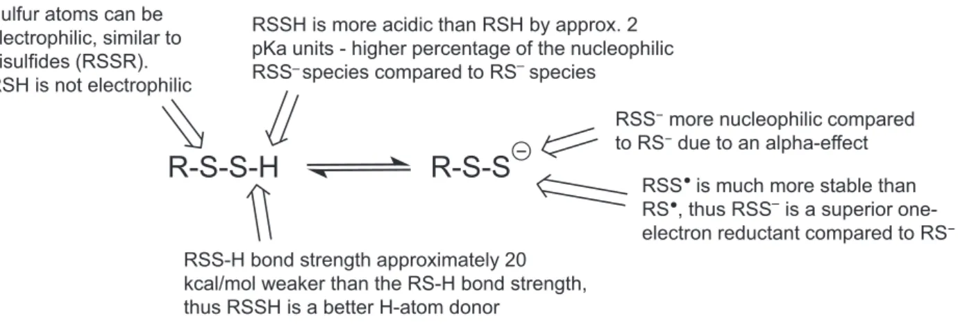
distinct chemistry. Thus, in order to speculate as to the biological functions of hydropersulfides and poly- sulfides, it is essential to first have a discussion regard- ing the chemistry of hydropersulfides and polysulfides and compare these chemical properties to other biolog- ically relevant sulfur species. As mentioned above, hydropersulfides are superior to thiols as nucleophiles and reducing agents (Fig. 3). Although not yet estab- lished, it can also be predicted that the metal ligation properties are distinct. It has been proposed that RSSH is equivalent to a chemically hyperactivated RSH which led to the idea that “anything a thiol can do, a hydropersulfide can do better” [25]. Although this can be why hydropersulfides are made in some cases (to enhance the activity of thiol proteins), it is at least safe to say that hydropersulfides possess distinct chemistry from that of simple thiols and that this may be important to the biological utility of hydropersul- fides. One important distinction between thiols and hydropersulfides is the fact that thiols can be nucle- ophilic while hydropersulfides can be either nucle- ophilic or electrophilic depending on the protonation state. That is, a persulfide anion (RSS) is extremely nucleophilic while the protonated RSSH species is elec- trophilic (akin to an oxidized sulfur species like a disulfide or sulfenic acid). Thus, hydropersulfides have the properties of both thiols and disulfides. It is also noteworthy that RSSH is oxidized relative to RSH (RSSH is at the same oxidation state as RSSR) and yet RSSH/RSS is a superior reductant compared to RSH/RS. It may seem counterintuitive that the oxi- dation of a molecule results in the generation of a superior reductant, but this is exactly the case with the conversion of a thiol to a hydropersulfide. Indeed, this situation led to the idea that RSSH generation can be a biological response to cellular oxidative stress since an oxidized species generated under this stress is actu- ally a superior reductant capable of possibly protecting against potentially deleterious oxidation events (due to its reducing capabilities, vide infra).
Thiyl radicals (RS) are used biologically as potent oxidants capable of, for example, breaking a C-H bond viahydrogen atom abstraction [6] and thus initi- ating radical chemistry. It has been postulated that RS species may be involved in fundamental cellular redox signaling as well [40]. The fact that RS is a fairly potent one-electron oxidant is an indication of the relatively poor ability of RSH to serve as a reduc- tant. On the other hand, perthiyl radicals (RSS) are relatively weak oxidants and are readily formed from one-electron oxidation of RSSH/RSS [41,42]. RSSH/
RSS is a good reductant due to the stability of the oxidized RSS species. The stability of RSS is akin to
the stabilization of analogous nitroxide systems (R2NO). In both cases, the stability of the unpaired electron is likely due to an overlap of the orbital con- taining the unpaired electron with the adjacent orbital containing a lone pair of electrons [43]. The lack of reac- tivity of RSS is evidenced by the fact that it does not readily react with O2 and NO, both established scav- engers of other radical species [41,44]. Although it has been proposed that the one-electron redox properties of RSSH/RSS may have biological function (i.e., can serve as a “redox gate” [41]), this has yet to be shown.
Thus far, only the chemical biology of hydropersul- fides has been discussed. As briefly mentioned above, polysulfides are also prevalent (albeit at lower levels than, for example, Cys-SSH or GSSH). Like disulfides, polysulfides are electrophilic and thus readily react with nucleophiles. As “soft” electrophiles, these sulfur- based species react primarily with “soft” nucleophiles, such as thiols, phosphines and cyanide [45,46]. The reaction of a nucleophile with a polysulfide presum- ably occursviadirect displacement of a sulfur contain- ing leaving group (Fig.4). For example, considering the reaction of an electrophilic disulfide with an appro- priate nucleophile (e.g., a thiolate), the site of nucle- ophilic attack is primarily governed by the quality of the corresponding leaving group. That is, the most electrophilic sulfur, which is the site of nucleophilic attack, is the one that is attached to the best leaving group (i.e., the thiolate with a conjugate acid having the lowest pKa) [47]. In the case of higher order poly- sulfides, the same trend is expected. Importantly, the pKa of an RSSH species is typically 1–2 pKa units lower than the corresponding RSH species [42] (and possibly much lower [32]), indicating that reactions with an RSS leaving group should predominate over a reaction with an RS leaving group. Thus, an RSSnR (n>1) species should be more electrophilic
than the corresponding disulfide. It is also important to note that increasing the length of the polysulfur chain should also increase the inherent nucleophilicity of the sulfur atoms (due to an extended alpha-effect [19,48]). Thus, an RSSSR species should be more nucleophilic compared to a simple disulfide. Although this is chemically expected and justifiable, quantitation of this phenomenon has not been reported. Regardless, the chemical biology of higher order polysulfides can be distinct from disulfides and these differences may be important to their biology (although this has not been specifically addressed thus far).
Physiological implications and future work
Hydropersulfides are biosynthesized, incorporated into proteins and present as small molecules/peptides.
Numerous descriptive studies have appeared in the lit- erature reporting that the presence of hydropersulfides in proteins can elicit a functional change. Despite this, the overriding question remains: What is it that hydropersulfides specifically do? What is unique about hydropersulfides that prompted Nature to adopt them and evolve numerous synthetic processes to generate them (both as small molecule species and in proteins)?
Is there something unique and novel about the hydropersulfide functional group that distinguishes it
R-S-S-H
Sulfur atoms can be electrophilic, similar to disulfides (RSSR).
RSH is not electrophilic
RSSH is more acidic than RSH by approx. 2 pKa units - higher percentage of the nucleophilic RSS–species compared to RS–species
R-S-S
−RSS-H bond strength approximately 20 kcal/mol weaker than the RS-H bond strength, thus RSSH is a better H-atom donor
RSS−more nucleophilic compared to RS−due to an alpha-effect
RSS is much more stable than RS , thus RSS–is a superior one- electron reductant compared to RS−
Fig. 3.Comparative properties of RSS/RSSH versus RS/RSH.
R-S-S
n-R
−
R-S-Nuc +
−S
n-R
n > 1
Nuc:
Fig. 4.Reaction of a nucleophile with a polysulfur species.
from, for example, thiols and/or selenols and are these unique properties a necessary aspect of fundamental biological/physiological signaling? At present, there are no clear or definitive answers to these questions, although some speculative proposals have been for- warded based on what is currently known regarding the distinct and novel chemistry of these species (vide infra). For example, the enhanced reductive capa- bilities and nucleophilicity of hydropersulfides com- pared to thiols may predict that their formation could be the result of cellular exposure to oxidative and/or electrophilic stress. Interestingly, proteins involved in Cys-SSH biosynthesis can be induced under oxidative/
electrophilic stress, consistent with the idea that their synthesis can be a response to these stresses [34]. In this regard, it is important to reiterate that a hydroper- sulfide is an oxidized thiol and yet is a superior reduc- tant compared to a thiol. That is, the oxidation of a thiol to a hydropersulfide, which could occur under oxidizing cellular conditions, generates a superior reductant. The seemingly incongruous and counterin- tuitive situation that a superior reductant is made via oxidation may be an important factor to the biological utility of hydropersulfides. To be sure, this is not a completely novel chemical phenomenon. For example, oxidation of an amine can generate a hydroxylamine, which is a superior one-electron reductant compared to the corresponding amine. The generation of a hydropersulfide in the face of oxidative/electrophilic stress may serve to counteract the deleterious effects associated with these stresses, akin to the way glu- tathione/glutathione transferase scavenges elec- trophiles, and antioxidants such as ascorbate or the tocopherols limit oxidative radical damage. Impor- tantly, Paul and Snyder proposed that hydropersulfide formation in proteins may possibly protect the protein cysteine thiol from oxidation under oxidative stress [49]. However, the chemical nature of this protection was not discussed and it should be understood that conversion of a thiol to a hydropersulfide does repre- sent an oxidation. More recently, it has been proposed that conversion of a protein thiol to a hydropersulfide results in a species that when oxidized further is easily reversible (since oxidation of the sacrificial sulfane sul- fur would be expected) under cellular reducing condi- tions [25,50]. Thus, irreversible oxidative modifications of protein hydropersulfides would not occur as they would for thiol functions in proteins. Importantly, selenoproteins function similarly in that they are also resistant to overoxidation to irreversibly modified spe- cies [38,51].
Clearly, other aspects of hydropersulfide chemistry may be important to their biological utility. Although
not unequivocally demonstrated, the formation of a hydropersulfide at a protein active site cysteine could confer increased nucleophilic reactivity, possibly increas- ing enzyme activity. Thus, thiol proteins that may have important function in an oxidative and/or electrophilic stress environment may have increased activity if con- verted to a hydropersulfide. Also, as alluded to previ- ously, the more physiologically accessible redox couple for RSS/RSS compared to RS/RS may allow the oxidation of a protein cysteine to the corresponding hydropersulfide to serve as an electron transfer “redox gate” [41]. That is, electron transfer through an RS/ RScouple is prohibitive since RS is too oxidizing and RS is too poor a reductant – however, oxidation of the protein thiol to a hydropersulfide could allow elec- trons to flow since RSS is a reasonably good reduc- tant and RSS is not nearly as oxidizing as RS.
Moreover, RSS has been shown to be relatively unre- active toward the paramagnetic signaling species NO and O2, allowing the RSS/RSScouple to be usable in aerobic and NO-rich environments [41,44]. It should be stressed that this idea is speculative at this time and there are no documented examples of this phenomenon.
This is presented only because the chemical properties would predict such a scenario.
Clearly, there is a great deal of work to be done to fully elucidate hydropersulfide physiology and the associated mechanisms. The recent work of the Akaike group demonstrating translational incorporation of cysteine hydropersulfides into proteins reveals the importance of hydropersulfides in fundamental bio- chemistry and provides ample impetus for the further elucidation of hydropersulfide chemistry and biology.
Some of the more pressing questions regarding biologi- cal hydropersulfides are the following:
How is hydropersulfide incorporation into proteins regulated?
Protein hydropersulfides are translationally incorporated via the actions of CARS. What regulates this activity (i.e., what are the factors that determine whether CARS incorporates Cys-SH or Cys-SSH into proteins)? Also, protein hydropersulfides can conceivably be incorpo- rated into proteins via sulfur exchange chemistry (i.e., protein cysteines reacting with hydropersulfides or oxi- dized protein cysteines reacting with H2S). With this in mind, it will be important to determine which process, translational incorporation or post-translational sulfur exchange chemistry, is the most relevant and is the rele- vance variable depending on cellular conditions? It will also be important to determine the specific roles (direct or indirect) of the transsulfuration enzymes such as 3-
mercaptopyruvate sulfurtransferase (3MST), which is another protein capable of synthesizing hydropersulfides, in hydropersulfide production.
The temporal aspects of hydropersulfides in pro- teins represent an important issue in delineating the signaling and function of this modification. Thus, it is essential to determine how and when hydropersulfides are generated as well as determine how and when they are removed. In this regard, Doka and cowork- ers [28] have reported that intracellular protein hydropersulfide levels are highly dependent on the presence and activity of thioredoxin (Trx), an impor- tant enzyme involved in the reduction of oxidized thiol proteins. Thus, it will be important to consider the expression, activity, and regulation of Trx (and possibly other reductases) when speculating on the function and signaling associated with protein hydropersulfides.
What determines which protein cysteines end up as hydropersulfides?
It appears that only certain cysteine residues in proteins are “sulfurated” ([33] for a discussion of this term) dur- ing translation, and possibly during sulfur exchange pro- cesses. Since amino acid incorporation into proteins is governed by codons, it will be important to discern how CARS interprets codons as a mechanism for specific hydropersulfide incorporation into proteins. With respect to post-translational sulfuration, it is appropriate to mention studies of the enzyme rhodanese, a protein with a catalytic cysteine that is known to be sulfurated. Rho- danese is capable of transferring a sulfane sulfur from a donor molecule to cyanide ion (CN) to make thio- cyanate (SCN) via an intermediary catalytic cysteine hydropersulfide. Numerous proteins have been identified to possess a “rhodanese homology domain” associated with their catalytic cysteine residues, implicating these residues as potential sites of sulfuration [14,18,52]. For example, the phosphatase Cdc25, involved in cell-cycle regulation, possesses a catalytic rhodanese homology domain [53], possibly implicating persulfide formation as a regulatory mechanism in this system.
What cellular conditions are conducive to hydropersulfide formation?
It has been hypothesized that oxidative and/or elec- trophilic stress can lead to increases in hydropersul- fide generation. This idea is largely based on the fact that hydropersulfides are superior to thiols as reduc- tants and nucleophiles; chemical attributes that may be important in combating oxidative and electrophilic
stressors. In partial support of this idea, it has been shown that increased expression of CSE in A549 cells (a lung epithelial cell line) led to increased intracellular hydropersulfides and increased resistance to H2O2 toxicity [27]. Although this result is consis- tent with the idea that hydropersulfides are protective against oxidative/electrophilic stress, clearly further work must be performed to fully address this hypothesis.
Why are there multiple biosynthetic pathways for hydropersulfides?
The recent discovery of CARS-mediated Cys-SSH gen- eration begs the question of why there are multiple and biochemically distinct processes capable of making the same molecules (i.e., CSE/CBS, CARS, etc.).
Clearly, some rationale can include compartmentaliza- tion of Cys-SSH for disparate biochemical purposes, differential regulation for accommodating varying cel- lular stresses/signals or simple redundancy to ensure the availability of a crucial biological effector. Answer- ing this fundamental question will greatly assist in determining the physiological significance of hydroper- sulfides. Regardless, the fact that there are multiple processes to generate Cys-SSH is consistent with the idea that hydropersulfides are important biological species with varied and essential functions.
How does hydropersulfide incorporation into a protein affect protein activity/function?
As mentioned above, numerous proteins have been reported to possess hydropersulfide residues, at least part of the time. However, it is not clear how hydrop- ersulfides affect the activity and function of all of these proteins. Based on the current literature, it appears that hydropersulfide formation mostly confers a loss or decrease in enzyme activity. For example, the cys- teine-hydrolase papain [22] and the cysteine dehydro- genase GAPDH [26] are inhibited when sulfurated.
Sulfuration of erythrocyte enzymes via exposure to a hydropersulfide generating system led to the inhibition of numerous enzymes [54]. Similarly, liver enzymes treated with CSE/cystine (a system that generates Cys- SSH) were also found to be either inhibited or unaf- fected [55]. However, hydropersulfide formation in the protein parkin has been reported to enhance its cat- alytic activity, possibly leading to a protection against the development of Parkinson’s disease [56]. Further studies are clearly warranted to investigate the possible alteration of protein activity as well as determination of possible functional changes.
What are the chemical properties that are important to the biological utility of hydropersulfides?
As discussed previously, hydropersulfides should pos- sess all of the chemical properties of a thiol, albeit enhanced. Although this is clear, it is not known which, if any, of these chemical properties are impor- tant to hydropersulfide function in biological systems.
The enhanced chemistry may not yet be realized at the active sites of proteins. It should also be noted that the chemistry of a hydropersulfide function is, to a cer- tain extent, partly disulfide and partly selenol. Since selenols and disulfides are already established biolog- ical effectors, it will be important to determine how and why the functions of hydropersulfides are dis- tinct from the functions of these species. Also, the one-electron redox chemistry of hydropersulfides and thiols are distinct, with RSS/RSSH being superior reductants compared to RS/RSH and RSS being much poorer oxidants compared to RS. Nature has found a use for RSas an oxidant in numerous enzy- matic systems [6]. Since RS is a much stronger oxi- dant than RSS, the use of RSS as an oxidant in analogous systems is not expected. The question remains, however, whether Nature has also found a use for RSS or the RSS/RSS redox couple. The stability of RSSindicates that RSS/RSSH can con- ceivably act as a one-electron reductant similar to the antioxidants ascorbate and tocopherols. Indeed, the antioxidant properties of RSSH have already been described [42] and it is intriguing to speculate that endogenously generated hydropersulfides also function in this manner. Interestingly, numerous enzymes proposed to possess hydropersulfides also have redox activity. For example, xanthine oxidase, an enzyme that catalyzes the oxidation of (hypo)xan- thine with subsequent reduction of O2 to superoxide ( O2 ), was among the first to be proposed to contain a hydropersulfide [10]. Regardless, it remains to be determined whether the RSS/RSS couple has bio- logical utility.
What are the biological roles and functions of the higher order polysulfide species?
Although this article has focused mostly on RSSH spe- cies, it has been mentioned that polysulfides (RSSnR, n>1, R=hydrogen or alkyl) are also prevalent and related to hydropersulfides. Indeed, the generation of these higher order polysulfides is a result of H2S and hydropersulfide formation since all of these species can be relatedvia a series of equilibria. In fact, it is going
to be difficult to distinguish among the possible bio- logical activities of all these species since they are related to each other via equilibrium reactions and, therefore, can all be present simultaneously in a bio- logical milieu. Regardless, it is entirely possible, if not probable, that polysulfides are not merely storage forms of H2S or RSSH species, but rather possess important biological activities and properties on their own. Discerning these activities/properties will be important in the full elucidation of the chemical biol- ogy and physiology of the hydropersulfides.
Are hydropersulfides involved in metal/
metalloprotein biology?
Thus far, much of the focus on hydropersulfide bio- chemistry has primarily involved interactions with other sulfur species (vide supra). Due to the presumed novel ligation and redox properties of hydropersulfides, met- als and metalloproteins are also likely sites of biological reactivity. Previous studies have demonstrated that the RSSH species can reduce ferric heme proteins such as cytochrome c under conditions where thiols or H2S do not [22] (and, as mentioned above, the biological rele- vance/utility of the one-electron redox properties of hydropersulfides are worthy of further investigation). It also seems possible, if not likely, that hydropersulfides will interact with biologically nonredox active metal ions such as Zn2+ as well. As a borderline “soft” ion, Zn2+ can coordinate a wide array of biological ligands containing S, N, and O atoms. For example, Zn-finger proteins bind Zn2+ viahistidine nitrogens and cysteine thiols [57]. The regulation of Zn2+“redistribution” (i.e., movement of Zn2+ from storage sites to protein bind- ing sites) is an important field due to the prevalence and of Zn-containing proteins in signaling fundamental biochemistry [58]. Indeed, one mechanism of mobiliza- tion of Zn2+ from sites of sequestration is thought to beviaredox chemistry at Zn-binding thiolates, coupling redox signaling with Zn-dependent signaling/biochem- istry. It is intriguing to speculate that cysteine persulfide anions can also be involved in Zn2+ coordination, allowing a different redox couple (compared to thiolate) to affect Zn2+ binding, release and/or mobilization.
Moreover, it is highly likely that the coordination chemistry of a persulfide anion versus a thiolate will be significantly different, thus altering the affinity of pro- teins for Zn2+ and possibly resulting in a chemically distinct pool of Zn2+ (e.g., where the redox chemistry of the Zn2+ ligands allows for release under different redox conditions).
One aspect of the interaction of hydropersulfides with metals in a biological system that has been
examined is the effect of persulfides on electrophilic heavy metal toxicity [59]. Abiko and coworkers [60]
found that detoxification of methyl mercury can occur viainteraction with H2S, hydropersulfides and polysul- fides. Similarly, Cd2+ toxicity can also be abated by the presence of hydropersulfides [61]. Importantly, Zn2+and Cd2+are chemically and biochemically sim- ilar in that they both readily bind thiol proteins and indeed a facet of Cd2+ toxicity is thought to involve interference with Zn2+homeostasis [62]. The fact that Cd2+and Zn2+ are chemically similar and persulfides alter the pathobiology of Cd2+ is consistent with the idea that endogenous persulfides can also be involved in Zn2+regulation.
Are hydropersulfides involved in the etiology of disease or involved in redox signaling?
One of the most important aspects of hydropersulfide/
polysulfide chemical biology is with respect to their possible roles in disease. As alluded to above, hydrop- ersulfides possess the proper chemical properties to protect cells from certain chemical stresses, although this remains to be established. Moreover, hydropersul- fides possess chemical properties that are distinct from thiols (and other derived species), allowing them to perform biochemistry not available to simple thiols.
Considering all of this, these species may be involved in many diseases, disorders or developmental pathways whereby “oxidative” or electrophilic stress and/or sig- naling have been implicated and/or where thiol modifi- cation processes are proposed. Moreover, if hydropersulfides are fundamental to cellular protec- tion, they may also be important aspects of, for exam- ple, cancer cell viability, neuronal cell degeneration, regulation of apoptosis, and/or redox-mediated devel- opmental pathways. These are highly speculative ideas, but it is clear that the impact of hydropersulfides on disease states, disorders, developmental signaling, or cellular protection is potentially significant.
The points listed above likely represent only the
“tip-of-the-iceberg” with regard to pressing questions in this field. Moreover, details of the physiological implications of hydropersulfides and polysulfides remain to be addressed once some of the above ques- tions are answered. It appears likely that the current ideas regarding the biological signaling associated with thiol sulfur modification will need to be revisited and redefined in the light of the recent findings pertaining to the prevalence of hydropersulfides and polysulfides.
Moreover, hydropersulfide/polysulfide-derived species that are analogous to established thiol-derived species will also be essential to include in any conversation
involving sulfur species. For example, thiol oxidation to the corresponding sulfenic acid is a well-known and established process under certain biological conditions [63]. It will now be equally important to consider the analogous reaction with hydropersulfides under those same conditions. That is, oxidation of a hydropersul- fide to the corresponding perthiosulfenic acid (RSSOH) is an equally likely, if not more likely, fate for hydrop- ersulfides [64]. It is unnecessary to list all of the biologi- cally relevant thiol modification reactions but yet important to indicate that any type of thiol modifica- tion reaction (many of which have been associated with thiol-based signaling) can occur and are likely with hydropersulfides, possibly with important signaling/bio- chemical consequences. Regardless, the potential impor- tance and impact of hydropersulfides and polysulfides on the fields of fundamental biochemistry and (patho)- physiology, among many others, appear significant and burgeoning and, in our opinion, it would be unwise not to consider these novel species as important molecules in the chemical biology of nature.
References
1 Go YM and Jones DP (2013) Thiol/disulfide redox states in signaling and sensing.Crit Rev Biochem Mol Bio48, 173–191.
2 Paulsen CE and Carroll KS (2013) Cysteine-mediated redox signaling: chemistry, biology and tools for discovery.Chem Rev113, 4633–4679.
3 Winterbourn CC and Hampton MB (2008) Thiol chemistry and specificity in redox signaling.Free Radic Biol Med45, 549–561.
4 Reddie KG and Carroll KS (2008) Expanding the functional diversity of proteins through cysteine oxidation.Curr Opin Chem Biol12, 746–754.
5 Jacob C, Giles GI, Giles NM and Sies H (2003) Sulfur and Selenium: the role of oxidation state in protein structure and function.Angew Chem Int Ed42, 4742–4758.
6 Stubbe J and van der Donk WA (1998) Protein radicals in enzyme catalysis.Chem Rev98, 705–762.
7 Bonnet D, Stevens JM, de Sousa RA, Sari MA, Mansuy D and Artaud I (2001) New inhibitors of iron- containing nitrile hydratases.J Biochem130, 227–233.
8 Munchberg U, Anwar A, Mecklenburg S and Jacob C (2007) Polysulfides as biologically active ingredients of garlic.Org Biomol Chem5, 1505–1518.
9 Calabrese L, Federici G, Bannisterm WH, Bannister JV, Rotilio G and Finazzi-Agro A (1975) Labile sulfur in human superoxide dismutase.Eur J Biochem56, 305–309.
10 Massey V and Edmondson D (1970) On the mechanism of inactivation of xanthine oxidase by cyanide.J Biol Chem245, 6595–6598.
11 Branzoli U and Massey V (1974) Evidence for an active site persulfide residue in rabbit liver aldehyde oxidase.
J Biol Chem249, 4346–4349.
12 Nagahara N, Nagano M, Ito T and Suzuki H (2016) Redox regulation of mammalian 3-mercaptopyruvate sulfurtransferase.Methods Enzymol554, 229–254.
13 Zheng L, White RH, Cash VL and Dean DR (1994) Mechanism for the desulfurization of L-cysteine catalyzed by the nofS gene product.Biochemistry33, 4714–4720.
14 Cipollone R, Ascenzi P and Visca P (2007) Common themes and variations in the rhodanese superfamily.
IUBMB Life59, 51–59.
15 Mueller EG (2006) Trafficking in persulfides: delivering sulfur in biosynthetic pathways.Nat Chem Biol2, 185–194.
16 Toohey JI (1989) Sulphane sulphur in biological systems: a possible regulatory role.Biochem J264, 625–632.
17 Wood JL (1987) Sulfane sulfur.Methods Enzymol143, 25–29.
18 Toohey JI (2011) Sulfur signaling: is the agent sulfide or sulfane.Anal Biochem412, 1–7.
19 Nagy P (2015) Mechanistic chemical perspective of hydrogen sulfide signaling.Methods Enzymol554, 3–29.
20 Blackstone E, Morrison M and Roth MB (2005) H2S induces a suspended animation-like state in mice.
Science308, 518.
21 Cavallini D, Federici G and Barboni E (1970)
Interactions of proteins with sulfide.Eur J Biochem14, 169–174.
22 Francoleon NE, Carrington SJ and Fukuto JM (2011) The reaction of H2S with oxidized thiols: generation of persulfides and implications to H2S biology.Arch Biochem Biophys516, 146–153.
23 Mustafa AK, Gadalla MM, Sen N, Kim S, Mu W, Gazi SK, Barrow RK, Yang G, Wang R and Snyder SH (2009) H2S Signals through protein S-sulfhydration.
Sci Sign2, 1–8.
24 Pan J and Carroll KS (2013) Persulfide reactivity in the detection of protein S-sulfhydration.ACS Chem Biol8, 1110–1116.
25 Ono K, Akaike T, Sawa T, Kumagai Y, Wink DA, Tantillo DJ, Hobbs AJ, Nagy P, Xian M, Lin Jet al.
(2014) The redox chemistry and chemical biology of H2S, hydropersulfides and derived species: implications of their possible biological activity and utility.Free Radic Biol Med77, 82–94.
26 Jarosz AP, Wei W, Gauld JW, Auld J, Ozcan F, Aslan M and Mutus B (2015) Glyceraldehyde 3-phosphate dehydrogenase (GAPDH) is inactivated by
S-sulfurationin vitro.Free Rad Biol Med89, 512–521.
27 Ida T, Sawa T, Ihara H, Tsuchiya Y, Watanabe Y, Kumagai Y, Suematsu M, Motohashi H, Fujii S, Matsunaga Tet al.(2014) Reactive cysteine persulfides
and S-polythiolation regulate oxidative stress and redox signaling.Proc Natl Acad Sci USA111, 7606–7611.
28 Doka E, Pader I, Biro A, Johansson K, Cheng Q, Ballago K, Prigge JR, Pastor-Flores D, Dick TP, Schmidt EEet al.(2016) A novel persulfide detection method reveals protein persulfide- and polysulfide- reducing function of thioredoxin and glutathione systems.Sci Adv2, e1500968.
29 Longen S, Richter F, Kohler Y, Wittig I, Beck KF and Pfeilschifter J (2016) Quantitative persulfide site identification (qPerS-SID) reveals protein targets of H2S releasing donors in mammalian cells.Sci Rep6, 29808.
30 Akaike T, Ida T, Wei FW, Nishida M, Kumagai Y, Alam M, Ihara H, Sawa T, Matsunaga T, Kasamatsu S et al.(2017) Cysteinyl-tRNA synthase governs cysteine polysulfidation and mitochondrial bioenergetics.Nat Comm8, 1–15.
31 Vasas A, Doka E, Fabian I and Nagy P (2015) Kinetic and thermodynamic studies on the disulfide-bond reducing potential of hydrogen sulfide.Nitric Oxide46, 93–101.
32 Cuevasanta E, Lange M, Bonanata J, Coitino EL, Ferrer-Sueta G, Filipovic MR and Alvarez B (2015) Reaction of hydrogen sulfide with disulfide and sulfenic acid to form the strongly nucleophilic persulfide.J Biol Chem290, 26866–26880.
33 Toohey JI (2012) The conversion of H2S to sulfane sulfur.Nat Rev Mol Cell Biol13, 803.
34 Alvarez L, Bianco CL, Toscano JP, Lin J, Akaike T and Fukuto JM (2017) The chemical biology of hydropersulfides and related species: possible roles in cellular protection and redox signaling.Antioxid Redox Signal27, 622–633.
35 Turanov AA, Xu XM, Carlson BA, Yoo MH, Gladyshev VN and Hatfield DL (2011) Biosynthesis of selenocysteine, the 21stamino acid in the genetic code, and a novel pathway for cysteine biosynthesis.Adv Nutr2, 122–128.
36 Lu J and Holmgren A (2009) Selenoproteins.J Biol Chem284, 723–727.
37 Srinivasan G, James CM and Krzycki JA (2002) Pyrrolysine encoded by UAG in Archaea: charging of a UAG-decoding specialized tRNA.Science296, 1459–1462.
38 Reich HJ and Hondal RJ (2016) Why Nature chose selenium.ACS Chem Biol11, 821–841.
39 Sen N, Paul BD, Gadalla MM, Mustafa AK, Sen T, Kim S and Snyder SH (2012) Hydrogen sulfide-linked sulfhydration of NF-jbmediates its antiapoptotic actions.Mol Cell45, 13–24.
40 Winterbourn CC (2015) Are free radicals involved in thiol-based redox signaling?Free Radic Biol Med80, 164–170.
41 Bianco CL, Chavez TA, Sosa V, Nguyen QNN, Tantillo DJ, Ichimura AS, Toscano JP and Fukuto JM
(2016) The chemical biology of the persulfide (RSSH)/
perthiyl (RSS) redox couple and possible role in biological redox signaling.Free Rad Biol Med101, 20–31.
42 Everett SA and Wardman P (1995) Perthiols as antioxidants: radical-scavenging and prooxidative mechanisms.Methods Enzymol251, 55–69.
43 Mahoney LR, Mendenhall GD and Ingold KU (1973) Calorimetric and equilibrium studies on some stable nitroxide and iminoxyl radicals. Approximate O-H bond dissociation energies in hydroxylamines and oximes.J Am Chem Soc95, 8610–8614.
44 Chauvin JR, Griesser M and Pratt DA (2017) Hydropersulfides: H-Atom transfer agents par excellance.J Am Chem Soc139, 6484–6493.
45 Saund SS, Sosa V, Henriquez S, Nguyen QNN, Bianco CL, Soeda S, Millikin R, White C, Le H, Tantillo DJ et al.(2015) The chemical biology of hydropersulfides (RSSH): chemical stability, reactivity and redox roles.
Arch Biochem Biophys588, 15–24.
46 Pearson RG (1987) Recent advances in the concept of hard and soft acids and bases.J Chem Educ64, 561–567.
47 Hiskey RG and Carrol FI (1961) Chemistry of aliphatic disulfides. II. Cyanide cleavage of unsymmetrical disulfides.J Am Chem Soc83, 4644–4647.
48 Fina NJ and Edwards JO (1973) The alpha effect. A review.Int J Chem Kin5, 1–23.
49 Paul BD and Snyder SH (2012) H2S Signaling through protein sulfhydration and beyond.Nat Rev Mol Cell Biol13, 499–506.
50 Millikin R, Bianco CL, White C, Saund SS, Henriquez S, Sosa V, Akaike T, Kumagai Y, Soeda S, Toscano JP et al.(2016) The chemical biology of protein
hydropersulfides: studies of a possible protective function of biological hydropersulfide generation.Free Rad Biol Med97, 136–147.
51 Snider GW, Ruggles E, Kahn N and Hondal RJ (2013) Selenocysteine confers resistance to inactivation by oxidation in thioredoxin reductase: comparison of selenium and sulfur enzymes.Biochemistry52, 5472–5481.
52 Bordo D and Bork P (2002) The rhodanese/Cdc25 phosphatase superfamily.EMBO Rep3, 741–746.
53 Hofmann K, Bucher P and Kajava AV (1998) A model of Cdc25 phosphatase catalytic domain and Cdk-interaction surface based on the presence of a
rhodanese homology domain.J Mol Biol 282, 195–208.
54 Valentine WN, Toohey JI, Paglia DE, Nakatani M and Brockway RA (1987) Modification of ertythrocyte enzyme activities by persulfides and methanethiol:
possible regulatory role.Proc Natl Acad Sci USA84, 1394–1398.
55 Ogasawara Y, Suzuki T, Ishii K and Tanabe S (1997) Modification of liver cytosol enzyme activities promotedin vitroby reduced sulfur species genertated from cystine withc-cystathionase.Biochim Biophys Acta1334, 33–43.
56 Vandiver MS, Paul BD, Xu R, Karuppagounder S, Rao F, Snowman AM, Ko HS, Lee YI, Dawson VL, Dawson TMet al.(2013) Sulfhydration mediates neuroprotective actions of parkin.Nat Commun4, 1626.
57 Laity JH, Lee BM and Wright PE (2001) Zinc finger proteins: new insights into structural and functional diversity.Curr Opin Struct Biol11, 39–46.
58 Krezel A, Hao Q and Maret W (2007) The zinc/thiolate redox biochemistry of metallothionein and the control of zinc ion fluctuations in cell signaling.Arch Biochem Biophys463, 188–200.
59 Kumagai Y and Abiko Y (2017) Environmental electrophiles: protein adducts, modulation of redox signaling and interaction with persulfides/polysulfides.
Chem Res Toxicol30, 203–219.
60 Abiko Y, Yoshida E, Ishii I, Fukuto JM, Akaike T and Kumagai Y (2015) Involvement of reactive persulfides in biological bismethylmercury sulfide formation.Chem Res Toxicol28, 1301–1306.
61 Shinkai Y, Masuda A, Akiyama M, Xian M and Kumagai Y (2017) Cadmium-mediated activation of the HSP90/HSF1 pathway regulated by reactive persulfides/
polysulfides.Toxicol Sci156, 412–421.
62 Martelli A, Rousselet E, Dycke C, Bouron A and Moulis JM (2006) Cadmium toxicity in animal cells by interference with essential metals.Biochemie88, 1807–1814.
63 Conte ML and Carroll KS (2013) The redox
biochemistry of protein sulfenylation and sulfinylation.
J Biol Chem288, 26480–26488.
64 Heppner DE, Hristova M, Ida T, Mijuskovic A, Dustin CM, Bogdandi V, Fukuto JM, Dick TP, Nagy P, Li J et al.(2018) Cysteine perthiosulfenic acid (Cys-SSOH):
a novel intermediate in thiol based redox signaling?
Redox Biol14, 379–385.