The operative treatment of unstable pelvic ring injuries:
biomechanical and clinical rewiew Ph.D thesis
Dr. Tamás Bodzay
Clinical Medicine Doctoral School Semmelweis University
Supervisor:Dr. László Hangody MD, D.Sc
Official reviewers:
Dr. Sándor Pintér MD, Ph.D.
Dr. Attila Pavlik MD, Ph.D
Head of the Final Examination Committee:
Dr. Emil Monos MD, D.Sc
Members of the Final Examination Committee:
Dr. Andrea Szabó MD, Ph.D.
Dr. Zoltán Detre MD, Ph.D.
Introduction:
The treatment of pelvic ring injuries is a challenge for trauma surgeons. With the development of motorization, there is an increase in the number of high energy traumas, including pelvis injuries. Since 1987, the research group at our department has been analyzing the treatment of pelvic ring injuries. The author entered the research group in 1994. The following dissertation will account for the biomechanical experiments performed on classical human cadaver pelvic dissections, a computerized finite element pelvic model which we developed including the results from our research, as well as our conclusions of the analysis of patients we treated between 1995 until 2005.
Pelvic ring injuries result from high energy trauma. Earlier, they were rare injuries, Magaigne observed 0.3% prevalence (Malgaigne 1847, 1859). In the past few decades, the frequency has risen to about 3-6% (Melton et al. 1981, Mucha et al. 1984, Ragnarsson et al. 1993). Until the end of the 1970’s, the majority of pelvis injuries were treated conservatively, and most patients died (Rothenberger et al. 1978, Henderson 1989). Although intensive care and surgical methods have significantly improved recently, the mortality of unstable pelvic ring fractures is nonetheless 35% (Rieger et al. 1993, Varney et al. 1990). If the mechanically unstable pelvic injury is combined with hemodynamic instability, the primary care involving stabilization of the pelvis is vital to prevent further bleeding.
The long term consequences of unstable pelvis injuries are aggravating: close to 75 percent of all cases (Ahlers et al. 1976, Probst et al. 1976) have complaints while standing, walking and sitting. The reasons are due to poor fracture position union leading to limb shortening and secondary spine deformities as well as malunion. Recent articles describe substantial deterioration in the quality of life in this injury type (Tornetta et al. 1996, Pohlemann et al.
1996).
The other reason for later complaints is due to accompanying injuries associated with pelvic ring injuries: injuries of the urogenital tract, blood vessels, nerves and the muscles of the pelvis (Hersche 1992, Moorehouse 1988).
Objective:
Vertically unstable, Type C pelvis injuries are an absolute indication for operative treatment.
Since these types of injuries result from high energy trauma, we can presume that there are many associated injuries (multiple injury patients), and that the soft tissue surrounding the pelvis and hip is not uncommon to be in critical condition (decollement). In both cases, it is important to perform a minimally invasive surgical procedure causing less strain to the patient, which can be easily learned, is not time consuming, and does not require any special implant. Previously, a “cobra” plate technique was used in the therapy of pelvic ring fractures.
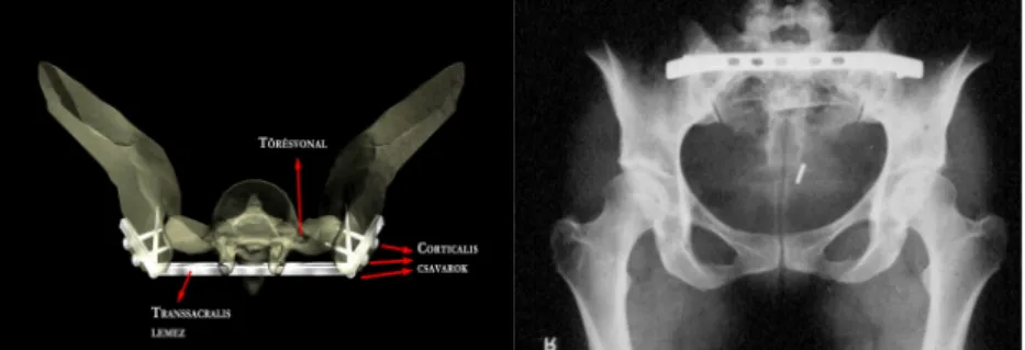
The question is whether the previous technique along with our modifications (indirect reduction, extrafocal approach, iliac crest grove, subcutaneous tunnel technique) provides enough stability next to the less operative strain it offers. (Figure 1)
Figure 1: schematic and radiograph of transsacral plate synthesis
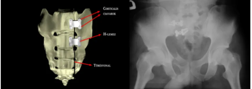
We chose the following method to answer the above mentioned question: Using fresh, non- frozen cadaver bone-ligament pelvis dissections, we modeled a Type C unstable pelvic ring injury (syphyseolysis + Type I Denis sacrum fracture) and examined the stability of a transsacral plate synthesis. For comparison, we chose a theoretically rather stable surgical procedure (direct plate synthesis) rarely used due to the large invasiveness required. (Figure 2)
Figure 2: schematic and radiograph of direct plate synthesis (seen on the right side of the radiograph)
Parallel to this, we developed a computerized finite element pelvic model. We simulated the cadaver bone-ligament injury on the computerized model, and measured the stability of the previously mentioned two operative techniques. We compared the results of the finite element model to the bone-ligament dissections, and verified the realistic and practical qualities of the computerized model. We then examined the stability of two other surgical techniques – KFI- H plate synthesis (Figure 3) and iliosacral screw fixation (Figure 4) – on the validated computerized model, as well as the stability of all four operative techniques on another type of pelvic injury (symphyseolysis + Denis Type II sacrum fracture.
Figure 3: schematic and radiograph of KFI-H plate synthesis
Figure 4: schematic and radiograph of iliosacral screw fixation
We performed the retrospective analysis of pelvic ring injuries at our department between the years 1995 until 2005. We chose this interval because there were at least two years between the time of the injury and the examination, therefore the observed condition can be considered a functional final state. We used the standard accepted international scoring system for the analysis of the retrospective study. According to the results of the biomechanical examinations, we give recommendations regarding which implant and operative technique we
find the most optimal according to the localization of the posterior pelvic ring injury. The goal was to examine our therapeutic protocol for pelvic ring injuries and suggest any necessary changes to that protocol.
Methods:
Our cadaver experiments were performed at the Biomechanical Research Laboratory of Debrecen University, Hungary, Faculty of Medicine and Health Sciences. The experiments were approved by the DEOEC Ethics Committee and Research Ethical Subcommittee.
Twelve cadaver pelvises were used for biomechanical investigations. The specimens were not frozen; biomechanical tests were performed within one week following death. We modeled a Type-C pelvic ring injury: anteriorly, a symphysis pubis rupture was produced, which was stabilized with a 4-hole narrow DC plate (Synthes), while posteriorly, a Denis type I fracture was produced. The first stabilization technique was the direct plate synthesis, in which the anterior side of the sacrum is stabilized with two 3-hole reconstruction plates positioned less than 90º to each other on the linea terminalis. The second stabilization technique was the transsacral plate synthesis. Biomechanical testing was performed in a single limb stance model. The femur on the injured side was fixed to the biomechanical test instrument.
While we developed a computerized finite element model, we first simulated the cadaver experimental injuries on the computerized model. Then we compared the amount of dislocation on the finite element model to the cadaver results, thus validating the values. The finite element model is based on simple geometry; the bones, joints and ligaments were each individually represented. We differentiated between cancellous and compact bone surrounding the injury and the implants.
We used acknowledged data from literature for material properties. We measured load capacities of intact sacrum, Denis type I and a Denis Type II fractures while standing on both feet, using direct plate, transsacral plate, KFI-H plate synthesis and iliosacral screw fixations.
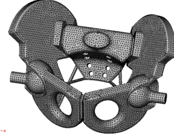
Finite element analysis was performed using the ALGOR system, in which four-node tetrahedron elements were used with 4 mm mean element size. We thus obtained a finite element model of about 90000 elements (Figure 5).
Figure 5: The finite element web
Clinical examinations: We analyzed patients treated at our department between the years 1995 and 2005. We chose this interval because in order to provide at least two years between the time of the injury and the examination, therefore the observed condition can be considered a functional final state. The objective of the retrospective analysis was to examine the radiological outcome as well as the general condition of the patient, their reintegration into their workplaces and to their families, not merely complaints localized in the pelvic area. We used the “outcome score” recommended by Pohlemann for the retrospective analysis of the study.
Results:
Cadaver experiments: Of the 12 specimens, we were not able to obtain results from 3 specimens due to mechanical failure, therefore we analyzed 9 specimens. The mean difference of movements on each side of the fracture at cyclic loads between 100 and 250 N was 0.19 mm in the two stabilization techniques. Statistical analysis was achieved using the ANOVA test: p=0.18, i.e. the difference was not significant.
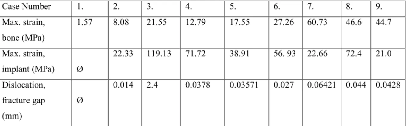
Results of the finite element analysis can be observed in Table 1.
Case Number 1. 2. 3. 4. 5. 6. 7. 8. 9.
Max. strain, bone (MPa)
1.57 8.08 21.55 12.79 17.55 27.26 60.73 46.6 44.7
Max. strain,
implant (MPa) Ø
22.33 119.13 71.72 38.91 56. 93 22.66 72.4 21.0
Dislocation, fracture gap (mm)
Ø
0.014 2.4 0.0378 0.03571 0.027 0.06421 0.044 0.0428
Table 1: Maximum strain in bones, implants and dislocations in the fracture gap at 500 N vertical loading.
Clinical examinations: Between 1995 and 2005, we treated 725 pelvis injured patients at our department. In 591 cases, operative treatment was not necessary, 133 patients underwent surgery and one patient died without surgical treatment. 115 patients survived the operation.
The gender ratio of the operated patients was 72 male and 61 female. Of the 133 operative cases, 60 were type B injuries, and 73 were type C injuries. The distribution of type B injuries was as follows: 33 cases type B1, 20 cases type B2, and 7 cases were type B3 injuries. The distribution of type C injuries was as follows: 48 cases type C1, 11 cases type C2, and 14 cases were type C3 injuries. The mean ISS was 28 (12-63).
Conclusions:
We examined the operative treatment of unstable, type C pelvic injuries with various methods. One of the methods was the standard biomechanical analysis of the bony- ligamentous cadaver specimens. We modeled the pelvic injury by bringing about a symphysis rupture and a Denis type I sacrum fracture. We experimented with two different operative stabilization techniques, in which the symphyseolysis was fixed with a DC plate and the posterior ring injury was stabilized first with a direct plate synthesis, then with a transsacral plate synthesis. The results demonstrate that the transsacral plate synthesis we used gives less stability, however in clinical practice it has not caused any disturbance in bone healing, implant loosening, nor implant breakage. In our opinion, the movements which occur when transsacral fixation is applied fall into the category of micromovements and - similar to other
type I sacrum fracture. Similar to the cadaver experiments, the two fixation methods used were “direct” plate and transsacral synthesis technique. We fixed the symphysis rupture with a narrow DC plate in both cases on the model. We then validated the model, i.e. we measured the amount of dislocation in the fracture gap on the computerized finite element model and compared those outcomes to the results of the cadaver experiments. As in the cadaver experiments, we modeled a standing position bearing weight on the injured extremity. The amount of movement in the fracture gap on the computerized model was in the same range as measured in the cadaver specimens. We thus found the model to be functional, and eligible for further analysis. We then imitated a symphysis rupture and Denis type I or type II sacrum fracture, standing on both feet, with the fixation methods we applied, such as direct plate synthesis, transsacral plate synthesis, KFI-H plate synthesis, and iliosacral screw fixation. The amount of movement between the bones surrounding the fracture was greater in a Denis type I injury than in a Denis type II fracture. The direct plate synthesis proved to be the most stable synthesis technique. The movements recorded using the KFI-H plate syntheses were somewhat greater than that of the iliosacral screw technique, but there was hardly any difference between the two techniques. Transsacral plate synthesis demonstrated the least amount of stability in Denis type I and II fractures.
Due to the ethical, hygienic, and technical difficulties of using cadaver bone-ligament pelvis specimens for analysis, only a limited amount of results could be gained. Therefore, the demand for computerized finite element modeling is increasing. An intact pelvis finite element model has been previously presented in literature (Dalstra et al. 1995); however, we were able to examine and compare the stability of the operative stabilization of two different types of injuries, possessing the previous results of our cadaver experiments. The finite element model can only produce approximate results compared to reality; therefore we must take into consideration the following sources of error: the fractured surfaces are not plane and we do not know its frictional coefficient, the finite element pelvis geometric model is only approximate, and the applied substance parameters in literature are not standardized.
In order to analyze the clinical data, the therapeutic protocol of our department must be demonstrated: injuries of the pelvic ring must be treated with surgery if they are mechanically unstable. We must distinguish those injuries, where mechanical instability is associated with hemodynamic instability. In type C pelvis injuries, we can count on 1500-2000 ml blood loss due to the pelvis injury alone. If the mechanically unstable pelvis injury is combined with hemodynamic instability, the goal of the primary care is to control the bleeding. In a hemodynamically unstable patient, the urgent reduction and stabilization of the pelvic ring is
necessary. The associated injuries - most importantly intraabdominal injuries – influence the course of treatment at this stage. If FAST or peritoneal lavage confirms intraabdominal bleeding, then laparotomy is performed after the reduction and temporary fixation of the pelvis. During the course of the operation, after the abdominal injuries are treated, the retro- infraperitoneum is approached. If the posterior pelvic ring injury can be visualized, then a plate synthesis is applied onto the pelvic cavity façade. It is emphasized, that the above mentioned stabilization technique through a transperitoneal approach is a “last resort”
operation. During the exploration of the extraperitoneal space of the pelvic cavity, bleeding is surgically controlled and the presacral area is tamponaded. If the anterior ring injury involves symphysiolysis, then at the end of the laparotomy, a plate synthesis is applied. If the posterior ring injury cannot be stabilized with a plate synthesis from the pelvic cavity during the laparotomy and tamponade, a temporary surgical fixation is applied. Similarly, if laparotomy is not necessary during the course of primary care, the reduction of the pelvic injury is also performed with a temporary surgical fixation. There exist two instruments for temporary fixation: the pelvic clamp, and external fixator device. Last, but not least, we must mention the primary definitive surgical treatment during the primary care of pelvic ring injuries. Many authors (Varga 1999) justify this tactic of treatment. Our opinion is that if the general condition of the patient is good, i.e. blood pressure is stabile and coagulation status is adequate, then the definitive operative treatment can be performed. However, this is only true in most cases of “monotrauma”. In addition, adequate material and personnel background is required. We insist that every trauma center should be capable of treating an unstable pelvis injury to an extent of saving a life, using a pelvic clamp or external fixator, or if necessary, tamponade. It is not expected however, that the sometimes difficult definitive operative treatment can be given by any hospital or any surgeon. The algorithm in figure 6 demonstrates the previously mentioned treatment protocol.
CIRCULATION UNSTABLE + PELVIS UNSTABLE ↓
NON-INVASIVE FIXATION (WRAPPED BEDSHEET or PELVIC BINDER) ↓
X-RAY+FAST→ POSITIVE→ CLAMP, LAPAROTOMY, TAMPONADE → ICU
CIRCULATION UNSTABLE → TAMPONADE→ CIRCULATION STABLE → ICU ↓
CIRCULATION UNSTABLE → ANGIOGRAPHY
Figure 6: Primary care tactic of hemodynamically and mechanically unstable pelvis injury
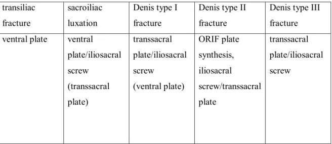
Table 2 demonstrates the definitive treatment of pelvic injuries. It is recommended that ventrally positioned plate synthesis should be used transiliac fractures and SI luxations, while local ORIF plate synthesis should be applied in cases of Denis type II fractures in which radicular lesion is observed, and iliosacral screw fixation or transsacral plate synthesis should be used in all other types of injuries.
transiliac fracture
sacroiliac luxation
Denis type I fracture
Denis type II fracture
Denis type III fracture ventral plate ventral
plate/iliosacral screw
(transsacral plate)
transsacral plate/iliosacral screw
(ventral plate)
ORIF plate synthesis, iliosacral
screw/transsacral plate
transsacral plate/iliosacral screw
Table 2: Localization of fracture and the recommended operative procedure
In relation to the functional outcome, more than half the cases with type B injuries belong to the excellent to good category, while more than half the cases suffering type C injuries have satisfactory to poor results.
Type B Type C
Excellent (7 points) 6 3
Good (6 points) 18 8
Satisfactory (4-5 points) 9 17
Poor (3 points) 2 10
Table 3: cumulative retrospective outcomes Author’s publications related to the topic of the dissertation:
1. Bodzay T, Burján T, Váradi K: Finite Element Modeling of Operative Treatment of Pelvic Ring Fractures
First Hungarian Conference on Biomechanics, 2004. Abstract Book
2. Bodzay T, Flóris I, Vendégh Zs, Szita J: Treatment of unstable pelvic injuries- a review of 10 years
European Journal of Trauma Vol 32. S 1
3. Bodzay T-Váradi K: Csont-implantátum kapcsolatok végeselemes modellezése
Halász Gábor, szerk: Modellezés a biomechanikában, BME, egyetemi tankönyv, 2007. 9, 371-395
4. Bodzay T - Burján T - Bagdi C - Flóris I - Vendégh Zs - Váradi K: Evaluation of stabilization methods of pelvic ring injuries by finite element modeling
Joint Diseases and Related Surgery 2007; 18(3) 108-115 2007 (BME egyetemi tankönyv) 9, 371-395
5. Bodzay T - Asbóth L - Szita J - Váradi K: Medencegyűrű-sérülések műtéti rögzítésének végeselemes modellezése; Biomechanica Hungarica 1: 37-47 2008
6. Bodzay T - Szita J - Flóris I: Medencetörések ellátásának modern szemlélete- minimálinvazív lehetőségek és kiterjesztett rekonstruktív ellátás
Orvosképzés 2010; 3 251-261
7. Flóris I - Bodzay T - Balázs P - Gál T - Karsay P: Acetabulum törések késői szövődményei miatt végzett csípőízületi arthroplasticák eredményei; Magy. Traumatol. Ortop.
2011;54(1):13-26
8. Bodzay T - Flóris I - Váradi, K: Comparison of stability in the operative treatment of
Other publications:
1. Bodzay T, Szita J, Laczkó T, Sződy R: A primer femur velőűrszegezés kontraverziói Magy. Traumatol. Ortop. 1995
2. Bodzay T, Érdi A, Csuha A P, Fockter V: Colorectalis sérülések ellátása: colostomia v.
primer zárás?
Magyar Sebészet, 1995
3. Bodzay T, Szita J, Sződy R:Femurtörések ellátása polytraumában Polytrauma, Kongresszusi Kiadvány, 1996
4. Bodzay T, Szita J, Vánkos Z, Kecskeméti Á: Bridge-Plating bei distalen Unterschenkelfrakturen mit geringen Weichteilschaden
GKK, Kongresszusi Kiadvány, 1999
5. Bodzay T, Szita J, Sződy R: Distalis radius „romtörések” ellátása MTT, Kongresszusi Kiadvány, 1999